Cell and tissue engineering for liver disease
The MIT Faculty has made this article openly available.
Please share
how this access benefits you. Your story matters.
Citation
Bhatia, S. N., G. H. Underhill, K. S. Zaret, and I. J. Fox. “Cell and
Tissue Engineering for Liver Disease.” Science Translational
Medicine 6, no. 245 (July 16, 2014): 245sr2–245sr2.
As Published
http://dx.doi.org/10.1126/scitranslmed.3005975
Publisher
American Association for the Advancement of Science (AAAS)
Version
Author's final manuscript
Citable link
http://hdl.handle.net/1721.1/110764
Terms of Use
Creative Commons Attribution-Noncommercial-Share Alike
Cell and Tissue Engineering for Liver Disease
Sangeeta N. Bhatia1,2, Gregory H. Underhill3, Kenneth S. Zaret4, and Ira J. Fox5
1Institute for Medical Engineering & Science at MIT, Department of Electrical Engineering and
Computer Science, David H. Koch Institute at MIT, and the Howard Hughes Medical Institute, Cambridge, MA, 02139
2Division of Medicine, Brigham and Women’s Hospital, Boston, MA, 02115
3Department of Bioengineering, University of Illinois at Urbana-Champaign, Urbana, IL 61801 4Institute for Regenerative Medicine, Perelman School of Medicine, University of Pennsylvania,
Philadelphia, PA, 19104
5Department of Surgery, Children’s Hospital of Pittsburgh of UPMC, University of Pittsburgh
School of Medicine, and McGowan Institute for Regenerative Medicine, Pittsburgh, PA, 15224
Abstract
Despite the tremendous hurdles presented by the complexity of the liver’s structure and function, advances in liver physiology, stem cell biology and reprogramming, and the engineering of tissues and devices are accelerating the development of cell-based therapies for treating liver disease and liver failure. This State of the Art Review discusses both the near and long-term prospects for such cell-based therapies and the unique challenges for clinical translation.
Introduction
Liver disease and the subsequent loss of liver function is an enormous clinical challenge, and is currently the twelfth most frequent cause of death in the United States and the fourth most frequent for middle-aged adults (1). The situation is progressively worsening, prompted by several factors including the emergence of new liver diseases such as non-alcoholic fatty liver disease (NAFLD) and steatohepatitis, the lack of a hepatitis C vaccine, and an aging population of hepatitis patients at risk for progression to hepatocellular carcinoma (2, 3). Liver transplantation is the primary treatment for liver failure and is the only therapy shown to directly alter mortality. In order to expand the supply of available livers for transplant, numerous surgical options have been pursued, including split liver transplants and living-related partial donor procedures (4). In spite of these surgical advances and improvements in organ allocation, organ shortages remain acute, suggesting that it is unlikely that liver transplantation procedures alone will ever meet the increasing demand. Cell-based therapies have long held promise as an alternative to organ
transplantation. In this State of the Art Review, we will describe both near and long-term prospects for cell-based treatments, including the use of stem cells and other non-hepatocyte sources and tissue engineering, within the context of clinical manifestations of liver disease. We will discuss the unique potential and big challenges that exist for cell-based approaches
HHS Public Access
Author manuscript
Sci Transl Med. Author manuscript; available in PMC 2015 July 16.
Published in final edited form as:
Sci Transl Med. 2014 July 16; 6(245): 245sr2. doi:10.1126/scitranslmed.3005975.
Author Manuscript
Author Manuscript
Author Manuscript
and will provide an overview of fundamental biological questions, technological tools, and future directions for the field.
The Liver in Health and Disease
The liver is the largest internal organ in the body, accounting for 2–5% of body weight, and performs a complex array of over 500 functions including metabolic, synthetic,
immunologic, and detoxification processes. The liver also exhibits a unique capacity for regeneration, with the potential for full restoration of liver mass and function even after massive damage in which less than one-third of the cells remain uninjured (5, 6). In fact, procedures such as partial liver transplants take advantage of this significant regenerative potential combined with the body’s finely tuned homeostatic regulation of liver mass. However, the potential for liver regeneration is often difficult to predict clinically and criteria for identifying patients that may resolve liver failure complications due to
regenerative responses remain poorly defined. As a result, efforts have been made towards the development of liver support technologies that could provide temporary function for patients with liver failure, thereby enabling sufficient time for regeneration of the native liver tissue or serving as a bridge to transplantation. These measures include extracorporeal support devices that act in a manner analogous to kidney dialysis systems, processing the blood or plasma of liver failure patients (7, 8). Initial designs based on non-biological exchange/filtering systems have showed limited clinical success, likely due to the
insufficient level of hepatocellular functions exhibited by these devices. In order to provide a larger complement of important liver functions, including synthetic and regulatory processes, support devices incorporating living hepatic cells have been developed, although these systems remain primarily experimental to date (9). In addition to temporary
extracorporeal platforms, the development of cell-based therapies aimed at the replacement of damaged or diseased liver tissue is an active area of research. For instance, the
transplantation of isolated liver cell types, such as mature hepatocytes, has been extensively explored (10) and has potential as an attractive therapeutic option particularly for inherited single gene metabolic deficiencies. Moreover, liver tissue engineering approaches, wherein preformed cellular constructs are implanted as therapeutics, are under development. Finally, these engineered tissues are also being explored as in vitro model systems for fundamental and applied studies of liver function in healthy and diseased states.
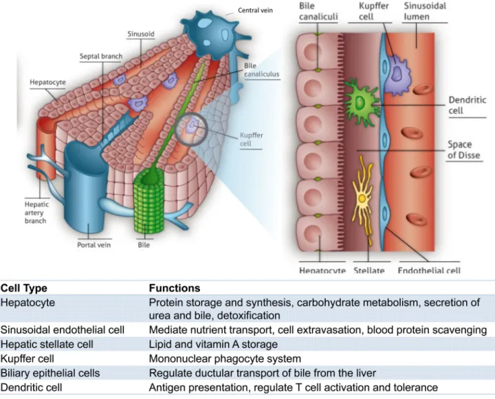
The development of liver cell-based therapies poses unique challenges, largely stemming from the scale and complexity of liver structure and function. The organ displays a repeated, multicellular architecture, in which hepatocytes, the main parenchymal cell of the liver, are arranged in cords that are sandwiched by extracellular matrix in the space of Disse (Figure 1). The space between cords is also home to a multitude of supporting cell types such as sinusoidal endothelial cells, Kupffer cells, biliary ductal cells, and stellate cells. Due to this architectural arrangement and cellular heterogeneity, the hepatocytes are exposed to gradients of nutrients, hormones, and growth factors delivered via the combined blood supply of the portal vein and hepatic artery. In particular, a major challenge that has hindered the advancement of cell-based therapeutic strategies is the propensity of hepatocytes to lose liver-specific functions and the ability to replicate when isolated from the normal in vivo microenvironment. Microenvironmental signals including soluble factors,
Author Manuscript
Author Manuscript
Author Manuscript
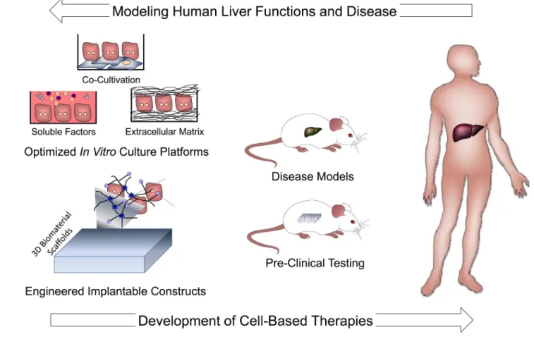
extracellular matrix components, and heterotypic cell-cell interactions have all been implicated in the regulation of hepatocyte survival and phenotypic stability. The success of cell-based therapies hinges on the capacity to replace or overcome such necessary signals in order to promote the survival and function of hepatocytes transferred to patients in a clinical setting. Research efforts to achieve this goal have included studies at the interface of microtechnology and cell biology, and have led to the ability to manipulate the hepatocyte phenotype by the controlled presentation of environmental cues. Such systems exhibit long-term stabilized hepatocyte function and also enable fundamental studies investigating drug-induced liver injury and hepatotrophic infections, in addition to providing insight into how clinically transplanted hepatocytes might be supported in an in vivo setting. The continued evolution of in vitro liver platforms through the incorporation of additional liver cell types and capabilities, such as perfusion and control over tissue architecture in three dimensions, will be critical for the improved understanding of mechanisms underlying hepatocyte processes and the enhanced functionality of cell-based therapeutics.
Another obstacle in the progress of cell-based approaches is the limited availability of human hepatocytes. Only a small supply of human hepatocytes is currently available from organs determined to be inappropriate for transplantation. Despite the significant
proliferative capacity exhibited during regeneration in vivo, as noted above, mature human hepatocyte proliferation in culture is limited. Hence, the elucidation of molecular mediators that regulate hepatocyte proliferation and that could potentially promote expansion in vitro is an active area of investigation. In addition, significant research efforts are focused on the potential of alternative cell sources, most notably leveraging stem cell differentiation and reprogramming. Based on knowledge of developmental mechanisms, recent advances in pluripotent stem cell differentiation protocols have illustrated that highly proliferative pluripotent stem cells can give rise to hepatocyte-like cells derived from a single donor (i.e. normal/disease genotype) (11–14). Yet it remains to be seen whether it will be feasible to produce these cells on a clinically-relevant scale and whether lingering safety concerns will be overcome for transplantation purposes.
In the case of hepatic failure, in particular hepatic encephalopathy, which is classified by graded alterations in mental status, the liver community faces a greater challenge – at least in some ways – than in other cases of organ failure, in that the clinical priorities for
achieving functional improvement (ie. cardiac output for heart, filtration rate for kidney), are not available due to our lack of fundamental understanding of the pre-existing disease state. That is, we have limited ‘biomarkers’ that are predictive of clinical response in a liver failure patient. For example, despite the use of metabolic readouts and parameters, such as timing of jaundice, bilirubin levels, prothrombin time and age, the causes of hepatic encephalopathy are not completely understood, with distinct clinical indicators in acute versus chronic liver disease and marked patient-to-patient variability. Thus, any animal models used to assess candidate therapeutics must be very carefully chosen given that etiologies of liver failure from trauma, metabolic liver disease, and cirrhotic disease are each very distinct, and any clinical trial design must be tailored, based on the particular clinical setting in question. Despite the many challenges that remain, substantial progress has been made in the understanding of liver development, regeneration, and the development of
Author Manuscript
Author Manuscript
Author Manuscript
instructive animal models. These insights, together with recent innovations in engineered in
vitro culture platforms and in vivo transplantation approaches, form a strong foundation for
future advancements and the ultimate implementation of cell-based therapeutics for liver disease.
Clinical manifestations of liver disease
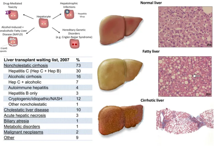
The vast majority of liver functions are mediated by the hepatocyte, the functional metabolic unit of the liver that constitutes about two-thirds of its cell mass (15). Though liver disease does not routinely result in abdominal pain, it does lead to a variety of life-threatening metabolic and physiologic abnormalities. For example, the absence of these functions leads to bleeding abnormalities, accumulation of neurotoxins causing altered consciousness, low blood sugar and accumulation of serum ammonia, and jaundice from elevation of serum bilirubin. Unfortunately, although patients with liver disease can be medically supported through therapies targeted at features such as portal hypertension and coagulopathy, there are no therapeutic strategies that collectively augment the range of impacted functions, and thus an organ transplant has been the only permanently successful therapy to date. This approach contrasts with other organ systems, such as the heart and the kidney, in which patients with failing tissues can be given ionotropes to improve contractility, or diuretics to improve fluid balance, respectively, without the need for immediate transplantation. The most appropriate approaches to future treatment design for patients with liver damage depend largely on the particular etiology of the organ damage in an individual case. Types of liver disease can be broadly grouped into three categories: chronic liver disease due to metabolic dysfunction, i.e., in the absence of trauma or tissue scarring; acute liver failure that does not damage normal tissue architecture but is associated with direct injury and loss of hepatocytes; and chronic liver failure that is accompanied by widespread tissue damage and scar-based remodeling, or cirrhosis (Figure 2). Whereas there will always be
circumstances that call for an alternate approach to clinical intervention, these three categories are typically best targeted by distinct therapeutic strategies, as outlined below.
Metabolic-based chronic liver disease
Patients with life-threatening, inborn liver-based metabolic disorders, often caused by defects in single enzymes or transport proteins, are not typically accompanied by changes in liver architecture. However, even in the absence of structural change to the liver, these inborn deficits often cause injury to other organ systems, such as the brain. This is the case with urea cycle disorders, Crigler-Najjar syndrome,, and phenylketonuria. In oxalosis, the liver-based genetic abnormality leads to accumulation of oxalate crystals in the kidney and renal failure. To date, although patients suffering from this category of liver diseases are treated with organ transplantation, their clinical manifestations offer an ideal scenario for cell transplantation therapy. Given that the tissue architecture is unscarred and patients can be identified early and treated over time – prior to an acute episode - transferred hepatocytes may have both time and opportunity to engraft and replace endogenous, functionally inadequate cells.
Author Manuscript
Author Manuscript
Author Manuscript
Acute liver injury
Acute liver failure is defined as a rapid deterioration of liver function over a period of less than 26 weeks in patients who have no preexisting liver disease (16). Acute injury initially leads to necrosis of hepatocytes, which causes release of liver-specific enzymes, such as alanine transaminase (ALT) and aspartate transaminase (AST) into the blood. An increase in ALT and AST indicate hepatocyte injury but are not indicators of hepatic failure in and of themselves. Acute liver failure carries a very high mortality in many, but not all, cases, thus it is important to identify its cause as some forms can be treated or will resolve
spontaneously. Clinically, the dominant findings are onset of fatigue and jaundice, a transient increase in serum concentrations of ALT and AST, followed by progressive increases in serum bilirubin, worsening coagulation, and development of altered
consciousness (hepatic encephalopathy), leading to coma and brain swelling. Histologically, the liver architecture is intact except for loss or necrosis of the hepatocytes. As it is often difficult to know which patients will recover spontaneously, liver transplantation is currently the only available treatment option for severe, even if transient, acute hepatic failure. It remains unclear if hepatocyte transplantation procedures could be sufficiently optimized such that cell engraftment would occur on a clinically relevant time scale due to the urgent, acute status. The advent of extracorporeal artificial liver devices may eventually address this need (see next section) in that they could offer a clinical ‘bridge’ to address acute symptoms during a period in which the endogenous liver could be given an opportunity to recover spontaneously.
Chronic liver failure with accompanying cirrhosis
Liver failure more commonly occurs in the setting of cirrhosis, as an acute decompensation or abrupt loss of liver function, in patients with chronic liver disease. Cirrhosis of the liver caused approximately 1 million deaths worldwide in 2010 (17). Histologically, cirrhosis is characterized by expansion of the extracellular matrix with capillarization of the sinusoidal endothelium and loss of fenestrae with production of regenerative hepatic nodules (18) (Figure 2). In addition to producing symptoms of hepatic failure, the scarring from cirrhosis results in resistance to flow in the portal circulation. The increased pressure in what is normally a low pressure conduit leads to gastrointestinal bleeding, severe accumulation of abdominal ascites, and can lead to secondary dysfunction of the kidneys (hepatorenal syndrome) and lungs (hepatopulmonary syndrome). Some patients with cirrhosis are completely asymptomatic, whereas others with a similar histological picture exhibit severe symptoms of hepatic failure. Cirrhosis may be caused by hepatitis B or C infection, autoimmune processes, chronic alcohol abuse, or inflammation and fat accumulation from chronic metabolic syndromes. Additionally, in contrast to examples discussed above, some inborn errors of metabolism can lead to structural liver damage with cirrhosis and liver failure. Examples include alpha-1-antitrypsin deficiency, hemochromatosis, Wilson’s disease, hereditary tyrosinemia, and cystic fibrosis. As is currently the case for acute liver failure, the only definitive therapy for end-stage cirrhosis and liver failure is orthotopic (i.e. in the natural position) liver transplantation. While it may one day be possible for these non-acute patients to benefit from cell transplantation or implantable tissue engineered grafts, given that their symptoms should provide sufficient time for both engraftment and vascularization, there are significant barriers to overcome that may preclude these
Author Manuscript
Author Manuscript
Author Manuscript
therapeutic advances from being applied. Specifically, neither the site of implantation – in this case the high-pressure portal system or the scarred microenvironment – may be amenable to accepting infiltrating cells or engineered grafts. Thus, ectopic sites of transplantation are being considered for these sorts of interventions.
Design and development of existing cell-based therapeutic interventions
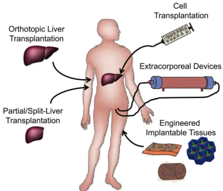
Clinical effectiveness of hepatocyte transplantationIn the clinical scenarios of liver damage presented above, there are a variety of cases in which hepatocyte transplantation – either in the context of an extracorporeal device, a tissue engineered graft, or as individually engrafted cells – may offer a clinical alternative to orthotopic organ transplantation (Figure 3). Since the development of techniques for isolation of individual hepatocytes by collagenase digestion (19), investigators have studied whether hepatocyte transplantation could be used to treat liver diseases, first in the
laboratory and then in patients. As hepatocyte transplant would classically involve simple infusion of isolated cells into the liver through the portal vein, this form of therapy is far less invasive than orthotopic liver transplantation and could be performed safely in severely ill patients. In the presence of normal host liver architecture, some fraction of the transplanted cells should cross the endothelium to integrate into the host liver (20–22). Because the native liver is not removed, the transplanted hepatocytes only need to provide replacement of defective enzyme activity or enough hepatic function to overcome the liver dysfunction. Other sites of transplantation have been explored in animal models including the mesentery, spleen and the renal capsule (23–25). In these settings, the survival and persistence of cells at ectopic sites seems to depend minimally upon nutrient availability through local vascular beds; however, cells may not require perfusion specifically with portal blood containing ‘hepatotrophic’ factors, as was once believed. Unexpectedly, the lymph node, a highly vascularized site has been a useful location for promoting remarkable growth and function of transplanted hepatocytes in animal models (26), adding further support for the potential clinical utility of adult hepatocyte transplantation. Regardless, in acute liver failure approximately 40% of patients with advanced symptoms recover spontaneously with only medical management (27). As there is no effective means to distinguish patients who will survive without transplantation from those who will not (16), treatment with transplanted hepatocytes could potentially obviate the need to perform an irreversible organ transplant on a patient who could recover spontaneously. However, in an acute setting, it remains to be established whether functional engraftment could be accomplished on a clinically relevant time scale.
Numerous studies in rodents done over the last 30 years indicate that adult hepatocyte transplantation can reverse hepatic failure and can correct various metabolic deficiencies of the liver (28). Although clinical trials of hepatocyte transplantation have demonstrated the long-term safety of the procedure, only partial correction of metabolic disorders has been achieved, and the degree to which donor hepatocytes have restored failing livers has not yet been adequate to circumvent the need for organ replacement (10, 29, 30). Because of the limited availability of fresh donor hepatocytes, or effective alternatives, such as
cryopreserved hepatocytes or in vitro expanded cells, trials have been limited to case reports
Author Manuscript
Author Manuscript
Author Manuscript
or anecdotes involving few patients and no untreated control patients (23, 24, 31–36). However, this limited clinical experience with hepatocyte transplantation has helped to identify barriers to its successful application and potential pathways for overcoming these challenges. One explanation for the failure to translate laboratory studies to the clinic is the limited number of animal models that fully recapitulate human disease and the challenges of mapping animal model data to clinical outcomes and action plans. A re-evaluation of the results of cell transplantation in animal studies has shown that attaining adequate levels of engraftment and technical issues such as cell availability will need further attention. In addition, immunological graft rejection is more difficult to manage in patients than expected because it is not possible to diagnose rejection of donor hepatocytes by conventional biopsy techniques. This gap most likely explains why successful correction of metabolic diseases by transplantation has been transient, rarely lasting more than a year or two. Through the development of improved animal models, pretreatment strategies, and engineered delivery platforms, ongoing efforts in the field aim to build on these preliminary findings in patients towards an improvement in transplantation efficiency and clinical efficacy.
Mature hepatocytes as sources for cellular therapies
Despite the lack of correlation between some animal model experiments and the minimal success of hepatocyte transfer to patients, there is sufficient support for their clinical potential to prompt the field to discover strategies to obtain, maintain and expand human hepatocytes. Access to sufficient functional cell numbers is a major hurdle despite evidence from serial transplantation using genetically marked, mature donor hepatocytes showing that such cells have a remarkable ability to replicate in vivo, many times more than that seen following partial hepatectomy (37). Thus, unlike many other fully differentiated cell types, hepatocytes have the intrinsic capacity for robust replication, at least under a set of poorly defined circumstances. Still, despite decades of research and partial successes with culture conditions, hepatocytes in vitro rapidly lose their differentiated characteristics and proliferate poorly (38, 39). It is currently not feasible to routinely obtain hepatocytes from healthy human donor livers, amplify the cell populations in vitro while maintaining their functional capacities, and obtain sufficient cells for transplantation or use in extracorporeal devices. This situation will be helped by further research on the hepatocyte
microenvironment including the roles of extracellular signaling, matrix, cell-cell interactions, physical forces and soluble factors (40, 41), in order to explore diverse and potentially unexpected ways to generate a replicative environment for the hepatocyte. A different approach to controlling hepatocyte replication is to manipulate the intrinsic cellular mechanisms that either keep hepatocytes quiescent in the liver or allow the cells to enter the cell cycle after loss of tissue mass (42). In the past decade, substantial inroads have been made in identifying how different cellular networks converge on the proteins and complexes that govern the hepatocyte cell cycle during hepatic regeneration (43). The FoxM1b transcription factor (44) activates Cdc25B, which in turn is needed for Cdk1 activation and entry into mitosis after partial hepatectomy. The anaphase promoting complex restrains hepatocytes in vivo from entering the cell cycle (45) and the Hippo/YAP signaling pathway promotes cell cycle re-entry (46). Recently, the kinase MKK4 was found to restrain hepatocyte proliferation, such that experimental inactivation of MKK4 stimulated
Author Manuscript
Author Manuscript
Author Manuscript
hepatocyte regeneration in mouse models of acute and chronic liver failure (47). These disparate findings and others could be brought to bear in a united fashion to better understand how the hepatocyte cell cycle can be controlled.
Extracorporeal bioartificial liver devices
With improved understanding of the signals needed to expand and/or maintain long term function of isolated hepatocytes, it may be possible to provide appropriate architecture-based and soluble-architecture-based signals to populate extracorporeal liver devices. Extracorporeal liver devices are principally aimed at providing temporary support to patients with liver failure. Early attempts at developing extracorporeal support technologies were based on non-biological mechanisms such as hemodialysis, hemoperfusion, hemodiabsorption, plasmapheresis, and plasma exchange (48), in part due to the lack of available hepatocytes needed to populate such devices. More recent configurations of artificial, non-biological support systems have focused on the elimination of albumin-bound toxins utilizing a method termed albumin dialysis. These devices, such as the Molecular Adsorbent Recirculating System (MARS; Gambro, Sweden) and the Prometheus (Fresenius Medical Care, Germany) platform have been shown to be effective for the reduction of plasma bilirubin, bile acids, and other albumin-bound molecules (49, 50). Although some reports point to the potential utility of these approaches as bridge treatments prior to organ transplantation (51–53), improvements in patient outcome have not been fully demonstrated (54, 55), suggesting that additional trials are required to fully evaluate their effectiveness. In particular, initial clinical studies have illustrated beneficial effects on organ function without an associated
improvement in transplant-free survival (55, 56), which further underscores the complexity of liver failure events and the need for controlled clinical trials. In light of the broad range of vital synthetic functions carried out by the liver and the lack of a liver ‘biomarker’ around which to design these devices, much of the field has gravitated towards cellular rather than purely device-based solutions.
Accordingly, to provide a more complete array of synthetic and biochemical functions, which is lacking in strictly artificial support systems, considerable efforts have been placed in the development of bioartificial liver (BAL) devices containing hepatic cells. Following the initial studies, such as the study by Matsumura et al. in 1987 (57), a broad range of BAL device designs have been reported, and have highlighted the importance of several criteria in the development of an effective device (Figure 4). These include issues of cell sourcing, maintenance of cell viability and hepatic functions, sufficient bidirectional mass transport, and scalability to therapeutic levels. As discussed earlier, the sourcing of hepatic cells is a fundamental challenge for all liver cell-based therapies. As a result, xenogeneic sources (primarily porcine) or transformed/immortalized human hepatocyte cell lines have formed the basis for the majority of BAL devices (which typically include approximately 1 × 10¹⁰ cells) tested in the clinic to date (58–61). However, lack of hepatocyte function in
transformed cell lines (62) and cryopreserved cells, and the potential risk for porcine endogenous retroviruses have hampered attempts to demonstrate efficacy. More recently, studies have begun to incorporate primary liver cells by exploiting advances in
cryopreservation, fetal cell procurement, and stem cell differentiation (60, 63–66). Magnifying the cell sourcing challenge are the scalability requirements for translation.
Author Manuscript
Author Manuscript
Author Manuscript
Furthermore, the design of an effective BAL device is dependent on the incorporation of the appropriate environmental and organizational cues that enable maximal survival and function of the hepatocellular component. Hollow fiber devices are the most common BAL design and contain hepatic cells within cartridge units (67), with the hollow fiber
membranes serving as a scaffold for cell attachment and compartmentalization (Figure 4). A range of modifications aimed at optimizing cellular performance have been explored. In particular, due to the enhanced function of hepatocyte aggregates relative to single cell suspensions, many device configurations contain either attached or encapsulated hepatocellular spheroids (67–71). In the modular extracorporeal liver support (MELS) system (Charite, Germany), hepatocytes are aggregated in co-culture with liver non-parenchymal cells resulting in the formation of tissue-like organoid structures (72). Furthermore, exposure of hepatocytes to plasma of a sick patient may necessitate specific alterations in hepatocyte culture conditions. For example, the supplementation of plasma with amino acids has been shown to increase albumin and urea synthesis (73), and preconditioning with physiological levels of insulin (lower than standard culture medium), has been demonstrated to prevent abnormal lipid accumulation in hepatocytes (74). Overall, environmental conditions within a BAL device, such as oxygen tension and fluid shear forces can significantly affect hepatocyte functions (75). In addition, both the convective and diffusive properties of the systems must be optimized to provide vital nutrients to the cells while simultaneously allowing export of therapeutic cellular products. Currently, although clinical efficacy of BAL devices remains limited, improvements in device and trial design continue to be implemented. It is anticipated that parallel progress towards the development of highly functional in vitro platforms will provide a reciprocal benefit for the advancement of BAL approaches. For example, small-scale in vitro bioreactor systems have been utilized to systematically examine the effects of shear stress and oxygen tension on hepatocyte function (76, 77). BAL device development is in desperate need of predictive biomarkers that point to the degree of hepatic function that is represented by a device. For example, the ability to monitor a single protein or metabolite in the perfusate as a broader indicator of reduced accumulation of systemic toxic metabolites, and hence the
neuroprotective effects of a device, would be invaluable. Applying ‘omics’ methods in an effort to evaluate the state of these devices might provide clues towards the development of appropriate biomarker readouts.
In vitro strategies for improving hepatocyte viability and function
A major research area is the development of improved in vitro culture models, which would improve BAL design as well as provide platforms for the study of human hepatic function. Although systems employing single enzymes, liver slices or liver cell lines have found utility for addressing focused questions, each has limitations. For example, liver slices have limited viability and are not amenable to high-throughput screening; cell-free microsomes lack the dynamic gene expression and intact cellular machinery required for cellular responses (i.e. cytotoxicity); and carcinoma-derived cell lines and immortalized hepatocytes display an abnormal repertoire of liver proteins and limited liver-specific functions (5). For these reasons, isolated primary hepatocytes are considered to be the most suitable platform for a range of in vitro applications (3, 6). As a result, extensive research over several decades has been focused on identifying specific culture configurations and molecular
Author Manuscript
Author Manuscript
Author Manuscript
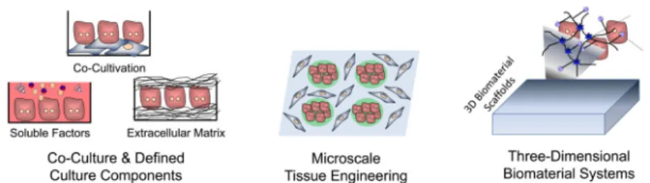
stimuli that can maintain the phenotypic functions of hepatocytes (Figure 5). In general, the phenotype of an isolated hepatocyte is quite plastic and is exquisitely sensitive to its microenvironment. Many different manipulations regulate aspects of the differentiation program, although not necessarily in equivalent ways (the plethora of hepatocyte functions and the absence of a clinical biomarker of rescue means that many parameters must be measured). Such manipulations include culture medium, extracellular matrix, and interactions with non-parenchymal cells. Engineers and biologists have also tried manipulations that are not explicitly ‘biomimetic’ or obviously physiological but still manage to influence hepatocyte fate and function, presumably through some of the existing adult or developmental signaling pathways. For example, additives such as hormones, amino acids, corticosteroids, growth factors, as well as non-physiological small molecules, have been demonstrated to affect hepatocyte functions (78–81). Additionally, both extracellular matrix composition and topology can modulate hepatocyte morphology and phenotype. In the classic “double gel” culture format, hepatocytes are sandwiched between two layers of collagen gel (82). In this configuration, hepatocytes exhibit improved morphological features such as polarized bile canaliculi, and stabilized functions, including albumin and transferrin secretion, for weeks. However, key detoxification pathways such as oxidation and conjugation reactions have been shown to become imbalanced over time in this system (83, 84). Studies investigating the potential added benefits of complex mixtures of
extracellular matrix components on hepatocyte function have utilized strategies including extracellular matrix preparations from native liver tissue (85, 86), or conversely, screening methods to systematically investigate defined extracellular matrix combinations (40, 87). Synthetic surface modifications, such as polyelectrolyte chemistries, also influence hepatocyte function in vitro (88, 89), and a polyurethane matrix identified by polymer library screening was shown to support hepatocellular differentiation and function (90). In addition to strictly 2D systems, hepatocyte culture under conditions that promote
aggregation into 3D spheroids can affect functionality, with spheroid configurations displaying superior hepatocyte function compared to standard collagen monolayer cultures (91, 92). Potential mechanisms underlying these effects include the increased number of homotypic cell-cell contacts between hepatocytes, the retention of a 3D cytoarchitecture, and the asymmetric presentation of extracellular matrix and other signals surrounding the spheroids (93). Numerous technologies including arrays, rotational cultures, and
encapsulation methods have been developed for the optimization and scale-up of hepatocyte spheroid culture (94–96).
In both 2D and 3D formats, the addition of secondary supportive cells for heterotypic interactions is one of the most robust approaches for preserving the viability, morphology, and function of cultured hepatocytes. In addition to modulating cell fate through cell-cell sensing pathways such as cadherins, stromal cells additionally modify the extracellular matrix and the paracrine signals in the microenvironment. Specifically, beginning with the initial experiments by Guguen-Guillouzo and colleagues (97), substantial research efforts have demonstrated that both liver-derived cell types including liver biliary epithelial cells and non-parenchymal cells (e.g. stellate cells, sinusoidal endothelial cells), as well as numerous non-liver-derived cells (e.g. embryonic fibroblasts), are capable of supporting hepatocyte function in co-culture contexts (98). Although varied culture conditions have
Author Manuscript
Author Manuscript
Author Manuscript
been explored, typically, primary hepatocyte functions can be rescued if co-cultures are initiated within 3–7 days following isolation. Transcriptional and post-transcriptional mechanisms have been implicated in the co-culture stabilization of hepatocyte-specific genes including albumin, transferrin, pyruvate kinase, and glutathione S-transferase (99, 100). Furthermore, cytochrome P450 (CYP) detoxification enzymes are elevated in co-cultures compared to hepatocytes cultured alone (101).
Co-culture systems have formed the basis for investigations into a broad range of hepatocellular processes such as the acute phase response, oxidative stress, mutagenesis, lipid and drug metabolism, and xenobiotic toxicity (98). For example, co-cultures of hepatocytes and Kupffer cells (the liver’s resident macrophages) have been used to examine mechanisms of hepatocellular damage (102, 103). Meanwhile co-cultures with liver sinusoidal endothelial cells have highlighted the importance of hepatocyte-endothelial cell interactions in the bidirectional stabilization of these cell types (104–109). Research efforts continue to employ co-culture platforms as in vitro models aimed at dissecting physiological cell-cell interactions in the liver, and as a tool for optimizing engineered liver
microenvironments. In particular, for certain tissue engineering applications, replacing supportive cell types with acellular components that do not consume nutrients and occupy limited space may be advantageous. Studies focused on the underlying mechanisms of hepatocyte stabilization in co-culture have identified several cell surface and secreted factors that play a role including T-cadherin, E-cadherin, decorin, TGF-β1, and liver-regulating protein (110–116). Although such factors are currently incapable of supporting hepatocyte function at a level comparable to supportive stromal cell types, they represent a proof-of-concept for highly functional hepatocyte-only culture platforms.
Collectively, a set of in vitro strategies is emerging that may aid in BAL design as well as inform the development of in vitro liver models for discovery. Because the hepatocyte phenotype is quite plastic once the cell is isolated from its native environment, much of the focus has been in rescuing the hepatocyte phenotype ex vivo. There seems to be a shift in the field from strategies that are solely biomimetic (e.g. culturing hepatocytes in sandwich extracellular matrix, or under gradients of oxygen tension, or in co-culture with non-parenchymal hepatic cells) to culture models that do not necessarily mimic the native hepatic microenvironment (e.g. spheroids and co-cultures with embryonic fibroblasts). Strategies that optimize hepatic function, duration of rescue, accessibility to perfusate, and amenability to scale-up may or may not ultimately resemble native liver architecture. A systems-level picture of molecular signals that influence phenotypic stability is emerging and will aid these efforts (117).
Stem cells as sources of hepatocytes for cellular therapies
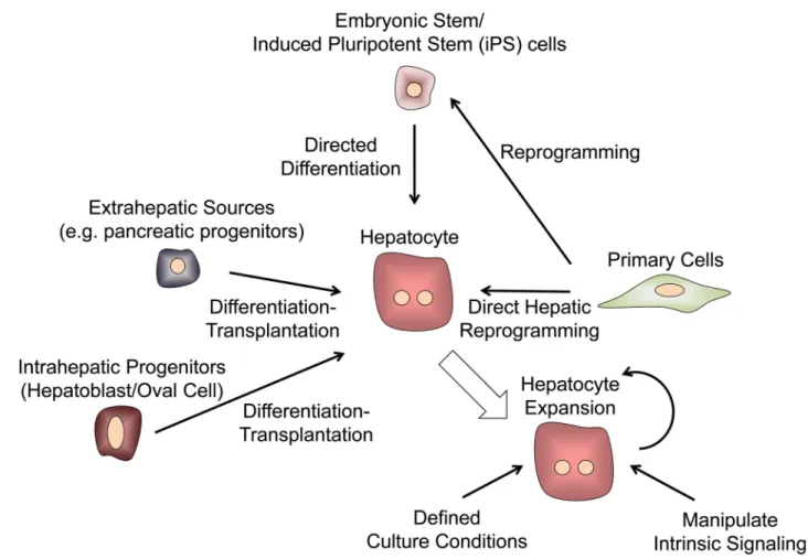
An independent approach to generating hepatocytes for therapeutics is to use stem and/or progenitor cells, which by definition have a high capacity for expansion (Figure 6), and may be sourced from a variety of tissues. Theoretically, such cells could be amplified, induced to differentiate, and used in diverse applications. Whereas progress has been made in
genetically or immunologically identifying tissue-resident stem cell-like populations that arise in chemically damaged livers (42, 118–122), further work is needed to understand the
Author Manuscript
Author Manuscript
Author Manuscript
role of these cells in normal liver physiology and repair (123) and to assess whether these populations represent a clinically-relevant source of hepatocytes. Other cell sources, including mesenchymal stem cells have been discussed recently in this context (124). Another current interest is on the potential of pluripotent stem cells, including human embryonic stem cell (hESC) and induced pluripotent stem cell (iPSC) lines, which have a high proliferative capacity and can differentiate into diverse lineages in vitro and in vivo (125). Various directed differentiation strategies have been applied to hESC and iPSC cultures, and have yielded populations that exhibit many phenotypic and some functional traits of mature hepatocytes, earning the derived cells the label of ‘hepatocyte-like cells” (11, 126–136). Yet hepatocyte-like cells exhibit numerous characteristics, such as distinctive CYP activities as well as expression of alpha-fetoprotein, that more closely resemble fetal-stage hepatocytes rather than mature adult cells (137). To further characterize these cells, some studies have tested the maturation and repopulation potential of both mouse and hESC-derived hepatic cells in rodent transplantation models, such as immune-deficient recipient mice that harbor genetic defects in resident hepatocytes (13, 138, 139). This form of inquiry helps to support the conclusion that hepatocyte-like cell populations can support at least some liver function in a replacement setting, but the best choice of animal models for these studies is still under debate (140). Attempts to identify small molecules that can induce the maturation of hepatocyte-like cells have suggested that ex vivo maturation may be possible (41).
One study utilized partially differentiated human iPSCs and admixed supportive stromal and endothelial cells to mimic aspects of early liver development. The resulting liver ‘buds’ were able to vascularize upon ectopic transplantation and provide functional rescue of mouse models of liver injury (141). Furthermore, recent progress suggests that hepatocyte-like cells derived by directed differentiation of fibroblasts also exhibit more adult hepatic traits, and, like iPSCs, might provide an autologous stem cell source that would bypass a need for clinical immune suppression (142–146). In light of this progress, the strategy of utilizing pluripotent cells as a source material for generating hepatocytes---either for clinical transplantation, as a cellular component in BAL devices, or in various model platforms to study disease and drug development---holds promise. Considerable experimental energy continues to be applied to find methods that improve the efficiency and extent of
differentiation of these expansion-ready precursor cells (Figure 6). More needs to be done to obtain expansion of human stem cell- derived hepatocytes that have been implanted into mouse livers and evaluate their performance and safety. Regardless, the capacity to populate mouse model livers with human hepatocyte-like cells both from normal individuals and patients offers an experimental system of “humanized” mouse livers for use in toxicological studies on human hepatocytes (141, 147–150).
Translating existing technology into pre-clinical and therapeutic
applications
Implantable therapeutic constructs for preclinical testing
Delivery of hepatocytes by cell transplantation requires cells to home to the liver from the portal blood stream and extravasate across the space of Disse into the hepatic lobular
Author Manuscript
Author Manuscript
Author Manuscript
compartment. Early ‘tissue engineers’ sought to support these adhesion-dependent cells with biomaterials that could alleviate the need for homing and attachment. These seminal studies used available biomaterials (such as poly-lactic co-glycolic acid, PLGA) and demonstrated an improvement in the survival of the transplanted cells. Nonetheless, animal studies suggested that the persistence of hepatocyte phenotype and survival were dependent on the site of implantation, at least in this delivery context. For example, some studies
demonstrated an improvement in the function of transplanted hepatocytes when bathed in the so-called hepatotrophic factors draining from the gut to the portal vein (151). Because the portal vein is an unattractive site for transplantation in chronic liver failure patients with elevated portal pressures, investigators have begun to explore ectopic sites (spleen,
subcutaneous, renal capsule, intraperitoneal) (64, 152, 153). In these ectopic sites,
manipulating the cellular microenvironment using biomaterials, peptides, or even other cells has led to a diminished dependence on the portal circulation, setting the stage for ectopic transplantation of engineered hepatic tissues (as is currently the practice for renal
transplantation). Under strong regenerative stimulation such as that found in genetic mouse models of tyrosinemia, the transplanted cells can even repopulate host lymph nodes ectopically, suggesting that either the cells in these sites receive some hepatotrophic stimuli or the reliance on portal hepatotrophic factors may be mitigated in some regenerative contexts (26, 154). These findings are consistent with the classic parabiosis experiments of Bucher and colleagues, suggesting that regenerating liver produces bloodborne factors that stimulate hepatocyte expansion in a conjoined animal (155).
Since the early experiments with PLGA, a synthetic degradable polyester found in suture material, the community has explored many porous scaffold materials ranging from both natural (e.g. collagen, alginate) and synthetic (e.g. PLGA, PLLA) sources. Scaffold traits including porosity, material and chemical characteristics, and 3D architecture are among the parameters that are customizable in 3D platforms, and these properties play important roles in dictating cellular function and facilitating the transport of nutrients and secreted
therapeutic factors. In addition, synthetic hydrogel systems based on poly(ethylene glycol) (PEG), which have been extensively utilized for tissue engineering studies (156), have found recent utility in 3D liver platforms (157–159). Hydrogel platforms offer the advantage of polymerization in the presence of cells, thereby enabling the fabrication of 3D networks with uniform cellular distribution without the need for cell migration or expansion in situ. Modifications in polymer chain length and the conjugation of bioactive factors such as adhesive peptides improve the survival and function of PEG hydrogel-encapsulated hepatic cells (157, 158, 160). Moreover, to generate scaffolds with a highly defined architecture that provide better control over the 3D environment at the microscale, a range of rapid
prototyping and patterning strategies have been developed and tested for liver applications (161–164). For hydrogel systems, these include photolithography-based techniques for dictating the size and shape of constructs and for building multilayer geometries (157, 159, 165–167), and dielectrophoresis methods for controlling cellular positioning (168). Notably, cell-cell interactions (both homotypic and heterotypic) have been suggested to substantially affect hepatocellular survival and function in 3D biomaterial scaffolds (158, 167, 169, 170). As a result, similar to 2D culture models, the controlled integration of additional cell types
Author Manuscript
Author Manuscript
Author Manuscript
or acellular factors that mimic key cell-cell interactions will be critical for the advancement of 3D liver platforms.
Early studies also uncovered a scale-up issue that paralleled that found with BAL devices: transplanting large numbers of cells in biomaterials without a sufficient nutrient supply rapidly leads to necrosis of the engineered tissue. Hepatocytes are highly metabolic and are normally in close contact with an extensive sinusoidal vasculature. As a result, significant efforts must be made in the design of implantable liver systems to avoid transport limitations that can greatly diminish tissue function. In particular, the site of implantation is a critical parameter in liver tissue engineering studies, with more highly vascularized sites such as the peritoneum or renal capsule generally promoting improved engraftment and function (158, 171, 172). Thus, another area of focus is the establishment of a vascular network that supports a large number of hepatocytes. Priming of the implantation site through pre-vascularization is a particularly effective strategy due to the minimization of early transport restrictions that occur prior to the establishment of functional vasculature. Microfabrication approaches, such as polymer molding with etched silicon, dissolvable sugar lattices, and microtissue molding of aligned endothelial cords have been employed as strategies to preform capillary-sized channel networks (163, 173, 174). Additionally, the integration of angiogenic growth factors into the implantable scaffolds has been shown to promote the recruitment of host vessels (175–178). Alternative strategies to achieve this goal include multilayer microfluidic networks, prevascularization through release of angiogenic growth factors, and inclusion of non-parenchymal cells that promote angiogenesis and vascular stabilization with pericytes. For instance, preceding hepatocyte delivery with the
implantation of scaffolds that release angiogenic vascular endothelial growth factor (VEGF) enhanced capillary density and improved engraftment in rat liver lobules (179). Similarly, fibroblast growth factor 2 (FGF2) coated scaffolds served as a supportive environment for mouse ESC-derived hepatocyte inoculation in an in vivo hepatic failure mouse model (64). Furthermore, several recent studies aimed at engineering skeletal or cardiac muscle tissue have illustrated that significant improvements in survival and host vasculature connections can be achieved following in vitro formation of vessel structures within optimized tri-culture systems (muscle, endothelial and mesenchymal cells) (173, 180–182).
In addition to vascular integration, an improved understanding of multicellular organization and morphogenesis in the liver could also aid in the formation of functional biliary transport systems. Various in vitro models have been developed that exhibit organized bile canaliculi (183–185) or artificial duct structures (186), but their incorporation into implantable systems has yet to be fully explored. Although early work demonstrated engrafted bile ducts in ectopic sites (187), the degree to which the biliary tree must be reconstructed has not yet been established; in ectopic cell transplantation experiments the hepatocytes do not appear cholestatic, and biliary products do appear to find their way to the digestive tract. One hypothesis is that the biliary products are redirected or ‘leak’ into the bloodstream where they circulate and are processed by the remnant liver into bile. This scenario would argue against removal of the diseased liver in the setting of transplantable tissue engineered constructs, and is consistent with the functional outcome achieved in peritoneal
transplantation of mature hepatocytes and hepatocyte-like cells that lacked biliary networks
Author Manuscript
Author Manuscript
Author Manuscript
(26, 141). The need for reconstruction of the nervous and lymphatic systems is likewise even less well understood, although, if clinical transplantation is a guide, these are likely to be less critical.
Similar to whole organ transplantation, the host immune response following the introduction of tissue engineered constructs is a critical determinant of successful engraftment.
Accordingly, the immune isolation capabilities of polymer scaffolds is a highly active research area (188), and represents the foundation of numerous tissue engineering approaches such as pancreatic islet transplantation (189–191). Recent liver tissue engineering studies demonstrate that hydrogel encapsulation enabled detectable human hepatic function in transplanted immunocompetent mouse strains for greater than one week (158), suggesting that aspects of immune isolation may find utility in various liver
applications. In general, approaches to tune the degree of inflammatory cell recruitment to engineered implants could substantially aid in preventing foreign body responses that severely impair engraftment, while potentially maintaining the positive remodeling effects that inflammatory cells provide in numerous normal tissue regeneration contexts (192). For example, liver regeneration proceeds with a sequence of remodeling processes including protease expression and extracellular matrix deposition (193–195). The incorporation of protease-sensitive domains into hepatic hydrogel systems could allow for the degradation of the constructs following implantation and subsequent changes in ligand presentation and gel mechanics (196). Tailoring the degradation properties of constructs based on the kinetics of cell proliferation, inflammation, and angiogenesis could provide a means for efficient integration. Overall, the development of new biomaterials platforms that can collectively modulate host cell recruitment and material-resident cell function, as well as engraftment and release of implanted cell types (197), is a major focus and should advance liver tissue engineering approaches.
Animal models for pre-clinical testing of candidate interventions and barriers to overcome in clinical translation
Transplantation of liver cells has been shown to improve the survival of animals with chemically and surgically induced acute liver failure (198–207), end-stage liver failure secondary to cirrhosis (208), and to correct metabolic deficiencies and prolong survival in numerous models of liver-based metabolic diseases (199, 209). Whereas animal models have provided a foundation for clinical translation, the results of clinical trials using liver cell transplantation have been disappointing. The most promising results have come from the treatment of children with liver-based metabolic diseases, where evidence of functional replacement of the deficient enzyme has been documented and post-transplant biopsies have shown engrafted cells expressing a corrected form of the deficient enzyme (10).
Most disappointing has been the failure to successfully reverse acute hepatic failure in patients by hepatocyte transplantation. As the normal hepatic architecture remains intact in most cases of acute liver failure, transplanted hepatocytes, which in animals are infused through the portal vein or directly into the spleen, would be expected to translocate to the liver, engraft, and provide life-saving metabolic support while residual host hepatocytes regenerate. Yet, the field lacks an animal model that adequately recapitulates clinical hepatic
Author Manuscript
Author Manuscript
Author Manuscript
failure such that the potential clinical effectiveness of cell transplantation or a liver assist device may be predicted. In the most used chemical and surgically-induced models of acute liver failure, animals develop severe histologic injury to the liver and elevated serum levels of ALT, AST, and bilirubin; however, the majority of animals do not die as a result of intracranial hypertension, which is the most common cause of death in acute liver failure. In addition, if the animals can be kept alive for as little as 72 hours after the injury,
regeneration is rapid enough to completely correct the liver injury and animal survival approaches one hundred percent. In contrast, experience with auxiliary liver transplant in patients with acute liver failure indicates that native liver recovery could take weeks to months (210–212). Thus, the dramatic results obtained in animal studies may result from the short-term metabolic support provided by the transplanted hepatocytes rather than from stable replacement of hepatic function. Finally, it has not been possible to determine how many of each of the immediately available sources of functioning hepatocytes, which include cryopreserved cells, xenografts, or stem cell-derived hepatocytes, need to be transplanted to reverse clinical liver failure.
In addition to acute hepatic failure, intrasplenic hepatocyte transplantation to treat mouse models of chronic hepatic failure secondary to cirrhosis has been effective but transient, and the clinical experience has produced only anecdotal reports of improvement in some aspects of hepatic function. This inability to successfully reverse human liver failure associated with cirrhosis is not unexpected, as the cause of hepatic failure in cirrhosis is not completely understood (18, 195, 213). Furthermore, changes in liver architecture inhibit entry of transplanted hepatocytes into the abnormal cirrhotic environment, thus supporting the exploration of ectopic transplantation sites (195, 214). Cirrhosis represents a final phase of liver disease characterized by advanced liver fibrosis and nodular architecture. The two most commonly used animal models of experimental fibrosis are toxic damage (using the
chemical CCl4) and bile duct ligation (18). Other mouse models that mimic specific liver diseases, including non-alcoholic steatohepatitis, may require special diets to induce injury (215) or they are genetic knockout models, such as Mdr2-null mice, that spontaneously develop fibrosis (216). Notably, the degree of reversibility of liver fibrosis in rodents following the discontinuation of the toxic agent varies between experimental model systems (208, 217). In addition, changes in hepatic function may result from acute effects of the intoxicating agent rather than from chronic injury to the liver. Reversal of human cirrhosis and secondary hepatic failure is not so easily accomplished (18, 218, 219). In rats, treatment with CCl4 for 28–32 weeks can reliably recapitulate many physiologic abnormalities found in patients with advanced cirrhosis and liver failure (217). In this model, hepatocytes transplanted into the spleen, to bypass vascular changes in the liver including portal hypertension, minimized hepatic encephalopathy and supported survival for a period of months (208, 220, 221). Alternatively, rodents that receive portacaval shunts and ammonium chloride treatments exhibit neurobehavioral changes similar to those associated with
cirrhosis, without other hallmarks of hepatic failure. In this setting, hepatocyte transplantation into the spleen markedly improves hyperammonemia-induced hepatic encephalopathy and amino acid imbalances, and prevents development of hepatic coma (222, 223).
Author Manuscript
Author Manuscript
Author Manuscript
The failure of isolated hepatocyte transplantation to affect liver function and survival in patients with cirrhosis may be attributed to their infusion through the splenic artery, rather than by direct injection through the splenic capsule, which is the route of engraftment successfully used in rodents (224). Given that it appears that cells need to engraft in an extrahepatic site, treatment of chronic liver failure might also benefit from work in tissue engineering and whole organ liver bioengineering. Decellularized human or animal livers could serve as a biological scaffold for transplanted cells forming engineered internal auxiliary liver grafts (225–229).
Interestingly, transplantation of mesenchymal stem cells (MSCs) derived from bone
marrow, adipose tissue, amniotic fluid, and other tissues has been associated in clinical trials with correction of portal hypertension and decompensated hepatic function. Evidence that this effect is mediated by replacement of diseased hepatocytes by MSC-derived cells, however, is lacking. [The complex role of MSCs in the treatment of liver disease is reviewed in (230, 231)]. Of course, any therapy that allows the native cirrhotic liver to remain in place will leave unresolved the management of coexisting portal hypertension and the risk of developing hepatocellular carcinoma unless the rescue is sufficient to ultimately allow explant of the cirrhotic liver.
Finally, beneficial improvement in liver function has been reported after hepatocyte transplantation in several models of life-threatening liver-based metabolic disease. Multiple animal models faithfully represent this class of diseases in man. These include the Gunn rat, a model of Crigler-Najjar syndrome type 1 (209); the fumarylacetoacetate-hydrolase (FAH)-deficient mouse, a model of tyrosinemia type I (232); the Long Evans cinnamon rat, a model of Wilson’s disease (233); the mdr2 null mouse, a model of progressive familial intrahepatic cholestasis type 3 (216); an arginosuccinate synthetase deficient mouse (234), the spf-ash mouse, a model of ornithine transcarbamylase (OTC) deficiency (235, 236); a liver arginase null mouse (237), the Agxt−/− mouse, a model for primary hyperoxaluria-1 (238); the Watanabe heritable hyperlipidemic (WHHL) rabbit, a model of familial
hypercholesterolemia (239); the hyperuricemic Dalmatian dog (240) and the PiZ transgenic mouse, models of α-1-antitrypsin deficiency (241). Unfortunately, transplantation of liver cells has resulted in only partial correction of the genetic abnormality in most of these animal models, and the experience in humans has mirrored these results. In most cases of liver-based inherited diseases, the life-span and regenerative capacity of the host hepatocytes are normal, and transplanted hepatocytes do not compete successfully for survival in the host liver.
Transplantation studies in animal models of liver-based metabolic disease have also shown that it is not possible to engraft enough hepatocytes in a 24–48 hour time period to
completely correct an enzyme deficiency even with multiple cell infusions. The number of donor cells that can be safely transplanted into the liver at any one time via the portal vein is usually less than 5% of the liver mass, or approximately 2 × 10⁸ cells/ kg, as transplantation of greater numbers leads to either portal hypertension or translocation of cells out of the liver into the systemic circulation and embolization of cells in the lungs. Whereas replacement of 5% enzyme activity should be adequate to correct most metabolic liver diseases, it is thought that only 10 to 20% of transplanted cells engraft. Thus, improving
Author Manuscript
Author Manuscript
Author Manuscript
engraftment rates with biomaterials, use of ectopic sites that do not harbor ongoing injurious stimuli, and the need for expansion of engrafted cells in response to regenerative cues are all strategies worth exploring further.
Experience in a selected group of animal models of human metabolic disease, however, has highlighted a strategy that could improve the outcome of hepatocyte transplantation in patients. In the FAH-deficient mouse model of hereditary tyrosinemia (242), the albumin-uPA transgenic mouse (243–245), and the transgenic mouse model of human α1-antitrypsin deficiency (241), host hepatocytes exhibit markedly reduced survival due to the inherited metabolic defect. In these cases, transplanted wild-type hepatocytes spontaneously replace the host cells over time, leading to near-complete replacement of host liver cells by donor hepatocytes. Recent advances in genome editing techniques suggest that in situ gene correction may even be beneficial in this model. In addition, another chimeric mouse model has been recently developed, which is based on thymidine kinase transgene expression in the liver of an immunodeficient mouse strain (TK-NOG) (246). In this model, brief exposure to ganciclovir causes a time-limited toxicity to host liver cells, which opens a window for chimerism in the absence of continuous injury during the repopulation phase. Partial repopulation by engrafted hepatocytes also occurs in the LEC rat model of Wilson’s disease and in the mdr2-deficient mouse, where the hepatocellular injury to native liver cells is more modest. Strategies have been developed to try to recapitulate this effect by exogenous means. For example, a selective growth advantage can be given to transplanted hepatocytes by preconditioning the liver using drugs that impair native liver regeneration, or that damage hepatocytes or sinusoidal endothelial cells, followed by a proliferation stimulus for the transplanted cells. Plant alkaloids, such as retrorsine, prevent hepatocellular proliferation (247), as does preparative irradiation of the liver; partial hepatectomy can provide a proliferative stimulus to the engrafted cells (248). Irradiating as little as 35% of the liver mass prior to allogeneic hepatocyte transplantation results in complete correction of hyperbilirubinemia in the Gunn rat model of Crigler-Najjar syndrome (249). Whereas this approach may resolve the problem concerning the adequacy of engraftment to treat metabolic liver disease, an additional barrier must be resolved, and that is the inability to directly monitor the status of the graft. As a result, rejection or other potential sources of graft loss cannot be determined, and inappropriate immune suppression can lead to rejection of the graft. New approaches for monitoring disease progression in the liver may offer a path to the identification of biomarkers that improve clinical decision-making (250).
Because of the proliferative advantage of donor hepatocytes in FAH-deficient, albumin-uPA transgenic mice, and in thymidine kinase transgenic mice during ganciclovir treatment, these animals, when crossed onto an immune-deficient background, have become the most frequently used animal models to study engraftment and expansion of primary human hepatocytes (251). Nearly complete replacement of the native liver can be achieved when primary human hepatocytes are transplanted into these models. In addition, in immune deficient FAH-deficient mice, serial transplantation of human hepatocytes produces several generations of animals with humanized livers (242). Serum concentrations of human albumin or α1-antitrypsin correlate with the degree of engraftment in these animals, as determined by immunohistochemistry of tissue sections. Use of these serum measurements is necessary to confirm the extent of human cell repopulation because autofluorescence in
Author Manuscript
Author Manuscript
Author Manuscript
the liver can lead to misinterpretation of immunofluorescence data. Thus far, stem cell-derived hepatocyte-like cells have been shown to engraft and expand to a small degree in albumin-uPA SCID mice (13, 252), and in PiZ SCID mice (253). Transplanted human iPSC-derived hepatocytes have also been shown to engraft and expand in the livers of Gunn rats (254). Transplantation leads to partial correction of hyperbilirubinemia when part of the host was irradiated; hepatocyte growth factor was provided to stimulate expansion of
transplanted cells and Tacrolimus was used for immune suppression. Similar results have been obtained in the mouse model of oxalosis (255). As mentioned above, recent studies indicate growing success in not only differentiating human ESCs and iPSCs into
hepatocytes, but also the direct reprogramming of human fibroblasts into hepatocytes (144– 146). Collectively, these results indicate that it may be possible to achieve an expandable, patient-specific source of transplantable human hepatocytes, and also bypass the need to generate iPSCs, which carry a measure of clinical risk of teratoma. No matter which animal model is utilized, is it important to apply more than one assay to monitor engraftment, as models of liver disease and techniques for cell delivery and transplantation are not yet standardized across most laboratories, and can generate results that have been confusing to investigators.
Clinical translation and scale-up
Clinical translation of candidate cell-based therapeutics that exhibit promise in faithful, robust preclinical models will require solutions to a range of practical challenges such as appropriate patient diagnosis and selection, detailed trial design, overcoming potential immune barriers, accounting for the impact of immune suppression regimens, as well as establishing ‘Good Manufacturing Practices’ or GMP conditions for the generation of any allogeneic cellular materials. The success of cell-based therapy will depend on the ability to scale the approach to a level that provides clinical effectiveness. BAL devices tested clinically have used between 0.5 × 10⁹ and 1 × 10¹¹ hepatoblastoma cells or porcine hepatocytes (69). The target capacity of most BAL designs is approximately 1 × 10¹⁰ hepatocytes corresponding to approximately 10% of total liver weight, the minimal mass estimated to be required to support vital metabolic functions such as gluconeogenesis. Hepatocyte transplantation experiments in rodent models and human subjects have demonstrated improvements in blood parameters following transplantation of cell numbers that are 1%–10% of total liver mass (256–258). Although distinct categories of liver diseases (acute liver failure, end-stage cirrhosis, inherited metabolic disorders) will have different scale requirements, it is expected that scale-up will represent a significant translational challenge for all cell-based liver therapies. To achieve clinical efficacy will require (i) efficient nutrient transport within the scaled-up systems and (ii) an expandable cell source. To address nutrient limitations in large-scale engineered tissues, efforts have focused on improving integration with the host vasculature as well as microfabrication approaches to develop constructs with 3D architectures and improved diffusion characteristics (173, 174). For extracorporeal approaches, increasing the number of
cartridges (259) and fiber cartridge size (260), have been explored to increase the capacity of hollow fiber-based devices. Expanded configurations of flat plate or perfusion BAL device designs have been suggested, but these modifications may introduce heterogeneous flow distributions or large fluid volumes, respectively (261). Efforts exploring the utility of