HAL Id: tel-01552175
https://tel.archives-ouvertes.fr/tel-01552175
Submitted on 1 Jul 2017
HAL is a multi-disciplinary open access archive for the deposit and dissemination of sci-entific research documents, whether they are pub-lished or not. The documents may come from teaching and research institutions in France or abroad, or from public or private research centers.
L’archive ouverte pluridisciplinaire HAL, est destinée au dépôt et à la diffusion de documents scientifiques de niveau recherche, publiés ou non, émanant des établissements d’enseignement et de recherche français ou étrangers, des laboratoires publics ou privés.
Dynamic of nuclear changes occurring during the
conversion between naïve (ESCs) and primed (EpiSCs)
pluripotent cells
Matteo Tosolini
To cite this version:
Matteo Tosolini. Dynamic of nuclear changes occurring during the conversion between naïve (ESCs) and primed (EpiSCs) pluripotent cells. Subcellular Processes [q-bio.SC]. Université Paris Saclay (CO-mUE), 2016. English. �NNT : 2016SACLS511�. �tel-01552175�
NNT: 2016SACLS511
T
HESE DE DOCTORAT
DE
L’U
NIVERSITE
P
ARIS
-S
ACLAY
PREPAREE A
L'UNIVERSITE
PARIS-SUD
E
COLED
OCTORALE N° 577
Structure et dynamique des systèmes vivants (SDSV)
Spécialité de doctorat: Sciences de la Vie et de la Santé
Par
Mr. Matteo Tosolini
Dynamique de la réorganisation nucléaire accompagnant la conversion
entre deux états pluripotents: l'état naïf (ESCs) et amorcé (EpiSCs)
Thèse présentée et soutenue à Jouy-en-Josas, le 12 Décembre 2016: Composition du Jury :
Dr. Sébastien Bloyer Professeur, Université Paris-Sud Président Dr. Véronique Azuara Professeur, Imperial College London Rapporteur Dr. Antoine H.F.M. Peters Professeur, University of Basel Rapporteur Dr. Pablo Navarro-Gil Chargé de Recherche, Institut Pasteur Examinateur
Dr. Amélie Bonnet-Garnier Chargé de Recherche, INRA Co-encadrant de thèse Dr. Alice Jouneau Chargé de Recherche, INRA Directeur de thèse
“Per quanto lontano scorra l’acqua, non dimentica mai la sua fonte.”
“Je n'ai pas peur de la route, faudrait voir, faut qu'on y goûte.”
“When you talk, you are only repeating what you already know. But if you listen, you may learn something new.”
ACKNOWLEDGMENTS
First of all I would like to thank the members of the jury for accepting to read and evaluate my PhD manuscript and thesis defense. Many thanks to Dr. Véronique Azuara and Dr. Antoine H.F.M. Peters the reviewers of my manuscript and to the PhD examinators Dr. Sébastien Bloyer, Dr. Pablo Navarro-Gil and Dr. Nathalie Beaujean.
All my gratitude to my PhD supervisor, Dr. Alice Jouneau, for her true commitment to science, her profound knowledge and expertise in the ESCs and EpiSCs world and her infinite passion in her work, all of which she passed on to me in these years.
The word “thanks” is not enough to express my appreciation to my co-supervisor Dr. Amélie Bonnet-Garnier for her unconditional support, for believing in me and pushing me to always do my best. She introduced me to the world of epigenetic and heterochromatin and it is has been a real pleasure to work with her.
A very big thank to Dr. Nathalie Beaujean for her constant support, even from far away. I really appreciated.
Many thanks to all the members of the BDR-ER1 EPEE group: first of all Dr. Veronique Duranthon, you always had a good word to say to make me smile. Dr. Pierre Adenot I will never thank you enough for the help you gave me, for all the constructive discussions at the building 212 and all the time you invested in me. Martine Chebrout and Tiphaine Aguirre-Lavin thank you for the help you provided me and all the tips you gave me. The list is very long: thanks to Eugenie Canon, Nathalie Daniel, Dr. Sophie Calderari, Nathalie Peynot, Catherine Archilla, Magali Monnoye, Dr. Fabienne Nuttinck, Linda Maulny, Dr. Juliette Salvaing, Claire Boulesteix, Renaud Fleurot, Silvie Ruffini, Ludivine Laffont, Dr. Brigitte Le Guienne and Dr. Alline De Paula Reis for the good times at coffee breaks and at the canteen, for the discussions and all the support you gave me in these three years. Special thanks to Dr. Laurent Boulanger for having repaired all the things that I broke during my PhD, I am so sorry!
Vincent, you deserve a thanking paragraph all for yourself. I do not know how I can thank you for all your help in these years: with the cell culture, qRT-PCR, western-blot, driving back and forth home-lab an infinite number of times and driving to IKEA to buy furniture for the apartment. I have no idea how I would have made all this without you, thanks a lot.
Many thanks to the people of the Student Room - Office 55 past and present, in particular Anne-Clemence Veillard, Sophie Veniel, Violette Navia, Luc Maillet, Melanie
Bernard-Cacciarella, Anais Carvalho and Delphine Dube. Delphine I owe you a special thank as you supported me from the beginning till the end. It has been a pleasure to do the PhD "adventure" side by side with you, sharing the good and bad moments and helping each other.
Thanks to all the students of the BDR unit: Maxime, Jean-Philippe, Mouna, Clara, Audrey P, Sofiane, Audrey L, Sarah, Lessly and all the others for the good times we have spent together.
I am grateful to you, Polina, as you helped me since the first day I arrived in the lab. You forced me to switch from English to French and you reintroduced me to volleyball, my favourite sport, after many years of not playing. I will never forget all of our discussions about science and much more while going from the lab to the Vauboyen station and during the RER C trips to Massy.
Thank you, to everyone in the BDR unit who has somehow helped me during my PhD. Many thanks to the guys of the Monday's volley at lunchtime: you made me start each week in a so positive and smiling way!
I apologize to Dr. Pierre Adenot and Luc Jouneau for not including the work we have done together in this thesis. We have all invested so much time in my "side" project and I am so grateful to you, but for evident reasons I did not have the time to write this part here. I am confident we will pursue this work and publish it next year.
I would like also to thank the PhD committee that has followed me during these three years, especially the president Dr. Claire Rougeulle and all the other members: Dr. Anne Gabory, Dr. Christian Muchardt and Dr. Anselme Perrier for the scientific help in leading my project.
I thank the collaborators for the technical and scientific help: Dr. Claire Francastel, Dr. Guillame Velasco and Giacomo Grillo.
I must thank all the people outside the lab that helped me through my life in France all along my PhD studies. Many thanks to the italian friends of the "Magistere European de Genetique", in particular Maddalena, Giacomo & Francesca and Giulia, still here in Paris, as well as Boris and Debora that are oversea. I would have never made it without you guys!
Thanks to my volleyball team in Massy. Guys you have been so important for my integration in France, thank you for teaching me so many French words (official and unofficial), and thank you for helping me distress after the hard days in the lab. Aleksandar, Xavier, Boris, Thibault, Johnnie, Simon, Sebastian, and all the others: I will never forget you!
Special thanks the "Cratere team" for the nice indoor and summer volleyball tournament we made in these three years and also to the people of the beach-volley in "Supelec".
It is more than four years that I am not living in Italy but you are still there to discuss, hang out and have a good time together every time I am back in Udine. I am so grateful for the long-standing friendship and support from my friends of "La Banda": Alessandro, Luca, Stefano, Martina e Gaia. I miss you guys and I thank you for everything.
Last but not least, I am thankful to my family. I love you mum, dad and sister. I know that these years have been very hard for you, as well as for me. I really appreciate your long-distance unconditional support. There are not enough words to thank you. I know you are proud of my life choices even if these have brought me far from you.
1
TABLE OF CONTENTS
Abbreviations ... 5
INTRODUCTION ... 9
1 EPIGENETICS AND GENOME ORGANIZATION ... 9
1.1 Organization of the eukaryotic genome ... 9
1.2 Epigenetics ... 11
1.2.1 DNA methylation ... 11
1.2.1.1 DNA methylation machinery ... 13
1.2.2 Histone modifications ... 16
1.2.2.1 Active histone modifications ... 19
1.2.2.1.1 H3K4me3 and H3K9ac and their enzymes ... 19
1.2.2.2 Repressive histone modifications ... 19
1.2.2.2.1 H3K9me3, H4K40me3 and their enzymes ... 19
1.2.2.2.2 H3K27me3, H2AK119ub and their enzymes ... 21
1.2.3 Histones variants ... 23
1.2.4 Euchromatin and Heterochromatin ... 24
1.2.4.1 Constitutive heterochromatin: the repetitive sequences of mammalian genome….. ... 25
1.2.5 Non-coding RNAs (ncRNAs) ... 28
1.2.5.1 Satellite non-coding RNAs ... 29
2 PLURIPOTENCY ... 32
2.1 Totipotency and Pluripotency ... 32
2.2 Early mouse embryo development ... 32
2.3 In vitro pluripotency in mouse ... 35
2.3.1 The core pluripotency factors: OCT4, SOX2 and NANOG ... 36
2.3.2 Signaling pathways in pluripotency ... 38
2.3.3 The naïve state of mouse pluripotency ... 40
2.3.3.1 Naïve-metastable mESCs in serum/LIF ... 40
2.3.3.2 Ground-naive mESCs in 2i/LIF ... 42
2.3.4 The primed state of mouse pluripotency: EpiSCs in ActivinA/FGF2 ... 45
2
2.4 In vitro human pluripotency ... 51
2.4.1 The primed state of human pluripotency: conventional hESCs ... 51
2.4.2 The naïve state of human pluripotency: naïve hESCs ... 52
3 HETEROCHROMATIN ORGANIZATION IN MOUSE PLURIPOTENCY ... 55
3.1 Chromatin organization during early mouse embryonic development ... 55
3.1.1 Dynamic organization of constitutive heterochromatin during early mouse embryo development ... 55
3.1.2 DNA methylation dynamics of heterochromatin in early mouse embryo development ... 58
3.1.3 H3K9me3 dynamics in early mouse embryo development ... 60
3.1.4 H3K27me3 dynamics in early mouse embryo development ... 60
3.1.5 Satellite non-coding transcription in early mouse embryo development ... 61
3.2 Chromatin plasticity in mouse in vitro pluripotency ... 63
3.2.1 Chromatin bivalency in naïve mESCs ... 64
3.2.2 Heterochromatin organization in mouse in vitro pluripotency ... 65
3.2.3 Plasticity of heterochromatin in mouse naive pluripotency ... 68
OBJECTIVES OF THIS THESIS ... 75
MATERIALS AND METHODS ... 79
1 Cell culture ... 79
2 Western-blot ... 79
3 Immunostaining ... 80
4 DNA-FISH ... 81
5 In situ Proximity Ligation Assay (PLA) ... 82
6 Three-dimensional structured image acquisition and analysis ... 82
7 Southern-blot ... 83
8 qRT-PCR ... 83
9 Bioinformatics analysis of ChIP-Seq datasets for satellite repeats ... 85
RESULTS ... 89
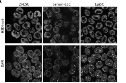
1 Global epigenetic organization in the different types of mouse pluripotent stem cells 89 1.1 EUCHROMATIN IN MOUSE PLURIPOTENCY ... 89
1.2 BIVALENCY IN MOUSE PLURIPOTENCY ... 91
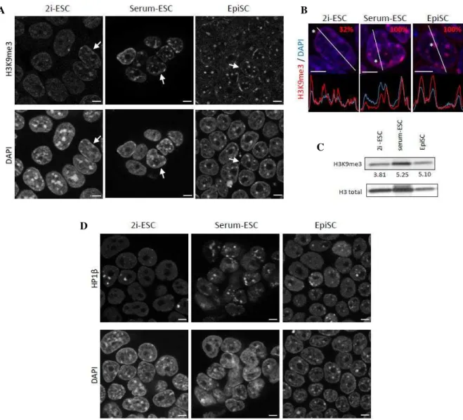
1.3 HETEROCHROMATIN IN MOUSE PLURIPOTENCY ... 93
1.3.1 Heterochromatin domains are characterized by different epigenetic histone marks depending on the pluripotent stem cell type ... 93
3 1.3.2 Pericentromeric heterochromatin is subjected to an epigenetic switch in
2i-ESCs compared to serum ones and EpiSCs. ... 98
1.3.3 DNA methylation state of repetitive sequences reflects the pluripotent stem cell type.. ... 100
1.3.4 2i condition leads to decondensation of the well-organized structure of the chromocenter ... 103
1.3.5 The transcriptional state of major and minor satellites depends on pluripotent cell type………...105
1.3.6 Absence of Suv39h1/2 induces different phenotypes depending on the pluripotent stem cell type ... 107
1.3.7 Absence of DNA methylation has limited effects on satellite transcription 112 1.3.8 Reduced levels of H3K27me3 do not up-regulate satellite transcription ... 114
2 Conversion from naive to primed pluripotency in mouse ... 118
DISCUSSION ... 129
1 Uncoupling epigenetic state of naïve ESCs with transcription regulation of major and minor satellites ... 129
2 The problem of major satellite repeats quantification ... 131
3 Cross-talk between H3K27me3 and 5-meC in mouse pluripotency ... 132
4 Hypothetical SUV39H-independent H3K9me3 deposition at PCH in 2i-ESCs ... 133
5 EpiSC: a pluripotent cell with a somatic epigenetic state ... 134
6 Major and minor satellites sequences respond to different epigenetic pathways ... 135
7 Genome stability in ESCs ... 137
8 Does the epigenetic status of in vitro pluripotency reflect in vivo pluripotency of mouse embryo? ... 137
9 In vitro conversion: why such an inefficient process compared to in vivo? ... 139
10 Bivalent Domains ... 141
11 Nomenclature ambiguity: ground, naive, primed in mouse and human pluripotency 142 12 Concluding remarks of this thesis ... 144
RESUME SUBSTANTIEL DE LA THESE EN FRANÇAIS ... 149
1 INTRODUCTION ... 149
2 OBJECTIFS ... 151
3 RESULTATS ... 152
3.1 L’hétérochromatine est caractérisée par différentes modifications d’histones selon le type de cellules pluripotentes. ... 152
4 3.3 Le PCH est décondensé dans les ESCs en 2i mais transcriptionnellement réprimé……. ... 155 3.4 L’absence des SUVγ9H1/β induit des phénotypes différents en fonction du type de cellule pluripotente. ... 156 3.5 L'absence de méthylation de l'ADN augmente le dépôt d’HγKβ7meγ mais a un effet limité sur la transcription des séquences satellites. ... 159 3.6 La réduction des niveaux d’HγKβ7meγ n’induit pas une sur-expression des séquences satellites ... 160 4 DISCUSSION ... 162 4.1 Dialogue entre H3K9me3, H3K27me3 et 5-meC dans la pluripotence chez la souris……… ... 162 4.2 EpiSC: une cellule pluripotente avec un état épigénétique somatique ... 163 4.3 Découplage de l’état épigénétique des ESCs avec régulation de la transcription des satellites ... 163 REFERENCES ... 167 APPENDIXES ... 189
5
Abbreviations
2i Two inhibitors 5h-meC 5-hydroxymethylcytosine 5-meC 5-methylcytosine AP Alkaline phosphatase BCA Bicinchoninic acid BMP Bone morphogenic factor BSA Bovine serum albumin CAF-1 Chromatin assembly factor 1 CDM Chemically define medium CENP-A Centromere protein A cEpiSC Converted epiblast stem cell CH Centromeric heterochromatin ChIP Chromatin immunoprecipitation CpG CG dinucleotideDAPI 4',6-diamidino-2-phénylindole DMEM Dulbecco's Modified Eagle Medium dn double null
DNMT DNA methyl transferase EB Embryoid body
EGA Embryonic genome acrivation Epi Epiblast
EpiLC Epiblast stem cell-like EpiSC Epiblast stem cell
ERK Extracellular signal-regulated kinase ESC Embryonic stem cell
ExE Extra embryonic endoderm EZH2 Enhancer of Zeste
FBS Fetal bovine serum FGF Fibroblast growth factor
FISH Fluorescence in situ hybridization GSK-3 Glycogen synthase kinase 3
H2AK119ub Ubiquitylation on the lysine 119 of the histone H2A H3K27me3 Trimethylation on the lysine 27 of the histone H3
6 H3K4me3 Trimethylation on the lysine 4 of the histone H3
H3K9ac Acetylation of the lysine 9 of the histone H3 H3K9me3 Trimethylation on the lysine 9 of the histone H3 HMT Histone methyl transferase
HP1 Heterochromatin protein 1 ICM Inner cell mass
iPSC induced pluripotent stem cell KDM Lysine demethylase
LIF Leukemia inhibitory factor MAPK Mitogen-activated protein kinase MEF Mouse mebryonic fibroblast miR micro RNA
ncRNA Non-coding RNA NOD Non-obese diabetic NPB Nucleolar precursor body NPC Neural progenitor cell
NuRD Nucleosome Remodeling Deacetylase PCH Pericentromeric heterochromatin PFA Paraformaldehyde
PLA Proximity ligation assay PRC Polycomb repressive complex PrE Primitive endoderm
PTGS Post transcriptional gene silencing
qRT-PCR Quantitative retro-transcribed polimerase chain reaction RIPA Radioimmunoprecipitation assay buffer
RNAi RNA interference
SCID Severe combined immunodeficiency
STAT Signal Transducer and Activator of Transcription SUV39H Suppressor of variegation
TBS Tris-borate saline TE Trophoectoderm
TET Ten eleven translocation
TGF Transforming growth factor beta TKO Triple knock-out
7
9
INTRODUCTION
1
EPIGENETICS AND GENOME ORGANIZATION
1.1
Organization of the eukaryotic genome
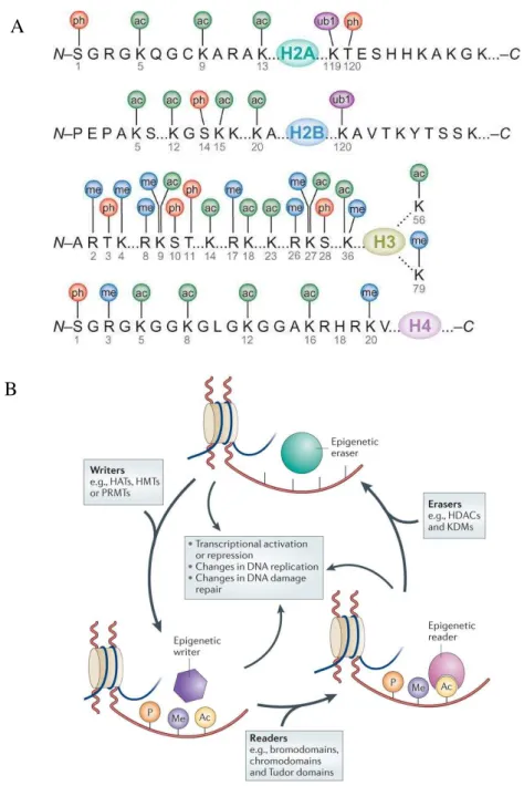
Eukaryotic cells have to pack their immense genome made by a long linear DNA molecule into the confined volume of their nucleus. This problem is solved by wrapping the DNA around proteins called histones in order to form the nucleosome which is the structural unit of the chromatin (DNA-protein complex). One nucleosome is composed by two turns of double-helix DNA (146bp) around the core histone octamer made by two copies of heterodimers of histones H2A - H2B, and a tetramer of H3 and H4 (Figure 1) (Luger et al., 1997; Rhodes, 1997). Histones have a globular portion and a tail that is extruding from the nuclesomal core.
Figure 1: Nucleosome core and tails (From Mattiroli et al., 2015)
Canonical nucleosome structure made by histone H3 (blue), H4 (green), H2A (yellow), H2B (red), and DNA (white). Tails of each histone are extruding from the nucleosome core.
10 Two successive nucleosomes are separated by 20-80bp of DNA linker (Routh et al., 2008) giving rise to the 10nm-fiber (bead on a string structure). The histone H1 is the only one that does not take part in the nuclesome core and its role is bringing together linkers DNA between nucleosomes creating the 30nm-fiber, which is the one found in interphase. The chromatin could be then further compacted till the maximum condensation of mitotic chromosomes (Figure 2) (Happel and Doenecke, 2009; Izzo et al., 2008).
Figure 2: Organization of the eukariotic genome (From Felsenfeld and Groudine, 2003)
In eukaryotes the double helix of DNA is wrapped on nucleosomes which are separated one another by a stretch of DNA linker. Successively the string of nucleosomes is folded into a fiber about 30 nm in diameter. These fibers are then further folded into higher-order structures till the hyper-condensation of a mitotic chromosome.
11
1.2
Epigenetics
The term Epigenetics has been coined by Conrad Waddington in 194β to describe “processes by which genotype gives rise to phenotype”. In more recent times epigenetics has been defined as mitotically and/or meiotically heritable changes in gene functions that do not entail changes in DNA sequence and in the absence of the factor responsible for these changes (Wu Ct and Morris, 2001). However the “gene” concept in epigenetics must be extended also to non-coding sequences.
There are many mechanisms by which epigenetics can act, the first described is DNA methylation where a methyl-group is added and covalently bound to nucleotides principally on cytosines. Others are the covalent modification of histones, in particular on C- or N-tails, or the substitution of histone in toto (histone variants), both influencing the DNA indirectly as its helix is wrapped on the protein of the histone core. Finally a non-negligible portion of epigenetic inheritance and maintenance is mediated by RNAs and particularly non-coding RNAs (ncRNA) (Allis and Jenuwein, 2016).
1.2.1 DNA methylation
DNA methylation is an epigenetic mark that consists in the covalent binding of a methyl group on the carbon 5 of the pyrimidine ring of cytosine producing a 5-methylcytosine (5-meC) using as a donor S-adenosyl methionine (SAM) (Figure 3). This mark seems to be inherited through cell divisions via a mechanism that recognizes an hemimethylated palindrome CpG and induces the DNA methylation in the newly synthesized strand to obtain a fully methylated CpG (Bird, 2002). These CpGs are not randomly distributed into the genome but enriched in specific short regions (around 1kb) called CpG Islands found near the majority of vertebrate genes (Deaton and Bird, 2011). CpG sites are not the only one susceptible to DNA methylation, particularly in embryonic stem cells (ESCs) compared to somatic tissue there is a significant presence of CpA methylated sites and to a lesser extent CpT (Ramsahoye et al., 2000).
12
Figure 3: The mechanism of DNA methylation (From Strathdee and Brown, 2002)
5-Methylcytosine is produced by the action of the DNA methyltransferases (DNMT1, 3A or 3B), which catalyze the transfer of a methyl group (CH3) from S-adenosylmethionine (SAM) to the carbon-5 position of cytosine.
DNA methylation has been historically associated with transcriptional repression but the position of the DNA methylation in the sequence of the transcriptional unit strongly influences the final effect (Jones, 2012). DNA methylation in close proximity of the transcription start site (TSS) or promoter region is known to repress the initiation of transcription (Kass et al., 1997), but when present in the gene body the effect is opposite and it could even stimulate the elongation of transcription (Jones, 1999). Moreover CpG methylation in the gene body could also influence the splicing (Jones, 2012). DNA methylation plays also key role in the stability of the genome allowing correct chromosomal segregation during mitosis when present on centromeric repeat sequences or suppressing the expression of transposable elements (Jones, 2012).
The majority of CpGs islands are unmethylated, however CpGs islands within promoters are methylated for stable repression of the corresponding gene. This is the case of genes subjected to imprinting, inactivated X chromosome- and germ-cell lineage-genes that undergo DNA methylation to suppress their inappropriate expression. DNA methylation seems to be the final lock of already silenced genes (Jones, 2012).
13 Methylated DNA is recognized by specific proteins with Methyl Binding Domains, such as MBD1 and MBD3 (Wade and Wolffe, 2001) and methyl-CpG binding protein 2 (MeCP2) (Fuks et al., 2003) that promote a compacted heterochromatic state and prevent from transcription factor bindings (Trojer and Reinberg, 2007).
1.2.1.1 DNA methylation machinery
Mammals have three different DNA methyltransferases (DNMTs) which catalyze the methylation (Okano et al., 1999) and one enzymatic inactive cofactor Dnmt3-like (Bourc’his et al., 2001) (Figure 4). DNMT3A and DNMT3B are the de novo DNMTs which are able to methylate the naked DNA in concert with the cofactor DNMT3L, thus establishing the DNA methylation pattern during development (Denis et al., 2011). Dnmt3a encodes for two isoforms DNMT3A1 and DNMT3A2 with the second being shorter at the N-terminal part than the first one and specifically interacting with DNMT3L at heterochromatin foci in ESCs (Nimura et al., 2006). The DNA methylation pattern is maintained through cell divisions by DNMT1 and it copies the methylation from hemimethylated DNA during replication (Cheng and Blumenthal, 2008).
Figure 4: DNMTs, their functions and structures (From Cheng and Blumenthal, 2008 and Nimura et al., 2006)
A: de novo methylation is firstly established on unmodified CpG by DNMT3A and DNMT3B in concert with their cofactor DNMT3L. The maintenance role of DNMT1 is then to fully methlylate the hemi-methylated sites and to maintain this state thought DNA replication and DNA repair. B: schematic representation of the DNMT family in mouse. NLS (nuclear localization signal), PWWP (PWWP DNA- and protein-binding domain) PHD (the cystein-rich PHD zinc-finger domain), I, IV, VI, IX and X correspond to conserved methyltransferase catalytic motifs. Of note DNMT3L lacks motif X.
To accomplish this process DNMT1 localizes in the replication fork and interact with Proliferative Cell Nuclear Antigen (PCNA) and UHRF1 which binds hemimethylated DNA (Sharif et al., 2007). However the maintenance role of DNMT1 alone is not sufficient as
14
Dnmt3a-/- and Dnmt3b-/- embryonic stem cells (ESCs) gradually lose their DNA methylation pattern after several passages (Chen et al., 2003; Jackson et al., 2004). In the absence of
Dnmt1, ESCs show reduced global level of 5-meC, but some sequences remained fully methylated such as major satellites (Arand et al., 2012). These observations showed that the separation in de novo and maintenance function of DNMTs is not as strict as it seems, as they can partially compensate each other. All the enzymatic DNMTs are strictly necessary for mammalian development (Li et al., 1992; Okano et al., 1999), as only Dnmt3l knock-out mice are viable even though males are sterile (Bourc’his et al., β001). Indeed Dnmt1-/- embryos die at E8.5-9.0 and showed only one third of 5-meC compared to wild-type condition (Li et al., 1992). Dnmt3b mutant embryo develop even further but after E9.5 they show multiple defects and do not go to term, while Dnmt3a-/- develop to term but die one month after (Okano et al., 1999) (Figure 5).
DNA methylation is not an irreversible epigenetic mark and can be reverted through two principal ways: passive by replication and active via the action of specific enzymes. Demethylation could simply occur by the lack of maintenance after DNA replication inducing a dilution effect and a progressive loss of methylation after numerous rounds of cell division (Hill et al., 2014). However an active enzymatic mechanism of demethylation has been discovered in recent years thanks to the action of the Ten Eleven Translocation enzymes (TETs) which catalyze the oxidation of 5-methylcytosine (5-meC) into 5-hydroxymethylcytosine (5h-meC) (Tahiliani et al., 2009). 5h-meC could be then transformed in 5-formylcytosine fC) also by TETs enzymes and subsequently in 5-carboxylcytosine (5-caC). 5-caC finally can enter in the Base-Excision Repair process (BER), removed by Thymine DNA Glycosidase and replaced by new unmodified cytosine (He et al., 2011; Ito et al., 2011). Function of 5h-meC is still controverted as it could be considered simply as a short-lived entity or as an epigenetic modification on its own. Its genome profiling shows a distinct distribution compared to 5-meC and it is associated to gene transcription (active promoters) as well as gene silencing (Polycomb-mediated) (Branco et al., 2012).
15
Figure 5: DNMTs and histone modifiers knock-out effects in mouse development and ESCs (From Meissner, 2010)
A: Stage of embryonic lethality of knock-out in vivo embryos for different epigenetics modifiers. B: in vitro knock-out ESCs for different epigenetics modifiers.
# (lethal), ## (normal ESCs maintenance, but differentiation defects), *(Dnmt3a knockout mice die around 3 weeks postnatal and are smaller/runted), ** (No observed phenotype), *** (Mice are viable, but have hematopoietic and neural abnormalities), **** (Homozygous mice are sterile, offspring of homozygous female mice and heterozygous crosses show imprinting defects and die), ***** (Wild-type ES cells cannot differentiate into trophectodermal cells), d.p.c. (days post coitum), PN (pronuclei), EN (endoderm), ME (mesoderm), EC (ectoderm), TE (trophectoderm), ICM = inner cell mass.
16
1.2.2 Histone modifications
Another mechanism by which the epigenetics can act is through post-translational covalent modification of histone tails that are extruding from the core octamer. Many different post-translational modifications of histones exist, mainly methylation, acetylation, phosphorylation and ubiquitylation (Kouzarides, 2007) (Figure 6A). The chemical group attached, the number of these modifications, the position in the histone tail and the type of residues accepting the modification could lead to different effects (Jenuwein and Allis, 2001; Strahl and Allis, 2000). Histone modifications are reversible and dynamic marks under the control of chromatin "writers", that establish the modification catalyzing the deposition of the chemical group, and "erasers", which conversely remove the mark. The function and the "message" of an histone modification are then transmitted by chromatin "readers" that have specific protein binding motifs in their aminoacid sequence that recognize and bind to specific histone modifications (Figure 6B) (Allis and Jenuwein, 2016; Yun et al., 2011). Altogether the sequential combinations of one or more histone modifications, that are read by proteins influencing down-stream events, form the so called histone code or histone language (Strahl and Allis, 2000). “Readers” can have different function such as architectural proteins, chromatin remodelers and modifiers (Figure 7) (Yun et al., 2011).
Histone methylation could occur on arginine (R) and on lysine (K), but while arginines can only accept up to two methyl groups, lysines can be even tri-methylated (Greer and Shi, 2012). The function of histone methylation is strictly dependent on the position in the tails and could even have opposite effect, for example tri-methylation on lysine 4 of the histone H3 (H3K4me3) and H3K36me3 are active chromatin marks that promote the transcription, while H3K27me3, H3K9me3 or H4K20me3 are repressive marks leading to transcriptional silencing. Histone methylation is deposed by writers called histone methyl transferases (HMTs) and more specifically lysine methyl transferases (KMTs), while the erasers are histone lysine demethylases (KDM). The readers for histone methylation are proteins containing the chromodomain (CD) binding site (Sims et al., 2003). CD-proteins can recognize different type of methylated histone and generally recruits, in collaboration with RNAs and DNA-binding proteins, other proteins forming a larger complex (Tajul-Arifin et al., 2003).
17
Figure 6: Histone code and chromatin modifiers: Writers, Erasers and Readers (From Advanced BioDesign and Falkenberg and Johnstone, 2014)
A: possible modification on the different residues of the tails of each histone. S (Serine), K (Lysine), R (Arginine), T (Threonine). Ph (Phosphorylation), ac (acetylation), ub1 (ubiquitylation), me (methylation). B: dynamics of epigenetic regulations: chromatin writers depose the histone mark which is then recognized by chromatin readers or eventually reversed by chromatin erasers.
A
18
Figure 7: Chromatin readers functions (From Yun et al., 2011)
Chromatin readers can have different roles, acting as: architectural protein (inducing compaction for example), remodeling (increasing accessibility of nucleosomal DNA, energy dependant (use of ATP), chromatin modifiers (adding a new mark) or adaptors (recruiting other protein machineries such as transcription factors).
Histone acetylation occurs on lysine residues and it induces the neutralization of the positive charge of histone tail. This leads to decompaction of the nucleosomal structure by a less intimate interaction with the negatively charged DNAs. For that reason hyperacetylation of histones is generally associated with a more decondensed and open conformation of the chromatin which is more prone to active transcription of genes due to increased accessibility of the transcriptional machinery to the DNA. Conversely hypoacetylated regions correlate with compact and silent domains (Sterner and Berger, 2000). The writers in this case are the histone acetyltransferases (HATs) while the erasers are the histone deacetylases (HDACs) (Berndsen and Denu, 2008). Readers must contain a bromodomain to recognize histone acetylation (Kouzarides, 2007). Bromodomain proteins play a key role anchoring complexes to the acetylated chromatin in order for example to remodel chromatin and recruit transcription factors (Josling et al., 2012).
Histone ubiquitylation is a less common epigenetic mark that consists in the binding of the ubiquitin (a polypeptide of 76-amino acids) to lysines via the successive action of E1, E2 and E3-ligases. The most studied is the monoubiquitylation of the lysine 119 of the histone H2A (H2AK119ub) which is linked to gene silencing (Wang et al., 2004; Zhang, 2003).
19
1.2.2.1 Active histone modifications
1.2.2.1.1
H3K4me3 and H3K9ac and their enzymes
Trimethylation of lysine 4 on histone H3 (H3K4me3) is the hallmark of actively transcribed genes as it is specifically enriched at the 5’ region of these genes (promoter), while H3K4me2 is more distributed along the gene body (Martin and Zhang, 2005). However H3K4me3 when present in association to H3K27me3 at the promoter level (bivalent domain) leads to the loading of the RNA Polymerase II on the gene promoter that stalled in a paused condition without elongating the transcription (Azuara et al., 2006; Bernstein et al., 2006). H3K4 is principally methylated by the KMT called mixed-lineage leukemia 1 (MLL1) which is a SET-domain protein belonging to the Tritorax complex (TrxG). Mll expression is necessary for the correct regulation of the Hox genes during embryo development, in fact Mll-/- embryo died early during development (Schuettengruber et al., 2011; Terranova et al., 2006).
Another active histone modification is the acetylation of the lysine 9 on the histone H3 (H3K9ac) present at open regions of the genome that are sensitive to DNase I and enriched in active transcription factors (Hezroni et al., 2011). H3K9ac is enriched at the TSS region of active genes, highly correlating with H3K14ac, H3K27ac, H3K4me2 and H3K4me3 (Hezroni et al., 2011; Karmodiya et al., 2012). In mouse the main HATs responsible for H3K9ac is GCN5/PCAF as its deletion dramatically reduces the global level of H3K9ac with little or no effect on the level of H3K14ac or other histone H3 and H4 acetylations (Jin et al., 2011). On the other hand HDAC1 is the main enzyme responsible for deacetylation of histones and
Hdac1-/- leads to embryonic lethality before E10.5 with embryos presenting proliferation defects and retard in development (Lagger et al., 2002). Moreover mutant ESCs showed reduced proliferation rates that cannot be compensated by the expression of HDAC2 and HDAC3 (Lagger et al., 2002).
1.2.2.2 Repressive histone modifications
1.2.2.2.1
H3K9me3, H4K40me3 and their enzymes
Trimethylation of the lysine 9 of the histone H3 (H3K9me3) is found from fission yeast to human on repeat-rich centromeric, pericentromeric and telomeric regions (Peters et al., 2001), but also in block of tissue-specific genes (Becker et al., 2016). It is thought that H3K9me3 protects clusters of repetitive genes and non-coding repeats from illicit recombination,
20 suppressing as well their transcription. This histone modification generally prevents the binding of transcription factors and this is probably why H3K9me3-chromatin blocks are the last to be reprogrammed during induced pluripotent stem cell (iPSC) generation being the less accessible (Becker et al., 2016). All H3K9-KMTs have a SET-domain and can be divided into two groups: the first comprises G9a (Ehmt2) and GLP (Ehmt1) that are the KMTs necessary to catalyze H3K9me1 and H3K9me2, while the trimetylation is achieved by the second group composed by SET domain bifurcated 1 (SETDB1), SUV39H1 and SUV39H2 (Mozzetta et al., 2015). This repressive mark is known to induce heterochromatin compaction and spreading via the recruitment of heterochromatin proteins 1 (HP1), thanks to the chromoshadow domain (Bannister et al., 2001; Lachner et al., 2001). In mammals there are three HP1 proteins: α, and (Jones et al., 2000). HP1α and seem to share the same function accumulating together over H3K9me3 (Figure 8), while HP1 has a diffuse genome localization (Dialynas et al., 2007). HP1 α and can self-oligomerize and recruit other repressive machineries like DNMTs to depose DNA methylation and SUV4-20H2 that specifically catalyze H4K20me3, another repressive histone modification, inducing further more compaction and repression (Figure 8; Schotta et al., 2004; Wongtawan et al., 2011).
In mouse SUV39H KMTs are encoded by two gene loci Suv39h1 and Suv39h2 which are both expressed in embryogenesis. Suv39h double null (Suv39hdn) condition impairs severely the viability of mice (which are growth retarded and infertile), inducing chromosomal instability and increased risk of tumorigenesis. However mice deficient for either Suv39h1 or Suv39h2
are fertile and showed normal viability showing redundant functions of the two enzymes (Peters et al., 2001). Interestingly Suv4-20h2 -/- mice have no apparent defects and develop normally however Suv4-20hdn display peri-natal lethality and are smaller (Schotta et al., 2008).
SETDB1 is described as the principal KMT responsible for H3K9me3 in the genome outside centromeric, pericentromeric and telomeric repeats (Schultz et al., 2002). However its loss in mice is associated with a substantial reduction of H3K9me3 also at pericentromeric heterochromatin (Mozzetta et al., 2015). It is not clear if this reduction is a result of SETDB1 mono-methyltransferase activity necessary for successive trimethylation by SUV39H1 and SUV39H2 or potentially direct trimethyltransferase activity of SETDB1 at pericentromeric repeats.
21
Figure 8: Conserved pathway SUV39H1/2-HP1-SUV4-20H2 (From Schotta et al., 2004)
Proposed model of sequential induction of H3K9me3 deposition by SUV39H1/2. H3K9me3 is then recognized by HP1α and inducing compaction and recruiting SUV4-20H2 that finally deposits H4K20me3 condensing furthermore the chromatin.
1.2.2.2.2
H3K27me3, H2AK119ub and their enzymes
Trimethylation of the lysine 27 on the histone H3 (H3K27me3) is another repressive histone mark present principally in tissue-specific gene regions and on the inactive X chromosome (Boyer et al., 2006; Margueron and Reinberg, 2011). H3K27me3 is not enriched in focal peak but on larger and broader regions in the genome, and these H3K27me3 blocks are negatively correlated with transcription such as the Hox cluster in differentiated cells (Pauler et al., 2008). Interestingly during iPSCs derivation H3K27me3-block are reprogrammed earlier than H3K9me3 ones and in general H3K27me3 and H3K9me3 blocks are largely exclusive (Becker et al., 2016).
Polycomb group proteins (PcGs) were originally identified as important regulators of developmentally related genes like the Hox cluster in Drosophila. In mammals there are two PcG called Polycomb repressive complex 1 and 2 (PRC1 and PRC2, Figure 9). Enhancer of
22 zeste homolog 2 (EZH2) is the catalytic subunit of PRC2, which contains a SET domain and it is thought to be the only KMT for di- and tri-methylation of H3K27 (Trojer and Reinberg, 2007). However in Ezh2-/- ESCs a residual H3K27me3 is found in the genome and it is likely due to EZH1 that seems to partially complement the absence of EZH2 (Margueron et al., 2008; Shen et al., 2008). Ezh2 transcription is up-regulated after fertilization and it is highly expressed all along the pre-implantation development. Ezh2-null condition is lethal at early stages of mouse development as these embryos die between pre- and post-implantation development (Figure 5) (Becker et al., 2016). Ezh2-null blastocysts have an impaired potential to outgrowth preventing the establishment of mutant ESCs (O’Carroll et al., β001). The other PcG complex is PRC1 that mediates via, its catalytic subunit RING1B (E3-ubiquitin ligase), the ubiquitylation of lysine 119 of histone H2A (H2AK119ub) which is also a repressive mark. Mouse RING1B is coded by the Rnf2 gene which when ablated causes an arrest during gastrulation with developmental defects occurring in both embryonic and extra-embryonic tissues (Voncken et al., 2003) (Figure 5).
PcG complexes are composed by different combination of subunits. The most common and studied PRC1 is called the canonical PRC1 (cPRC1) and it is recruited on PRC2 targets thanks to Cbx subunit that recognize H3K27me3 (Cao et al., 2002). However more recently it has been discovered that a variant or non-canonical PRC1 (vPRC1 or ncPRC1) complex could be firstly recruited at unmethylated CpG island by KDM2B and only in a second time PRC2 is enrolled via H2AK119ub recognition (Blackledge et al., 2014; He et al., 2013).
23
Figure 9: Mechanism of PcG recruitment to chromatin (From Aranda et al., 2015)
Upper panel: PRC2 complex deposes H3K27me3 that mediates the recruitment of PRC1 complex by interacting with Cbx (its chromatin “reader” subunit).
Lower panel: KDM2B binding to CpG could induce recruitment of non-canonical PRC1 complexes deposing H2AK119ub. This mark is then recognized by PRC2 complexes that will successively appose H3K27me3. Canonical PRC1 (cPRC1), non-canonical PRC1 (ncPRC1), CpG island (CGI).
1.2.3 Histones variants
The histone H3 variants, CENP-A (Centromeric protein A) or CenH3 is highly enriched at centromere, defining it and directing kinetochore assembly (Müller et al., 2014). Its functionality is very well conserved among Eukaryotes and it is the epigenetic determinant of centromeres. The regulation of CENP-A deposition by the histone chaperon HJURP (Holliday junction recognition protein) is crucial for fidelity of chromosome segregation and cell division (Rop et al., 2012).
H3.3 is another histone variant of H3 differing only in five residues and it is principally found at pericentromeric and telomeric region where it is specifically deposited by DAXX (Death domain-associated protein 6) (Mattiroli et al., 2015; Santenard et al., 2010). However H3.3 is present also at transitionally active regions of the genome where it is deposited by another histone chaperone (HIRA) (Mattiroli et al., 2015).
24
1.2.4 Euchromatin and Heterochromatin
Historically, inside an eukaryotic nucleus the chromatin can be divided into two compartments based on the degree of compaction: euchromatin and heterochromatin (Tamaru, 2010). Euchromatin is mainly made by active transcriptional gene-rich regions of the genome, marked by H3K4me3, H3K36me3 and hyperacetylated histones, replicating in early S phase, highly dispersed and diffused in interphase (not stained in electron microscopy) and compacted only during mitosis. On the other hand heterochromatin is composed by gene-poor regions (especially for constitutive heterochromatin) which is supposed to be not transcribed, hypoacetylated in histones, highly condensed and compacted (even in interphase), replicated in late S phase and heavily stained in electron microscopy (Heinz, 1928) (Allis and Jenuwein, 2016) (Figure 10).
Figure 10: Eukaryotic cell under transmission electron microscopy (TEM).
From a TEM image of an eukaryotic nucleus it is possible to distinguish two different types of heterochromatin: heterochromatin blocks (black) around nucleoli (a), at the nuclear periphery (b) and dispersed into the nucleoplasm (c) and more diffuse heterochromatin (light gray).
The heterochromatin could be further divided in facultative and constitutive parts. Facultative heterochromatin can go from local gene to genomic regions (Hox gene cluster) up to entire chromosomes (X-chromosome in female mammalian cells). It is composed by regions of the genome that have the opportunity to adopt an open and dynamic or closed and compact conformation depending on space and time (Trojer and Reinberg, 2007). Facultative heterochromatin is silenced and generally marked by H3K27me3, but also by H2AK119ub, (Trojer and Reinberg, 2007). Conversely constitutive heterochromatin consists in large blocks
a
b c
25 made by repetitive sequences (especially present near centromeres and telomeres) which maintain their characteristics on both homologous chromosomes (Dimitri et al., 2005). Compared to facultative, constitutive heterochromatin is generally marked by H3K9me3 and H4K20me3 and it is recognized by HP1 proteins (Trojer and Reinberg, 2007).
1.2.4.1 Constitutive heterochromatin: the repetitive sequences of
mammalian genome
Only 4% of the mouse genome encodes for proteins while the majority of the DNA is made by repetitive sequences (44%) and non-coding sequences (52%) (Martens et al., 2005). The human genome content is similar to the mouse one with only 2% of protein coding sequences and the remaining (98%) is made up of transposable elements and tandem repeats (López-Flores and Garrido-Ramos, 2012). Now it is known that this “junk DNA” plays a role in the formation of specialized structure but, due to its repetitive characteristic, this DNA is also an issue for genome stability as more easily subjected to recombination, deletion or translocation than single-copy sequences (Jaco et al., 2008).
Mouse chromosomes are all acrocentric while human chromosomes can be divided in three groups: metacentric, sub-metacentric and acrocentric. However chromosomes of both species present around their centromeres the constitutive heterochromatin which is cytologically visible (Figure 11A) (Padilla-Nash et al., 2007). Constitutive heterochromatin consists principally in centric, pericentric and telomeric region. At the end of each linear chromosome there is the telomeric region composed from hundreds to thousand repeats of TTAGGG sequence, which is conserved between all the mammals, and coated with protecting proteins in order to prevent DNA damage and inappropriate recombinant events (Calado and Dumitriu, 2013). In addition to these large tandem arrays of repeats, the mouse genome contains single repetitive elements that are all along the chromosomes, such as DNA transposons that represent 1% of this interspersed repetitive element, while RNA transposons or retrotransposons represent about 25%. Retrotransposons include: LTR (Long terminal repeats) transposons, principally intracesternal A particle (IAP), non-LTR transposons or LINEs (Long Interspersed Nuclear Element) which represent the largest fraction with 19% of the genome and the Short Interspersed Nuclear Element (SINE) (Martens et al., 2005) (Figure 11B).
26
Figure 11: The different repetitive sequences in the mouse genome (From Martens et al., 2005)
(A) Schematic representation of a mitotic mouse chromosome illustrating the distribution of major and minor satellite repeats, respectively pericentromeric and centromeric heterochromatin and of the various interspersed repetitive elements. (B) Summary of repetitive elements in mouse with repeat organization, length, copy number and overall abundance in the mouse genome. Specific primers (black arrows) can be designed to generate PCR fragments from within one or more successive repeat of the repetitive elements.
In mouse the core of the centromere is made by tandem arrays of minor satellite repeats (123bp) in approximately 2000 copies (Martens et al., 2005). Minor satellites are heavily methylated at the DNA level, transcriptionally silenced and characterized by the H3 histone variant CENP-A (Scott, 2013). Juxtaposed to the centromere, the pericentromeric region is composed by major satellite repeats (234bp) which are A/T-rich sequences that are divided in four sub-repeats and present in more than 10000 copies. Major satellites are characterized by H3K9me3, HP1 and H4K20me3. Like minor satellites, they are characterized by an hypermethylation at the DNA level and a repressed transcriptional state (Lehnertz et al., 2003). Together centromeric and pericentromeric regions represent more than 3.5% of the mouse genome, with major satellites alone representing 3% (Martens et al., 2005). A
27 characteristic of mouse nuclei is the clusterization of pericentromeric and centromeric heterochromatin in chromocenters. These structures are formed by the coalescence of major satellites coming from different chromosomes with the corresponding minor satellites that are located in surrounding separate domains at the periphery (Guenatri et al., 2004) (Figure 12). These prominent heterochromatin domains being rich in A/T sequences (major satellites) are well discernible in the nucleus as 4',6-diamidino-2-phenylindole (DAPI)-dense foci (Dambacher et al., 2013).
Figure 12: Mouse chromosomes organize together their pericentromeric region to form Chromocenters (From Guenatri et al., 2004)
(A) Schematic organization and DNA-FISH images of major and minor satellites along the mouse chromosome. DAPI DNA counterstaining (blue), majors satellites (green), minor satellites (red).
(B) DNA-FISH images for major and minor satellites revealing the chromocenter organization inside a mouse interphase nucleus. DAPI DNA counterstaining (blue), majors satellites (green), minor satellites (red).
(C) Schematic organization of mouse chromosome during cell-cycle. (1) In interphase major satellites from different chromosomes associate in clusters (chromocenters). (2) In prophase major satellites from different chromosomes dissociate. (3) In metaphase minor satellites from sister chromatids dissociate, whereas the major satellite sister chromatids still cohere. (4) In anaphase finally major satellites from sister chromatids separate.
28 In human these regions are also composed by satellite repeats, however the DNA sequence of these repeats is different. Human centromeric region is made by alpha satellite repeats of 171bp that are present in all chromosomes, while the closer pericentromeric region is composed by Satellite 2 and 3 principally with different size and composition between the chromosomes (Saksouk et al., 2015).
1.2.5 Non-coding RNAs (ncRNAs)
The discovery that mammalian genome is transcribed quite entirely even if only a small fraction codes for protein, introduces the concept of the existence of non-coding RNAs (“Dark matter” RNA) (Mattick, 2007). ncRNAs are divided in two groups based on their size: small and long non-coding RNAs.
The small non-coding RNAs are RNA species of less than 200nt that are in many cases associated with 5’ or γ’ regions but also in introns of protein-coding genes (Mattick and Makunin, 2006). The most studied are Dicer-dependent microRNA (miRNA) and small interfering RNA (siRNA), and the Dicer-independent PIWI-interacting RNA (Carthew and Sontheimer, 2009). These ncRNA are largely involved in post-transcriptional gene silencing (PTGS) (Agrawal et al., 2003).
The long non-coding RNAs (lncRNA) are defined as transcripts of more than 200nt that lack an open reading frame (Cao, 2014). LncRNAs are mainly transcribed by RNA polymerase II, they can undergo splicing and can also contain a poly-A tail. They can be developmentally-regulated and/or tissue-specific, being implicated in alternative splicing, modulation of protein activity, alternative protein localization, epigenetic regulation, transcriptional silencing. LncRNAs can act as signals, guides, decoy and scaffold (Sana et al., 2012).
While for many years the repetitive constitutive heterochromatin has been considered as a transcriptionally inactive domain due to its compacted organization, high DNA methylation content and presence of repressive histone mark like H3K9me3, it is now clear that in many cases these sequences are transcribed giving rise to satellite non-coding RNAs (Biscotti et al., 2015).
29
1.2.5.1 Satellite non-coding RNAs
Transcripts homologous to centromeric and pericentromeric repetitive sequences have been identified in several organism from yeast to human (Saksouk et al., 2015).
In yeast the RNA polymerase II transcribes satellites repeats and these ncRNA are involved in the heterochromatin formation, maintenance and silencing via the RNA interference machinery (RNAi). Paradoxically the heterochromatin needs to be transcriptionally active to maintain its inactive state by the recruitment of H3K9-KMTs (Biscotti et al., 2015).
In mouse the implication of the RNAi pathway in the heterochromatin is controverted (Kanellopoulou et al., 2005; Murchison et al., 2005) and still under debate with no real proof (Plohl et al., 2014). However transcription of ncRNA from both major and minor satellites is observed in physiological conditions as well as in pathological conditions (Figure 13) (Saksouk et al., 2015). Interestingly it has been shown that an RNA component is involved in the high structured three-dimensional chromatin at pericentromeric regions, as RNase treatment disrupts the H3K9me3-HP1 foci (Maison et al., 2002). Whether this RNA component is also made by pericentromeric satellite transcripts is still unknown.
Non-coding pericentromeric RNA are produced by RNA polymerase II in both orientation in mouse as well as in human: sense or forward (T-rich in mice) and antisense or reverse (A-rich) (Figure 13) (Saksouk et al., 2015) and can be also polyadenylated (Lehnertz et al., 2003).
30
Figure 13: Pericentromeric satellite transcription in different context (From Saksouk et al., 2015)
Physiological expression of pericentromeric satellite repeats has been reported during cell cycle, senescence, development and differentiation. Pathological expression has been observed upon cellular stress and in cancer. The size, orientation and putative functions of the transcripts are indicated when known.
In the major satellite repeat sequence many binding sites for transcription factors were found (Figure 14). For example these repeats contain sites for PAX3 and PAX9 Paired box-transcription factors, which were found to be necessary to repress non-coding RNA transcription from these regions and also to help the recruitment of SUV39H enzymes needed for the deposition of H3K9me3 (Bulut-Karslioglu et al., 2012).
Figure 14: Transcriptional factor binding sites on major satellite repeat sequence (From Bulut-Karslioglu et al., 2012)
Consensus sequence of a full-length major satellite repeat. Transcription factor binding sites are highlighted above or below the DNA sequence according if their binding motif is respectively sense or antisense.
31 Different populations of satellite transcripts seem to be generated according to the cell cycle stage. First major satellite transcription is Cdk-dependent because cells do not transcribe if they are not proliferating or maybe in this condition there are extremely short-lived (Lu and Gilbert, 2007). During mitosis small RNA species (less than 200nt) are produced and their half-life is very short (less than one hour). Conversely a more abundant, large and heterogeneous population of transcripts (from 1kb to more than 8kb) is produced between late G1 and early S, and strongly down-regulated after mid-S phase when pericentric heterochromatin is replicated (Lu and Gilbert, 2007). Major satellites are found to be transcribed in adult mice only in highly proliferative tissues as liver and testes. In particular in the liver transcription occurs only in sense orientation while in testes it is antisense in immature germ cells and sense in the mature ones (Saksouk et al., 2015). During replicative senescence and aging, pericentromeric transcripts were detected especially from the sense orientation and concomitant to reduction of methylation levels and decondensation of constitutive heterochromatin (Figure 13) (Saksouk et al., 2015). Similar observations were made in several cancer cells with an associated genetic instability and chromosomal disorder (Frescas et al., 2008).
Minor satellites have also been shown to produce heterogeneous populations of transcripts and to be cell-cycle regulated. In in vitro cultured mouse cells, centromeric transcripts are present in two long forms of 2kb and 4 kb but also in a smaller form around 120nt. These small minor satellite transcripts accumulate with culture-time (Bouzinba-Segard et al., 2006). No centromeric transcripts were found at the range of size for siRNAs (22-30nt) suggesting no RNAi involvement in mouse. The 120nt population of minor satellites increased during stress condition, using a DNA demethylation agent or inducing apoptosis, leading to an impaired centromeric function during mitosis and promoting defects in chromosomal segregation (Bouzinba-Segard et al., 2006). In addition minor satellites are lowly expressed in G1 phase and increased with cell-cycle progression picking at G2/M phases just prior the kinetochore assembly (Ferri et al., 2009). Furthermore centromeric transcripts were found to be an RNA component of the CENP-A associated complex in concert with Chromosomal Passenger Complex (CPC), Aurora B kinase and Survivin providing implications of minor satellite transcripts in chromosome segregation (Ferri et al., 2009).
32
2
PLURIPOTENCY
2.1
Totipotency and Pluripotency
Pluripotency is defined as the capacity of cells to self-renew (auto-maintain themselves) in vitro and to differentiate into the three embryonic lineages (mesoderm, endoderm and ectoderm) that will give rise to all the different tissues of an adult organism, but not to extra-embryonic tissues. Totipotency is the capability of a cell to give rise to a fertile adult individual, so generating also the extra-embryonic tissues (like placenta or yolk-sack). In mouse, totipotency is restricted to the zygote (1-cell stage) and to blastomeres of a 2-cell stage embryo (Condic, 2014). In the following pre-implantation embryo stages, cells are pluripotent up to the blastocyst stage where we assist to the first real differentiation process: the inner cell mass (ICM) and the trophectoderm (TE). At this stage only ICM cells are still pluripotent. Pluripotent cells are also present after the implantation in the epiblast up to E7.5 (Nichols and Smith, 2009; Osorno et al., 2012).
2.2
Early mouse embryo development
The early mouse embryo development goes from totipotency to pluripotency. It begins with fertilization, followed by dividing pre-implantation embryo stages that lead to the blastocyst formation (Figure 15A) which then will implant into the uterus starting the post-implantation development and gastrulation. The pre-implantation development starts in the oviduct where the oocyte is fertilized by the sperm giving rise to the zygote (fertilized egg) (Wang and Dey, 2006).
At the blastocyst stage E3.5 two types of cells can be observed: the pluripotent inner cell mass (ICM) and the trophectoderm (TE), distinguished by Oct4-Sox2-Nanog or Cdx2-Eomes-Gata3
expression, respectively (Chazaud et al., 2006). At this stage Oct4 repressed TE lineage and
Cdx2 conversely ensures the repression of Oct4 and Nanog (Figure 15B). In E3.5 early blastocyst individual ICM cells show exclusive expression of epiblast genes (such as Nanog) or primitive endoderm genes (Gata6 and Gata4) in a “salt and pepper” manner (Chazaud et
33
Figure 15: Pre-implantation development and first gene specifications (From Wang and Dey, 2006)
A: Schematic representation of mouse pre-implantation embryo development: once the egg is fertilized by the sperm successive cell divisions will give rise at E3.5 to the blastocyst.
B: Gene expression pattern governing blastocyst specification. Oct4 (dark pink) is expressed throughout the embryo before the late morula stage. Nanog (light pink) is specifically induced in the inside cells of late morula. Cdx2 (blue) is expressed in the outer layer of cells in late morula and is required for the repression of Oct4 and Nanog in the trophectoderm of the blastocyst. Gata6 (green) is expressed in the primitive endoderm of the late blastocyst, where Oct4 and Nanog are repressed. Oct4 represses Cdx2 expression, which in turn represses Oct4 expression allowing segregation of the ICM and trophoectoerm of the blastocyst. Nanog and Gata6 antagonized each other segregating epiblast and primitive endoderm within the ICM.
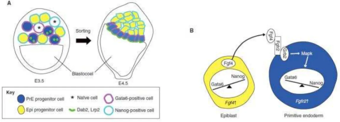
Figure 16: Epiblast vs. Primitive endoderm specification in early- to late-blastocyst stage (From Takaoka and Hamada, 2012)
(A) Primitive endoderm (blue) and epiblast (yellow) progenitors are randomly positioned in the inner cell mass (ICM) at E3.5. At this stage, some of the cells in the ICM are still naïve (asterisk) for Nanog or Gata6 expression. By E4.5, the primitive endoderm cells migrate at the surface of the blastocoel cavity, whereas the Epiblast cells are confined to the inner part of ICM. Dab2 and Lrp2 are localized to the apical surface of PrE cells. (B) FGF signaling guides primitive endoderm formation. Fgf4 secreted by epiblast progenitors interacts with Fgfr2 and thereby activates Grb2 and Mapk in primitive endoderm progenitors. Activated Mapk induces the expression of PrE-specific genes, such as Gata6. Fgf4 simultaneously represses Nanog expression, further promoting PrE fate while inhibiting Epi fate. Epiblast (Epi), primitive ectoderm (PrE), adaptor protein disabled (Dab2), lipoprotein receptor-related protein 2 (Lrp2), fibroblast growth factor 4 (Fgf4), Fgf receptor 2 (Fgfr2), growth factor receptor bound protein 2 (Grb2), microtubule-associated protein kinase (Mapk).
A
34 FGF signaling is involved into primitive endoderm specification (Figure 16) (Artus and Chazaud, 2014). By E4.5, just prior to implantation, this mosaic pattern is resolved in a late blastocyst where ICM cells are sorted and relocated in epiblast cells (Epi) (Figure 16) which are pluripotent and will give rise to the fetus, and primitive endoderm (PrE) from which will predominantly originates the yolk sac. The PrE cells form a layer at the surface of the ICM facing the blastocoel cavity and are positive for Gata6. Conversely Epi cells are positive for
Nanog and are surrounded by PrE cells and TE cells (Rossant and Tam, 2009).
At this stage (E4.5) the blastocyst invades the uterine tissues and implants. Mural TE surrounding the blastocyst cavity will first makes contacts with maternal tissues and differentiates into primary TE giant cells which mediate the mother-embryo exchanges. Conversely polar TE surrounding the outer face of the ICM proliferates and differentiates in extra-embryonic ectoderm (ExE) and ectoplacental cone (EPC) that will build the proximal half of the egg cylinder and later on the placenta (Figure 17). After implantation at E5.0 there is a burst of cell proliferation and growth resulting in the expansion of embryonic as well as extra-embryonic lineages into the blastocyst cavity. From PrE derives the visceral endoderm cells that cover both Epi and ExE. The elongating egg cylinder grows, reorganizes and forms the primitive streak in order to initiate the gastrulation under the control of TGF /Activin/Nodal pathway. Nodal play a key role in the maintenance of pluripotency of the epiblast and the posterior cell fate. Indeed in Nodal -/- condition the epiblast is precociously differentiated into the neuronal fate (Camus et al., 2006). During peri-implantation stages the Epi reorganized itself from a compact ball non-polarized to a rosette-like organization, at the time of implantation, and finally to a cup-shaped polarized epithelium surrounding the pro-amniotic cavity in 24 hours (Figure 17) (Bedzhov et al., 2014). The anterior-posterior axis of the mouse embryo is established when distal visceral endoderm (DVE) will form the anterior visceral endoderm (AVE) on the future anterior side of the E6.5 embryo. Once established the AVE will secrete signal like Nodal antagonists (Lefty1) to the Epi for anterior specification (Perea-Gomez et al., 2002).
35
Figure 17: Early mouse development from 1-cell stage to gastrulation (From Takaoka and Hamada, 2012)
Kinetics of morphological changes and cell-specification from zygote to late egg cylinder. The cell types in the embryos are coded with different colors: epiblast (Epi), primitive ectoderm (PrE), visceral endoderm (VE), extra-embryonic ectoderm (Exe), distal visceral endoderm (DVE), anterior visceral endoderm (AVE), primitive streak (PS).
2.3
In vitro pluripotency in mouse
The attractive potential of embryo-pluripotent stem cells has fascinated scientists who attempted derivation of these cells as a stable line in vitro. In 1981 two groups established for the first time an in vitro culture of pluripotent stem cells starting from 129 strain mouse blastocyst-embryos E3.5 (Evans and Kaufman, 1981; Martin, 1981). These cells have the capacity to differentiate into all the three primary germ layers and were called embryonic stem cells (ESCs) in order to distinguish them from embryonal carcinoma cells (ECCs), that were isolated from teratocarcinoma induced by transplantation of mouse embryos in an extra-uterine site of a host. Many years later in 2007 two other groups established another type of pluripotent stem cells this time using epiblast of the egg-cylinder of a post-implantation embryo E5.5 (Figure 18) (Brons et al., 2007; Tesar et al., 2007). These cells were called epiblast stem cells (EpiSCs) in order to distinguish from the ESCs and while they form
36 teratomas, as ESCs, they cannot form chimeras when injected in the ICM of a blastocyst. These two type of cells are clearly distinct at the molecular point of view (intracellular and extracellular signaling) and they have been defined as naive pluripotency for mouse ESCs and primed pluripotency for mouse EpiSCs (Figure 18) (Nichols and Smith, 2009). However these states share some characteristics like the expression of the transcription factors Oct4, Sox2
and Nanog which represent the molecular foundation of pluripotency.
Figure 18: From in vivo to in vitro mouse pluripotency (From Chenoweth et al., 2010)
Pluripotent cells from a developmental window in the pre-implantation embryo grow as mouse embryonic stem cells (ESCs) and those from the post-implantation epiblast grow as epiblast stem cells (EpiSCs).
2.3.1 The core pluripotency factors: OCT4, SOX2 and NANOG
OCT4 is a POU domain-containing transcription factor which is encoded by Pou5f1 gene. It binds to the octamer DNA sequence motif ATGCAAAT and it controls the expression of many genes related to pluripotency in tandem with SOX2 (Loh et al., 2006). The role of OCT4 in pluripotency is crucial and it is one of the transcription factors (“Yamanaka factors”) that are needed to induce the pluripotent state from differentiated cells such as fibroblasts (Takahashi and Yamanaka, 2006). Oct4 is expressed all along the pre-implantation development (Figure 19) but at the blastocyst stage it becomes restricted to ICM cells while