HAL Id: tel-01514167
https://tel.archives-ouvertes.fr/tel-01514167
Submitted on 25 Apr 2017
HAL is a multi-disciplinary open access
archive for the deposit and dissemination of sci-entific research documents, whether they are pub-lished or not. The documents may come from teaching and research institutions in France or abroad, or from public or private research centers.
L’archive ouverte pluridisciplinaire HAL, est destinée au dépôt et à la diffusion de documents scientifiques de niveau recherche, publiés ou non, émanant des établissements d’enseignement et de recherche français ou étrangers, des laboratoires publics ou privés.
Regulation of the tumor suppressor LKB1 by the
acetyltransferase GCN5
Maya Ghawitian
To cite this version:
Maya Ghawitian. Regulation of the tumor suppressor LKB1 by the acetyltransferase GCN5. Cancer. Université Grenoble Alpes, 2015. English. �NNT : 2015GREAV039�. �tel-01514167�
THESIS
To obtain the degree of
DOCTOR FROM UNIVERSITY OF GRENOBLE ALPES
Specialty: Cellular biology
Ministerial order: August 7, 2006
Presented by
« Maya GHAWITIAN»
PhD thesis supervised by « Jean VIALLET » and co-supervised by « Marc
BILLAUD »
Prepared in team 3 of the Albert Bonniot Institute at the Ecole Doctorale Chimie et Sciences du Vivant
Regulation of the tumor suppressor
LKB1 by the acetyltransferase GCN5
Defended in public « June 18, 2015 », In front of the jury composed of: Mrs. Christine PERRET
DR1 (INSERM) Cochin Institute INSERM U1016 - CNRS UMR8104 - Paris Descartes University (President)
Mr. Benoit VIOLLET
DR2 (INSERM) Cochin Institute INSERM U1016 - CNRS UMR8104 - Paris Descartes University (Reviewer)
Mr. Guy MOUCHIROUD
DR1 (INSERM) CG(phi)MC UMR 5534, Lyon 1 University (Reviewer)
Mr. Saadi KHOCHBIN
DRCE (CNRS) Albert Bonniot Institute CRI INSERM/UJF U823, Grenoble/La Tronche (Examiner)
Mr. Jean VIALLET
MCU (UJF) Albert Bonniot Institute CRI INSERM/UJF U823, Grenoble/La Tronche (Supervisor)
Mr. Marc BILLAUD
DR1 (CNRS) Albert Bonniot Institute CRI INSERM/UJF U823, Grenoble/La Tronche (Co-supervisor)
THÈSE
Pour obtenir le grade de
DOCTEUR DE L’UNIVERSITÉ GRENOBLE ALPES
Spécialité: Biologie cellulaire
Arrêté ministériel : 7 août 2006
Présentée par
« Maya GHAWITIAN»
Thèse dirigée par « Jean VIALLET » et co-dirigée par « Marc BILLAUD »
Préparée au sein de l´équipe 3 de l´Institut Albert Bonniot à l'École Doctorale Chimie et Sciences du Vivant
Régulation du suppresseur de tumeur
LKB1 par l´acétyltransférase GCN5
Thèse soutenue publiquement le « 18 juin 2015 », devant le jury composé de :
Mme Christine PERRET
DR1 (INSERM) Institut Cochin INSERM U1016 - CNRS UMR8104 – Université Paris Descartes (Présidente)
Mr. Benoit VIOLLET
DR2 (INSERM) Institut Cochin INSERM U1016 - CNRS UMR8104 – Université Paris Descartes University (Rapporteur)
Mr. Guy MOUCHIROUD
DR1 (INSERM) CG(phi)MC UMR 5534, Université Lyon 1 (Rapporteur)
Mr. Saadi KHOCHBIN
DRCE (CNRS) Institut Albert Bonniot CRI INSERM/UJF U823, Grenoble/La Tronche (Examinateur)
Mr. Jean VIALLET
MCU (UJF) Institut Albert Bonniot CRI INSERM/UJF U823, Grenoble/La Tronche (Directeur de thèse)
Mr. Marc BILLAUD
DR1 (CNRS) Institut Albert Bonniot CRI INSERM/UJF U823, Grenoble/La Tronche (Co-directeur de thèse)
“Important thing in science is not so much to obtain new facts as to discover new ways of thinking about them”
(Sir William Bragg)
“The secret of life, though, is to fall seven times and to get up eight times.” (Paulo Coelho)
”The roots of education are bitter, but the fruit is sweet.” (Aristotle)
Acknowledgements
It is time for me to embark on a new adventure, and the past years, especially the ones I spent working on my PhD, have armed me with enough knowledge, strength and patience to embrace my future path. This PhD was for me a scientific but also a personal experience that made me grow in different ways. Therefore, I would like to thank all the people who´ve crossed my path, and maybe without knowing it, have contributed to making me the person I am today.
First, I would like to thank the members of the jury, namely:
Mrs. Christine Perret, Mr. Benoit Viollet and Mr. Guy Mouchiroud, for kindly accepting to evaluate my thesis, as well as for your comprehension and patience when needed for the organization of my PhD defense.
Mr. Saadi Khochbin, special thanks, not only for accepting to be part of this jury, but also for your tireless availability, support and kindness as well as for all the enriching scientific discussions and advices you gave me throughout my PhD.
Jean Viallet, for initiating me, supporting me and giving me the opportunity to develop my teaching experience at the university.
Last but not least, I would like to thank Marc Billaud, for welcoming me in the new team that he was founding in Grenoble, a team with which I´ve also progressed. Thank you Marc for your support, your advices, the opportunities that you gave me, for the time you spent with me on grants applications and for accompanying me at the end of this PhD in order to get the best of it. I hope that today, when you see the road I´ve travelled, you´re proud of what I´ve learned and the person I´ve become. You´re an admirable scientist and I wish you the best for your future projects.
I also thank our collaborators and members of my thesis committee, namely:
Mrs. Sophie Creuzet, for your advices and encouragements. It was an honor for me to have a scientific collaboration with you.
Mr. Vincent Mirouse, for our discussions during the advisory PhD commitees and for kindly providing us with your home-made antibody, the main tool that allowed me to conduct my project.
I also thank the ARC (association pour la recherche sur le cancer) and FRM (foundation pour la recherche médicale) associations for funding my third and fourth years of PhD respectively.
Special thanks to the members of our team:
Thank you Sakinou, for welcoming me when I first arrived in this team, for always taking care of my nutrition and well-being, and especially for your delicious home-made “cornes de gazelle”. Thank you for opening up your heart to me and for giving me rides to the train station at 4 a.m! You´re so giving and caring and I advise no one to get on your “nerves”… Thank you Chantalou, or should I say “TAAZ”, for your extraordinarily tireless dynamism, for our mutually enriching scientific afternoon discussions, for your collaboration on the GCN5 project and of course your daily smile. You´re a model for hard work and ambition! My dear sweet Anca… there´s a bright future for you out there…hopefully Coca (cola)-free! Thank you for being the freshness of the team, for your hard work and your smile. You´re a beautiful person inside and out.
Sandrine, thanks for your help and your pleasant presence. You´ve learned a lot in a short time, always with pleasure, enthusiasm and curiosity. Make good use of the small “tricks” (tin, tin, tin)!
Chloé, I will always remember shooting that famous “scene” with Anne, my ribs still hurt from laughing. I wish you good luck with your PhD, and cross my fingers for your future publication.
Laurence, thank you for your daily communicative good mood. You´re a remarkable person, a model of courage, leadership and scientific success for all women.
Jacques, “Mr. in situ”, the team´s painter, a multi-talented researcher… Clotilde, I will remember the happy moments we shared in the lab.
Thanks to the former members of the team: Nico, the perfect devotion as a father and as a scientist. Audrey, “the free electron” and “zen-attitude mistress”. Caroline, the strawberry milkshake culture medium specialist. Renaud, our chemistry genius. Nouna, our sunshine and microscopy expert, thanks for the Friday evening craziness, your humor and our “fexe” discussions. My dear Véro, thanks for your trust, your support, your kindness and for never failing me. I will always admire your courage and the devoted mother you are. Emilien, alias “Milou”, thanks for being my bench-mate, for your humor, your “boeuf” attitude and our weekly “game of thrones” debates.
I also thank the friendly and pleasant trainees Déborah, Aurélia, Dimi, Christopher and Sylvain for passing-by bringing freshness and renewal to our team, as well as Bruno who spent a few months in our team as a collaborator, it was a pleasure meeting you and debating over some LKB1 mysteries with you after long working days.
Thanks to the members of team 1:
Our sister-team…It was a pleasure to greet you all every morning, to work next to your lab, and very often in your lab…
Special thanks: To Sandra, you´re a sunshine, kind, caring and helpful. To Christiane, thanks for being my molecular biology teacher! To Ingrid, thanks for your kindness and for letting me use your benchtop pump for my numerous IPs! To Christos, my PhD mate. To my dear Sanela, although it was for a short time, I was extremely happy and lucky to meet you. Your overflowing motivation is inspirational. Thanks for being of great support. To my dear Sacho, thanks for being there, for being so helpful in every way without expecting anything in return…but BEERS!!! Ok, THAT WAS EASY! Thanks for being so crazy and so funny. You ROCK!
Thanks to Christian Villiers and the administrative staff without whom we could not work… Charlotte, Aude, Céline, Grégory, Natacha and Dalenda, thanks for your daily help and for always doing your best to find suitable solutions. And thanks Johanna for your cheerful attitude. It was great working with you all.
…and the technical staff as well, namely: Michail, Benjamin, Norbert and Liliane.
Thanks to all the persons from the other teams with whom I´ve interacted:
Team 6, and more particularly Karin, “vielen Dank” for all our scientific debates and your suggestions. My dear Emilie, thanks for being so sweet and for all out little scientific and non-scientific PhD students secret chitchats! I have no doubts that you´ll be a successful researcher.
Team 4, and more particularly to Daniel, thanks for being a great and humble scientist, always available, ready for sharing your experience and discussing scientific topics in many different fields.
Team 10, and especially to my dear Lydia alias “Didi”, thanks for being available, so kind and advising. You deserve the best!
Team 7, especially to my PhD coadventurers Néda and Azadeh, good luck for your future projects, as well as Sam for the teasing funny jokes and daily smile.
The microscopy platform team: Alexei, Jacques and Mylène, for your availability and for always trying to innovate and to adapt to each person´s needs.
Thanks to the friends that I´ve encountered during my academic path…
Especially Rémy, you´ve been like a big brother to me, crazy, caring, creative and with an outstanding passion for science…I wish that you find what you´re looking for, you´re on the right path! Amandine, I hope that your lucky star will always shine and I wish you a lot of success in your teaching career.
…as well as the teachers and supervisor who believed in me, encouraged me and gave me a chance, namely:
Vincent Laudet, Laure Bernard, François Morlé, Joëlle Starck, Edmund Derrington, Katherin Giesler, Sandrine Gonin-Giraud and Dominique Le Guellec.
Thanks to the people who kept me alive…literally:
Sam and Babette, thanks for your delicious Lebanese sandwiches and plates that I could enjoy eating almost every night after long exhausting days, for being so kind to me and becoming friends. Thanks for being a daily reminder of my beloved Lebanon.
Thanks to my closest friends:
Reine, alias “matouti”, the words friendship and loyalty were named after you. Since school, you´ve always been there for me despite the distance. You´re my sister of heart and always will be.
Maïlys, alias “muchemuche”, the beautiful, clever, caring and funny girl who took me under her wing since my first day in Grenoble like a big sister. We´ve shared SO MUCH, “beside our common passion for sushi”, that even the longest distances will never tear us apart. Thank you for the wonderful adventure in Disneyland, for always being there and for simply being YOU!
Joji: some people stay around you for a long time but you do not notice them. Some others only need to spend 2 days around you before changing your life forever. My pink angel, my friendly thunderbolt, you have a very special place in my heart. Pizza and ice-cream do not taste the same without you! I promised to come visit you soon in South Africa and I will! Justyna, alias “tittina”, your beautiful voice reflects the beautiful soul you have. I am so lucky to have a reliable and trustworthy friend like you.
I am infinitely thankful to my family:
To my French family: my aunt Annie, my second mother, I will never thank you enough for offering me a loving home for 6 years, for everything that you´ve taught me, for your infinite kindness, and most of all for believing in me. I hope that today you´re proud of me. I will be forever in your debt because without you, this adventure would not have been feasible. My three wonderful cousins, Azo, Clauda and Tamar: thank you for showing your support through small things that you´ve done without noticing but that meant a lot to me.
To my other half Jim, my lucky star, who now knows what a western blot is, I could not have done it without you. You gave me the strength to fight, every day, thanks to your unconditional love, support and patience. You´ve made me laugh and wiped my tears, you´ve brought the best out of me because you´re simply my happiness, my love, the best Husband in the whole world.
To my sister Sola: thank you for your help in making my journey possible. You´re the most giving sister I´ve ever seen, your altruism has no limit. To my brother Serge: thank you for your support, your encouragements and for always teasing your little sister. You both deserve all the happiness in the world.
To my loving parents: Mom, Dad, being away from you has never been easy, neither for you, nor for me. Thank you for accepting this separation to give me my best chances in this life. Thank you for your unconditional support and tireless prayers that helped me achieve my goal and make you proud. I love you so much.
Table of Contents
I. Introduction ... 1
1 The tumor suppressor LKB1 ... 1
1.1 The Peutz-Jeghers syndrome ... 1
1.1.1 Germline mutations of LKB1 ... 2
1.1.2 LKB1 mutations in sporadic cancers ... 2
1.1.3 Murine models of Lkb1 inactivation ... 4
1.1.3.1 Role of LKB1 during embryogenesis and in tissue homeostasis ... 4
1.1.3.2 Understanding the PJS ... 7
1.1.3.3 Tumor models ... 8
1.2 LKB1: from the gene to the protein ... 11
1.2.1 Splice variants of LKB1 ... 11
1.2.2 The protein LKB1 ... 14
1.2.2.1 Structure and subcellular localization of LKB1 ... 14
1.2.2.2 Post-translational modifications ... 14 1.2.2.2.1 Phosphorylation ... 14 1.2.2.2.2 Prenylation ... 16 1.2.2.2.3 Ubiquitination ... 17 1.2.2.2.4 Acetylation ... 17 1.3 LKB1 binding partners ... 20
1.3.1 The LKB1/STRAD/MO25 complex ... 20
1.3.2 Regulation of the subcellular localization of LKB1 ... 23
1.3.3 Regulation of LKB1 stability ... 25
1.3.4 Regulation of LKB1 activity ... 26
1.4 LKB1 regulates downstream kinases ... 28
1.4.1 The energy sensor AMPK: structure and regulation by upstream kinases ... 31
1.4.2 AMPK regulating drugs ... 34
1.4.2.1 AICAR (5-aminoimidazole-4-carboxamide riboside) ... 34
1.4.2.2 The A-769662 compound ... 35
1.4.2.3 Biguanides ... 35
1.4.2.4 Thiazolidinediones ... 36
1.4.3 AMPK functions downstream of LKB1 ... 38
1.4.3.1 LKB1/AMPK maintains lipid homeostasis ... 38
1.4.3.2 LKB1/AMPK regulates glucose homeostasis ... 38
1.4.3.3 LKB1/AMPK regulates cell growth, autophagy and metastasis ... 42
1.4.3.4 Role of LKB1/AMPK in primary cilia ... 47
1.5 Role of LKB1 in cellular polarity and migration ... 49
1.5.1 LKB1 regulates polarity in invertebrates ... 49
1.5.2 LKB1 regulates epithelial and neuronal cell polarity... 53
1.5.2.1 ARKs mediate LKB1 function in polarity establishment ... 56
1.5.3 LKB1 regulates polarized cell migration ... 60
1.6 Role of LKB1 in cancer ... 64
1.6.1 LKB1/AMPK in cancer and metabolism ... 64
1.6.2 A dual role of LKB1 in tumor progression ... 65
1.7 Conclusion ... 67
2 The acetyltransferase GCN5 ... 68
2.1.1 Acetylation regulates protein stability ... 68
2.1.2 Acetylation regulates protein localization ... 70
2.1.3 Acetylation regulates protein activity ... 70
2.2 Histone acetyltransferases ... 71
2.2.1 The GNAT family ... 71
2.2.2 The MYST family ... 73
2.2.3 The “orphan” family ... 73
2.3 GCN5 from the gene to the protein ... 76
2.3.1 Identification of GCN5 and its conservation across evolution ... 76
2.3.2 Structure of GCN5 ... 77
2.3.2.1 The AT domain ... 77
2.3.2.2 The Bromodomain ... 79
2.3.2.3 The PCAF-HD ... 79
2.3.3 GCN5 isoforms and homolog ... 80
2.3.3.1 2 isoforms of GCN5 in metazoans ... 80
2.3.3.2 The GCN5 homolog: PCAF ... 82
2.3.3.2.1 GCN5 and PCAF different features ... 82
2.3.3.2.2 GCN5 and PCAF shared features ... 84
2.4 GCN5-containing multiprotein complexes ... 85
2.4.1 The SAGA complex ... 85
2.4.2 The ADA complex ... 92
2.4.3 The SLIK complex ... 92
2.4.4 The ATAC complex ... 93
2.5 GCN5 functions ... 96
2.5.1 GCN5 acetylates histone proteins ... 96
2.5.2 GCN5 acetylates non-histone proteins ... 96
2.5.3 Role of GCN5 in growth and development ... 101
2.6 Conclusion ... 103
3 The neural crest ... 104
3.1 Neural crest cells ... 104
3.1.1 Neural crest cells subpopulations ... 107
3.1.1.1 Cranial (cephalic) neural crest cells ... 107
3.1.1.2 Trunk neural crest cells ... 107
3.1.1.3 Vagal and sacral neural crest cells ... 110
3.1.1.4 Cardiac neural crest cells ... 110
3.2 Neural crest cells ontogenesis: from induction to migration ... 111
3.2.1 Neural crest induction and specification networks ... 111
3.2.1.1 Neural Crest induction ... 111
3.2.1.2 The neural plate border specifiers ... 113
3.2.1.3 The neural crest specifier genes ... 114
3.2.1.4 Effector genes ... 116
3.2.2 Neural crest cells survival ... 116
3.2.3 Delamination ... 117
3.2.4 Cell cycle control ... 117
3.2.5 Epithelial-to-mesenchymal transition ... 119
3.2.5.1 Tight-to-gap junctions transition ... 119
3.2.5.2 Changes in cadherins expression ... 119
3.2.6 Extracellular matrix remodeling ... 122
3.2.7.1 Streams and cell-free zones ... 123
3.2.7.2 Contact inhibition of locomotion (CIL) and Planar cell polarity (PCP) ... 125
3.3 Neurocristopathies ... 128
3.4 Cranial neural crest cells ... 130
3.4.1 Formation of cranio-facial structures: fate of cephalic neural crest cells ... 130
3.4.2 Molecular mechanisms of CNCCs positional identity and migration during craniofacial morphogenesis ... 132
3.4.2.1 CNCCs positional identity is orchestrated by transcriptional programs... 132
3.4.2.2 Environmental signals involved in craniofacial development ... 134
3.4.2.2.1 FGF and BMP crosstalk ... 134
3.4.2.2.2 Sonic hedgehog signaling in craniofacial development ... 135
3.4.2.3 Cranial neural crest cells migrational cues ... 136
3.5 Conclusion ... 138
II. Results: Project 1 ... 139
Scientific context and results summary ... 139
Contexte scientifique et résumé des résultats ... 141
II. Results: Project 2 ... 143
Scientific context and results summary ... 143
Contexte scientifique et résumé des résultats ... 145
1 LKB1 is a substrate of the acetyltransferase GCN5 ... 147
1.1 Validation of a home-made antibody ... 147
1.2 LKB1 is acetylated by GCN5 on lysine 48 ... 148
1.3 LKB1 interacts with GCN5 ... 148
2 GCN5 modulates the subcellular localization of LKB1 ... 154
2.1 The acetylated form of LKB1 on K48 is mainly nuclear ... 154
2.2 The non-acetylated form of LKB1 is present in both the nucleus and the cytoplasm ... 154
2.3 GCN5 leads to the translocation of LKB1 into the cytoplasm, independently from its acetyltransferase activity ... 155
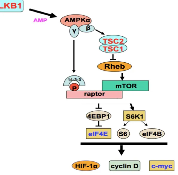
3 The acetylation of LKB1 at K48 is required for the regulation of the mTOR signaling pathway ... 158
4 GCN5 regulates the RNA levels of LKB1 ... 160
5 The GCN5/LKB1 signaling is essential for head formation ... 163
5.1 GCN5 is expressed in cephalic neural crest cells... 163
5.2 Functional interaction between GCN5 and LKB1 in vivo ... 164
III. Discussion and perspectives... 169
1 The acetylation state of LKB1 correlates with its subcellular localization and consequent activity ... 169
1.1 Two GCN5-dependent modes of LKB1 localization modulation ... 170
1.1.1 Summary of the results ... 170
1.1.2 The HAT activity of GCN5 is dispensable for LKB1 shuttling into the cytoplasm ... 171
1.1.3 GCN5-dependent translocation of LKB1 into the cytoplasm ... 173
1.1.4 Retention of acetylated LKB1 in the nucleus ... 174
1.2 LKB1 acetylation at K48 is required for its functions ... 176
1.2.1 Summary of the results ... 176
1.2.2 Complementary experiments ... 176
1.2.3 Role of acetylated LKB1 in the nucleus ... 177
1.2.3.2 Regulation of lipid homeostasis ... 180
1.2.3.3 Potential role of nuclear LKB1 in response to cellular stress ... 184
1.2.3.4 Role of nuclear LKB1 in cancer ... 184
2 A new regulatory mode of LKB1 expression levels ... 186
IV. Conclusion ... 188
Table of Figures
Figure 1: PJS characteristics ... 1
Figure 2: Mutations identified in the human LKB1 gene in patients with PJS and sporadic cancer. ... 3
Figure 3: Characterization of developmental arrest in Lkb1−/− embryos. ... 4
Figure 4: Non-tumorigenic phenotypes following Lkb1 targeting in mice. ... 6
Figure 5: Tumorigenic phenotypes following Lkb1 targeting in mice. ... 10
Figure 6: LKB1 splice variants: the long and short isoforms ... 12
Figure 7: A novel LKB1 isoform generated by alternate splicing encodes a protein called N-LKB1 ... 13
Figure 8: Posttranslational modification sites of the mouse LKB1 protein. ... 19
Figure 9: Human LKB1 acetylation sites ... 19
Figure 10: The PMSE syndrome: dysmorphic features ... 21
Figure 11: STRAD allosterically activates the kinase domain of LKB1. ... 22
Figure 12: Activation and translocation of LKB1 ... 24
Figure 13: Molecular modeling of the Hsp90–Cdc37–LKB1 complex. ... 27
Figure 14: Members of the AMPK and AMPK-related kinase (ARK) family... 28
Figure 15: Optimal substrate motif for LKB1 phosphorylation in ARKs ... 30
Figure 16: Graphic structure of AMPK. ... 32
Figure 17: Regulation of AMPK ... 33
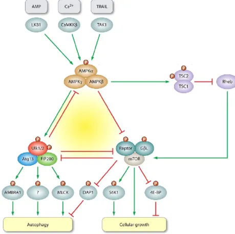
Figure 18: Regulation of AMPK by drugs and the principal metabolic pathways it regulates. 37 Figure 19: Role of AMPK in the regulation of metabolism in response to events such as nutrient- or exercise-induced stress. ... 39
Figure 20: LKB1/AMPK controls gluconeogenic gene expression ... 40
Figure 21: Schematic diagram showing LKB1–SIK pathway in the control of hepatic gluconeogenesis. ... 41
Figure 22: The LKB1/AMPK/mTORC1-dependent regulation of protein translation. ... 43
Figure 23: Fine adjustment of autophagy by the AMPK-mTORC1-Ulk1/2 kinase network ... 46
Figure 24: Regulation of Cell Size by the Primary Cilium ... 48
Figure 25: Introducing the PAR family. ... 50
Figure 26: Asymmetric division of Drosophila neuroblast ... 52
Figure 28: Model of the Kinase Pathway Identified in this Study... 59
Figure 29: A model for a role of LKB1-GSK3β-APC pathway in centrosomal forward movement. ... 61
Figure 30: Model presenting the role of LKB1 in regulating cell invasion ... 63
Figure 31: Lysine acetylation is involved in diverse cellular processes ... 69
Figure 32: Structure of the GNAT and MYST HAT families ... 74
Figure 33: HAT domain motifs ... 75
Figure 34: Crystal structure of Tetrahymena Gcn5 with bound coenzyme A and histone H3 peptide. ... 76
Figure 35: Schematic representation of the structure of GCN5 homologs ... 78
Figure 36: Schematic representation of hGCN5 isoforms ... 80
Figure 37: The hGCN5 mRNA is alternatively spliced ... 81
Figure 38: The overall structure of the GCN5 and PCAFenzymes in vertebrates, Drosophila and yeast. ... 83
Figure 39: The modular nature of ySAGA supports multiple activities ... 87
Figure 40: Schematic representation of yeast and human SAGA ... 89
Figure 41: GCN5 acetylates nucleosomes when in complex with Ada2 and Ada3 ... 90
Figure 42: The overall three-dimensional structure of the yeast and human SAGA complexes are evolutionarily conserved. ... 91
Figure 43: Schematic representation of the ADA complex ... 92
Figure 44: A model illustrating the role of Snf2 acetylation by GCN5 in vivo ... 97
Figure 45: Model showing the regulation of CDC6 by sequential modification (acetylation and phosphorylation) in early S phase ... 98
Figure 46: GCN5-mediated regulation of hepatic gluconeogenesis ... 100
Figure 47: GCN5hat/hat embryos exhibit defects in neural tube closure and exencephaly. ... 102
Figure 48: A gene regulatory network orchestrates neural crest formation ... 105
Figure 49: Neural crest cells subpopulations and derivatives ... 106
Figure 50: Skeletal fate of cranial neural crest cells in vertebrates. ... 108
Figure 51: Trunk neural crest cells progression and fate ... 109
Figure 52: Neural crest formation and migration during development. ... 112
Figure 53: Putative gene-regulatory and signaling interactions at the neural plate border of vertebrates ... 115
Figure 55: Neural crest cells epithelial-to-mesenchymal transition (EMT) ... 120
Figure 56: Migration of cranial and trunk NC. ... 124
Figure 57: NCCs migration: planar cell polarity and contact inhibition of locomotion ... 127
Figure 58: Absence of cephalic NC entails abnormal craniofacial development. ... 130
Figure 59: Intrinsic transcriptional programs underlying cranial NCC positional identity. .... 133
Figure 60: Segmental and directional migration of cranial neural crest cells. ... 137
Figure 61: Regulation of LKB1 acetylation and localization by GCN5 ... 170
Figure 62: GCN5 inhibits IFN- production in an HAT-independent manner ... 172
Figure 63: GCN5 induces the phosphorylation of LKB1 at Ser 428. ... 173
Figure 64: GCN5 modulates LKB1 localization independently from its HAT activity... 174
Figure 65: Sox-9 and Sox-10 associated abnormalities ... 179
Figure 66: SIRT6 interacts with LKB1 and promotes its acetylation and phosphorylation .... 180
Figure 67: SREBPs regulate hepatic lipogenesis ... 181
Figure 68: Model of the regulation of hepatic lipogenesis by SIRT6 ... 182
Figure 69: Hypothetic model of the SIRT6/GCN5/LKB1/AMPK signaling in hepatic lipogenesis regulation ... 183
Table 1: Reported role of the ARKs downstream of LKB1 ... 30
Table 2: HAT family groups and their histone substrates ... 72
Table 3: Composition of the 2 MDa and 700 kDa GCN5- and PCAF-containing multiprotein complexes ... 86
Table 4: Composition of GCN5 and PCAF SAGA and ATAC complexes ... 94
1
I. Introduction
1 The tumor suppressor LKB1
1.1 The Peutz-Jeghers syndrome
The Peutz-Jeghers syndrome (PJS) is an autosomal inherited disease firstly described by Johannes Peutz in 1921 and further characterized by Harold Jeghers in 1948. This rare disease is characterized by hamartomatous polyposis in the gastrointestinal tract, mucocutaneous hyperpigmentation of oral mucosa, of the lips, of the nose, of fingers and toes (Figure 1). The patients have also a high risk to develop malignant tumors affecting various organs such as the digestive tract (Banno et al., 2013), breast, testis and pancreas (Gan & Li, 2014).
Figure 1: PJS characteristics
The PJS is characterized by hyperpigmentation of oral mucosa and the lips (A)
(http://www.medicinenet.com/image-collection/peutz-jeghers_syndrome_picture/picture.htm), of toes (B) (http://www.ijdvl.com/articles/2008/74/2/images/ijdvl_2008_74_2_154_39705_2.jpg), of fingers (D)
(http://www.dermis.net/dermisroot/en/51592/image.htm) as well as polyposis in the gastrointestinal tract (C) (http://4.bp.blogspot.com/-ULLXZnuvP5k/UdPi0Zi4TJI/AAAAAAAADPU/B3s7qxY7nzo/s720/18.jpg).
2 1.1.1 Germline mutations of LKB1
Genetic linkage analysis led to the localization of a predisposing locus located on chromosome 19p13.3. Positional cloning allowed the identification of mutations in the LKB1 gene (also called STK11) in the germline of PJS patients. These mutations encompass all types of loss of function mutations including deletions of the LKB1 locus, as well as nonsense and frameshift mutations (Launonen, 2005). Missense mutations are mostly located in the catalytic domain and they disrupt the LKB1 kinase activity. A few mutations have also been observed in the C-terminal tail of LKB1, which impairs the biological activity of LKB1 (Forcet et al., 2005) but no point mutations in the N-terminal non-catalytic region have been identified (Alessi, Sakamoto, & Bayascas, 2006) (Figure 2). LKB1 is the major gene involved in PJS and mutations have been found in more than 80% of the families worldwide. However, linkage with other locus than LKB1 has been mapped in a few families without LKB1 mutations. Recently, a germline mutation of the gene coding MYH11 (myosin heavy chain) was found in a PJS patient but the significance of this observation is not clear (Alhopuro et al., 2008). Thus, despite circumstantial evidence of a genetic heterogeneity, no additional genes besides LKB1 have been ascribed to PJS.
LKB1 is a bona fide tumor suppressor since this gene is the target of a double mutational hit
that disrupts both LKB1 alleles. Genetic analysis of PJS cancers has shown that the wild type allele is frequently lost and leads to a loss of heterozygosity (LOH) (Banno et al., 2013). Several reports indicate that additional somatic mutations in other genes such as -catenin and p53 also contribute to the conversion of hamartomatous polyps into adenomatous and carcinomatous lesions (Miyaki et al., 2000).
1.1.2 LKB1 mutations in sporadic cancers
LKB1 mutations have also been described in sporadic cancers, more precisely in 4 to 7% of
pancreatic cancers, 20% of cervical cancers and 30% of human non-small cell lung cancer (NSCLC) (Banno et al., 2013; Sahin et al., 2003; Hardie & Alessi, 2013). It is now well established that the loss of LKB1 function is critical to pulmonary tumorigenesis, being involved in different stages from tumor initiation to metastasis spreading (Ji et al., 2007).
3
Figure 2: Mutations identified in the human LKB1 gene in patients with PJS and sporadic cancer.
Schematic representation of the mutations predicted effects on the primary structure of the LKB1 protein.The genomic organization of the coding sequence of the LKB1 gene is shown on the top, and the functional domains of the protein are shown below with (a) stop mutations, in-frame deletions, splicing mutations, and deletions; (b) point mutations; (c) frameshift mutations. Abbreviations used: NRD, N-terminal regulatory domain; CRD, C-terminal regulatory domain (white boxes); Δ, in-frame deletion; fs, frameshift; Ref, reference; X, point mutation. The protein kinase domain (blue boxes) and amino acid sequence introduced by the frameshift (green boxes) are also indicated (Alessi et al., 2006).
4 1.1.3 Murine models of Lkb1 inactivation
1.1.3.1 Role of LKB1 during embryogenesis and in tissue homeostasis
In order to study the physiological role of LKB1 in mammals, homozygous and heterozygous inactivation of Lkb1 were conducted in mice. These models have shown that inactivation of
Lkb1 through homologous recombination or ‘knock-out’ (KO) does not always lead to
tumors. This observation is partly due to essential functions of Lkb1 in development and partly demonstrates the tissue-specificity of Lkb1 functions.
Following Lkb1 inactivation in the murine germ line, heterozygous (Lkb1+/−) intercrosses resulted in both Lkb1+/+ and Lkb1+/− animals at expected frequencies, whereas Lkb1−/− mice
died in utero. Lkb1−/− embryos developed normally up to E8.0. However, macroscopic
analysis beyond E8.25 revealed multiple abnormalities including neural tube closure defects and an absence of the first branchial arch which form some facial stuctures at later stages (Figure 3). The embryos also exhibited defective somitogenesis, excessive mesenchymal cells death and vascular abnormalities associated with increased vascular epidermal growth factor (VEGF) production (Ylikorkala et al., 2001). Altogether, these observations show the essential role of LKB1 in early development for angiogenesis and head formation in mice.
Figure 3: Characterization of developmental arrest in Lkb1−/− embryos.
Light microscopy of E9.25 Lkb1+/+ andLkb1−/− embryos showing unturned embryos with open neural folds, a
5 The role of LKB1 during angiogenesis remains controversial. Deletion of Lkb1 in vascular endothelial cells using Tie1-Cre and analysis of heterozygous Tie2-Cre;Lkb1flox/+ mice suggest a proangiogenic role of Lkb1. Tie2-Cre;Lkb1flox/+ mice exhibit a normal phenotype, including
vasculature. However, they display reduced revascularization after hind-limb ischemia in adult mice (Londesborough et al., 2008; Ohashi, Ouchi, Higuchi, Shaw, & Walsh, 2010). In contrast, during embryonic development, increased VEGF signaling upon Lkb1 deletion rather suggests an antiangiogenic role for Lkb1 (Ylikorkala et al., 2001). This antiangiogenic effect is also present in the context of PJS polyps where a loss of Lkb1 leads to increased vasculature. Thus, the role of Lkb1 in angiogenesis seems to be context-dependent (Shackelford et al., 2009).
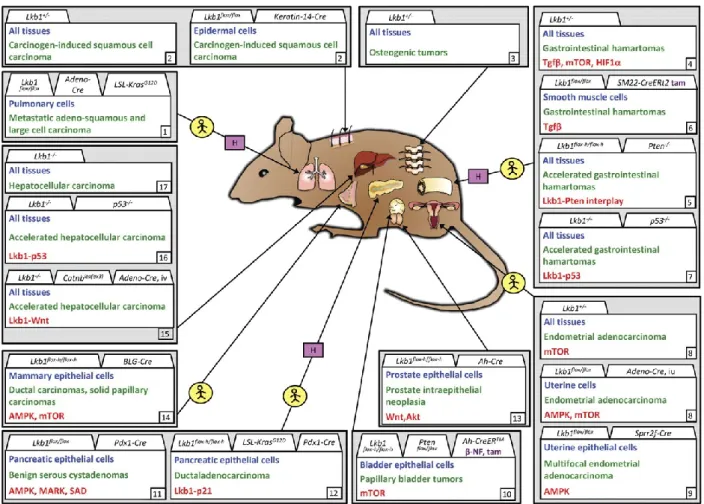
Other studies have reported the role of Lkb1 in maintaining the homeostasis of other tissues such as liver, skeletal muscle, pancreas and nervous system. Non-tumorigenic mouse phenotypes following Lkb1 targeting in different tissues are presented in Figure 4.
For instance, conditional hepatocyte-specific Lkb1 deletion using Adeno-Cre resulted in impaired glucose metabolism in the mice as demonstrated by elevated blood glucose levels and expression of the PGC-1 (peroxisome proliferator-activated receptor gamma coactivator 1-alpha) gluconeogenic target genes encoding the G6Pase (glucose-6-phosphatase) and PEPCK (phosphoenolpyruvate carboxylase). Thus Lkb1 is involved in the regulation of hepatic glucose production (Shaw et al., 2005). The increase in the expression of the genes encoding PGC-1α, G6Pase, and PEPCK was also observed in another study in
Lkb1-deficient hepatocytes (Foretz et al., 2010). Other reports of liver-targeted deletions of Lkb1 also demonstrate that Lkb1 is required for lipid, bile, and cholesterol metabolism
6
Figure 4: Non-tumorigenic phenotypes following Lkb1 targeting in mice.
Phenotypes (green) are grouped according to tissue type, cell type affected/analyzed (blue), and alleles used for targeting. When appropriate, activator of deletor is indicated in purple. Noted signaling change(s) indicated in red. Alleles as displayed in original publications except for Lkb1 flox−h/flox−h hypomorphic Lkb1 (Sakamoto et al, 2005). (1) Londesborough et al., 2008; (2) Ohashi et al., 2010; (3) Cao et al., 2010; Tamas et al., 2010; (4) Shorning et al., 2009; (5) Woods et al., 2011; (6) Shaw et al., 2005; (7) Sun et al., 2010a; (8) Sun et al., 2011; (9) Granot et al., 2009; Fu et al., 2009; (10) Koh et al., 2006; (11) Sakamoto et al., 2005; (12) Sakamoto et al., 2006; Jessen, et al., 2010; (13) Ikeda et al. 2009; (14) Gurumurthy et al., 2010; Nakada et al., 2010; (15) Gan et al., 2010; (16) Barnes et al., 2007; (17) Ylikorkala et al., 2001. tam, tamoxifen; β-NF, β-naphtoflavone; pIpC, polyinosinic–polycytidylic acid; iv, intravenous. (Ollila & Mäkelä, 2011).
7
1.1.3.2 Understanding the PJS
Since homozygous inactivation of Lkb1 has proven to be lethal, heterozygous Lkb1 mice were generated and have proven to be useful for understanding Lkb1-induced polyp formation.
The development of large occluding hamartomatous polyps in the gastrointestinal (GI) tract is a major characteristic in PJS. Different laboratories have established that biallelic Lkb1 inactivation is not required for polyp development. Indeed, Lkb1 monoallelic inactivation was sufficient for the development of polyps in mice (Bardeesy et al., 2002; Jishage et al., 2002; Miyoshi et al., 2002; Rossi et al., 2002). The Lkb1 +/- mice are viable and fertile and show no apparent phenotype until the adult age. Polyps are first detected at around 5 months and are particularly prominent in the pyloric region rather than in the small intestine like in human, in line with recent reports on gastric polyposis in human. They cause premature lethality from 8 months onwards, presumably due to intestinal obstructions (Katajisto et al., 2008; Udd et al., 2004). Polyps from both patients and murine models were shown to retain the Lkb1 wild-type allele suggesting that haploinsufficiency triggers polyp formation (Jishage et al., 2002; Miyoshi et al., 2002; Rossi et al., 2002). Cyclooxygenase 2 (COX2) was shown to be up-regulated in the mouse and PJS patient polyps (Rossi et al., 2002), and COX2 inhibitors have been shown to be efficient suppressors of PJS polyps (Udd et al., 2004).
Another study suggests that LOH is required for polyp development. In this study, 25% of polyps which were isolated from Lkb1 +/- mice retain the Lkb1 wild-type allele in the stromal compartment but lose it in the epithelial compartment. However, in half of the polyps which conserved the Lkb1 wild-type allele, Lkb1 was not detected at the protein level suggesting epigenetic modifications. The mouse model used in this study exhibits polyps within the gastrointestinal tract in addition to the gut. The differences between this model and the previous ones might be due to the genetic background (Bardeesy et al., 2002).
To date, the implication of a p53-deficient background in malignant transformation of murine Lkb1 +/− polyps remains controversial (Jansen, Ten Klooster, Offerhaus, & Clevers, 2009) vs (Ollila & Mäkelä, 2011). However, inactivating Lkb1 in the smooth muscle lineage, using a tamoxifen-inducible SM22-CreERt2 line, is sufficient for GI polyposis (Katajisto et al.,
8 2008). In this model attenuated TGF signaling from stromal cells to epithelial cells was associated with increased epithelial proliferation. The endothelium-specific deletion of Lkb1 resulted in loss of TGF secretion, followed by loss of supporting smooth muscle cells (Londesborough et al., 2008). Finally, Lkb1 deficiency in MEFs leads to attenuated TGF signaling resulting in defects in smooth muscle cell lineage differentiation. These observations suggest that stromal Lkb1 acts as a ‘landscaper’ tumor suppressor gene (Vaahtomeri et al., 2008) and that stromal TGF signaling is an important growth restrictive signal to GI epithelial cells.
Despite these latest reports, the mechanism whereby the cell carcinomas occur in PJS and
Lkb1 +/- mice remains unclear. However, understanding the mechanisms underlying this
cellular transformation is worth further investigation in order to better understand the tumor suppressor function of LKB1.
1.1.3.3 Tumor models
In several tissues, Lkb1 inactivating mutations result in the development of cancer. Targeted inactivation of Lkb1 in mice, sometimes in combination with other tumorigenic mutations, have led to the development of various tumors in multiple tissues, sometimes modeling human cancers in very useful ways as discussed below (some examples) and summarized in Figure 5. Investigating the role of Lkb1 during malignant tumor progression has firmly established its tumor suppressive activity.
In mice, both homo- and heterozygous loss of Lkb1, combined with Kras activation, promote lung carcinogenesis and metastasis (Ji et al., 2007).
As for cervical cancer, to date, no targeted Lkb1 mouse models have been reported. However, Contreras group has generated three mouse models with Lkb1 deficiency. In the first model, (53%) of surviving Lkb1−/+ females spontaneously developed endometrial adenocarcinomas by 55 weeks of age and the tumors were highly invasive. In the second model, they injected an Adeno-Cre virus into the uterine lumen of female mice homozygous for a floxed Lkb1 allele (L). Again, 65% of the mice developed endometrial adenocarcinomas which were confined to the uterus, but one was diffusely metastatic within the peritoneum. The fact that not all of the mice developed uterine tumors and the focal nature of these
9 neoplasms suggest that cooperating genetic events are required for Lkb1-driven neoplasia. Interestingly, the team has also observed that decreased expression of Lkb1 correlates with invasiveness in human tumors, presenting the first evidence that Lkb1 similarly drives human endometrial carcinogenesis (Contreras et al., 2008). Finally, Contreras team has generated a third mouse model allowing conditional inactivation of Lkb1 only in the epithelial cells of uterine lumen and endometrial glands. These mice died rapidly of invasive endometrial adenocarcinomas. These mouse models developing endometrial adenocarcinomas show that the frequent uterine cancer in PJS patients can be recapitulated in mice, and targeting therapies can be developed to treat LKB1-deficient endometrial cancer(Contreras et al., 2010).
One study has reported that 70% of aged Lkb1 +/− male mice develop hepatocellular carcinoma (HCC) in contrast to 20% of female mice. These neoplasms recapitulated human HCC types and were shown to have lost the wild-type copy of Lkb1 (Nakau et al., 2002). The tumor development was accelerated upon crossing Lkb1+/- mice with p53 mutant mice, or forced activation of Wnt signaling (Miyoshi et al., 2009; Takeda, Miyoshi, Kojima, Oshima, & Taketo, 2006). However, complete deletion of Lkb1 in hepatocytes resulted in metabolism impairment without tumor development (Shaw et al., 2005; Angela Woods et al., 2011). This observation may reflect the time-span required for the lesions to occur.
10
Figure 5: Tumorigenic phenotypes following Lkb1 targeting in mice.
Tumor types (green) are grouped according to tissue type, cell type affected/analyzed (blue), and alleles used for targeting of Lkb1 and possible other alleles. When appropriate, activator of deletor is indicated in purple. Noted signaling change(s) indicated in red. Tissues where LKB1 deficiencies noted in human tumors are marked with yellow circle. Tumorigenic phenotypes resulting from Lkb1 haploinsufficiency indicated with a pink H box. Alleles as displayed in original publications except for Lkb1 flox−h/flox−h, hypomorphic Lkb1 (Sakamoto et al., 2005). (1) Ji et al., 2007; (2) Gurumurthy et al., 2008; (3) Robinson et al., 2008; (4) Bardeesy et al., 2002; Jishage et al., 2002; Miyoshi et al., 2002; Rossi et al., 2002; Katajisto et al., 2008; Shackelford et al., 2009; (5) Huang et al., 2008; (6) Katajisto et al., 2008; (7) Wei et al., 2005; Takeda et al., 2006; (8) Contreras et al., 2008; (9) Contreras et al., 2010; (10) Shorning et al., 2011; (11) Hezel et al., 2008; (12) Morton et al., 2010; (13) Pearson et al., 2008; (14) McCarthy et al., 2009; (15) Miyoshi et al., 2009; (16) Takeda et al., 2006; (17) Nakau et al., 2002. tam, tamoxifen; β-NF, β-naphtoflavone; pIpC, polyinosinic–polycytidylic acid; iv, intravenous; iu, intrauterine; LSL, lox-stop-lox. (Ollila & Mäkelä, 2011)
11
1.2 LKB1: from the gene to the protein
LKB1 is a tumor suppressor gene which is evolutionarily conserved. The human gene spans
23 kb and comprises nine coding exons and one non-coding exon (Mehenni et al., 1998). The murine Lkb1 gene is located on chromosome 10, also contains 10 exons, and the encoded protein shares more than 90% with the human protein sequence. Besides, murine Lkb1 has a conserved prenylation motif (Cys433–Lys–Gln–Gln436) at the carboxyl-terminus, directly downstream from a consensus cAMP-dependent protein kinase (PKA) phosphorylation site (Arg428–Arg–Leu–Ser431) (Collins, Reoma, Gamm, & Uhler, 2000).
The LKB1 gene is ubiquitously expressed in fetal and adult tissues as well as tumors. During embryonic development in mouse, Lkb1 (mRNA) is mainly expressed in the gastrointestinal tract, lung and testis. Human LKB1 is predominantly expressed in epithelia and the seminiferous tubules of the testis, with higher levels in fetal than in adult tissues. However, the ortholog of LKB1 in Xenopus, XEEK1, seems to be restricted to early embryogenesis while in C. elegans, the protein Par-4 can be found in the gonads, oocytes and early embryos. Finally, some malignant tumors express high levels of LKB1 while some cancer cells show no expression of LKB1 which suggests a dual role of LKB1 at different stages of tumorigenesis (Gan & Li, 2014).
1.2.1 Splice variants of LKB1
The 23 kb human LKB1 gene is transcribed in the telomere-to-centromere direction and the 3 kb mRNA can be alternatively spliced to produce two isoforms: LKB1L (long isoform) and
LKB1S (short isoform). The alternative splicing occurs at exon 9; the C-terminal sequence of
LKB1L is encoded by exon 9B, while in LKB1S this sequence is encoded by exon 9A of the
mRNA leading to the replacement of the last 63 amino-acids (a.a.) of the long isoform by 39 different amino acids in the short isoform (Towler et al., 2008) (Figure 6). Thus, the corresponding proteins are distinguishable by their molecular weights which are 50 KDa for LKB1L and 48 KDa for LKB1S. LKB1L is ubiquitously expressed, whereas LKB1S is predominant
in the testis where it appears to be involved in spermiogenesis. Indeed, male knockout mice for this isoform are sterile and show a decrease in the number of mature spermatozoa. The major cause of the infertility phenotype is a defect in the release of mature spermatids from
12 the seminiferous epithelium (spermiation) during spermatozoan development (Denison et al., 2011; Denison, Hiscock, Carling, & Woods, 2009; Reuben J Shaw, 2008).
Figure 6: LKB1 splice variants: the long and short isoforms
(A) The arrangement of exons (E1 through E10) and introns (I1 through I10) of the LKB1 gene are shown.
Protein coding regions common to both isoforms are shown in grey, while the regions in exons 9A and 9B encoding the unique C-terminal regions of LKB1S and LKB1L are shown with forward or backward cross-hatching respectively. (B) The deduced amino acid sequences of the C-terminal regions of human and mouse LKB1L and LKB1S are shown. A portion of the sequence encoded by exon IIIV, common to both forms, is represented in italics. Serine 428/431 (human/mouse) and cysteine 430/433 in LKB1L that have been shown to undergo post-translational modification are underlined. The number of amino acids in each of the full-length proteins is indicated. Adapted from (Denison et al., 2009; Towler et al., 2008).
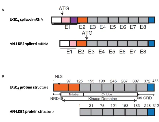
13 Recently, a third isoform of LKB1 was identified by the team of Christine Perret (Institut Cochin, Paris) in collaboration with our team. This isoform of LKB1 is generated by alternate splicing and encodes a protein called LKB1. This novel isoform is truncated in its N-terminal domain and results from an alternative splicing in exon 1 and internal initiation of translation of the mRNA in exon 3 (Figure 7). The 42 KDa corresponding protein is mainly expressed in the heart and skeletal muscle. It has pro-oncogenic properties and may account for some of the paradoxical effects of LKB1 during tumorigenesis (Dahmani et al., 2014).
Figure 7: A novel LKB1 isoform generated by alternate splicing encodes a protein called N-LKB1
(A) The initiation of translation of the N-LKB1 isoform starts in exon 3. (B) Structure of the human LKB1
isoforms LKB1L and N-LKB1. The alternative splicing in exon 1 and internal initiation of translation in exon 3 result in a novel isoform called N-LKB1, which is truncated in its N-terminal domain. CRD: C-terminal-regulated domain; NLS: nuclear localization signal; NRD: N-terminal-C-terminal-regulated domain. Adapted from (Dahmani et al., 2014).
14 1.2.2 The protein LKB1
1.2.2.1 Structure and subcellular localization of LKB1
LKB1 encodes a serine/threonine kinase comprising 433 amino acids (a.a) in human (436 in
mouse). Its kinase catalytic domain is localized between a.a. 49 and a.a. 309 (44-309 for murine Lkb1). The N-terminal region of LKB1 contains a nuclear localization signal (NLS) which is localized between a.a. 38 and 43 and which regulates LKB1 shuttling between the nucleus and the cytoplasm (Figure 7). As for the C-terminal region, it contains a prenylation site (Cys433) which is associated to the localization of LKB1 at the plasma membrane.
Thus, LKB1 can be localized within the nucleus, the cytoplasm, the plasma membrane in living cells but can translocate into mitochondria during apoptosis (Alessi et al., 2006; Jansen et al., 2009; Karuman et al., 2001).
1.2.2.2 Post-translational modifications
LKB1 undergoes post-translational modifications such as phosphorylation, prenylation, ubiquitination as well as acetylation.
1.2.2.2.1 Phosphorylation
So far, nine LKB1 Ser/Thr residues were found to be phosphorylated (Figure 8). The major autophosphorylation site of LKB1 is on Thr336. Mutation of this residue to Glu (to mimic phosphorylation), but not Ala (to abolish phosphorylation), prevents LKB1 from inhibiting the growth of G361 melanoma cells. This observation indicates that this phosphorylation seems to inhibit the tumor suppressor function of LKB1 (Alessi et al., 2006). LKB1 is also autophosphorylated on Thr185, Thr189 and Ser404 (murine)/Ser402(human) but no functional role has been attributed to these residues (Alessi et al., 2006; A F Baas et al., 2003; Gopal P Sapkota, Boudeau, et al., 2002).
LKB1 is also phosphorylated by upstream kinases on Ser31, Ser307, Ser325, Thr366 and Ser428 (human)/431(mouse). Mutation of Ser31, Ser325 and Thr366 had no effect on the catalytic activity of subcellular localization of LKB1 and the autophosphorylation on Ser31 and Ser325 remain controversial (Gopal P Sapkota, Boudeau, et al., 2002). However, the phosphorylation of Thr366 by ATM (ataxia-telangiectasia-mutated), in response to oxidative
15 stress, induces LKB1-dependant autophagy (Alexander et al., 2010; Gopal P Sapkota, Deak, et al., 2002). LKB1 is also phosphorylated on this residue by DNA-PK (DNA-dependant protein kinase) in vitro (Gopal P Sapkota, Deak, et al., 2002).
Ser428 is phosphorylated by PKC- (protein kinase C-), PKA (protein kinase A) and RSK (ribosomal protein S6 kinase). Mutating Ser428 into an Ala resulted in an LKB1 mutant which is retained in the nucleus, indicating that the phosphorylation of this residue regulates the subcellular localization of LKB1 (Song et al., 2008). Indeed, the LKB1 S428A mutant cannot translocate into the cytoplasm following metformin treatment, nor associate with its cytoplasmic substrate AMPK (AMP-activated protein kinase) in HeLa-S3 cells. However, this mutation has no effect on the catalytic activity of LKB1 (Xie, Dong, Scholz, Neumann, & Zou, 2008). Interestingly, the short isoform of LKB1 (LKB1S) lacks this residue in its C-terminus, but
can still activate the LKB1 substrate AMPK, suggesting that the phosphorylation of Ser428 is dispensible for the subcellular distribution of LKB1 and its downstream signaling. Surprisingly, a team of researchers has shown that in this short isoform, PKC- phosphorylates LKB1 in its C-terminus on Ser399, which is also essential for the nucleocytoplasmic export of LKB1S and subsequentAMPK activation, but has no effect on the
catalytic activity of LKB1 (Zhu, Moriasi, Zhang, Zhao, & Zou, 2013).
The phosphorylation of LKB1 on Ser428 is important for the role of LKB1 in axon specification in the developing nervous system in response to BDNF. In Drosophila, loss-of-function mutations in the LKB1 gene cause defects in the oocyte polarity. Although this defect is rescued by low level expression in the germ line of wild-type LKB1, the Ser535 (homologous of Ser428) LKB1 mutant fails to rescue the polarity defects in the oocyte. Thus, the phosphorylation of LKB1 on Ser428 is also important for LKB1 functions in vivo (Houde et al., 2014).
Despite the available data on the importance of LKB1 phosphorylation on Ser428 for the translocation of LKB1 into the cytoplasm and subsequent AMPK activation, these data remain controversial. Indeed, a homozygous Lkb1S431A/S431A (homologous of Ser428 in human)
knockin mouse model was recently generated (Houde et al., 2014). These mice were viable, fertile and displayed no overt phenotypes. As expected, the S428A mutation had no effect on LKB1 kinase activity in vitro. However, in contrast to the previous data, AMPK was activated normally in Lkb1S431A/S431A tissues and MEFs, raising doubts regarding the importance
16 of this phosphorylation on LKB1 functions in vivo. Despite these intriguing observations, the authors draw our attention to the fact that these mice might actually display a phenotype that they did not notice. They also suggest that the phosphorylation of LKB1 on Ser428 is important for the regulation of oocyte polarity in Drosophila, but that mammalian embryonic development is not significantly affected by ablation of this phosphorylation site. Besides, we cannot ignore the possibility of the presence of compensatory mechanisms in mammalian cells in vivo. Finally, the residue equivalent to Ser428 is conserved in all species where LKB1 has been reported, even in C. elegans where the prenylation motif is not conserved, which strongly suggests that phosphorylation of this residue must indeed have a significant function. Thus, the relevance of this phosphorylation site regarding LKB1 subcellular localization and functions requires further investigation.
LKB1 phosphorylation by PKC- on Ser307 also induces the nucleocytoplasmic export of LKB1 and subsequent AMPK activation, without affecting the catalytic kinase activity of LKB1. This phosphorylation promotes the LKB1-dependant regulation of cell cycle progression, proliferation, angiogenesis and apoptosis (Xie et al., 2009).
The LKB1 Thr336, Thr366, and Ser428 phosphorylation sites and the residues surrounding them are highly conserved in Drosophila, Xenopus, and mammalian LKB1, but not in C.
elegans (Gopal P Sapkota, Boudeau, et al., 2002).
1.2.2.2.2 Prenylation
LKB1 is prenylated in cultured cells and invertebrates (Collins et al., 2000; Martin & St Johnston, 2003a; G P Sapkota et al., 2001; J. Watts, Morton, Bestman, & Kemphues, 2000). As previously mentioned, murine Lkb1 has a conserved prenylation motif (Cys433–Lys–Gln– Gln436) at the carboxyl-terminus (Figure 8). Cys433 is prenylated in vivo, and PKA-mediated LKB1 prenylation targets it to the cellular membrane (Collins et al., 2000). This prenylation occurs by the addition of a farnesyl moiety to Cys433, and mutation of this residue prevents LKB1 prenylation (G P Sapkota et al., 2001). Interestingly, point mutation of the homologous residue to Cys433 in Drosophila constitutes an allele with severely reduced rescue activity. These data indicate that the C-terminus of LKB1 is important for its function (Jansen et al., 2009).
17 More recently, an LKB1C433S/C433S
knockin mouse model was generated. These mice were viable, fertile and displayed no overt phenotypes. This study showed that the majority, if not all, of the endogenous LKB1 is prenylated. In these mice, the levels of LKB1 localized at the membrane of the liver cells and fibroblasts were reduced compared with the wild-type mice, confirming that farnesylation plays a role in mediating membrane association. In addition, in all of the examined tissues and cells taken from these mice, LKB1 failed to activate its substrate AMPK. Thus, these data confirm that farnesylation of LKB1 is important for its localization to the cell membrane and present the first evidence that this farnesylation is required for downstream signaling of LKB1 (Houde et al., 2014).
1.2.2.2.3 Ubiquitination
Two groups, including our team, have reported that the molecular chaperone heat shock protein 90 (Hsp90) binds to and stabilizes LKB1. Disrupting the LKB1-Hsp90 complex favors the recruitment of both Hsp/Hsc70 and the U-box dependent E3 ubiquitin ligase CHIP (carboxyl terminus of Hsc70-interacting protein) that triggers LKB1 degradation by the proteasome. Thus, LKB1 can be ubiquitinated (Nony et al., 2003) (Gaude et al., 2012).
1.2.2.2.4 Acetylation
Several teams have shown that LKB1 can be acetylated (Figure 9) and deacetylated (Calamaras et al., 2012; Lan, Cacicedo, Ruderman, & Ido, 2008; M.-J. Lee et al., 2010; Yi Yang et al., 2014; Z. Zheng et al., 2012; Zu et al., 2010). Although the enzymes that acetylate LKB1 remain unknown, a team has shown that Sirtuin 1 (SIRT1) is responsible for deacetylation of LKB1 on lysine 48 (K48) which allows its translocation from the nucleus to the cytoplasm (Lan et al., 2008). This team, as well as ours, has identified lysine residues that undergo acetylation using mass spectrometry (Figure 9).
SIRT1 is a conserved NAD+ -dependent deacetylase which increases LKB1 phosphorylation on Ser428 and Thr 336 and activation of AMPK in HEK293T cells (Lan et al., 2008). However, in primary porcine aortic endothelial cells, SIRT1 promotes proliferation and prevents senescence through targeting LKB1, thus acting as a negative regulator for LKB1/AMPK signaling (Zu et al., 2010). Finally, in HepG2 cells and mouse liver, over-expression of SIRT1 can stimulate basal AMPK signaling via phosphorylation and activation of LKB1 (Hou et al.,
18 2008). Although these data are controversial, LKB1 acetylation/deacetylation seems to modulate its kinase activity and subcellular localization.
Our team has identified the acetyltransferase GCN5 (detailed in section 2) as an enzyme which acetylates LKB1. During my PhD, I have focused on studying the role of the acetylation of LKB1 by the acetyltransferase GCN5 on the functions and subcellular localization of LKB1 (chapter II-project 2).
19
Figure 8: Posttranslational modification sites of the mouse LKB1 protein.
Autophosphorylation sites are depicted in red, and the sites phosphorylated by other kinases are in black. The Cys433 farnesylation site is depicted in green. The agonists and upstream protein kinases postulated to phosphorylate each site are indicated. Residues Thr366, Ser404, Ser431, and Cys433 in the mouse sequence correspond to human LKB1 residues Thr363, Thr402, Ser428, and Cys430, respectively. The noncatalytic domains are in white, and the kinase domain is light blue. Adapted from (Alessi et al., 2006).
Figure 9: Human LKB1 acetylation sites
Schematic representation of LKB1 acetylated lysine residues identified by mass spectrometry analysis in Lan´s team (Lan et al., 2008) as well as our team (unpublished data). Numbers in blue red and black indicate lysine residues identified by Lan´s team, our team and those common to both studies respectively.
20
1.3 LKB1 binding partners
The stability, subcellular localization and kinase activity of LKB1 are modulated by its association with several partners.
1.3.1 The LKB1/STRAD/MO25 complex
Yeast two-hybrid analysis and affinity purification from mammalian cells have shown that LKB1 forms a heterotrimeric complex with two other proteins: the pseudokinase STE20-related adaptor (STRAD) and the scaffolding protein MO25 (CAB39, calcium binding protein 39). The formation of this complex results in the activation and translocation of LKB1 from the nucleus to the cytoplasm (Alessi et al., 2006).
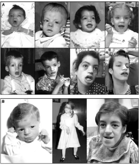
Two isoforms of STRAD ( and ) and MO25 ( and ) were identified and they all interact with LKB1. STRAD α and β possess high sequence similarity to protein kinases and can bind ATP. However, they lack several residues required for catalytic function and are therefore qualified as pseudokinases (Alessi et al., 2006). The kinase domain and the region between a.a. 319 and 343 of LKB1 are essential for its interaction with STRAD (A F Baas et al., 2003). Homozygous deletion of STRADα (LYK5) is associated with the PMSE syndrome (polyhydramnios, megalencephaly, symptomatic epilepsy) which is a severe human developmental and epileptic disorder. PMSE patients suffer from craniofacial abnormalities, severe mental retardation, gross movement disorders, and childhood mortality (Puffenberger et al., 2007) (Figure 10).
MO25 has no identifiable sequence similarity with other proteins. It stabilizes the complex by interacting with the conserved Trp–Glu–Phe sequence (WEF motif) at the C-terminus of STRAD which enhances the binding of STRAD to LKB1 and further stimulates STRAD-induced kinase activity and nucleocytoplasmic transport of LKB1 (Jérôme Boudeau, Baas, et al., 2003).
21
Figure 10: The PMSE syndrome: dysmorphic features
(A) Young persons with homozygous LYK5 deletions had a long face, large forehead, peaked eyebrows, broad nose and wide-set eyes. As patients got older (from upper left to lower right), the mouth became enlarged and the lips thickened. (B) Individual craniofacial structure changed considerably with age. Serial pictures of one affected individual show macrocephaly, frontal bossing, hypertelorism and broad nasal bridge shortly after birth (left panel), lengthening of the face during early childhood (middle panel), and overgrowth of the mandible, enlargement of the mouth and thickening of the lips during adolescence (right panel). (Puffenberger et al., 2007).
22 Recently, the first structure of the STRADα pseudokinase and its interaction with MO25α was described. Interestingly, STRADα could interact with MO25α through its WEF motif as previously described, but could also form an extensive network of interactions with the highly conserved concave surface of MO25α. This study has revealed that despite of being catalytically inactive, STRADα can adopt a closed active-like conformation, with an ordered activation loop similar to active protein kinases, which is stabilized through binding of ATP and/or MO25α. STRADα mutants that cannot interact with both ATP and MO25α are unable to activate LKB1, while STRADα mutants that retain the ability to bind either ATP or MO25α still activate LKB1. Thus, it is rather the closed active-like conformation of STRADα, rather than a catalytic phosphoryltransferase activity, that triggers the activation of the tumor suppressor LKB1 by STRADα. A model presenting the interaction of STRADα/MO25α with LKB1 based on known mutagenesis and structural data is presented in Figure 11 (Zeqiraj, Filippi, Deak, Alessi, & van Aalten, 2009).
Figure 11: STRAD allosterically activates the kinase domain of LKB1.
The kinase domain of STRAD is presumed to be maintained in an inactive conformation in its isolated state. The binding of ATP and MO25 to STRAD transitions its kinase domain to an active-like kinase conformation that is characterized by the extended conformation of its A-loop. This active-like conformation allows STRAD to bind LKB1 as a pseudosubstrate. STRAD binding allosterically induces the kinase domain of LKB1 to adopt an active kinase conformation, which is further stabilized by the binding of MO25 to the A-loop of LKB1 that positions the loop in an extended conformation (Rajakulendran & Sicheri, 2010).
23 The PMSE-causing mutation in humans results in the truncation of the pseudokinase STRADα at residue 251 and consequent loss of the last 180 amino acids (almost half of the C-terminal lobe of the pseudokinase domain). The expression levels of the PMSE-STRADα (residues 1– 251) mutant in 293 cells are significantly lower than full-length STRADα. Thus, this mutant is unstable and fails to interact with or activate LKB1 in vitro. These results indicate that the STRADα mutation found in PMSE patients represents a loss-of-function mutation that would destabilize STRADα and prevent it from binding to and activating LKB1 (Zeqiraj et al., 2009). 1.3.2 Regulation of the subcellular localization of LKB1
The cytoplasmic localization of LKB1 has been demonstrated to be essential for its functions. Indeed, mutant forms of LKB1 found in PJS patients localize exclusively in the nucleus and are catalytically inactive and unable to suppress cell growth (J Boudeau et al., 2003). Mutant LKB1 which lacks the nuclear localization signal (NLS) located in its N-terminal non-catalytic region (residues 38– 43) (Figure 9) is still able to suppress cell growth (Tiainen, Vaahtomeri, Ylikorkala, & Mäkelä, 2002).
LKB1 lacks a nuclear export domain of its own and is retained in the nucleus in the absence of metabolic stress (low glucose, low oxygen). STRADα and MO25 diffuse passively into the nucleus or are actively imported by importins α and β. Once synthesised, the LKB1 protein is imported to the nucleus by importins α and β. Only STRADα allows LKB1 to translocate from the nucleus to the cytoplasm by its binding to the nuclear export proteins CRM1 and exportin-7. MO25 stabilizes the heterotrimeric complex. STRADα also competes with the importins binding site on LKB1 to prevent it from re-entering the nucleus (van Veelen, Korsse, van de Laar, & Peppelenbosch, 2011) (Figure 12). STRADβ lacks the binding sites to exportin-7 and CRM1 and cannot transport LKB1 to the cytoplasm suggesting unique functions of the LKB1/STRADβ complex in the nucleus (Dorfman & Macara, 2008). Co-expression of LKB1 and STRADα targets the majority of LKB1 to the cytoplasm, but a significant amount remains nuclear. However, expression of LKB1, STRADα and MO25 fully localizes LKB1 to the cytoplasm (Alessi et al., 2006). Thus, the formation of LKB1/STRADα/MO25 complex is essential for LKB1 localization.
The subcellular localization of LKB1 is also modulated by its phosphorylation on several residues. As previously mentioned, LKB1 phosphorylation on Ser428, Ser307 by PKC-