HAL Id: hal-01004601
https://hal.archives-ouvertes.fr/hal-01004601
Submitted on 11 Jun 2014
HAL is a multi-disciplinary open access
archive for the deposit and dissemination of
sci-entific research documents, whether they are
pub-lished or not. The documents may come from
teaching and research institutions in France or
abroad, or from public or private research centers.
L’archive ouverte pluridisciplinaire HAL, est
destinée au dépôt et à la diffusion de documents
scientifiques de niveau recherche, publiés ou non,
émanant des établissements d’enseignement et de
recherche français ou étrangers, des laboratoires
publics ou privés.
Dynamics of a bacterial multidrug ABC transporter in
the inward- and outward-facing conformations.
Shahid Mehmood, Carmen Domene, Eric Forest, Jean-Michel Jault
To cite this version:
Shahid Mehmood, Carmen Domene, Eric Forest, Jean-Michel Jault. Dynamics of a bacterial multidrug
ABC transporter in the inward- and outward-facing conformations.. Proceedings of the National
Academy of Sciences of the United States of America , National Academy of Sciences, 2012, 109 (27),
pp.10832-6. �10.1073/pnas.1204067109�. �hal-01004601�
Dynamics of a bacterial multidrug ABC transporter in
the inward- and outward-facing conformations
Shahid Mehmooda,b,c, Carmen Domened, Eric Foresta,b,c,1, and Jean-Michel Jaulta,b,c,1
aInstitut de Biologie Structurale, Université Joseph Fourier Grenoble 1, 38027 Grenoble Cedex 1, France;bCommissariat à l’Energie Atomique et aux Energies
Alternatives Grenoble, 38027 Grenoble Cedex 1, France;cUnité Mixte de Recherche 5075, Centre National de la Recherche Scientifique, 38027 Grenoble
Cedex 1, France; anddDepartment of Chemistry, Physical and Theoretical Chemistry Laboratory, University of Oxford, Oxford OX1 3QZ, United Kingdom
Edited by H. Ronald Kaback, University of California, Los Angeles, CA, and approved May 18, 2012 (received for review March 8, 2012)
The study of membrane proteins remains a challenging task, and approaches to unravel their dynamics are scarce. Here, we applied hydrogen/deuterium exchange (HDX) coupled to mass spectrometry to probe the motions of a bacterial multidrug ATP-binding cassette (ABC) transporter, BmrA, in the inward-facing (resting state) and outward-facing (ATP-bound) conformations. Trypsin digestion and global or local HDX support the transition between inward- and outward-facing conformations during the catalytic cycle of BmrA. However, in the resting state, peptides from the two intracellular domains, especially ICD2, show a much faster HDX than in the closed state. This shows that these two subdomains are very flexible in this conformation. Additionally, molecular dynamics simulations sug-gest a large fluctuation of the Cα positions from ICD2 residues in the inward-facing conformation of a related transporter, MsbA. These results highlight the unexpected flexibility of ABC exporters in the resting state and underline the power of HDX coupled to mass spec-trometry to explore conformational changes and dynamics of large membrane proteins.
ATPase cycle
|
structural biology|
detergentI
mport of nutrients and export of signaling molecules or noxious compounds are essential processes for life, and selectivity for crossing membrane is usually governed by dedicated transporters. The ATP-binding cassette (ABC) is one of the largest families of transporters involved in these “checkpoints.” They are found in all living species and use ATP hydrolysis to transport a wide va-riety of substrates (1). The dysfunctions of several ABC trans-porters cause severe pathologies, such as adrenoleukodystrophy, hyperinsulinemic hypoglycemia, or cystic fibrosis (2). Other medically important ABC transporters are responsible for mul-tidrug resistance (MDR) phenotypes. Mulmul-tidrug transporters are capable of recognizing and expelling many unrelated organic compounds with quite dissimilar chemical scaffolds. Thus, al-though their original function is to protect healthy cells, the oc-currence of these transporters in malignant tissues, pathogenic microorganisms, or parasites confers a resistance toward the cu-rative drugs. The archetype of multidrug ABC transporters is the human MDR1 (or ABCB1); it is responsible for the failure of chemotherapeutic treatments in cancerous tissues. In bacteria, the identification of related transporters is relatively recent, compared with other families of multidrug transporters that use the proton gradient as the energy source (3). However, growing bodies of evidence support the implication of these multidrug ABC transporters in antibiotic resistance in many species, jeop-ardizing a successful therapy (4).All ABC transporters share a common architecture, with two transmembrane domains (TMDs) responsible for substrate translocation and two nucleotide-binding domains (NBDs). In most exporters, these four domains are either linked into a single polypeptide or function as dimers with one NBD fused to one TMD (homo- or heterodimers). As opposed to the TMDs, the NBDs are highly conserved, with several distinctive motifs, no-tably the Walker A and Walker B found in many ATPases and the ABC signature specific to this family (5, 6). NBD structures
have shown that during the catalytic cycle, the two NBDs interact transiently to form a sandwich dimer in a head-to-tail fashion, thereby trapping two ATP molecules at their interface (7–9). In this closed conformation, which can be stabilized either by ATPase-inactive mutations (10) or by vanadate-induced ADP trapping (11), each composite ATP-binding site is made of ele-ments from one NBD (i.e., Walker A and B motifs), whereas the ABC signature is provided in trans by the second NBD. The overall structure of exporters is less compact than that of importers with two large intracellular domains (ICDs), ICD1 and ICD2, which protrude from the TMD to interact with the NBD. Importantly, whereas ICD1 from one monomer interacts with its own NBD, ICD2 (or ICD4 for full-length exporters) interacts mainly with the NBD of the other monomer (or other moiety) (12–14), and this has been validated by cross-linking experiments (15, 16). Snapshots of 3D structures and electron paramagnetic resonance (EPR) spectroscopy suggest that large motions occur during the catalytic cycle of ABC exporters, with the transporter oscillating between an apo, open (or inward-facing), conforma-tion, with its two NBDs lying far apart, and an ATP-bound closed (or outward-facing) conformation, with the NBDs engaged in a tight interaction (12–14, 17). However, the open structures have raised some skepticism and were proposed to reflect non-physiological states of the proteins, possibly induced by the presence of detergent (18, 19). Importantly, in all of the struc-tures solved, the contacts between ICDs and NBDs are always maintained, because these two domains keep the same overall conformation and interactions. This suggests that NBD closure operates by a rigid body motion (12, 14). On the other hand, cross-linking experiments between ICDs and NBDs give support to alternative conformations for these two domains in different ABC exporters (15, 16, 20, 21).
To shed light on structural modifications that might occur during the mechanism of ABC transporters, we used a hydrogen/ deuterium exchange (HDX) approach coupled to mass spec-trometry (MS). HDX has been widely used on soluble proteins to probe the accessibility of backbone amides, thus highlighting secondary structure modifications at the peptide level (22, 23). For membrane proteins, this approach is only beginning to emerge (24–26), but its usefulness has been demonstrated, for example, to pinpoint the conformational changes associated with the binding of ligands, agonists, or antagonists, to the β-2-ad-renergic receptor GPCR (27). Given the importance of mem-brane proteins, our results emphasize the benefit of using HDX to address fundamental issues about their functioning. Here, it revealed the dynamics of a multidrug transporter homologous to Author contributions: C.D., E.F., and J.-M.J. designed research; S.M. and C.D. performed research; S.M., C.D., E.F., and J.-M.J. analyzed data; and E.F. and J.-M.J. wrote the paper. The authors declare no conflict of interest.
This article is a PNAS Direct Submission.
1To whom correspondence may be addressed. E-mail: eric.forest@ibs.fr or jean-michel. jault@ibs.fr.
This article contains supporting information online atwww.pnas.org/lookup/suppl/doi:10. 1073/pnas.1204067109/-/DCSupplemental.
MsbA, Sav1866, and to murine MDR1 (28, 29). In particular, it showed that the two ICDs of BmrA behave quite differently, depending on the conformational state considered, and are un-expectedly highly flexible in the resting state. These results ex-tend our vision of the functioning mechanism of ABC exporters and complement the snapshots of the catalytic cycle acquired from high-resolution 3D structures.
Results
BmrA Switches Between Two Opposite Conformations.3D structures
of ABC exporters solved in the presence of detergent have sug-gested that large conformational changes might occur during the ATPase cycle (12, 14). To investigate this, we checked whether the presence of detergent affected the conformational changes that take place during the catalytic cycle of BmrA. In agreement with our previous results (30), the ATPase activity of BmrA was con-served in a detergent solubilized state and sensitive to vanadate inhibition (Fig. S1). Then, global HDX was performed on the protein either purified in detergent or in a membrane environment previously shown to preserve the functional state of the transporter (29). The deconvoluted masses obtained from electrospray of BmrA, either embedded in membrane (66,341 ± 3 Da) or purified in n-dodecyl-β-D-maltoside (DDM) (66,339 ± 3 Da), are in very
good agreement with the theoretical mass of BmrA, possessing only one extra Ser amino acid on its N terminus (66,340 Da;Fig. S2). The other peaks of lower intensity were due to sequence heterogeneity (SI Results) and unidentified noncovalent adducts
on both forms of BmrA.
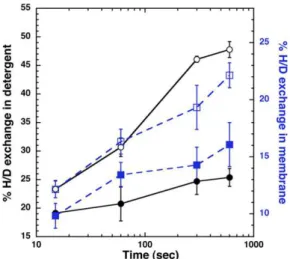
After incubation of BmrA preparations in deuterated buffers, the mass of deuterated BmrA was measured. The percentage of deuterated amides was calculated, taking into account the aver-age of total number of amide hydrogens in BmrA (585 for the native protein) and the mass increase. Curves for deuteration kinetics show the same behavior, with the exchange steadily in-creasing with time for BmrA in both states (Fig. 1). However, BmrA in membranes was approximately half less deuterated (13– 23%) than in its solubilized form (23–47%). This lower deuter-ation exchange in membrane could possibly reflect a more com-pact conformation of the BmrA dimer. However, when the same experiment was performed after prior incubation of BmrA with vanadate in the presence of ATP-Mg (closed symbols in Fig. 1) [i.e., conditions known to promote the formation of a closed di-mer (11)], a lower deuteration level was found for BmrA either in
membrane (squares in Fig. 1) or in detergent (circles in Fig. 1). Thus, although HDX occurs at approximately twice the rate in detergent compared with the membrane-bound BmrA, vanadate inhibition induced a significant decrease in HDX, to a similar extent for both environments. Therefore, this shows that in either state, BmrA can switch between two different conformations: an apo one whereby deuterium exchange readily occurs and a closed one more resistant to deuterium exchange.
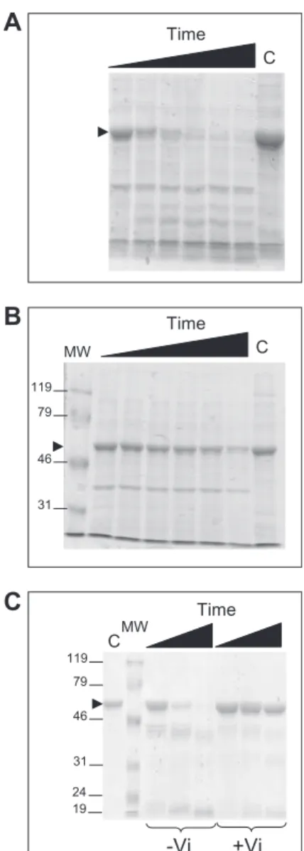
To bring further evidence for the similar behavior of BmrA in detergent and membranes, limited digestion with trypsin was carried out. BmrA in the apo conformation was rapidly digested by trypsin in membranes as well as in DDM (Fig. S3). In contrast, in the closed conformation (+Vi), BmrA was quite resistant to this protease in both environments. Thus, regardless of its en-vironment, BmrA adopts two different conformations with op-posite sensitivity to trypsin.
Peptide Mapping of BmrA. To achieve a sequence coverage as
large as possible, local HDX was performed on purified BmrA because (i) a much higher deuteration level was found for the protein in detergent, and (ii) the presence of Escherichia coli proteins in the membrane fraction would hamper the un-ambiguous identification, or the determination of deuterium incorporation, for some BmrA peptides. Optimized conditions led to a sequence coverage of 78% using pepsin (Table S1). It was lower in the TMD (60%), with the majority of the identified peptides belonging to the hydrophilic ICDs, ICD1 and ICD2 (Fig. S4). In contrast, the NBD sequence was almost fully cov-ered by the pepsin digestion (98%). Because deuteration in-duced the spreading of isotopic envelopes (i.e., the ensemble of isotopic peaks of a peptide), it often led to signal decrease and overlapping. Thus, a significant number of peptides were lost after deuteration, giving 45% and 92% coverage for TMD and NBD, respectively. Importantly, most of the two ICDs were still covered by HDX because these two subdomains are essential for the transmission of the conformational changes between the NBD and the TMD (13, 31, 32).
Comparison of the Closed Forms of BmrA Using HDX. Besides the
inhibition by vanadate, another way to promote the closed con-formation in ABC transporters is to use catalytic inactive mutants (8, 9, 33). For BmrA, we previously showed that the conserved glutamate residue E504 adjacent to the Walker B motif is very likely the catalytic base for ATP hydrolysis, because mutation of this residue fully abrogated the activity (10, 34). Deuterium incorporation was monitored as a function of time and was compared for each peptide generated from the three following forms of BmrA: wild-type in the resting state (apo form), wild-type inhibited by vanadate (in the presence of ATP-Mg), and E504A mutant in the presence of ATP-Mg (Fig. S5). In some cases (e.g., in Fig. 2), spectra from the vanadate-inhibited form showed two envelopes of isotopic peaks, thus giving two different kinetics, with the minor envelope corresponding to that
Fig. 1. Global deuteration kinetics of BmrA. HDX was followed for BmrA in membranes (squares) or purified in DDM (circles), either in the resting state (open symbols) or in a vanadate-inhibited state (filled symbols). SDs for two separate experiments are shown.
Fig. 2. Isotopic profiles of 203–215 peptide at two deuteration times. The mass spectra of peptide 203–215 from ICD2 is shown before (black peaks) or after 15 s (blue peaks) and 3,600 s (red peaks) of deuteration. This peptide was from the wild-type BmrA in the resting state (A) or vanadate-inhibited state (B) or from the E504A mutant incubated in the presence of ATP (C).
Mehmood et al. PNAS | July 3, 2012 | vol. 109 | no. 27 | 10833
BIOCHE
of the wild-type in the apo form. Presumably, this envelope originates from BmrA dimers not inhibited among the whole population, because the inhibition of ATPase activity by vana-date was not total (∼80%;Fig. S1) (see also refs. 17 and 30). In contrast, the E504A mutant showed practically only one isotopic envelope that was always similar to that of the low molecular mass observed in the vanadate-inhibited sample. Given the propensity of the E504A BmrA mutant to readily adopt the closed conformation of BmrA in the presence of ATP-Mg, and the homogeneity of HDX of its peptides, we focused on the differences between this mutant in the closed conformation (ATP-bound state) and the wild-type in the apo conformation (resting state).
Influence of the BmrA Conformation on HDX of the ICDs and NBDs.
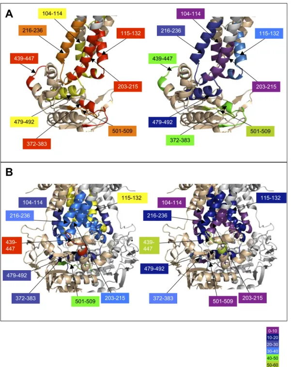
The major differences in HDX between both forms can be vi-sualized on the “heat map” presented inFig. S6. The regions in red or orange correspond to peptides with more than 45% and 30% HDX difference, respectively, after 1 h deuteration. They clearly belong to ICD1, ICD2, and the NBD, particularly the Walker A motif and the ABC signature, which are known to be involved in nucleotide binding. To highlight the HDX data, two BmrA models (28) based on the X-ray structures of homologous bacterial ABC transporters were used (Fig. 3). For the apo conformation of BmrA, the open structure of EcMsbA was used as a template, whereas the closed conformation of BmrA (mimicked by the ATP-bound E504A mutant) was modeled on the Sav1866 structure (13, 14). In agreement with the closed model, the regions participating in either NBD dimerization or direct interaction with ATP/Mg show a rather low level of HDX. This is the case, for instance, for the peptide 372–383, which entirely covers the Walker A motif (HDX of 29%), or for the peptide 479–492 encompassing the ABC signature (26%). In contrast, the former peptide became much more rapidly deu-terated in the apo conformation with a high deuteration level of the Walker A motif (82%), consistent with the presence of a large accessible loop, whereas the peptide covering the ABC signature motif also showed a substantial increase in its exchange rate (69%). The lowest value of HDX obtained for this second peptide suggests that in the open conformation, the region likely conserved an α-helical structure less prone to HDX than a loop. Thus, the exchange rates for these two peptides are in agreement with the open model used to depict the apo conformation
(Fig. 3). For other peptides of the NBDs known to be involved in ATP binding or NBD dimerization, similar trends were ob-served, with a faster exchange rate in the apo conformation compared with the ATP-bound conformation (e.g., peptide 501– 509 including most of the Walker B motif). Other regions of the NBDs not involved in either process gave similar HDX in both conformations with, for instance, a high deuteration level con-sistent with a loop structure (84%) for peptide 439–447.
A different picture emerges when one focuses on ICDs. In contrast to the two models suggesting that these domains have a similar conformation in the two states, open and closed, pep-tides from both ICDs showed a large difference in the HDX in these two conformations. Thus, ICD2 was highly deuterated in the apo conformation, with 82% and 79% for peptides 203–215 and 216–236, respectively, whereas in the closed conformation the HDX rate significantly decreased to 33% and 34%, re-spectively. Likewise, but to a lower extent, regions 104–114 and 115–132 from ICD1 were also fairly deuterated in the apo con-formation (66% and 81%, respectively). This value dropped to 27% in the closed state for the former peptide, whereas it remained highly deuterated for the latter (69%). Because the solvent accessibility of amide hydrogens from both ICDs is es-sentially unaffected upon the dimerization of the two NBDs (e.g., for peptides 104–114 and 203–215) (Fig. S7), the much faster rate of HDX observed in the resting state for these peptides must reflect a modification of conformation and/or flexibility of each ICD. It must be noted that in the closed state, some residues of ICD1 interact with ICD2 within the same subunit (see ref. 13) and thus will help to restrict the motion of the two ICDs.
To further assess the flexibility of these two domains in the 3D structures of the related transporters used to model BmrA, mo-lecular dynamics simulations of ∼100 ns were performed on EcMsbA and Sav1866, along with that of StMsbA that was in-cluded for comparison. After reaching a plateau for the rmsd of the Cα for each conformation, the fluctuation of the position of the Cα atoms were calculated for the last 20 ns of the trajectories, and the results are shown inFig. S8. Clearly, a minor fluctuation was observed in the closed conformations of either Sav1866 or StMsbA, regardless of the Cα considered (always below 2.5 Å). In contrast, some basal fluctuations occurred throughout the se-quence of EcMsbA and were much more pronounced (above 4 Å) for four areas, including in particular ICD2. This suggests that this latter domain must be quite flexible in this apo conformation.
Fig. 3. HDX of peptides using BmrA 3D models. 3D models of BmrA in the open conformation (Left) or in the closed conformation (Right). One subunit was drawn in wheat color and the other in light gray. Identified peptides are drawn with rainbow colors (in one monomer and in all ICDs) according to their percentage of deuterium exchange after 3,600 s (scale of exchange shown at right). Selected regions are also shown with arrows.
Discussion
The goal of this study was to get insights into the dynamics of a multidrug ABC transporter in two different conformations: the resting state, in which the transporter adopts presumably an open conformation (the extent of which remains to be determined), and the closed state, in which the two NBDs dimerize in a transient, ATP-bound, conformation. Overall, global HDX and trypsin di-gestion show that either in membrane or in detergent, BmrA can switch between two very different conformations during its catalytic cycle: a closed form rather resistant to trypsin digestion and HDX, and another one in the resting state quite sensitive to trypsin and more susceptible to HDX. Thus, our results support the postulate that multidrug ABC transporters can cycle between an open, in-ward-facing, conformation in the resting state and a closed, out-ward-facing, ATP-bound conformation. Similar conclusions were drawn for the catalytic cycle of MsbA from cross-linking experi-ments (35) or from EPR spectroscopy (17, 36, 37). On the basis of 3D structures of ABC exporters, the transition between these two opposite conformations was proposed to operate according to a rigid body motion (14). This entails that the same overall structure of the two monomers (or moieties for full-length transporters) would be maintained throughout the catalytic cycle. However, local HDX obtained for BmrA clearly challenges this assumption and suggests that in the resting state, BmrA samples different con-formations. However, these different conformations seem to be restricted to both ICDs. Indeed, apart from the peptides involved either in the interactions between the two NBDs or with ATP, no major changes are observed for the other peptides from either the NBD or the transmembrane helices, although limited information is available for the latter. Therefore, the interaction between the TMD and the NBD that is secured by the two ICDs in the closed state must become loose in the resting state owing to a greater flexibility of these ICDs. This would allow some reorientation of the whole NBD compared with the TMD and would explain some unexpected cross-links previously observed for BmrA. It was found that disulfide bonds were formed between ICD1 and the Q-loop of the NBD in the resting state only (20). However, in all structures of exporters, either closed or open, ICD2 is sandwiched between ICD1 and the Q-loop, thereby impeding the possible interaction between these two protein segments (12–14). Hence, some re-organization of BmrA domains would be required to allow the cross-link between ICD1 and the Q-loop (20). Other unpredicted cross-links questioning the conformation observed in 3D structures were also reported for two other ABC transporters, TAP1/TAP2 and Yor1p (15, 21). These results lend support to additional con-formations of these transporters, with different orientations of the NBD relative to ICDs or of the two ICDs with respect to each other. Other studies performed on homodimeric ABC transporters, LmrA and MsbA, using either solid-state NMR or EPR spectros-copy, reached similar conclusions of alternative conformations (or greater flexibility) of the NBD in the resting state, as opposed to the TMD (36, 38). In addition to the NBD flexibility, EPR experiments also support a large conformational entropy of residues from ICD1 in the resting state, as evidenced by the large distance distribution of the spin label probe tethered to residue 115 of MsbA (36). Un-fortunately, the flexibility of ICD2 could not be tested because the tethering of a spin probe to cysteine residues in this domain was detrimental to MsbA activity (37). For the cystic fibrosis trans-membrane conductance regulator (CFTR), although cross-link patterns support a global architecture similar to that found in Sav1866 or MDR1, a considerable flexibility at the interface of NBDs and ICDs was proposed to explain the formation of cross-links between these two domains, even with long cross-linker re-agent (up to 24.7 Å) (16). In addition, a recent report using a genetic screen with the yeast ABC multidrug transporter PDR5 supports the idea of a noncanonical coupling between the NBD and the TMD as being mainly achieved through ICD1 and not through ICD2 (39).
Whether this is a peculiar property of PDR5 or reflects an alternative conformation of the ICDs remains to be investigated. Interestingly, a molecular dynamics study of Sav1866 indicated that in a closed conformation and among residues whose Cα show the largest dis-placement between the ATP and ADP+Pi bound states, some be-long to ICD2 (residues 221–222) and to ICD1 (residues 110–113) (40). Here, molecular dynamics simulations suggest that in the apo conformation of EcMsbA, large fluctuations in the positions of the Cα atoms of ICD2 would take place. Conformational flexibility, and in particular of α-helices, has been reported for some membrane proteins, such as the lactose permease (41, 42), the osmoprotectant symporter ProP (43), and the glycerol facilitator (44), and this property likely pertains to the transport of their respective substrates (45). For multidrug ABC transporters, the flexibility of ICDs might not only be involved in the transport mechanism but possibly also permits the polyspecificity of drug recognition, provided that this flexibility could be propagated to the drug-binding site(s).
HDX coupled to MS is commonly used to probe the dynamics of soluble proteins, but this technique remains artful in the case of membrane proteins, especially because detergent often reduces the sequence coverage. Up to now, the only ABC transporter for which this technique was used was the soluble NBD1 of CFTR (46). However, our results on BmrA emphasize how useful this technique is, especially to study membrane proteins with large extramembranous domains. This includes, for example, secondary transporters of the resistance-nodulation-cell division family, such as AcrB (47), or primary transporters of the P-type family like the Ca2+-ATPase (48). The use of HDX coupled to MS to unravel different conformations of these membrane proteins should shed new light on their transport mechanism.
Materials and Methods
Site-Directed Mutagenesis and Purification of BmrA. The E504A BmrA mutant was constructed as previously described (10). The E504A mutant and wild-type BmrA were overexpressed as previously described (49), and BmrA-enriched membranes were either frozen in liquid nitrogen and kept at −80 °C until used or solubilized in 1% DDM. After solubilization, BmrA was purified by Ni-affinity chromatography as previously described (29).
HDX of BmrA. The HDX reaction was initiated by a 10× dilution into deu-terated buffer containing 50 mM NaCl and 0.05% DDM. Time course of the HDX was followed over a 1-h period by sequential withdrawing of 120 μL deuterated samples, which were immediately added to 26 μL of quenching buffer (8 M guanidium chloride, 500 mM glycine HCl, pH 2.2), rapidly mixed, and flash-frozen in liquid nitrogen.
HPLC Peptide Separation. After protease digestion in solution or on-column in an ice bath at 0 °C, peptides were loaded onto a peptide MicroTrap (Michrom Bioresources) column and washed with 0.03% TFA in water (HPLC solution A). They were then separated on a reversed-phase C12 column (1 × 50 mm, Ju-piter; Phenomenex) using a linear gradient of 15–45% (vol/vol) solution B (CH3CN 95% and TFA 0.03%) during 26 min. The column was connected to the
electrospray source of mass spectrometers until 30% (vol/vol) B and then disconnected to avoid pollution of the electrospray source by the detergent. The valves, trap cartridge and column were cooled to 0 °C by immersion in an ice bath.
Mass Spectrometric Analyses of Peptides. The tandem MS (mapping) analyses were performed on an ion trap mass spectrometer (Esquire 3000+; Bruker Daltonics) to enable the identification of the peptides after their sepa-ration on HPLC. Accurate mass measurements and the analysis of the local kinetics of deuteration were done on a time-of-flight (TOF) mass spec-trometer (6210; Agilent Technologies) equipped with an electrospray source. Data were processed with MassHunter Qualitative Analysis, and the deconvolution and calculation of the average masses were carried out in Magtran (50).
Electrospray Mass Spectrometry of the Full-Length Protein. The molecular mass of the intact protein was measured with the same electrospray TOF mass spectrometer coupled with HPLC. Protein was loaded onto a protein MicroTrap (Michrom Bioresources) column and washed with 0.03% TFA in water (HPLC
Mehmood et al. PNAS | July 3, 2012 | vol. 109 | no. 27 | 10835
BIOCHE
solution A). The protein and detergent were then separated on a reversed-phase C18 column (1 × 50 mm, Jupiter; Phenomenex) using a linear gradient of 5–60% (vol/vol) solution B (95% CH3CN, 4.97% water, and 0.03% TFA) during 3 min.
ACKNOWLEDGMENTS. We thank Serge Crouzy for his help with the naccess software. The Oxford Supercomputing Centre, the Centre Informatique
National de l’Enseignement Supérieur (France), and the UK national high-per-formance computing service facilities are acknowledged for providing compu-tational resources. This work was initiated with Grants ANR-06-Blan-0420 and ANR-PCV06-135269 from the Agence Nationale de la Recherche and is currently supported by Grant ANR-09-PIRI-0002-01 from this agency (all to J.-M.J.). C.D. received a university research fellowship from the Royal Society. S.M. received a doctoral fellowship from the Higher Education Commission of Pakistan.
1. Rees DC, Johnson E, Lewinson O (2009) ABC transporters: The power to change. Nat Rev Mol Cell Biol 10:218–227.
2. Borst P, Elferink RO (2002) Mammalian ABC transporters in health and disease. Annu Rev Biochem 71:537–592.
3. van Veen HW, et al. (1996) Multidrug resistance mediated by a bacterial homolog of the human multidrug transporter MDR1. Proc Natl Acad Sci USA 93:10668–10672. 4. Lubelski J, Konings WN, Driessen AJ (2007) Distribution and physiology of ABC-type
transporters contributing to multidrug resistance in bacteria. Microbiol Mol Biol Rev 71:463–476.
5. Geourjon C, et al. (2001) A common mechanism for ATP hydrolysis in ABC transporter and helicase superfamilies. Trends Biochem Sci 26:539–544.
6. Seeger MA, van Veen HW (2009) Molecular basis of multidrug transport by ABC transporters. Biochim Biophys Acta 1794:725–737.
7. Chen J, Lu G, Lin J, Davidson AL, Quiocho FA (2003) A tweezers-like motion of the ATP-binding cassette dimer in an ABC transport cycle. Mol Cell 12:651–661. 8. Smith PC, et al. (2002) ATP binding to the motor domain from an ABC transporter
drives formation of a nucleotide sandwich dimer. Mol Cell 10:139–149.
9. Zaitseva J, Jenewein S, Jumpertz T, Holland IB, Schmitt L (2005) H662 is the linchpin of ATP hydrolysis in the nucleotide-binding domain of the ABC transporter HlyB. EMBO J 24:1901–1910.
10. Orelle C, Dalmas O, Gros P, Di Pietro A, Jault JM (2003) The conserved glutamate residue adjacent to the Walker-B motif is the catalytic base for ATP hydrolysis in the ATP-binding cassette transporter BmrA. J Biol Chem 278:47002–47008.
11. Urbatsch IL, Tyndall GA, Tombline G, Senior AE (2003) P-glycoprotein catalytic mechanism: Studies of the ADP-vanadate inhibited state. J Biol Chem 278: 23171–23179.
12. Aller SG, et al. (2009) Structure of P-glycoprotein reveals a molecular basis for poly-specific drug binding. Science 323:1718–1722.
13. Dawson RJ, Locher KP (2006) Structure of a bacterial multidrug ABC transporter. Nature 443:180–185.
14. Ward A, Reyes CL, Yu J, Roth CB, Chang G (2007) Flexibility in the ABC transporter MsbA: Alternating access with a twist. Proc Natl Acad Sci USA 104:19005–19010. 15. Oancea G, et al. (2009) Structural arrangement of the transmission interface in the
antigen ABC transport complex TAP. Proc Natl Acad Sci USA 106:5551–5556. 16. Serohijos AW, et al. (2008) Phenylalanine-508 mediates a cytoplasmic-membrane
domain contact in the CFTR 3D structure crucial to assembly and channel function. Proc Natl Acad Sci USA 105:3256–3261.
17. Borbat PP, et al. (2007) Conformational motion of the ABC transporter MsbA induced by ATP hydrolysis. PLoS Biol 5:e271.
18. Gottesman MM, Ambudkar SV, Xia D (2009) Structure of a multidrug transporter. Nat Biotechnol 27:546–547.
19. Kerr ID, Jones PM, George AM (2010) Multidrug efflux pumps: The structures of prokaryotic ATP-binding cassette transporter efflux pumps and implications for our understanding of eukaryotic P-glycoproteins and homologues. FEBS J 277:550–563. 20. Dalmas O, et al. (2005) The Q-loop disengages from the first intracellular loop during
the catalytic cycle of the multidrug ABC transporter BmrA. J Biol Chem 280: 36857–36864.
21. Pagant S, Brovman EY, Halliday JJ, Miller EA (2008) Mapping of interdomain inter-faces required for the functional architecture of Yor1p, a eukaryotic ATP-binding cassette (ABC) transporter. J Biol Chem 283:26444–26451.
22. Zhang Z, Smith DL (1993) Determination of amide hydrogen exchange by mass spectrometry: A new tool for protein structure elucidation. Protein Sci 2:522–531. 23. Wales TE, Engen JR (2006) Hydrogen exchange mass spectrometry for the analysis of
protein dynamics. Mass Spectrom Rev 25:158–170.
24. Konermann L, Pan J, Liu YH (2011) Hydrogen exchange mass spectrometry for studying protein structure and dynamics. Chem Soc Rev 40:1224–1234.
25. Morgan CR, et al. (2011) Conformational transitions in the membrane scaffold pro-tein of phospholipid bilayer nanodiscs. Mol Cell Proteomics 10:M111.010876. 26. Rey M, et al. (2010) Conformational dynamics of the bovine mitochondrial ADP/ATP
carrier isoform 1 revealed by hydrogen/deuterium exchange coupled to mass spec-trometry. J Biol Chem 285:34981–34990.
27. West GM, et al. (2011) Ligand-dependent perturbation of the conformational en-semble for the GPCR β2 adrenergic receptor revealed by HDX. Structure 19: 1424–1432.
28. Do Cao MA, et al. (2009) Probing the conformation of the resting state of a bacterial multidrug ABC transporter, BmrA, by a site-directed spin labeling approach. Protein Sci 18:1507–1520.
29. Steinfels E, et al. (2004) Characterization of YvcC (BmrA), a multidrug ABC transporter constitutively expressed in Bacillus subtilis. Biochemistry 43:7491–7502.
30. Ravaud S, et al. (2006) The ABC transporter BmrA from Bacillus subtilis is a functional dimer when in a detergent-solubilized state. Biochem J 395:345–353.
31. Hrycyna CA (2001) Molecular genetic analysis and biochemical characterization of mammalian P-glycoproteins involved in multidrug resistance. Semin Cell Dev Biol 12: 247–256.
32. Kwan T, Gros P (1998) Mutational analysis of the P-glycoprotein first intracellular loop and flanking transmembrane domains. Biochemistry 37:3337–3350.
33. Oldham ML, Khare D, Quiocho FA, Davidson AL, Chen J (2007) Crystal structure of a catalytic intermediate of the maltose transporter. Nature 450:515–521. 34. Orelle C, et al. (2008) Conformational change induced by ATP binding in the
multi-drug ATP-binding cassette transporter BmrA. Biochemistry 47:2404–2412. 35. Doshi R, Woebking B, van Veen HW (2010) Dissection of the conformational cycle of
the multidrug/lipidA ABC exporter MsbA. Proteins 78:2867–2872.
36. Zou P, Bortolus M, McHaourab HS (2009) Conformational cycle of the ABC transporter MsbA in liposomes: Detailed analysis using double electron-electron resonance spectroscopy. J Mol Biol 393:586–597.
37. Zou P, McHaourab HS (2009) Alternating access of the putative substrate-binding chamber in the ABC transporter MsbA. J Mol Biol 393:574–585.
38. Siarheyeva A, et al. (2007) Probing the molecular dynamics of the ABC multidrug transporter LmrA by deuterium solid-state nuclear magnetic resonance. Biochemistry 46:3075–3083.
39. Ananthaswamy N, et al. (2010) The signaling interface of the yeast multidrug trans-porter Pdr5 adopts a cis conformation, and there are functional overlap and equiv-alence of the deviant and canonical Q-loop residues. Biochemistry 49:4440–4449. 40. Oliveira AS, Baptista AM, Soares CM (2011) Conformational changes induced by
ATP-hydrolysis in an ABC transporter: A molecular dynamics study of the Sav1866 ex-porter. Proteins 79:1977–1990.
41. Patzlaff JS, Moeller JA, Barry BA, Brooker RJ (1998) Fourier transform infrared analysis of purified lactose permease: A monodisperse lactose permease preparation is stably folded, alpha-helical, and highly accessible to deuterium exchange. Biochemistry 37: 15363–15375.
42. le Coutre J, Kaback HR, Patel CK, Heginbotham L, Miller C (1998) Fourier transform infrared spectroscopy reveals a rigid alpha-helical assembly for the tetrameric Streptomyces lividans K+ channel. Proc Natl Acad Sci USA 95:6114–6117. 43. Sayeed WM, Baenziger JE (2009) Structural characterization of the osmosensor ProP.
Biochim Biophys Acta 1788:1108–1115.
44. Pan Y, Piyadasa H, O’Neil JD, Konermann L (2012) Conformational dynamics of a membrane transport protein probed by H/D exchange and covalent labeling: The glycerol facilitator. J Mol Biol 416:400–413.
45. le Coutre J, Kaback HK (2000) Structure-function relationships of integral membrane proteins: Membrane transporters vs channels. Biopolymers 55:297–307.
46. Lewis HA, et al. (2010) Structure and dynamics of NBD1 from CFTR characterized using crystallography and hydrogen/deuterium exchange mass spectrometry. J Mol Biol 396:406–430.
47. Murakami S, Nakashima R, Yamashita E, Matsumoto T, Yamaguchi A (2006) Crystal structures of a multidrug transporter reveal a functionally rotating mechanism. Na-ture 443:173–179.
48. Toyoshima C, Mizutani T (2004) Crystal structure of the calcium pump with a bound ATP analogue. Nature 430:529–535.
49. Steinfels E, et al. (2002) Highly efficient over-production in E. coli of YvcC, a multi-drug-like ATP-binding cassette transporter from Bacillus subtilis. Biochim Biophys Acta 1565:1–5.
50. Zhang Z, Marshall AG (1998) A universal algorithm for fast and automated charge state deconvolution of electrospray mass-to-charge ratio spectra. J Am Soc Mass Spectrom 9:225–233.
Supporting Information
Mehmood et al. 10.1073/pnas.1204067109
SI Results
Electrospray Mass Spectrometry of the Full-Length Protein.Because
lipids and detergents either give strong signals in mass spec-trometry (MS) or disturb the protein ionization in the electro-spray source (1), they must be separated from the protein before reaching the source. Previous separations involved gel electro-phoresis and electroelution (2), protein precipitation and re-suspension in concentrated acidic solutions (3), or size exclusion chromatography after dissolution in a chloroform/methanol/acid solution (4). However, although efficient, all these methods were hardly compatible with the requirement of a fast preparation of the samples to limit as much as possible the back exchange re-action after deuterium incorporation into BmrA. Therefore, chromatographic parameters were first optimized to separate membrane lipids or n-dodecyl-β-D-maltoside (DDM) detergent
from the protein using classical reversed phase chromatography (Materials and Methods).
Because of the cloning strategy used (5), purified BmrA was expected to contain three additional residues on its N terminus (sequence MASMPT. . ., with the underlined residues corre-sponding to the beginning of the native protein) and 15 addi-tional residues on its C terminus, including the Histidine tag (sequence . . .KAGVDKLAAALEHHHHHH, with the under-lined residues corresponding to the end of the native protein). Thus, the deconvoluted mass obtained is in very good agreement with the calculated mass of BmrA, possessing only one extra Ser amino acid on its N terminus (66,340 Da). Masses of a second peak (66,412 Da, in membranes) are also in very good agreement with the calculated mass of BmrA, with two extra amino acids on its N terminus, Ala plus Ser (66,411 Da). Therefore, the first methionine and sometimes also the second amino acids (Ala) of the N terminus of the proteins were very likely cleaved off by proteolysis during the purification process. The high quality of each spectrum reflects the efficiency of the chromatographic separation upstream of the electrospray source for either sample.
Mass Spectrometric Analyses of Peptides.To localize the difference
in deuterium incorporation between the two conformations of BmrA, the protein digestion occurred at low pH and low tem-perature, and the peptide separations were optimized (Materials and Methods). This step was essential to reach a sequence cov-erage of the protein as large as possible with peptides as small as possible (6). However, DDM used for BmrA purification gave a very strong signal in MS. To enable observation of the mass spectra originating from the peptides, a protocol previously de-veloped in our group for another membrane protein purified in Triton X100 was first attempted without success (7). Reversed-phase separation was then adjusted so that all peptides eluted before the detergent, at low temperature to limit deuterium back-exchange. For peptides sequencing using tandem MS, the chromatographic parameters were chosen for a good separation to facilitate their identification, and the detergent still eluted after the last peptides. For the analysis of deuterated peptides, the elution times were also shortened as much as possible to limit deuterium back-exchange. Pepsin and rhizopuspepsin (8) were used independently to digest BmrA. The pepsin digestion in solution yielded only 31 peptides, covering 45% of the BmrA sequence (Table S1). However, the use of immobilized pepsin significantly increased the number of peptides (67), and thus the sequence coverage increased to 78%. These results were ob-tained in the presence of 1.5 M guanidinium hydrochloride (GndCl), a denaturant concentration that was optimized after
different trials between 0.5 and 4 M. However, the low coverage of the hydrophobic transmembrane helices is presumably due to the presence of the detergent that might afford protection against the protease digestion. Immobilized rhizopuspepsin was also used for on-line digestion of BmrA. The goal was to obtain complementary sequence coverage to improve spatial resolution by the occurrence of overlapping peptides. The number of peptides and the sequence coverage were significantly lower in the transmembrane domain (TMD) in comparison with immo-bilized pepsin (Fig. S4 and Table S1). In the nucleotide-binding domain (NBD), the coverage seemed quite complementary to the one obtained by pepsin. However, after deuteration a sig-nificant number of peptides were lost with each protease, a phenomenon that is frequently observed in hydrogen/deute-rium exchange (HDX) (9). This is due to a heterogeneous deuteration that induces a broadening of the MS signal, and thus a decrease in its intensity. The loss was much higher in the case of rhizopuspepsin, which was also less efficient than pepsin. SI Materials and Methods
Materials.Unless specified, chemicals and pepsin were purchased
from Sigma-Aldrich. DDM was purchased from Anatrace.
Site-Directed Mutagenesis and Purification of BmrA.Protein
con-centrations were estimated by using a modified Lowry method (10) for BmrA-enriched membranes, or the Bradford method (11) after purification. The accurate masses of the purified mu-tant and wild-type proteins were checked using electrospray MS.
Pepsin Digestion.All protein digestions in solution were performed
in an ice bath at 0 °C. Protease solutions were prepared in 500 mM glycine (pH 2.2) and cooled to 0 °C. BmrA was digested in the same buffer for 2–5 min using a protease/substrate ratio of 1:1 or 1:10 (wt/wt) for pepsin and rhizopuspepsin, respectively. Increase in digestion time did not have any effect on the pro-teolysis. On-line digestions in the presence of 0.5–4 M GndCl were also performed in an ice bath at 0 °C using columns packed with rhizopuspepsin or pepsin immobilized on POROS-AL resin (8).
Limited Proteolysis of BmrA.BmrA was submitted to trypsin
di-gestion at a protease/substrate ratio of 1:1,000 (wt/wt). For BmrA solubilized in detergent a reaction mixture of 100 μL was pre-pared, and aliquots of 10 μL were withdrawn after 5, 15, and 30 min. For the vanadate-inhibited state, 5 mM ATP, 5 mM MgCl2, and 3 mM vanadate were added to the protein solution and incubated for 15 min at room temperature before adding trypsin. For digestion of BmrA-enriched membranes, a reaction mixture of 100 μL was prepared, and aliquots of 10 μL were withdrawn after 5, 15, and 30 min, 1 h, 2 h, and 3 h. Each aliquot was acidified with 1 μL of 5% TFA and immediately frozen in liquid nitrogen. SDS/PAGE (12.5%) was resolved, followed by Coo-massie blue staining.
HDX of BmrA.The vanadate-inhibited state of the wild-type BmrA
was prepared as above for limited proteolysis experiments. The E504A mutant was first incubated for 10 min with 5 mM ATP and 5 mM MgCl2.
ATPase Activity. To measure the ATPase activity of BmrA in
detergent, an enzymatic coupled assay system was used, in the presence of 0.05% DDM and as described previously (12). The activity was measured in the presence of 10–20 μg of BmrA and followed at 37 °C for 10 min. When vanadate was present, it was
added at a final concentration of 3 mM from a 100-mM stock solution (see ref. 12).
Calculation of Accessibility Areas of Peptides.The solvent
accessi-bility area of BmrA peptides was calculated with the help of naccess. This software, based on the method of Lee and Richards (13), calculates the solvent-accessible area of a protein structure in a Protein Data Bank (PDB) format. Then, on the basis of the accessibility of each atom, the total access area of the main chain atoms was determined. To compare the solvent accessibility val-ues with HDX MS data, the mean solvent accessibility area of individual peptides was determined by taking into account the main chain atoms only. Because region from residue 325 to res-idue 332 shows the most solvent-accessible surface in both models, the values for all other peptides were normalized con-sidering the peptide 325–332 as a 100% solvent-accessible region.
Molecular Dynamics (MD) Simulations.MD trajectories were
simu-lated with version 2.7 of NAMD (14), using the CHARMM27 force field with CMAP corrections (15), and the TIP3 (transferable in-termolecular three-point) model for water molecules (16). All of the simulations were performed in the NpT ensemble. The pres-sure was maintained at 1 atm using a Nose-Hoover Langevin piston control (17), with period of 100 fs and damping time constant of 50 fs. Temperature was maintained at 300 K by coupling to a Langevin thermostat, with damping coefficient of 5 ps−1. Elec-trostatic interactions were treated by the Particle Mesh Ewald al-gorithm (18), with grid spacing lower than 1 Å. Smoothed cutoff (10–12 Å) was used for the van der Waals interactions. Equations of motion were integrated with a time step of 2 fs. The SETTLE algorithm was used to restrain hydrogen atoms (19).
Three systems were considered: (i) the Sav1866 multidrug transporter from Staphylococcus aureus at 3.0 Å in an outward-facing conformation reflecting the ATP-bound state (20), (ii) the MsbA lipid “flippase” either from Escherichia coli in the open apo-conformation at 5.3 Å, or (iii) from Salmonella typhimurium in the outward-facing conformation at 3.7 Å (21). The structures were defined according to PDB files 2HYD, 3B5W, and 3B60,
respectively, together with the crystallographic water molecules. Only the Cα-trace structure is available in 3B5W. Missing backbone and side-chain atoms were predicted using MAXSPROUT (22) and SCWRL (23). An acetyl group was attached to the N terminus residue to mimic the preceding peptide bond, and the C-terminal carboxylate was protonated. Default ionization states at pH 7 were set, except for buried residues whose ionization states were de-termined using WHATIF (24) and visual inspection. The proteins were centered in the x–y plane, with the permeation pathway aligned to the z axis, and embedded in a preequilibrated bilayer of 569 1,2-dioleoyl-sn-glycero-3-phosphocholine (DOPC) molecules. The upper layer of the lipid membrane was aligned to the center of mass along z of the aromatic residues of the extracellular side (Phe158, Phe157, Tyr39, and Tyr268 in Sav1866, and Trp165, Trp66, Tyr162, Tyr168, Phe157, Phe265, and Phe272 in MsbA). Lipid molecules closer than 1.2 Å to protein atoms were removed. Sodium and chloride ions were added to neutralize the system (up to a final concentration of 150 mM). The final systems contain ∼195,000 atoms in the Sav1866 simulation (∼41,800 TIP3P water molecules, 360 DOPC lipids, 61Cl−and 59Na+), ∼202,000 atoms in the simulation of MsbA PDB 3B60 (∼42,800 TIP3P water mole-cules, 402 DOPC lipids, 55Cl−and 63Na+), and ∼244 atoms in the simulation of MsbA PBD 3B5W (∼53,500 TIP3P water molecules, 472 DOPC lipids, 71Cl−and 79Na+).
To equilibrate the atoms around the transporters, 2,000 steps of energy minimization and 1 ns of MD were performed with restraints applied to the backbone atoms of the protein. Restraints were initially set to 10 kcal*mol−1*Å−2, and gradually reduced to zero. Unrestrained MD simulation followed for ∼82 ns (MsbA 3B5W), ∼88 ns (MsbA 3B60), and ∼93 ns (Sav1866). The atomic systems used for the analyses were from the last 20 ns of the unrestrained MD. A significant drift that stabilizes at 6.7 ± 0.2 Å after 60 ns and remains constant until the end of the simulation is observed in the rmsd of MsbA (3B5W). The rmsd values of the MsbA (3B60) and Sav1866 simulations reached a plateau in less than 3 ns, with average values of 2.4 ± 0.1 Å and 1.9 ± 0.1, respectively.
1. Barnidge DR, Dratz EA, Jesaitis AJ, Sunner J (1999) Extraction method for analysis of detergent-solubilized bacteriorhodopsin and hydrophobic peptides by electrospray ionization mass spectrometry. Anal Biochem 269:1–9.
2. le Maire M, Deschamps S, Møller JV, Le Caer JP, Rossier J (1993) Electrospray ionization mass spectrometry on hydrophobic peptides electroeluted from sodium dodecyl sulfate-polyacrylamide gel electrophoresis application to the topology of the sarcoplasmic reticulum Ca2+ ATPase. Anal Biochem 214:50–57.
3. Whitelegge JP, Gundersen CB, Faull KF (1998) Electrospray-ionization mass spectrometry of intact intrinsic membrane proteins. Protein Sci 7:1423–1430. 4. Whitelegge JP, et al. (1999) Toward the bilayer proteome, electrospray
ionization-mass spectrometry of large, intact transmembrane proteins. Proc Natl Acad Sci USA 96:10695–10698.
5. Steinfels E, et al. (2004) Characterization of YvcC (BmrA), a multidrug ABC transporter constitutively expressed in Bacillus subtilis. Biochemistry 43:7491–7502.
6. Zhang Z, Smith DL (1993) Determination of amide hydrogen exchange by mass spectrometry: A new tool for protein structure elucidation. Protein Sci 2:522–531. 7. Rey M, et al. (2010) Conformational dynamics of the bovine mitochondrial ADP/ATP
carrier isoform 1 revealed by hydrogen/deuterium exchange coupled to mass spectrometry. J Biol Chem 285:34981–34990.
8. Rey M, et al. (2009) Recombinant immobilized rhizopuspepsin as a new tool for protein digestion in hydrogen/deuterium exchange mass spectrometry. Rapid Commun Mass Spectrom 23:3431–3438.
9. Marcoux J, et al. (2010) p47phox molecular activation for assembly of the neutrophil NADPH oxidase complex. J Biol Chem 285:28980–28990.
10. Bensadoun A, Weinstein D (1976) Assay of proteins in the presence of interfering materials. Anal Biochem 70:241–250.
11. Bradford MM (1976) A rapid and sensitive method for the quantitation of microgram quantities of protein utilizing the principle of protein-dye binding. Anal Biochem 72: 248–254.
12. Galián C, et al. (2011) Optimized purification of a heterodimeric ABC transporter in a highly stable form amenable to 2-D crystallization. PLoS ONE 6:e19677. 13. Lee B, Richards FM (1971) The interpretation of protein structures: Estimation of static
accessibility. J Mol Biol 55:379–400.
14. Phillips JC, et al. (2005) Scalable molecular dynamics with NAMD. J Comput Chem 26: 1781–1802.
15. MacKerell AD, et al. (1998) All-atom empirical potential for molecular modeling and dynamics studies of proteins. J Phys Chem B 102:3586–3616.
16. Jorgensen WL, Chandresekhar J, Madura JD, Impey RW, Klein ML (1983) Comparison of simple potential functions for simulating liquid water. J Chem Phys 79:926–935. 17. Feller SE, Zhang YH, Pastor RW, Brooks BR (1995) Constant-pressure molecular
dynamics simulation—the langevin piston method. J Chem Phys 103:4613–4621. 18. Essmann U, et al. (1995) A smooth particle mesh Ewald method. J Chem Phys 103:
8577–8593.
19. Miyamoto S, Kollman PA (1992) Settle: An analytical version of the Shake and Rattle algorithm for wigid water molecules. J Comput Chem 13:952–962.
20. Dawson RJ, Locher KP (2006) Structure of a bacterial multidrug ABC transporter. Nature 443:180–185.
21. Ward A, Reyes CL, Yu J, Roth CB, Chang G (2007) Flexibility in the ABC transporter MsbA: Alternating access with a twist. Proc Natl Acad Sci USA 104:19005–19010. 22. Holm L, Sander C (1991) Database algorithm for generating protein backbone and
side-chain co-ordinates from a C alpha trace application to model building and detection of co-ordinate errors. J Mol Biol 218:183–194.
23. Canutescu AA, Shelenkov AA, Dunbrack RL, Jr. (2003) A graph-theory algorithm for rapid protein side-chain prediction. Protein Sci 12:2001–2014.
24. Vriend G (1990) WHAT IF: A molecular modeling and drug design program. J Mol Graph 8:52–56, 29.
ATPase a
ctivity
(%)
WT
WT+Vi
Fig. S1. ATPase activity of the wild-type BmrA. BmrA (10–20 μg) was injected into an ATPase assay medium in the absence (blue bar, WT) or presence of 3 mM vanadate (purple bar, WT + Vi). One hundred percent of ATPase activity corresponded to 1 μmol of ATP hydrolyzed/min/mg protein.
1 x10 0 0,2 0,4 0,6 0,8 1 1,2 1,4 1,6 1,8 2 2,2 750 800 850 900 950 1000 1050 1100 1150 1200 1250 1300 1350 1400 2 x10 0 0,1 0,2 0,3 0,4 0,5 0,6 0,7 0,8 0,9 1 1,1 66341
Deconvoluted Mass (amu)
50000 60000 70000 80000 90000 100000
A
Mass-to-Charge (m/z) 2 x10 0 0,1 0,2 0,3 0,4 0,5 0,6 0,7 0,8 0,9 1 750 800 850 900 950 1000 1050 1100 1150 1200 1250 1300 1350 1400 1021.592 2 x10 0 0,5 1 1,5 2 2,5 3 3,5 4 4,5 66339Deconvoluted Mass (amu)
60000 70000 80000 90000 100000
B
Mass-to-Charge (m/z)
50000
Fig. S2. Raw mass spectra of BmrA in membranes (A) or BmrA purified in DDM (B). Deconvoluted mass spectra are presented in First Insets with a close-up view in Second Insets.
MW C Time C Time
A
-Vi +ViB
Time CMWC
24 19 119 79 46 31 119 79 46 31Fig. S3. SDS/PAGE of BmrA-enriched membranes or BmrA purified in DDM and after limited proteolysis with trypsin. BmrA in membranes in the apo con-formation (A) or in the vanadate-inhibited concon-formation (B) and after incubation with trypsin for 5 min, 15 min, 30 min, 1 h, 2 h, and 3 h (from left to right lanes); (C) BmrA purified in detergent in the apo conformation (lanes 3–5) and in the vanadate-inhibited conformation (lanes 6–8) and after incubation with trypsin for 5, 15, and 30 min (from left to right lanes). C is the control lane with no trypsin added, with the black arrowhead showing the position of the full-length BmrA. The masses of the molecular weight markers (MW) are indicated in kDa.
TMH1 TMH2 ICD1 TMH4 ICD2 TMH3 TMH5 TMH6 WB ABC WA
Fig. S4. Peptide mapping of BmrA using immobilized pepsin (blue) or immobilized rhizopuspepsin (red). The predicted secondary structure (cylinders for α-helices and arrows for β-strands) and topology of BmrA based on the Sav1866 3D structure is shown above the sequence with the specific regions: Walker A (WA) and B (WB) motifs and the ATP-binding cassette signature. The transmembrane helices 1–6 (TMH1 to -6) are shown in green, and the positions of the intracellular domains 1 and 2 (ICD1 and ICD2, respectively) are indicated. The extracellular parts of BmrA are shown in gray. The boundary between the TMD and the NBD is shown by a vertical dotted line. The figure was drawn with the help of the MS Tools application (1).
1. Kavan D, Man P (2011) MSTools—Web based application for visualization and presentation of HXMS data. Int J Mass Spectrom 302:53–58.
Fig. S5. (Continued)
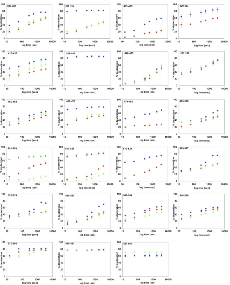
Fig. S5. HDX kinetics of all BmrA peptides identified after digestion with immobilized pepsin. The percentage of deuteration is plotted against different deuteration times for the wild-type BmrA in the apo state (blue circles), in the vanadate-inhibited state (green triangles), or in the mutated (E504A) form (red circles). SDs (for two experiments) are shown by vertical bars; in many cases, bars are so small that they are hidden by the symbols; in the vanadate-inhibited state, the first isotopic envelope was sometimes too tiny or too close to the second one to be deconvoluted, and the kinetics could not be drawn with certainty.
TMH4 ICD2 TMH3 TMH5 TMH6 WA WB ABC TMH1 TMH2 ICD1 0-15 15-30 30-45 45-60
Fig. S6. Heat map showing the difference in deuterium incorporation between the apo conformation (wild-type in the resting state) and the closed con-formation (E504A in the presence of ATP) after 1 h deuteration. The predicted secondary structure and topology of BmrA (see legend to Fig. S4) is indicated above the sequence. Colors defined in key at lower right.
203-215 216-236 372-383 104-114 439-447 479-492 501-509 115-132
A
B
439-447 479-492 115-132 501-509 372-383 216-236 203-215 104-114 0-10 10-20 20-30 30-40 40-50 50-60 60-70 70-80 80-90 439-447 479-492 115-132 501-509 372-383 216-236 203-215 104-114 203-215 216-236 372-383 104-114 439-447 479-492 501-509 115-132Fig. S7. Comparison of deuterium incorporation and solvent accessibility areas in both conformations of BmrA, open (A) or closed (B). Panels at Left are identical to those shown in Fig. 3 for the HDX, except that only the selected peptides are colored according to the deuterium exchange scale. Right: Solvent accessibility of the corresponding peptides deduced from each BmrA model and using naccess software (details in SI Materials and Methods).
Sav1866 EcMsbA StMsbA RMS fluctuations ( Å ) residue number ABCWB WA ICD1 ICD2
Fig. S8. Root mean square fluctuation of the Cα of Sav1866, StMsbA, and EcMsbA. Molecular dynamics simulations were performed on each structure and, once equilibrium was reached, the rms fluctuation in Å was calculated for the last 20 ns and is shown as a function of the residue number. The predicted topology of BmrA is also shown on top of the figure for comparison (see legend to Fig. S4 for color coding).
Table S1. Sequence coverage, number of peptides, and average size of the peptides for BmrA and for its domains TMD and NBD obtained in different digestion conditions and after deuteration and digestion with immobilized pepsin
Condition BmrA TMD NBD Pepsin column % coverage 78 60 98 Peptides 67 24 43 Average 7.0 8.0 6.4 Pepsin in solution % coverage 45 11 82 Peptides 31 3 28 Average 8.7 12.6 8.3 Rhizopuspepsin column % coverage 50 10 84 Peptides 52 8 44 Average 5.8 8.0 5.4 HDX % coverage 67.5 45 92 Peptides 44 15 29 Average 9.3 9.0 9.3