HAL Id: tel-02015610
https://tel.archives-ouvertes.fr/tel-02015610
Submitted on 12 Feb 2019HAL is a multi-disciplinary open access archive for the deposit and dissemination of sci-entific research documents, whether they are pub-lished or not. The documents may come from teaching and research institutions in France or abroad, or from public or private research centers.
L’archive ouverte pluridisciplinaire HAL, est destinée au dépôt et à la diffusion de documents scientifiques de niveau recherche, publiés ou non, émanant des établissements d’enseignement et de recherche français ou étrangers, des laboratoires publics ou privés.
Ultrasound Induced Blood-brain Barrier Opening on
Rodents : from Nanoparticles Delivery to a Therapy for
Alzheimer’s Disease
Matthieu Gerstenmayer
To cite this version:
Matthieu Gerstenmayer. Ultrasound Induced Blood-brain Barrier Opening on Rodents : from Nanoparticles Delivery to a Therapy for Alzheimer’s Disease. Biological Physics [physics.bio-ph]. Université Paris Saclay (COmUE), 2018. English. �NNT : 2018SACLS424�. �tel-02015610�
Ultrasound-Induced
Blood-Brain Barrier Opening on
Rodents: from Nanoparticles
Delivery to a Therapy for
Alzheimer’s Disease
Thèse de doctorat de l'Université Paris-Saclay Préparée à l’université Paris Sud
École doctorale n°575 : Electrical, Optical, Bio – physics and Engineering (EOBE) Spécialité de doctorat: Imagerie et Physique Médicale
Thèse présentée et soutenue à Gif-sur-Yvette, le 12 Novembre 2018, par :
Matthieu GERSTENMAYER
Composition du Jury :
M. Hervé Boutin
Directeur de recherche, University of Manchester (DNEP) Président M. Cyril Lafon
Directeur de recherche, Université Lyon 1 (LabTAU) Rapporteur M. Chrit Moonen
Professeur, University Medical Center Utrecht (Imaging Divison) Rapporteur Mme. Karine Cambon
Ingénieur chercheur, CEA (MIRcen) Examinateur
Me. Marie Sarazin
Professeur, Centre hospitalier Sainte-Anne (NML) Examinateur M. Cyril Poupon
Directeur de recherche, CEA (NeuroSpin) Directeur de thèse M. Benoit Larrat
« Voyage dans le temps et inverse le sens des flots de mon sang
Noyé dans l’océan, je caresse le flanc du grand Leviathan »
Remerciements
Je tiens tout d’abord à remercier Benoit Larrat, qui m’a encadré au quotidien durant ma thèse. Toujours bouillant d’idées, merci de m’avoir conseillé scientifiquement et soutenu moralement. Merci d’avoir été présent et disponible quand j’avais besoin de toi. Enfin merci pour ta bonne humeur et ton encadrement attentif qui m’ont permis de m’épanouir durant ces trois années.
J’exprime ma gratitude à l’égard de Cyril Poupon qui a accepté d’être mon directeur de thèse. J’adresse tous mes remerciements à Cyril Lafon et Chrit Moonen qui m’ont fait l’honneur d’être rapporteurs de ma thèse. Merci à Karine Cambon, Marie Sarazin et Hervé Boutin qui ont bien voulu en être examinateurs.
Je remercie également Sébastien Mériaux pour avoir patiemment répondu à toutes mes questions de reconstructions d’images et pour m’avoir finement conseillé lors de nombreuses répétitions de présentations. Enfin, un grand merci pour ton management constructif et bienveillant de l’équipe MIDAS.
Un grand merci à Erwan Selingue pour avoir subi avec moi les longues semaines d’expérimentation, pour ces journées au son de France Inter et pour ses mythiques blagues. Un grand merci à Françoise pour m’avoir gentiment approvisionné en images histologiques et pour nous avoir fait de délicieux gâteaux.
Un grand merci à tous mes collègues de l’open-space qui ont rendu l’ambiance de travail chaleureuse et agréable, Solenne, Jacques, Pauline, Laura, Anthony, Pavel, Benjamin, Hermès, Marianne, Gabrielle, Ashley, Ricardo. Merci pour toutes ces discussions à la pause-café et à la cantine 2, mais aussi pour ces pique-niques et ces gouters à l’ombre du grand châtaignier.
Merci Rémi de m’avoir formé, un début de thèse n’aurait pas pu se passer mieux. Merci Allegra de m’avoir guidé dans le monde de la recherche, qu’aurais-je fais sans toi perdu dans ces conférences internationales. Merci Venetia pour ton aide précieuse et merci de t’attaquer à toutes les questions auxquelles je n’ai pas pu répondre.
Merci à Sébastien, Sylvain, Emilie, Maud, Nicolas et Charles pour m’avoir chaleureusement accueilli au Service Hospitalier Frédéric Joliot. Merci à Denis et Jérémy pour m’avoir dépanné de nombreuses fois. Merci à Jonathan, Christel, Elena, Philippe, Florian, Florence, Jean-Christophe, Serge pour l’aide apportée sans laquelle ce travail n’aurait pas pu se faire.
Merci à tous mes amis de BSY, d’Orsay et de Paris, merci d’avoir été présents quand les résultats, eux, tardaient à venir. Merci à ma famille de m’avoir toujours fait confiance et de m’avoir toujours soutenu. Enfin, Alice, merci pour tout.
Je dédicace cette thèse à Théophile Parjadis de Larivière qui nous a quitté récemment. Esprit libre, classe incomparable, toujours à fond sur le terrain de rugby, toujours à fond pour faire la fête. Tu vas nous manquer.
Résumé en Français
Contexte de la thèse : l’ouverture transitoire de la barrière hémato-encéphalique par ultrasons Mon travail de thèse a consisté à développer des outils pour l’ouverture de la barrière-hémato encéphalique chez le rat et la souris, soit dans le but de délivrer des molécules dans le cerveau soit comme thérapie pour la maladie d’Alzheimer, qui se caractérise au niveau moléculaire par le dépôt de plaques amyloïdes et par la dégénérescence neuro-fibrillaire. Dans le reste du corps, pour permettre les échanges moléculaires entre le sang et les organes, les vaisseaux sanguins sont relativement perméables. Le cerveau est un organe à part. Les vaisseaux sanguins qui l’irriguent sont beaucoup plus imperméables. Cette barrière, limitant le transport de molécules entre le sang et le cerveau est appelée la barrière hémato-encéphalique (BHE). Son rôle est de maintenir l’homéostasie du cerveau, de finement réguler l’apport en sucre et en oxygène et de protéger le cerveau de pathogènes. Les cellules endothéliales constituant la BHE sont liées les unes aux autres par les jonctions serrées. L’espace entre ces cellules est très réduit, ce qui limite fortement le passage vers le cerveau des molécules ayant un poids moléculaire supérieur à 400 Da. La BHE est donc essentielle au bon fonctionnement du cerveau, mais devient une limite pour y délivrer des médicaments. En effet, la plupart des molécules thérapeutiques ont une taille nanométrique ce qui ne leur permet pas de franchir la BHE une fois injectées dans le sang.
L’action combinée de microbulles et d’ultrasons focalisés permet la perméabilisation transitoire de la BHE. Cette technique de perméabilisation de la BHE par ultrasons est une piste sérieuse pour la délivrance de médicaments, par exemple anti-tumoraux, qui ne peuvent pas ou trop peu franchir naturellement la BHE. Les variations de pression générées par les ultrasons compriment et dilatent les microbulles injectées dans le sang. Ce phénomène, appelé « cavitation », génère un stress mécanique sur les cellules endothéliales qui se contractent et relâchent leur jonctions serrées. L’espace entre les cellules endothéliales s’agrandit ce qui permet le passage de molécules de taille nanométrique. Cette technique peut être guidée par IRM. Avant la perméabilisation de la BHE, l’IRM peut informer sur la position du faisceau d’ultrasons dans le cerveau et choisir la région de perméabilisation. Après perméabilisation, l’injection d’un agent de contraste IRM, qui ne franchit pas naturellement la BHE mais qui peut la franchir après sa perméabilisation par ultrasons, permet de visualiser et de quantifier l’intensité de la perméabilisation.
L’imagerie précoce de la maladie est un enjeu majeur tant pour la prise en charge des patients que pour la planification et le suivi des essais thérapeutiques. Les plaques amyloïdes, chargées en fer, produisent un effet T2* qui permet de les visualiser avec des IRM haut champ. L’ouverture de la BHE
permet aussi de délivrer dans le cerveau des agents de contraste fonctionnalisés se liant aux plaques amyloïde pour améliorer leur détection. Enfin, l’ouverture la BHE par ultrasons, pourrait permettre en elle-même de diminuer la charge en plaques amyloïdes et d’améliorer les performances cognitives chez des souris modèles de la maladie.
Développements autour des ultrasons transcraniens
La première partie de ma thèse a été consacrée à des développements relatifs à la perméabilisation de la BHE par ultrasons. Cette technique nécessite de précisément doser l’intensité du faisceau ultrasonore dans le cerveau. Une intensité trop élevée peut causer des lésions alors qu’une intensité trop basse ne perméabilise pas la BHE. C’est pourquoi j’ai commencé par méthodiquement calibrer les émetteurs ultrasonores que j’allais utiliser. J’ai également développé un protocole pour perméabiliser la BHE dans une large région en déplaçant le transducteur pendant les tirs grâce à des moteurs. Sans déplacer le transducteur, la zone de perméabilisation est typiquement de 1x1x5 mm3.
En déplaçant le transducteur, il est possible de perméabiliser soit un hémisphère soit le cerveau entier (environ 15x10x10 mm3 chez la souris).
J’ai ensuite étudié le passage des ultrasons à travers le crâne. En effet, le crâne attenue les ultrasons et diminue leur intensité dans le cerveau. Pour atteindre une intensité voulue dans le cerveau, il faut donc augmenter l’intensité des ultrasons par rapport aux calibrations en cuve. Pour ce faire, les crânes sont placés dans une cuve entre le transducteur et l’hydrophone et la pression acoustique mesurée à travers le crâne est comparée à la pression sans le crâne. J’ai mené une étude extensive chez le rat où j’ai démontré une augmentation linéaire de l’atténuation des crânes avec la masse des animaux et une dépendance avec la région du crâne intersectée par le faisceau. Cette corrélation entre l’atténuation et la masse des animaux semble être due à une troisième variable, l’épaisseur des crânes, qui est proportionnelle à la fois à la masse des animaux et à l’atténuation.
Figure 1: Facteur de transmission des ultrasons à travers le crâne à 1.5 MHz en position « centrale » pour des rats Sprague Dawley.
J’ai investigué l’influence de l’intensité des ultrasons sur la perméabilisation de la BHE. J’ai proposé une nouvelle méthode pour mesurer l’intensité minimum nécessaire pour la perméabilisation grâce à une corrélation entre l’intensité utilisée lors de la sonication et la quantité d’agent de contraste pénétrant dans le cerveau. Enfin, je me suis penché sur une relation pas ou encore peu étudiée, l’influence de l’intensité des ultrasons sur la durée de la perméabilisation. En utilisant plusieurs conditions d’intensité et en injectant des agents de contraste à différents moments après la
perméabilisation, j’ai démontré que plus la pression acoustique était élevée, plus la perméabilisation était intense et longue.
Délivrance de nanoparticules dans le cerveau grâce à la perméabilisation de la BHE
Grâce à ces développements, j’ai pu participer à de nombreuses études portant sur la délivrance de molécules dans le cerveau.
En partenariat avec l’équipe de Nicolas Tournier du Service Hospitalier Frédérique Joliot (SHFJ), nous avons montré que la perméabilisation de la BHE par ultrasons chez le rat n’était pas suffisante pour délivrer des médicaments si ceux-ci étaient substrats des pompes d’efflux, qui transfèrent les molécules substrats du cerveau vers le sang. En collaboration avec plusieurs équipes du projet GRAVITY, nous avons pu délivrer des nanoparticules d’or dans le cerveau de souris et j’ai montré que le contraste des images IRM était proportionnel à la quantité d’agent de contraste mesurée par spectrométrie de masse. En collaboration avec Charles Truillet au SHFJ, nous avons également démontré que le passage d’un anticorps thérapeutique labellisé radioactivement était augmenté après perméabilisation de la BHE par ultrasons chez la souris, ceci mesuré par tomographie par émission de positrons.
Finalement, au sein de notre équipe, j’ai participé à des projets portés par Allegra Conti, postdoctorante dans notre équipe. Un premier projet dédié à l’étude de la diffusion d’agents de contraste dans le tissu cérébral après une perméabilisation localisée de la BHE. A l’aide d’une imagerie quantitative du T1, nous avons pu suivre la concentration en agent de contraste au cours du temps et
remonter à la tortuosité cérébrale. Nous avons observé, comme attendu, une diffusion plus rapide des agents de contraste de plus petite taille. Enfin, nous avons montré que perméabiliser la barrière hémato-tumorale permettait d’augmenter la quantité d’agent de contraste, et donc possiblement de médicaments, délivrée dans des tumeurs cérébrales.
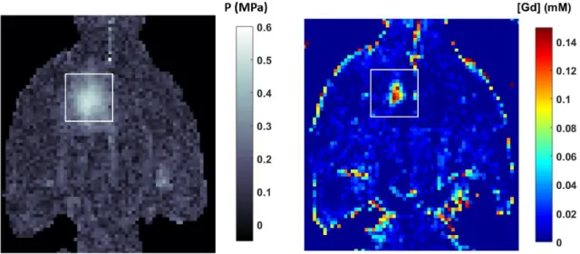
Figure 2: A – Image anatomique montrant la double implantation de tumeurs dans le cerveau d’un rat. B – Carte de concentration en agent de contraste gadoliné après perméabilisation par ultrasons de la tumeur de droite. La concentration dans la tumeur perméabilisée est plus importante et persiste
plus longtemps.
Imagerie des plaques amyloïdes
Une partie significative de mon travail fut dédiée à l’imagerie des plaques amyloïdes dans des modèles rongeurs de la maladie d’Alzheimer que nous élevons à NeuroSpin. Grâce à la perméabilisation de la BHE par ultrasons, j’ai pu délivrer des agents de contraste fonctionnalisés pour cibler les plaques amyloïdes. De nouveaux agents de contraste, les AguIX, produits par l’équipe de François Lux et Olivier Tillement à l’Institut Lumière-Matière à Lyon, ont été greffés avec du PEG pour augmenter leur biocompatibilité et avec du PIB pour qu’ils ciblent les plaques amyloïdes. J’ai procédé à leur délivrance, validée par IRM, dans le cerveau de souris modèles de la maladie d’Alzheimer. Nos partenaires, Jonathan Pansieriand Christel Marquette du CEA Grenoble, ont réalisé l’histologie de ces cerveaux et ont confirmé la présence d’AguIX greffées PIB dans le cerveau.
Une autre approche, cette fois-ci sans agent de contraste, a été développée par mes soins. J’ai optimisé une séquence IRM T2* ex vivo très haute résolution (40 µm isotrope) pour l’imagerie des
plaques amyloïdes, à 11,7 Telsa. Avec un traitement d’image approprié, cette séquence permet de quantifier la charge en plaques amyloïdes dans le cortex de souris modèle de la maladie d’Alzheimer. Cette technique mesure bien une charge en plaques plus élevée chez les souris plus âgées (qui ont de fait plus de plaques que les jeunes) et une charge quasiment nulle chez les souris sauvages (qui ne développent pas de plaques).
Figure 3 : A – Image anatomique haute résolution T2* (40 µm isotrope) d’un cerveau de souris
modèle de la maladie d’Alzheimer. Les hypo-signaux noirs dans le cortex sont des plaques amyloïdes. B – Détection automatique des plaques amyloïdes (en rouge) dans le cortex.
En collaboration avec Elena Longo et Philippe Zeitoun de l’ENSTA, nous avons étudié la délivrance de nanoparticules grâce à de l’imagerie par rayons X en contraste de phase. Après avoir délivré des nanoparticules dans le cerveau de souris de la maladie d’Alzheimer, nous avons amené ces cerveaux à l’European Synchrotron Radiation Facility à Grenoble pour l’imagerie. Cette technique s’est révélée très efficace pour l’imagerie des plaques. Des nanoparticules ont pu être retrouvées dans le tissu cérébral et j’ai pu corréler ces images avec des images IRM ex vivo haute résolution.
Pour finir, plusieurs marquages histologiques ont été mis au point pas Françoise Geoffroy, la responsable histologie de notre équipe, pour imager les plaques amyloïdes, la protéine tau et la neuroinflammation. J’ai mis au point le traitement d’image nécessaire pour quantifier la charge en
plaques amyloïdes. Ces développements pour quantifier les plaques amyloïdes ont ensuite été utilisés lors des protocoles de thérapie par perméabilisation répétée de la BHE de nos rongeurs modèles de la maladie d’Alzheimer.
Perméabilisations répétées de la BHE chez des modèles rongeurs de la maladie d’Alzheimer
Durant la dernière partie de ma thèse, je me suis servi de l’ensemble de ces développements pour étudier la perméabilisation répétée de la BHE par ultrasons comme thérapie pour la maladie d’Alzheimer. En effet, de récentes études ont montré que chez des souris modèles de la maladie d’Alzheimer, cette technique, utilisée de façon répétée (par exemple une perméabilisation par semaine pendant 2 mois), pouvait diminuer la charge en plaques amyloïdes et améliorer les performances cognitives. Cela serait vraisemblablement dû une neuroinflammation protectrice induite par les ultrasons.
Grâce au protocole de perméabilisation global développé précédemment, j’ai testé cette hypothèse sur un modèle rat et un modèle souris de la maladie. Après six semaines, avec une perméabilisation par semaine, les souris ont montré des signes d’amélioration lors des tests de comportement basés sur la mémoire. De plus, une analyse histologique montre une tendance vers une diminution de la taille des plaques amyloïdes chez les souris traitées par ultrasons. Les rats traités par ultrasons ont également montré des signes d’amélioration de leur mémoire ainsi qu’une diminution de leur anxiété. L’analyse histologique et biochimique n’est pas encore terminée.
Figure 4 : Distance parcourue pour retrouver la sortie du labyrinthe après 5 jours d’entrainement pour les souris (A) et 8 jours d’entrainement pour les rats (B). Dans les deux cas, les animaux modèles
d’Alzheimer traités par ultrasons (AD US) parcourent moins de distance que les animaux modèles d’Alzheimer non-traités (AD). Les animaux traités par ultrasons ont des performances semblables aux
animaux de type sauvage. Une distance parcourue plus courte indique une meilleure mémorisation de la sortie par rapport aux indices visuels extérieurs. Ces résultats montreraient un effet bénéfique
En collaboration avec Charles Truillet au service hospitalier Frédéric Joliot, nous avons étudié, par tomographie par émission de positrons, la neuroinflammation induite par la perméabilisation de la BHE par ultrasons. Grâce un radio-traceur de la neuroinflammation marqué au 18Fluor, nous avons
montré une augmentation significative de la neuroinflammation après six semaines, avec une augmentation plus importante chez les souris modèles de la maladie d’Alzheimer que chez les souris de type sauvage.
Conclusions et perspectives
En conclusion, cette thèse fut l’occasion d’approfondir les précédents développements de notre équipe en ce qui concerne la perméabilisation de la barrière hémato-encéphalique par ultrasons et de développer de nouvelles techniques telles que la perméabilisation globale. Ces nouvelles techniques m’ont permis de délivrer de diverses familles d’agents dans le cerveau de rongeurs dans le cadre de collaborations. Je me suis ensuite concentré sur la maladie d’Alzheimer en développant des techniques d’imagerie des plaques ainsi qu’en testant la permeabilisation de la barrière hémato-encéphalique par ultrasons comme thérapie pour cette maladie.
Les techniques de perméabilisation globale de la BHE et d’imagerie des plaques amyloïdes seront de nouveau utilisées lors d’études plus approfondies, en particulier sur les effets des perméabilisations répétées de la barrière hémato-encéphalique sur la maladie d’Alzheimer, dont l’étude que j’ai menée servira de base.
Table of Contents
INTRODUCTION ... 1
Focused ultrasound: a promising tool to deliver drugs to the brain ... 1
Medical use of ultrasound ... 1
The blood-brain barrier ... 3
Improving drug delivery to the brain ... 4
Ultrasound to increase the drug delivery to the brain ... 6
Optimum experimental parameters ... 7
Safety ... 11
Protocol for blood-brain barrier opening ... 13
Alzheimer’s disease ... 14
Epidemiology of Alzheimer’s disease ... 14
Physiopathology of AD and the amyloid cascade hypothesis ... 16
A difficult therapy ... 18
Animal models to study AD ... 20
Ethics of animal research ... 23
Magnetic resonance imaging ... 23
A brief history of MRI ... 23
Basic principles of MRI ... 25
Anatomical images... 28
Imaging contrast agents ... 30
Imaging the ultrasound beam ... 32
Presentation of the preclinical MR scanner of NeuroSpin... 33
Conclusion ... 34
METHODOLOGICAL ADVANCEMENTS ON ULTRASOUND-INDUCED BBB OPENING .. 37
Acoustic field calibration ... 37
Transducer calibration ... 37
a) Transducers to emit ultrasound ... 37
b) Setup for calibration ... 38
c) Single element transducer... 40
d) Multi element transducer... 40
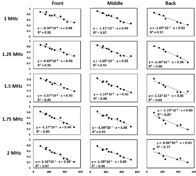
Transmission through rat skulls ... 42
a) Context and methods ... 42
a) Transmission factor decreases with frequency and body mass ... 45
b) Correlation with skull thickness ... 46
c) Spatial variation of the transmission factor ... 47
d) The different components of the insertion loss: aberration, attenuation and impedance mismatch ... 47
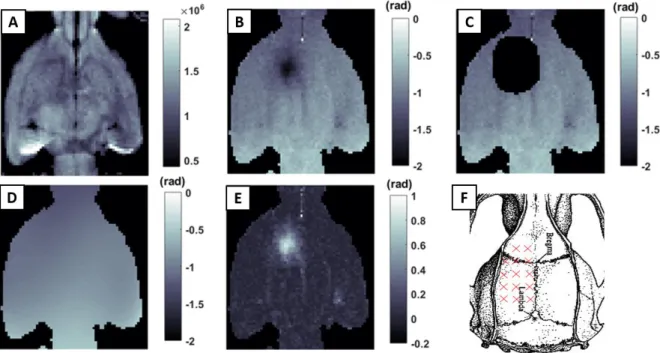
In vivo estimation of the transmission factor with MR-ARFI... 49
a) MR-ARFI mapping ... 49
b) Simulation of the viscoelastic response of the brain ... 51
c) Acoustic transmission mapping ... 52
Control of the position of the focal spot ... 54
MR-guided displacement of the transducer ... 54
a) Thermoguide ... 54
b) MR-guided global BBB opening in rats ... 55
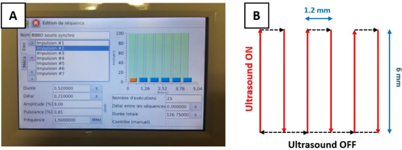
Motorized trajectories outside the MR scanner ... 57
a) Motorized positioning system ... 57
b) Validation and safety of a global BBB opening protocol in mice ... 58
Mapping of a rat skull with a transducer in pulse-echo mode ... 60
Influence of the acoustic pressure on BBB opening ... 61
An acoustic pressure threshold ... 61
a) Co-registration of MR-ARFI and concentration maps ... 62
b) Estimation of the acoustic pressure threshold ... 63
c) Different acoustic pressures in an unique BBB opening experiment ... 64
Higher pressures open the BBB longer ... 66
a) Square trajectory ... 66
b) Opening and follow-up of the permeabilisation ... 67
c) Estimation of a closing time constant depending on the pressure ... 68
Conclusion ... 70
PHYSICAL AND FUNCTIONAL PROPERTIES OF THE BRAIN AFTER ULTRASOUND-INDUCED BBB OPENING ... 73
Tortuosity of the brain ... 73
Free diffusion of contrast agent ... 74
In vivo restricted diffusion ... 76
Activity of the efflux pumps ... 78
Reminders on Positron Emission Tomography and efflux pumps ... 79
Protocol to follow erlotinib extravasation ... 80
a) Chemicals and radiochemicals ... 80
b) The ultrasound protocol ... 80
c) 11C-erlotinib PET study ... 82
d) 11C-N-desmethyl-loperamide PET study ... 83
e) Image analysis ... 83
FUS-induced BBB opening did not increase the brain exposure to 11C-erlotinib and 11 C-N-desmethyl-loperamide ... 83
Not only size matter ... 87
Increased delivery of therapeutic compounds with ultrasound... 88
a) BBB opening ... 89
b) Correlation between MRI and ICPMS ... 90
Delivery of an anti-cancer antibody followed by PET imaging... 91
a) BBB opening ... 91
b) PET imaging ... 92
FUS-induced blood-tumor barrier opening ... 93
Symmetrical double implantation of tumors in rats’ brains. ... 93
Ultrasound-induced opening of the blood-tumor barrier ... 94
An increased permeability after FUS-induced BTB opening ... 95
Conclusion ... 97
IMAGING OF ALZHEIMER’S DISEASE ... 99
A new functionalized contrast agent: AguIX-Peg-PIB ... 99
Characterization of AguIX ... 100
Delivery of AguIX-Peg-PIB in the brain of APP/PS1 mice ... 102
a) Context ... 102
b) Ultrasound protocol for AguIX-Peg-PIB delivery ... 103
c) MR imaging ... 103
Validation of the targeting with histology ... 104
Ex vivo imaging of Alzheimer’s disease ... 106
Automatic plaque detection ... 107
a) Optimization of sum of the echoes ... 108
b) Cortex segmentation ... 109
c) Detection of the hyposignals ... 110
Amyloid plaque load quantification ... 111
a) On mice ... 111
b) On rats ... 112
In vivo imaging ... 113
An efficient method to quantify amyloid load ... 115
Imaging AD with X-ray phase-contrast at the ESRF ... 115
Phase-contrast imaging ... 116
AguIX delivery... 116
Imaging of Alzheimer’s mouse brains ... 118
Correlation with MRI ... 120
Imaging Alzheimer’s disease with histology ... 121
Amyloid plaques imaging ... 121
a) Imaging of Aβ deposits: Thioflavin-S ... 121
b) Imaging of iron deposits: Perls’ ... 122
c) Correlation between iron and amyloid plaques ... 123
Other stains related to Alzheimer’s disease ... 125
b) Neuroinflammation imaging ... 125
Conclusion ... 126
BLOOD-BRAIN BARRIER OPENING AS A THERAPY FOR ALZHEIMER’S DISEASE ... 129
Ultrasound as a therapy for Alzheimer’s disease: promising studies ... 129
Preclinical studies ... 129
The first clinical trial ... 133
Assessment of memory deficits with behavior tests ... 134
The Open Field test ... 135
The Y-maze test ... 137
The Barnes Maze test ... 140
Therapeutic trials on mice ... 144
A first trial without motorization ... 144
a) Protocol ... 144
b) Safety ... 146
c) Behavior test ... 146
d) Ex-vivo imaging ... 149
e) Limits of this study ... 150
A safety study ... 150
a) Protocol and follow-up of the weights ... 150
b) Results of the behavior tests ... 152
A second trial on APP/PS1 mice ... 153
a) Behavior tests ... 154
b) Histology ... 157
Therapeutic trial on rats ... 160
Ultrasound protocol ... 160
Behavior tests results ... 161
Study of the neuroinflammation with PET ... 164
How to detect neuroinflammation ... 164
a) Neuroinflammation in Alzheimer’s disease ... 164
b) A marker of neuroinflammation: the TSPO protein ... 165
Ultrasound and PET protocol ... 166
Follow-up of the neuroinflammation ... 167
Conclusion ... 169
GENERAL CONCLUSION ... 170
SCIENTIFIC COMMUNICATIONS ... 172
1
Introduction
In this introduction chapter, I introduce the needed background knowledge to understand my PhD work. It starts with an overview of therapeutic ultrasound with a specific focus on the ultrasound-induced blood-brain barrier opening technique. I explain the functioning of this technique, list its possible applications and discuss its safety. Then, I continue with a broad introduction to Alzheimer’s disease and the possible underlying mechanisms. I present the animal models which are used to study the disease. Finally, I introduce Magnetic Resonance Imaging, its basic principles and how it can be used to study Alzheimer’s disease or to investigate ultrasound-induced BBB opening and even guide it.
Focused ultrasound: a promising tool to deliver drugs to the brain
Medical use of ultrasound
The French physicist Paul Langevin was the first person to ever use ultrasound when he designed the first sonar in the 1910’s, a technique that the animal kingdom has long mastered with dauphins or bats. A military technique at first, ultrasound became an industrial technique for detecting flaws in solid materials. In the 1940’s the Dussik brothers suggested that ultrasound could be used as a medical tool for detecting brain tumors by measuring the absorption through the head, so in a non-invasive way! Even if their images were mainly artifacts, they paved the way for ultrasound as an imaging tool for diagnosis. In 1957, the engineer Tom Brown and the gynecologist Ian Donald designed what can be named the first clinical ultrasound scanner for breast cancer imaging. To avoid fully immerging patients they used olive oil to ensure a good acoustic coupling between the skin and the transducer. Commercially developed during the 1960’s, ultrasound imaging is the best known clinical technique using ultrasound. Practiced routinely to follow pregnancies, cardiac diseases or liver diseases for example, this technique allows real-time imaging of moving structures in a non-invasive and safe way. Nowadays, ultrasound imaging provides not only tissue imaging but also blood flow imaging with Doppler, elastography to measure tissue stiffness, contrast enhanced perfusion imaging or it can be used to guide surgery. Its affordability, its portability and its ease of use make ultrasound imaging an essential imaging technique available for all clinical centers, hospitals or laboratories.
Best known for imaging, ultrasound can also be used as a therapeutic tool. Without impact on tissues when used for imaging, ultrasound can be focused to deliver more energy in a smaller volume. When focused, ultrasound can deliver enough energy to trigger bioeffects and so become therapeutic. The action mechanisms of ultrasound on tissues can be sorted in three main groups: the shock waves, the energy deposition inducing tissue heating and the cavitation mechanisms (Kiessling et al., 2012). The various therapeutic approaches associated with each mechanism of action will be described and cavitation mechanisms will be investigated in more details in the paragraph 1.1.4.
Chapter 1
2
Figure 1-1: Mechanisms of action of ultrasound on tissues (from O’Reilly & Hynynen 2016)
Shockwave therapy uses high pressures (tens of MPa) and short pulses (few microseconds) to deliver shockwaves at the target (Cleveland and McAteer, 2007). The shockwave results in a strong mechanical stress on the target tissues. Until the 1980’s, the treatment of kidney stones relied on open surgery and so was invasive. Ultrasound brought a revolutionary tool with extracorporeal shock wave lithotripsy (ESWL). In this non-invasive technique, the ultrasound transducer is coupled to the skin through ultrasonic gel and ultrasound are generated outside of the body (Bhojani and Lingeman, 2013). The shockwaves impacting the kidney stones lead to their mechanical destruction. At lower intensity, shockwave therapy is also used for tissue healing (Watson, 2008). By increasing the activity of cells, ultrasound start a pro-inflammatory response in the early repair phase. It has been shown to work on bone healing, scars and the vibrational movements have been shown to be able of improving the circulation and help break down adhesions between the muscles and their sheaths.
Ultrasound, when they are absorbed by tissues, lead to heating, mainly where they are focused. The degree of heating depends on the absorption coefficient of the tissue, the frequency of the ultrasound and the duration of the ultrasound pulse. The greater they are the higher is the heating. Focused ultrasound are capable of heating really local and small areas (millimeter-sized). Temperature rises of 10-15°C are enough to thermally ablate cells, typically cancer cells. Currently used to ablate cancer cells in the liver (Al-Bataineh et al., 2012) or the prostate (Pauly et al., 2006) using catheter-mounted ultrasound transducers, thermal ablation in the brain is more challenging. The Exablate Neuro (Insightec, Israel) can focus ultrasound behind the skull, thanks to dephasings based on the skull shape, and make use of thermal ablation in the brain (Elias et al., 2016). This protocol is now FDA approved for the treatment of essential tremor by ablation of the VIM nuclei in the thalamus. In Europe, the Exablate 4000 (Insightec, Israel) is also approved for the treatment of neuropathic pain and Parkinson disease. Several research teams are demonstrating the feasibility of thermal ablation of brain tumors thanks to High Intensity Focused Ultrasound (HIFU) (Coluccia et al., 2014; Macdonell et al., 2018). Hopefully, these could be the future technology to reach surgically inaccessible tumors. HIFU induced hyperthermia was also tried as a boost for radiotherapy of brain tumors (Guthkelch et al., 1991). This technique was limited to a phase I study but could now be pushed forward thank to the advance of MR-guided HIFU.
Ultrasound can be combined with microbubbles injected in the blood to make use of a phenomenon called cavitation. Ultrasound propagate through the tissue and encounter the microbubbles which
Focused ultrasound: a promising tool to deliver drugs to the brain
3
expand at low pressure and contract at high pressure. If the resulting size oscillation is stable, the cavitation is called “stable”. The oscillations produce micro-streams around the bubbles, the speed of the liquid is proportional to the amplitude of the ultrasound. If the acoustic pressure is increased, microbubbles can implode. This collapse can create liquid-jets and shockwaves, those violent phenomena concentrate the energy of the ultrasound and can break down membranes of nearby cells. This effect is called “inertial cavitation”. Inertial cavitation can for instance be used to clear artery or vein occlusions with a catheter approach (Crouch et al., 2008). In the brain, inertial cavitation is investigated as a promising tool for strokes (Ilyas et al., 2018). With MR guidance, HIFU could be transcranially focused in the brain to destroy clots. Cavitation is also used to enhance drug delivery. Drug delivery is enhanced by affecting either the drug carrier, which are drug-loaded microbubbles, or the cells surrounding the microbubbles. Cavitation can fragment the shell of microbubbles, made of liposomes or micelles, and trigger the release of their content, genes, proteins or drugs, in the targeted tissue (Pitt et al., 2004). Cavitation can also form temporary pores in the cell membrane surrounding the microbubbles, this phenomenon is called sonoporation (Pan et al., 2004). Cavitation is a very interesting tool but inertial cavitation has to be handled with care because it can damage the cells surrounding the microbubbles.
The blood-brain barrier
In the body, blood vessels are permeable to allow the transport of molecules from the blood to the organs. The brain is one special organ regarding molecular transport. Indeed, the blood vessels suppling the brain are a far less permeable structure. This barrier, limiting the diffusion of molecules to the brain, is called the blood-brain barrier (BBB). The BBB is made of endothelial cells, astrocyte end-feet and pericytes. An illustration of its structure is given on Figure 1-2. The endothelial cells, which forms the wall of the blood vessels, are, together with the tight junctions, the barrier itself whereas astrocytes are not thought to play directly a barrier role but they are key to ensure the maintenance of the tight junctions and provides a cellular link between the blood vasculature and the neurons (Ballabh et al., 2004). Pericytes seems to play an important role in the inhibition of properties normally associated with permeable vessels such as transcytosis (Davson et al., 2015).
This diffusion barrier plays a crucial role in maintaining the hemostasis of the brain (Engelhardt, 2003). The brain has a high energy consumption rate, 20% of the whole energy consumption of the body for only 2% of its mass. To properly function, the brains needs a precise oxygen delivery and metabolites supply, in particular glucose (Attwell et al., 2010), in the one hand and a protection from potential neurotoxic molecules and pathogens (Winkler et al., 2014) and waste disposal in the other hand. The BBB provides those functions by being highly impermeable to big molecules and allowing small molecules – metabolites, amino acids, hormones, vitamins etc. – to cross the BBB via transcellular mechanisms (Zhao et al., 2015). As illustrated on Figure 1-2, these transcellular transport of small molecules can be passive for lopothilic molecules or active, through transport proteins, for molecules such as glucose or amino acids (Abbott et al., 2010).
To summarize, brain endothelial cells differ significantly from non-brain endothelial cells by the presence of intercellular tight junctions, the low level of transcytosis and paracellular diffusion, a strong metabolic activity and the polarized expression of membrane receptors, and transporters
Chapter 1
4
which are responsible for the active transport of nutrients to the brain or the efflux of potentially toxic compounds from the brain to the blood vessels. Only drugs with a molecular weight smaller than 400 Da can naturally cross the BBB (Pardridge, 2005). This is the reason why huge efforts are done to overcome this barrier.
Figure 1-2:
On the left: Pathways across the blood–brain barrier (from Abbott et al. 2006). On the right: together with the endothelial cells, pericytes, glial cells (especially astrocytes), and the basal lamina are indirectly involved in the establishment and maintenance of the BBB (from Abbott et al. 2010).
A malfunctioning BBB has been associated with meningitis, epilepsy (Oby and Janigro, 2006), multiple sclerosis (Waubant, 2006), ischemia (Busto and Ginsberg, 1996), tumors (Weiss et al., 2009) or neurodegenerative disorders such as Alzheimer’s disease (Zenaro et al., 2016). The BBB is essential to the brain, but becomes a limit to deliver therapeutic molecules to the brain. Usually nanometer sized or larger, they are too big to naturally cross the BBB.
Improving drug delivery to the brain
Improving the delivery of therapeutic molecules to the brain allows to decrease the whole injected dose given to the patient in order to decrease peripheral toxicity. We already saw that only small lipophilic molecules can diffuse through the BBB (molecular weight under 400-500 Da) which disqualifies most therapeutic molecules. To overcome the BBB, one idea consists in the use of receptor-mediated endocytosis by conjugation of therapeutic molecules to ligands, such as antibodies and peptides, against receptors that are expressed on the surface of endothelial cells of the BBB allowing the drug to be transported into the brain. So far those receptors includes insulin receptors, transferrin receptors, LDL receptors and their related proteins, but more are being looked for (Gabathuler, 2010). The main drawback of this approach is the long and expensive design of each new compound. Moreover, modifications to the drug structure often result in the (at least partial) loss of
Focused ultrasound: a promising tool to deliver drugs to the brain
5
the drug’s biological activity. More recently several nano-carriers, which can be loaded with a drug, have been engineered such as liposome, micelles, carbon nano-tubes, dendrimers or gold nanoparticles (Figure 1-3). Those nano-carriers can be loaded with drugs and, as before, can be conjugated to ligands to target receptors of the BBB, acting like Trojan Horses. Moreover, loading the drug in a carrier protects the drugs and thus helps to further increase the concentration of the drugs to the target region. Of those nano-carriers, liposomes seem to be the most promising ones due to their capability to incorporate both hydrophilic and hydrophobic drugs, their low toxicity and because ligands can easily be attached to their surface to target biomarkers such as amyloid plaques in Alzheimer’s disease (Agrawal et al., 2017; Spuch and Navarro, 2011; Vieira and Gamarra, 2016). For more efficacy of the drug, its release can be triggered by degrading the shell of the carrier. For example lipid shells can be degraded by a pH change, by thiolysis or by heating (Kumari et al., 2014). Moreover, the diffusion of the drug in the brain parenchyma must be efficient. However, cytotoxicity generated by nanoparticles or their degradation products remains a major problem in drug development (Upadhyay, 2014).
Figure 1-3: Schematic representation of the main liposomal drugs and targeting agents that improve liposome affinity and selectivity for brain delivery (from Vieira & Gamarra 2016)
For drugs that do not naturally cross the BBB, two techniques have been developed to overcome this impermeability: direct injections (Krewson et al., 1995), which can be intracranial directly in the brain tissue, intracranial in the ventricles or trans-nasal, and chemical disruption of the BBB, with hyperosmotic solutions such as mannitol (Guillaume et al., 2010). In direct injections, the compound is delivered directly on site within the brain. Although direct injections showed therapeutic benefits in the treatment of brain tumors (Brem and Gabikian, 2001), it presents the clear disadvantage of being invasive, with a risk of infection, and neurosurgery is mandatory, making it really not suitable for repeated interventions. The drug release is also challenging to control as it is highly concentrated at the delivering site and the concentration decreases exponentially around this site (Voges et al., 2003), thus resulting in a non-homogeneous drug availability in the tissue. In the second method, the injection of hyperosmotic solutions causes the shrinkage of endothelial cells and the transient opening of the tight junctions (Rapoport, 1970) allowing drugs to pass through the BBB. Studies showed benefits of this method, especially in oncology (Hall et al., 2006). With mannitol the BBB remains open
Chapter 1
6
for 2 to 3 hours (Chi et al., 1996). But this method does not allow to choose the delivery site, since the hyperosmotic solution is injected in the blood flow and thus disrupts the BBB in the whole brain. The surgery is relatively serious because it requires general anesthesia and because the injection is intra-arterial and in order to target specific regions of the brain, researchers have used transient flow arrest, which causes a risk of strokes. So, once again, this technique can be considered invasive. Moreover some patients experienced hypotension or bradycardia (Bellavance et al., 2008). So although those techniques have shown their potential in clinical trials, they remains relatively invasive, difficult for the patients and not suitable for repeated treatments.
Ultrasound to increase the drug delivery to the brain
Ultrasound can be focused deep in the body, such ultrasound are called FUS for Focused Ultrasound, and can trigger bioeffects: hyperthermia or cavitation. As we saw, cavitation can enhance drug delivery using cavitation of microbubbles to permeate locally and temporarily the endothelial walls of the blood vessels in the brain we speak about FUS-induced BBB disruption.
Figure 1-4: two photon microscopy to follow the leakage of a dye from a blood vessel after FUS-induced BBB opening. Numbers are the time in second after the sonication (Cho et al., 2011)
First, it has to be said that the mechanisms ruling ultrasound-induced BBB opening are still unclear but several hypothesis prevail. Stable cavitation is thought to be the required regime of cavitation for BBB opening. Inertial cavitation even has to be avoided. It has often been associated with damages such as edema and extravasation of red blood cells suggesting hemorrhages. We will discuss in more details those effects and how to avoid them in paragraph 1.1.6 on safety. Micro-streams and direct contact generated by the stable cavitation mechanically stress the endothelial cells of the BBB (Krizanac-Bengez et al., 2004). The mechanical stress depends on the amplitude and frequency of the ultrasound, and also on the size of the microbubbles compared to the size of the blood vessels. The parameters affecting the efficacy of the BBB opening will be addressed in the paragraph 1.1.5. This mechanical stress generates cellular changes at the BBB. FUS have been shown to enhance at least 4 ways of molecular passage across the BBB: transcytosis using cellular vesicles, endocytosis, paracellular passage through widened tight junctions, and through the cytoplasmic channels in the
Focused ultrasound: a promising tool to deliver drugs to the brain
7
endothelium (Sheikov et al., 2004). The dominant way of passage is probably paracellular with the endothelial cells contracting under the stress and loosening the tight junctions between them (Sheikov et al., 2008). More recently, two photon microscopy has been used to characterize the BBB opening with a high temporal resolution (Cho et al., 2011), authors described two kinds of leakage, slow and fast. The fast one, reaching its maximum during the ultrasound application, corresponding to the paracellular way through the tight junctions and the slow one, reaching its maximum few minutes after the ultrasound application, corresponding to the transcellular way.
FUS-induced BBB opening for drug delivery has three main advantages. First, this technique is non-invasive. There is no need for surgery because ultrasound are shot from outside the body and the microbubbles are intravenously injected. The fact that this technique does not imply major surgery, and thus is relatively free from associated risks, makes FUS suitable for repeated treatments. Secondly, FUS can be MR-guided. Before the sonications, it is possible to visualize the focal spot of the ultrasound beam in the brain or to geometrically estimate it by visualizing the ultrasound probe and the brain on the same image. At the focus, the acoustic pressure is maximum. With an ultrasound beam properly calibrated, the cavitation will only take place at this location. The delivery site can be chosen by visualizing the focal spot using Acoustic Radiation Force Imaging sequences (Dervishi et al., 2013; Larrat et al., 2010a), thermometry (Kim, 2015) or with geometrical extrapolations by looking at the transducer surface. After BBB opening, MR-contrast agent that do not naturally cross the BBB can be intravenously injected. On T1-weighted images, the contrast of the MR images will be enhanced
only at the site of the BBB opening (Magnin et al., 2015). It is also possible to acquire T2- and T2
*-weighted images to assess the safety of the procedure. Indeed, T2 images have hypersignals if edema
are present (Sun et al., 2017) and T2* images have hyposignals in case of hemorrhages (Aoki et al.,
2014). Finally, FUS-induced BBB opening is transient, lasting for few hours (Marty et al., 2012a) and going back to a complete functionality of the BBB with no long term effects. FUS-induced BBB opening is currently used for drug or genes delivery (Al-Bataineh et al., 2012; Burgess et al., 2016; Huang et al., 2018) but also shows exciting results as a therapy by itself for Alzheimer’s disease (Burgess et al., 2014; Leinenga and Götz, 2015) as detailed in the Chapter 5.
Optimum experimental parameters
The intensity of the BBB opening is measured through the extravasation of molecular probes from the blood vessels to the brain. More intense (or stronger) BBB opening leads to more extravagated molecular probes (for the same injected dose). Those probe molecules can be: optically visible dyes like Evan’s Blue, where cutting the brain after an exsanguinous perfusion is enough to see the extravasation site, fluorescent dyes like the dextrans, observable under microscopy, MR-contrast agent leading to an increase of contrast on the MR images or radiotracers imaged in Positron Emission Tomography (PET) or Single Positron Emission computed Tomography (SPECT). The optimum acoustic parameters are the ones giving the stronger openings while remaining safe. So far, the vast majority of the pre-clinical research to optimize those parameters was done on small animal models, mainly rabbits, rats and mice. They demonstrate that several experimental parameters are essential.
Chapter 1
8
Figure 1-5: Illustration of an ultrasound shot made of two short pulses.
The impact of the critical experimental parameters are going to be detailed below. The parameters related to ultrasound are presented on Figure 1-5: the frequency of the ultrasound wave (f), the pulse length, the pulse repetition frequency (PRF), the acoustic pressure, which is proportional to the input voltage transmitted to the transducer, and the total sonication time.
Frequency
High frequencies are not suitable for clinical experiment as the thick, heterogeneous and curved human skull generates more attenuation and more aberration with increasing frequencies. For this reason, the frequency range suitable for clinical trials through skulls seems to be between 0.2 and 1.5 MHz but in rodents efficient BBB openings have been obtained from 28 kHz to 8 MHz.
Acoustic pressure
Several studies showed that higher acoustic pressures lead to stronger opening but that this relationship seems to saturate at high pressure (Hynynen et al., 2005). Numerous studies report the existence of an acoustic efficacy threshold for the minimal peak negative pressure (PNP) inducing some disruption (Aryal et al., 2014; Baseri et al., 2010; McDannold et al., 2006). But this threshold seems to depend on the frequency. McDannold’s team proposed to link this threshold to the mechanical index. This index is the ratio between the acoustic pressure and the square root of the frequency. They found that the different pairs frequency/pressure threshold give a constant mechanical index between 0.4 and 0.5 (McDannold et al., 2008), meaning that the higher the frequency the higher the acoustic pressure needed.
Focused ultrasound: a promising tool to deliver drugs to the brain
9
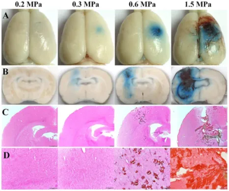
Figure 1-6: Evan’s Blue extravasation (A and B) shows an acoustic pressure threshold, around 0.3 MPa, for FUS-induced BBB opening. Histology (C and D) already shows damages at 0.6 MPa. At this frequency (515 kHz) the window for an efficient and safe BBB opening is somewhere between 0.3 and
0.6 MPa (Shin et al., 2018)
Pulse duration
Pulse durations from a few µs to 100 ms have been tested. Increase of the pulse duration seems to increase the intensity of the opening with no real benefit above 10 ms. Short pulses, under 3 µs, are appreciated for their capacity to reduce standing waves in the skull cavity (Choi et al., 2011).
Pulse repetition frequency and total sonication time
The effect of the pulse repetition frequency (PRF) is less clear as studies showed a better opening with higher repetition rate (O’Reilly et al., 2011; Shin et al., 2018) and others do not observe any dependency (Mcdannold et al., 2008). Finally total longer sonication time increase the intensity of the opening (Shin et al., 2018) but sonication time longer than the lifetime of microbubbles in the blood (few minutes) are useless and can lead to damages (Yang et al., 2011).
We just saw that the BBB opening is impacted by many acoustic parameters. So, performing the expected BBB opening requires to carefully characterize the ultrasound setup and also requires to precisely dose the ultrasound energy deposited in situ. Dosing the energy deposition can be achieved before the sonication for example by measuring the passage of the ultrasound beam through the skull or imaging the ultrasound beam in situ with acoustic radiation force imaging (these techniques will be detailed in Chapter 2) or during the sonication with passive cavitation detection (see the next paragraph on safety) . But this variety of acoustic parameters represents an advantage compared with other techniques. Indeed, it makes FUS-induced BBB opening tunable, in terms of spatial extend,
Chapter 1
10
intensity or duration. In addition to the acoustic parameters, two other experimental parameters can impact BBB opening: the microbubbles and the anesthesia.
Table 1-1: Reported effects of the experimental parameters on BBB disruption via FUS and microbubbles (Aryal et al., 2014)
Microbubbles
Microbubbles are usually made of an innocuous heavy gas (perfluorocarbon or sulfur hexafluoride) encapsulated in a shell (Martin and Dayton, 2014). The shell is made of lipids, proteins, or polymers and its role is to protect the gas in its inside. Used gases are heavy (heavier than air) in order to increase their lifetime in the blood and thus increase their availability. Indeed, heavier gas take longer time to diffuse through the shell which prevents dissolution in the blood. Their sizes range approximately from 1 to 5 µm. This diameter is smaller than brain capillaries, which prevents them from obstructing these vessels. The microbubbles I used during my PhD are Sonovue from Bracco (Schneider, 1999). Their mean diameter is 2.5 µm and their lifetime in the body is approximately 5 minutes long (data acquired in rabbit).
Microbubbles size and dose seems to have in important impact on BBB opening as larger doses lead to stronger openings (Yang et al., 2008) (Shin et al., 2018) (Choi et al., 2010). At last, it can be noticed that microbubbles injection rate does not seem to affect the intensity of the opening (O’Reilly et al., 2011) but attention must be payed to the diameter of the syringe used to injected the microbubbles as to small syringes can destroy the microbubbles (Talu et al., 2009).
Anesthesia
At last, the physiological state of the animals has an impact on the BBB opening, mainly temperature and anesthesia since they both affect perfusion, vasoconstriction and microbubble clearance rate from blood, three parameters that modify the efficacy of BBB opening. Two anesthesia protocols are commonly used: gas anesthesia with isoflurane mixed with oxygen (and sometime air) or chemical anesthesia with ketamine/xylazine intraperitoneally injected. McDannold’s team showed that, for identical acoustic parameters, the quantity of extravagated probe molecules was greater with ketamine/xylazine than with isoflurane (McDannold et al., 2012a). Indeed, isoflurane is a well-known vasodilator and it is possible that microbubbles cavitating in larger blood vessels apply less mechanical stress on the endothelial cells as they are further from them (especially capillaries which are
Focused ultrasound: a promising tool to deliver drugs to the brain
11
considered as being responsible for the major part of BBB leakage after ultrasound). This also results in lower acoustic pressure efficacy threshold when using ketamine/xylazine than when using isoflurane. When using isoflurane anesthesia, the percentage of oxygen in the carrier gas mixture also matters. It has been showed that BBB openings were stronger when isoflurane was mixed with air than when it was mixed with oxygen (Annold et al., 2017). Indeed, a faster clearance of microbubbles from the blood with higher amount of oxygen was observed. Those results prove that comparisons between groups using different anesthesia conditions must be done really carefully and that experimental conditions among animals in a group study needs to be highly reproducible.
Even if the main parameter characterizing the BBB opening is the quantity of extravagated probe molecules, it can be interesting to look at the duration of the opening. In their first experiment,
Hynynen’s team observed that the BBB was closed 48h after opening (Hynynen et al., 2001). With a
better temporal sampling, they observed that the BBB could close back as early as 6 hours after sonication. It is important to remember that the closure is a dynamic and progressive mechanism and that at one given time after the opening, the BBB can be closed to big molecules but open to smaller ones. As a consequence, the apparent duration of the increased vascular permeability can depend on the molecular probe used to quantify it. Marty et al. showed that, with identical BBB openings, the BBB was already closed at 2 hours for big molecules (around 60 nm hydrodynamic diameter) and that small molecules (around 1 nm) could cross the BBB for more than 10 hours (Marty et al., 2012a). BBB closure time seems to depend as well on the acoustic pressure used with opening lasting few hours more for higher pressure (Samiotaki and Konofagou, 2013). This influence need to be studied more in details.
Safety
The safety of FUS-induced BBB opening has been extensively investigated to help transfer this technique to the clinic. We will review in this paragraph the main studies. For given conditions, the pressure must be sufficient to trigger the stable oscillation of the microbubbles and thus a permeabilisation of the blood vessels, but a too high pressure might cause inertial cavitation with the associated damaging effects, such as edema or hemorrhages. These two thresholds, the efficacy threshold and the safety threshold, involved the existence of safety window.
The effects of BBB opening on tissues were first investigated on rodents. This investigation starts by looking at radiologically visible tissue lesions or reactions such as edema on T2 and diffusion images or
hemorrhages on T2* images. In most studies, histopathology was done after one sonication (Baseri et
al., 2010; Hynynen et al., 2005). The main reported deleterious effect is extravasation of few red blood cells or small petechial hemorrhages. Groups usually report damages for their higher pressure conditions and they associate it with inertial cavitation (McDannold et al., 2006). Repeated BBB opening sessions, once a week for 6 weeks, on rats show no or limited tissue damages (Kobus et al., 2015). Authors found small damages like micro-hemorrhages or scars and neuronal-necrosis for their higher pressure conditions. Finally, no effect on behavior or locomotion were observed on mice up to 6 months after BBB opening (Olumoladea et al., 2017).
Chapter 1
12
The best proofs of the safety of the technique were more recently brought with studies on non-human primates. The reference study for assessing the safety of the BBB opening was done by McDannold’s team (McDannold et al., 2012b). They identified a clear safety window for the acoustic pressure in which BBB opening could be performed without tissue damages. Histological results showed no effect on neurons or on white matter fibers. They only reported hemosiderin deposits in the meninges and hemorrhagic tissue in ventricles when the focal spot of the ultrasound was intersecting those tissues. Moreover, animals recovered from each ultrasound session without behavior deficit. Repeated sessions of BBB opening in non-human primates did not show negative long term physiological or neurological effects either (Downs et al., 2015).
Real-time monitoring is now commonly achieved using passive cavitation detectors (Arvanitis et al., 2012; O’Reilly and Hynynen, 2012). One or several small ultrasonic transducers, often placed at the center of the emission transducer, are used in reception mode to record and analyze the diverging pressure waves emitted by the oscillating microbubbles during FUS-induced BBB disruption. The spectral content allows to tell stable cavitation, emitting harmonics of the emission frequency, from inertial cavitation, emitting a broadband signal. Our group designed a real time feed-back controller for rodents and primates which increases the acoustic pressure for each pulse until internal cavitation appears (Kamimura et al., 2018), thus allowing to perform FUS-induced BBB opening without inertial cavitation, that is to say safely. The role of real-time monitoring is crucial for this technique as it is now moving to clinical trials.
Recently, more subtle tissue reactions were investigated. In particular, the effect of FUS-induced neuro-inflammation was studied. In an extensive study Kovacs et al. showed that sterile inflammatory responses to ultrasound with elevations in pro-inflammatory, anti-inflammatory, trophic factors, activated astrocytes and microglia (Kovacs et al., 2016). Another study reported activated microglia after FUS-induced BBB opening in an Alzheimer’s disease mouse model. The authors suggested that this activation is the mechanism responsible for the amyloid plaque clearance they observed (Leinenga and Götz, 2015). It is not clear if this inflammation is caused by the entry of endogenous compounds in the brain through an open BBB or if it is a direct mechanical effect due to the cavitation forces but
Kovacs et al. advocates for the second hypothesis.
The effects of BBB opening are numerous and depend on the experimental parameters previously listed. But FUS-induced BBB opening is a potential therapeutic tool and so what matters is the cost to benefit ratio. It can be anticipated that this technique will first be approved for brain tumors or neurodegenerative diseases where current treatment options are limited and the benefits are high. For these applications, the existing set of radiology, histopathology and behavior data acquired in non-human primates gives sufficient evidence for a clinical transfer. This is why FUS-induced BBB opening is already undergoing few clinical trials in France (Carpentier et al., 2016) and in Canada (Huang et al. 2016). Nevertheless, FUS-induced BBB opening triggers mechanisms in the brain which are not completely understood and need further investigations.
Focused ultrasound: a promising tool to deliver drugs to the brain
13
Figure 1-7: the first line shows microglia (Iba1) and nuclei (DAPI) in a sonicated region several hours after FUS-induced BBB opening and a control region (contralateral) and the second line shows astrocytes (GFAP) and nuclei. Microglia and astrocytes are both overexpressed after FUS-induced
BBB opening compare to the contralateral region (Kovacs et al., 2016)
Protocol for blood-brain barrier opening
Here, I briefly describe the BBB opening protocol I used during my PhD (Figure 1-8). I will firstly describe the full BBB opening protocol under MR-guidance and then adaptations of this protocols without MR guidance. The protocol under MR guidance was exclusively for rats as the MR coil is only suitable for them. The used anesthesia was always isoflurane in a mixture of air and oxygen. I used about 3% of isoflurane to get the animals asleep and this percentage was decreased to 1.5-2% once the animal was in the scanner. Animals had to be shaved to ensure a good coupling between the head and the water balloon of the transducer. We used an electrical razor and a depilatory cream. Then, a catheter was placed in a tail vein to inject the microbubbles and the contrast agent later on. To continue, the animals was placed in the MR bed, the head inside a specific coil which allows to position the transducer in its center. A temperature probe and a breathing probe were installed to monitor vitals parameters. Then, the bed was put inside the MR scanner. Before BBB opening, pre-scans can be acquired for positioning the ultrasound beam (MR ARFI) or for reference images. For BBB opening, a bolus of microbubbles was injected via the catheter in the tail vein, usually 200 µL for a rat, and ultrasound were shot. Finally, to validate or quantify the BBB opening, a contrast agent was injected via the catheter and MR images were acquired with a set of parameters tuned to detect the contrast agent.
Chapter 1
14
Figure 1-8: BBB opening protocols.
I used two other kinds of protocols without MR guidance. The first kind was without MR guidance but with a control after the BBB opening (protocol green and orange on Figure 1-8). The sonications were performed outside of the scanner and after contrast agent injection animals were placed in the scanner to validate the opening. In this protocol, BBB openings cannot be quantified due to the absence of references images, the description of the opening can only be quantitative. This protocol was used in particular for optimization of the BBB opening without MR guidance. Indeed, I performed repeated sessions of BBB openings on many animals for several weeks (see Chapter 5) and doing it without guidance and control (protocol green on Figure 1-8) is a huge gain of time. When using mice, microbubbles injection and contrast agent injection were always retro-orbital. More details on the protocols will be given when used in the next chapters. The last protocol was without MR guidance and without control, it was only used for the repeated sessions of FUS-induced BBB opening in Chapter 5.
Alzheimer’s disease
Epidemiology of Alzheimer’s disease
44 million people worldwide have Alzheimer’s disease (AD) and with aging of the population this number is expected to double by 2050 (Hebert et al., 2013). AD is the first cause of dementia (around 70% of the cases). Doctor Alois Alzheimer first described the symptoms of the disease in his patient August Deter in 1906. For a long time AD was wrongly considered as a natural effect of aging and not as a specific disease, thus explaining the limited resources governments have provided to its understanding, unlike other diseases such as cancer. It is only since the 2000s’ that governments launched ambitious research funding plans.
Alzheimer’s disease
15
The Organization for Economic Cooperation and Development (OECD) evaluates the cost of an AD patient at 20 000€ a year (Hebert et al., 2013) but other studies put the figure up to 90 000€ (Huang et coll., 1998). Due to the aging of the population, the Office parlementaire d’évaluation des politiques de santé (Opeps) anticipates a cost of 1 to 1.5 % of the PIB in 2040 for France. Wilkinson (Wilkinson, 2005) considers that in UK the cost of AD exceeds those of cardiac disease, cerebrovascular accidents and cancers while the budget on AD research is only 10% the budget for cardiac disease research and 3% the budget for cancer research. The cost of an AD patient is relatively high due to the loss of autonomy and socio-medical help needed to face it.
Figure 1-9: Change in the biomarkers of AD over time. Aβ accumulation is the first biomarker of the disease, appearing years before cognitive decline (Aisen et al., 2017)
AD is a neurodegenerative disease and is characterized by an alteration of the cognitive capacities. The only way to have an indisputable diagnosis of the disease is through postmortem analysis. Indeed, histopathology can reveal the presence of amyloid plaques and neurofibrillary tangles which are the hallmarks of the disease (NIA, 1997). Fortunately, physicians have developed ways to identify Alzheimer’s patients during their lifetime in order to analyze and prevent the progression of the symptoms. The clinical diagnosis is done in two steps. Firstly, dementia is diagnosed. Dementia is defined as a memory deficit associated with one cognitive function - language, praxis, gnosis… - strong enough to lead to a loss of autonomy. Then, various criteria allows to diagnose AD, the McKhann criteria (Mckhann et al., 1984) are the most used. They are based on the progressivity of the disease and the absence of other cerebral diseases. McKhann criteria have a good sensibility, around 80%, but a low specificity, around 70%, when compared to postmortem analysis. The term Mild Cognitive Impairment (MCI) is used to define the early stages of the disease, when the amnesic syndrome is non-disabling but progressive. At autopsy, 80% of the patient who received MCI diagnosis happened to have AD (JC et al., 2001). The goal of this classification is to better anticipate the evolution of the pathology. Those mental status tests conducted by interviewing patients should combined with brain imaging and CSF analysis (Waldemar et al., 2007). Brain imaging, mainly done with PET and MRI, is