HAL Id: hal-01772904
https://hal-amu.archives-ouvertes.fr/hal-01772904
Submitted on 20 Apr 2018
HAL is a multi-disciplinary open access
archive for the deposit and dissemination of
sci-entific research documents, whether they are
pub-lished or not. The documents may come from
teaching and research institutions in France or
abroad, or from public or private research centers.
L’archive ouverte pluridisciplinaire HAL, est
destinée au dépôt et à la diffusion de documents
scientifiques de niveau recherche, publiés ou non,
émanant des établissements d’enseignement et de
recherche français ou étrangers, des laboratoires
publics ou privés.
Distributed under a Creative Commons Attribution - NonCommercial - NoDerivatives| 4.0
International License
Genome sequence and description of Mannheimia
massilioguelmaensis sp. nov.
L. Hadjadj, Ahmed Aimen Bentorki, C. Michelle, K. Amoura, Abdelghani
Djahoudi, Jean-Marc Rolain
To cite this version:
L. Hadjadj, Ahmed Aimen Bentorki, C. Michelle, K. Amoura, Abdelghani Djahoudi, et al.. Genome
sequence and description of Mannheimia massilioguelmaensis sp. nov.. New Microbes and New
Infec-tions, Wiley Online Library 2015, 8, pp.131-136. �10.1016/j.nmni.2015.10.005�. �hal-01772904�
Genome sequence and description of Mannheimia massilioguelmaensis
sp. nov.
L. Hadjadj1, A. A. Bentorki2, C. Michelle1, K. Amoura3, A. Djahoudi3and J.-M. Rolain1
1) Unité de recherche sur les maladies infectieuses et tropicales émergentes (URMITE), UMR CNRS, IHU Méditerranée Infection, Faculté de Médecine et de Pharmacie, Aix-Marseille-Université, Marseille, France, 2) Laboratoire de Microbiologie, CHU Dorban and 3) Laboratoire de Microbiologie, Faculté de Médecine, Université Badji Mokhtar, Annaba, Algeria
Abstract
Strain MG13Tsp. nov. is the type strain of Mannheimia massilioguelmaensis, a new species within the genus Mannheimia. This strain was isolated from the exudate of a skin lesion of an Algerian man. Mannheimia massilioguelmaensis is a Gram-negative, facultative anaerobic rod, member of the family Pasteurellaceae. Here we describe this organism, together with the complete genome sequence and annotation. The 2 186 813 bp long genome contains 2048 protein-coding and 55 RNA genes, including eight rRNA genes.
New Microbes and New Infections © 2015 The Authors. Published by Elsevier Ltd on behalf of European Society of Clinical Microbiology and Infectious Diseases.
Keywords: Genome, human infection, infectious disease, Mannheimia massilioguelmaensis, taxonogenomics Original Submission: 7 August 2015; Revised Submission: 5 October 2015; Accepted: 7 October 2015 Article published online: 23 October 2015
Corresponding author: J.-M. Rolain, URMITE CNRS IRD UMR 6236, IHU Méditerranée Infection, Aix Marseille Université, Faculté de Médecine et de Pharmacie, 27 Bd J Moulin, 13385 Marseille Cedex 05, France
E-mail:[email protected]
Introduction
Mannheimia massilioguelmaensis sp. nov. strain MG13T(= CSUR P1431 = DSM 29915) is the type strain of M. massilioguelmaensis sp. nov. This bacterium is a Gram-negative, facultatively anaer-obic, nonhaemolytic, indole-negative rod-shaped bacillus. It was isolated from the exudate of a skin lesion of an Algerian patient. We recently proposed that genomic and proteomic data, which do not suffer from the lack of reproducibility and interlaboratory comparability that the reference standard DNA-DNA hybridization (DDH) and G+C content determi-nation does[1], be included in the official description of new
bacterial species[2,3].
The genus Mannheimia (Angen et al., 1999) formerly Pas-teurella, was created in 1999 [4] and currently comprises six
species, including M. haemolytica, M. granulomatis, M. glucosida, M. ruminalis, M. varigena and M. caviae. Mannheimia species are Gram-negative, non-spore-forming, nonmotile, facultative anaerobic rod-shaped bacilli. Some species of Mannheimia are commonly isolated in the gastrointestinal or upper respiratory tract of animals but are not associated with disease[4]. Others are pathogenic, such as Mannheimia haemolytica, which is one of the most important respiratory pathogens of domestic rumi-nants and causes serious outbreaks of acute pneumonia in neonatal, weaned and growing lambs, calves and goats[5]. In-fections are rare in humans but can be fatal when they do occur
[6,7].
Here we present a summary classification and a set of fea-tures for M. massilioguelmaensis sp. nov. strain MG13Ttogether with the description of the complete genome sequence and annotation. These characteristics support the circumscription of the species M. massilioguelmaensis.
Organism information
A pus sample was collected from a 90-year-old Algerian patient in Guelma, northeastern Algeria, with a cutaneous abscess of
New Microbe and New Infect 2015; 8: 131–136 New Microbes and New Infections © 2015 The Authors. Published by Elsevier Ltd on behalf of European Society of Clinical Microbiology and Infectious Diseases This is an open access article under the CC BY-NC-ND license (http://creativecommons.org/licenses/by-nc-nd/4.0/)
the left forearm. At the time of sample collection, he had been hospitalized for fever and multiple abscesses in his left arm. One week before hospitalization, the patient had afirst abscess in his left indexfinger after to pare one nail; this evolved into multiple abscesses of and a lymphangitis path in the left forearm. The patient signed informed consent, and agreement of the local ethics committee of the IFR48 (Marseille, France) was obtained (agreement 07-30). The bacterium was isolated in pure culture in September 2014.
When blasted to National Center for Biotechnology Infor-mation (NCBI) database, the 16S rRNA gene sequence of M. massilioguelmaensis strain MG13T (GenBank accession no. LN795822) exhibited an identity of 96.00% with Mannheimia haemolytica. This value was the highest similarity observed but was lower than 97.8% 16S rRNA gene sequence threshold recommended by Stackebrandt and Ebers [8] to delineate a new species without carrying out DNA-DNA hybridization.
Different growth temperatures (25, 30, 37 and 45°C) were tested. Growth occurred between 25°C and 37°C, but optimal growth was observed at 37°C, 24 hours after inoculation. Colonies were smooth, greyish and approximately 1 mm in diameter on 5% sheep’s blood–enriched agar (bioMérieux). Growth of the strain was tested in anaerobic and micro-aerophilic atmospheres using GasPak EZ Anaerobe Pouch (Becton Dickinson) and CampyGen Compact (Oxoid) systems, respectively, and in aerobic atmosphere, with or without 5% CO2. Growth was observed under aerobic (with and without
CO2), microaerophilic and anaerobic conditions. Gram staining
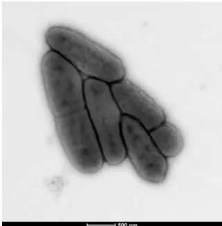
showed short Gram-negative rods unable to form spores (Fig. 1). A motility test was negative. The size of cells were determined by negative staining transmission electron micro-scopy on a Technai G20 Cryo device (FEI) at an operating voltage of 200 kV. The rods had a length ranging from 1.1 to 1.9 μm (mean 1.5 μm), a width ranging from 0.4 to 0.6 μm (mean 0.5 μm) and a diameter ranging from 0.4 to 0.8 μm (mean 0.6μm) (Fig. 2).
Differential phenotypic characteristics using API 50CH and API Zym system (bioMérieux) between M. massilioguelmaensis sp. nov. strain MG13T and other Mannheimia species [4] are
detailed inTable 1.
Susceptibility testing was performed by the Etest strip (bio-Mérieux) method. Minimum inhibitory concentration was expressed inμg/mL. M. massilioguelmaensis was susceptible to amoxicillin (0.19), amoxicillin–clavulanate (0.125), gentamicin (0.094), amikacin (1), imipenem (0.75), trimethoprim –sulfa-methoxazole (0.064), ciprofloxacin (0.012), ceftriaxone (1.5) and cholistine (0.19) but resistant to vancomycin (>256).
Extended features descriptions
Matrix-assisted laser desorption/ionization time-of-flight mass spectrometry (MALDI-TOF) protein analysis was performed as previously described [9] using a Microflex spectrometer
(Bruker). Twelve distinct deposits were done for strain MG13T
from 12 isolated colonies. The 12 MG13T spectra were im-ported into the MALDI BioTyper 2.0 software (Bruker) and analysed by standard pattern matching (with default parameter settings) against the main spectra of 4108 bacteria, including seven spectra from four Mannheimia species, used as reference data, in the BioTyper database. A score enabled the identifi-cation (or not) from the tested species: a score of >2 with a validated species enabled the identification at the species level; a score of >1.7 but <2 enabled the identification at the genus level; and a score of <1.7 did not enable any identification. No significant MALDI-TOF score was obtained for strain MG13T against the Bruker database, thus suggesting that our isolate was
FIG. 1.Gram staining of Mannheimia massilioguelmaensis strain MG13T.
FIG. 2. Transmission electron microscopy of Mannheimia massilio-guelmaensis strain MG13T using Technai G20 Cryo device (FEI) at operating voltage of 200 kV. Scale bar = 500 nm.
132 New Microbes and New Infections, Volume 8 Number C, November 2015
NMNI
New Microbes and New Infections © 2015 The Authors. Published by Elsevier Ltd on behalf of European Society of Clinical Microbiology and Infectious Diseases, NMNI, 8, 131–136 This is an open access article under the CC BY-NC-ND license (http://creativecommons.org/licenses/by-nc-nd/4.0/)
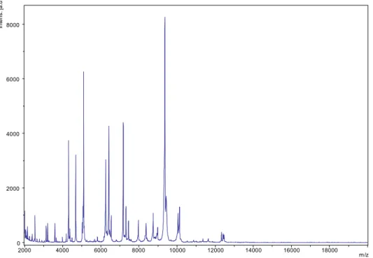
a new species. We incremented our database with the spec-trum from strain MG13T(Fig. 3).
Genome sequencing information
This strain was the 23rd genome of a Mannheimia species and the first genome of Mannheimia massilioguelmaensis sp. nov. (CDQL00000000).
After DNA extraction by the phenol–chloroform method, genomic DNA of Mannheimia massilioguelmaensis was sequenced on the MiSeq Technology (Illumina) with the mate-pair strategy.
For genome annotation, open reading frames (ORFs) were predicted using Prodigal[10]with default parameters, but the predicted ORFs were excluded if they spanned a sequencing gap. The predicted bacterial protein sequences were searched against the Clusters of Orthologous Groups (COGs) database and the GenBank database [11] using BLASTP. The tRNAS-canSE tool [12]was used to find tRNA genes, whereas ribo-somal RNAs were found by using RNAmmer[13]and BLASTn against the GenBank database. Transmembrane helices and li-poprotein signal peptides were predicted using the Phobius
Web server [14]. ORFans were identified if their BLASTP E
value was lower than 1e-03 for alignment length greater than 80 aa. If alignment lengths were smaller than 80 aa, we used an E value of 1e-05. Such parameter thresholds have already been used in previous works to define ORFans[2,3]. Finally, we used the online Genome-to-Genome Distance Calculator (GGDC;
http://ggdc.dsmz.de) to estimate of the overall similarity among the compared genomes and to replace the wet-lab DDH by a digital DDH[15,16]. GGDC 2.0 BLAST+ was chosen as the alignment method, and the recommended formula 2 was taken into account to interpret the results.
We compared the genome sequences of M. massilioguelmaensis strain MG13T (CDQL00000000) with those of Microbacterium granulomatis strain DSM1956 (JHZD00000000), Mannheimia haemolytica strain USDA-ARS-USMARC-183 (CP004752) and Mannheimia varigena strain USDA-ARS-USMARC-1312 (CP006944).
Digital DDH estimation of strain MG13T against the compared genomes ranged between 13.60 to 13.90. These values were very low and below the cutoff of 70%, thus again confirming the new species status of strain MG13T.
The genome is 2 186 813 bp long with 36.21% G+C content (Fig. 4andTable 2). It is composed of eight scaffolds (composed
TABLE 1.Differential characteristics of Mannheimia species
Characteristic M. ma M. ha M. gl M. gr M. ru M. va
Cell diameter (μm) 0.6 NA NA NA NA NA
Oxygen requirement Facultative anaerobic Facultative anaerobic Facultative anaerobic Facultative anaerobic Facultative anaerobic Facultative anaerobic
Gram strain − − − − − − Motility − − − − − − Endospore formation − NA NA NA NA NA Production of: Alkaline phosphatase + + + + + + Acid phosphatase w NA NA NA NA NA Catalase − NA NA NA NA NA Oxidase − NA NA NA NA NA Nitrate reductase + + + + + + Urease − − − − − − β-Galactosidase − v + v + v Indole − NA NA NA NA NA Esterase w NA NA NA NA NA Esterase lipase w NA NA NA NA NA Leucine arylamidase w NA NA NA NA NA Cystine arylamidase − NA NA NA NA NA Valine arylamidase − NA NA NA NA NA Utilization of: Mannitol − NA NA NA NA NA Trehalose − − − − − − L-Arabinose − − v − − + D-Sorbitol − + + + v − D-Xylose − + + v v + D-Ribose + NA NA NA NA NA D-Glucose + NA NA NA NA NA D-Mannose w − − − − − D-Fructose + NA NA NA NA NA Glycerol + NA NA NA NA NA N-Acetylglucosamine + NA NA NA NA NA G+C content (mol%) 36.2 43.6 41.6 39.2 NA 41.7 Habitat Human Bovine, ovine Ovine Bovine, leporine, deer Ovine Bovine, porcine Mannheimia massilioguelmaensis (M. ma) strain MG13T, Mannheimia haemolytica (M. ha) strain NCTC 980T, Mannheimia glucosida (M. gl) strain P925T, Mannheimia granulomatis (M. gr)
strain ATCC 49244T, Mannheimia ruminalis (M. ru) strain HPA92T, Mannheimia varigena (M. va) strain 177T.
+, positive result;−, negative result; v, variable result; w, weakly positive result; NA, data not available.
0 2000 4000 6000 8000 In te n s. [ a .u .] 2000 4000 6000 8000 10000 12000 14000 16000 18000 m/z
FIG. 3.Reference mass spectrum from Mannheimia massilioguelmaensis strain MG13T. Spectra from 12 individual colonies were compared and reference spectrum generated.
FIG. 4.Graphical circular map of chromosome. From outside to center: genes on forward strand (colored by COGs categories), genes on reverse strand (colored by COGs categories), RNA genes (tRNAs green, rRNAs red), G+C content, G+C skew.
134 New Microbes and New Infections, Volume 8 Number C, November 2015
NMNI
New Microbes and New Infections © 2015 The Authors. Published by Elsevier Ltd on behalf of European Society of Clinical Microbiology and Infectious Diseases, NMNI, 8, 131–136 This is an open access article under the CC BY-NC-ND license (http://creativecommons.org/licenses/by-nc-nd/4.0/)
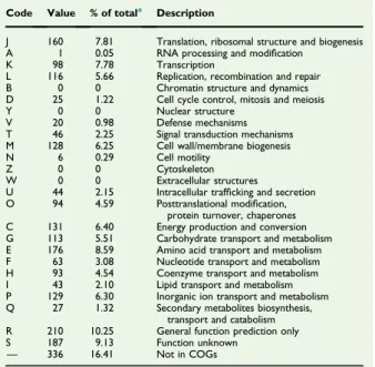
of eight contigs). Of the 2103 predicted genes, 2048 were protein-coding genes and 55 were RNAs (three genes are 5S rRNA, two genes are 16S rRNA, three genes are 23S rRNA and 47 genes are TRNA genes). A total of 1692 genes (82.62%) were assigned as putative function (by cogs or by NR blast). Twenty-five genes were identified as ORFans (1.22%). The remaining genes were annotated as hypothetical proteins (144 genes, 7.03%). The distribution of genes into COGs functional categories is presented inTable 3.
Conclusions
On the basis of phenotypic, phylogenetic and genomic analysis (taxonogenomics), we formally propose the creation of Man-nheimia massilioguelmaensis sp. nov. that contains the strain MG13T.
Taxonomic and nomenclatural proposals
Description of Mannheimia massilioguelmaensis sp. nov. Mannheimia massilioguelmaensis (ma.sil.io.guel.ma.en’sis. L. gen. masc. n. massilioguelmaensis, combination of Guelma, where strain MG13T was isolated, and Massilia, the Latin name of Marseille, where the strain was sequenced).
Colonies were moderately opaque and approximately 1 mm in diameter on 5% sheep’s blood–enriched agar. Cells are Gram-negative, nonhaemolytic, short, rod-shaped facultative anaerobic with a mean length of 1.5 μm, a mean width of 0.5 μm and a mean diameter of 0.6 μm. Growth occurred between 25°C and 37°C, but optimal growth was observed at 37°C.
Alkaline phosphatase and weak acid phosphatase, esterase (C4), esterase lipase (C8) and leucine arylamidase activities were present. The nitrate reduction was also positive, but catalase, oxidase, β-galactosidase and urease activities were negative. Positive reactions were obtained for D-glucose, D
-ribose, D-fructose, glycerol and N-acetylglucosamine and a
weak fermentation of D-mannose. M. massilioguelmaensis was susceptible to amoxicillin, amoxicillin–clavulanate, gentamicin, amikacin, imipenem, trimethoprim–sulfamethoxazole, cipro-floxacin, ceftriaxone and cholistine but resistant to vancomycin. The G+C content of the genome is 36.21%. The 16S rRNA and genome sequences are deposited in GenBank under accession numbers LN795822 and CDQL00000000, respec-tively. The type strain MG13T(= CSUR P1431 = DSM 29915) was isolated from a cutaneous abscess of a patient in Guelma in northeastern Algeria.
Con
flict of interest
None declared.
Acknowledgement
The authors thank the Xegen Company (http://www.xegen.fr/) for automating the genomic annotation process.
TABLE 2. Nucleotide content and gene count levels of genome
Attribute Value % of totala
Genome size (bp) 2 186 813 100 DNA coding (bp) 1 969 580 90.1 DNA G+C (bp) 791 841 36.2
DNA scaffolds 8 —
Total genes 2103 100
Protein coding genes 2048 97.4
RNA genes 55 2.6
Pseudo genes 8 —
Genes in internal clusters 590 — Genes with function prediction 1692 82.6 Genes assigned to COGs 1712 83.6 Genes with Pfam domains 1985 94 Genes with signal peptides 327 16 Genes with transmembrane helices 431 21
CRISPR 4 —
COGs, Clusters of Orthologous Groups database; CRISPR, clustered regularly interspaced short palindromic repeat.
aTotal is based on either size of genome (in base pairs) or total number of
protein-coding genes in annotated genome.
TABLE 3.Number of genes associated with 25 general COGs functional categories
Code Value % of totala Description
J 160 7.81 Translation, ribosomal structure and biogenesis A 1 0.05 RNA processing and modification
K 98 7.78 Transcription
L 116 5.66 Replication, recombination and repair B 0 0 Chromatin structure and dynamics D 25 1.22 Cell cycle control, mitosis and meiosis Y 0 0 Nuclear structure
V 20 0.98 Defense mechanisms T 46 2.25 Signal transduction mechanisms M 128 6.25 Cell wall/membrane biogenesis N 6 0.29 Cell motility
Z 0 0 Cytoskeleton W 0 0 Extracellular structures
U 44 2.15 Intracellular trafficking and secretion O 94 4.59 Posttranslational modification,
protein turnover, chaperones C 131 6.40 Energy production and conversion G 113 5.51 Carbohydrate transport and metabolism E 176 8.59 Amino acid transport and metabolism F 63 3.08 Nucleotide transport and metabolism H 93 4.54 Coenzyme transport and metabolism I 43 2.10 Lipid transport and metabolism P 129 6.30 Inorganic ion transport and metabolism Q 27 1.32 Secondary metabolites biosynthesis,
transport and catabolism R 210 10.25 General function prediction only S 187 9.13 Function unknown
— 336 16.41 Not in COGs COGs, Clusters of Orthologous Groups database.
aTotal is based on total number of protein-coding genes in annotated genome.
References
[1] Rossello-Mora R. DNA-DNA reassociation methods applied to mi-crobial taxonomy and their critical evaluation. In: Stackebrandt E, ed-itor. Molecular identification, systematics, and population structure of prokaryotes. Berlin: Springer; 2015. p. 23–50.
[2] Bendjama E, Loucif L, Diene SM, Michelle C, Gacemi-Kirane D, Rolain JM. Non-contiguousfinished genome sequence and description of Paucisalibacillus algeriensis sp. nov. Stand Genomic Sci 2014;9: 1352–65.
[3] Bendjama E, Loucif L, Diene SM, Michelle C, Gacemi-Kirane D, Rolain JM. Non-contiguousfinished genome sequence and description of Bacillus massilioalgeriensis sp. nov. Stand Genomic Sci 2014;9: 1046–61.
[4] Angen O, Mutters R, Caugant DA, Olsen JE, Bisgaard M. Taxonomic re-lationships of the [Pasteurella] haemolytica complex as evaluated by DNA-DNA hybridizations and 16S rRNA sequencing with proposal of Man-nheimia haemolytica gen. nov., comb. nov., ManMan-nheimia granulomatis comb. nov., Mannheimia glucosida sp. nov., Mannheimia ruminalis sp. nov. and Mannheimia varigena sp. nov. Int J Syst Bacteriol 1999;49(pt 1):67–86. [5] Ackermann MR, Brogden KA. Response of the ruminant respiratory
tract to Mannheimia (Pasteurella) haemolytica. Microbes Infect 2000;2: 1079–88.
[6] Lau JS, Omaleki L, Turni C, Barber SR, Browning GF, Francis MJ, et al. Human wound infection with Mannheimia glucosida following lamb bite. J Clin Microbiol 2015;53:3374–6.
[7] Punpanich W, Srijuntongsiri S. Pasteurella (Mannheimia) haemolytica septicemia in an infant: a case report. J Infect Dev Ctries 2012;6:584–7. [8] Stackebrandt E, Ebers J. Taxonomic parameters revisited: tarnished
gold standards. Microbiol Today 2006;33:152–5.
[9] Seng P, Drancourt M, Gouriet F, La Scola B, Fournier PE, Rolain JM, et al. Ongoing revolution in bacteriology: routine identification of bacteria by matrix-assisted laser desorption ionization time-of-flight mass spectrometry. Clin Infect Dis 2009;49:543–51.
[10] Prodigal.http://prodigal.ornl.gov.
[11] Benson DA, Karsch-Mizrachi I, Clark K, Lipman DJ, Ostell J, Sayers EW. GenBank. Nucleic Acids Res 2012;40(Database issue): D48–53.
[12] Lowe TM, Eddy SR. tRNAscan-SE: a program for improved detection of transfer RNA genes in genomic sequence. Nucleic Acids Res 1997;25:955–64.
[13] Lagesen K, Hallin P, Rodland EA, Staerfeldt HH, Rognes T, Ussery DW. RNAmmer: consistent and rapid annotation of ribosomal RNA genes. Nucleic Acids Res 2007;35:3100–8.
[14] Kall L, Krogh A, Sonnhammer EL. Advantages of combined trans-membrane topology and signal peptide prediction—the Phobius Web server. Nucleic Acids Res 2007;35(Web server issue):W429–32. [15] Meier-Kolthoff JP, Auch AF, Klenk HP, Goker M. Genome
sequence-based species delimitation with confidence intervals and improved distance functions. BMC Bioinformatics 2013;14:60.
[16] Meier-Kolthoff JP, Klenk HP, Goker M. Taxonomic use of DNA G+C content and DNA-DNA hybridization in the genomic age. Int J Syst Evol Microbiol 2014;64(pt 2):352–6.
136 New Microbes and New Infections, Volume 8 Number C, November 2015
NMNI
New Microbes and New Infections © 2015 The Authors. Published by Elsevier Ltd on behalf of European Society of Clinical Microbiology and Infectious Diseases, NMNI, 8, 131–136 This is an open access article under the CC BY-NC-ND license (http://creativecommons.org/licenses/by-nc-nd/4.0/)