HAL Id: hal-01274594
https://hal.univ-reunion.fr/hal-01274594
Submitted on 18 Jun 2018
HAL is a multi-disciplinary open access
archive for the deposit and dissemination of
sci-entific research documents, whether they are
pub-lished or not. The documents may come from
teaching and research institutions in France or
abroad, or from public or private research centers.
L’archive ouverte pluridisciplinaire HAL, est
destinée au dépôt et à la diffusion de documents
scientifiques de niveau recherche, publiés ou non,
émanant des établissements d’enseignement et de
recherche français ou étrangers, des laboratoires
publics ou privés.
Distributed under a Creative Commons Attribution| 4.0 International License
Evaluation of the Human IgG Antibody Response to
Aedes albopictus Saliva as a New Specific Biomarker of
Exposure to Vector Bites
Souleymane Doucoure, François Mouchet, Sylvie Cornélie, Jean-Sébastien
Dehecq, Abdul H. Rutee, Yelin Roca, Annie Walter, Jean-Pierre Hervé,
Dorothée Missé, François Favier, et al.
To cite this version:
Souleymane Doucoure, François Mouchet, Sylvie Cornélie, Jean-Sébastien Dehecq, Abdul H. Rutee,
et al.. Evaluation of the Human IgG Antibody Response to Aedes albopictus Saliva as a New Specific
Biomarker of Exposure to Vector Bites. PLoS Neglected Tropical Diseases, Public Library of Science,
2012, 6 (2), pp.e1487. �10.1371/journal.pntd.0001487�. �hal-01274594�
Aedes albopictus
Saliva as a New Specific Biomarker of
Exposure to Vector Bites
Souleymane Doucoure1*, Franc¸ois Mouchet1, Sylvie Cornelie1, Jean Se´bastien DeHecq2,
Abdul Hamid Rutee2, Yelin Roca3, Annie Walter1, Jean Pierre Herve´1, Dorothe´e Misse1, Franc¸ois Favier4, Philippe Gasque5, Franck Remoue1
1 Laboratoire Maladies Infectieuses et Vecteurs: Ecologie, Ge´ne´tique, Evolution et Controˆle, UMR 224 CNRS/IRD/UM1, Montpellier, France, 2 Direction Re´gionale des Affaires Sanitaires et Sociale, Saint Denis, La Re´union, France,3 Centro Nacional de Enfermedades, Santa Cruz, Bolivia, 4 Centre d’Investigation Clinique-Epide´miologie Clinique, Saint Pierre, La Re´union, France,5 Groupe de Recherche Immunopathologie et Maladie Infectieuses, Saint Denis, La Re´union, France
Abstract
Background:The spread of Aedes albopictus, a vector for re-emergent arbovirus diseases like chikungunya and dengue, points up the need for better control strategies and new tools to evaluate transmission risk. Human antibody (Ab) responses to mosquito salivary proteins could represent a reliable biomarker for evaluating human-vector contact and the efficacy of control programs.
Methodology/Principal Findings:We used ELISA tests to evaluate specific immunoglobulin G (IgG) responses to salivary gland extracts (SGE) in adults exposed to Aedes albopictus in Reunion Island. The percentage of immune responders (88%) and levels of anti-SGE IgG Abs were high in exposed individuals. At an individual level, our results indicate heterogeneity of the exposure to Aedes albopictus bites. In addition, low-level immune cross-reactivity between Aedes albopictus and Aedes aegypti SGEs was observed, mainly in the highest responders.
Conclusion/Significance:Ab responses to saliva could be used as an immuno-epidemiological tool for evaluating exposure to Aedes albopictus bites. Combined with entomological and epidemiological methods, a ‘‘salivary’’ biomarker of exposure to Aedes albopictus could enhance surveillance of its spread and the risk of arbovirus transmission, and could be used as a direct tool for the evaluation of Aedes albopictus control strategies.
Citation: Doucoure S, Mouchet F, Cornelie S, DeHecq JS, Rutee AH, et al. (2012) Evaluation of the Human IgG Antibody Response to Aedes albopictus Saliva as a New Specific Biomarker of Exposure to Vector Bites. PLoS Negl Trop Dis 6(2): e1487. doi:10.1371/journal.pntd.0001487
Editor: Michael J. Turell, USAMRIID, United States of America
Received March 4, 2011; Accepted November 30, 2011; Published February 21, 2012
Copyright: ß 2012 Doucoure et al. This is an open-access article distributed under the terms of the Creative Commons Attribution License, which permits unrestricted use, distribution, and reproduction in any medium, provided the original author and source are credited.
Funding: This work was funded by Centre de Recherche et Veille en Oce´an Indien –Project Nu PRAO/AIRD/CRVOI/08/01 (GIS CRVOI) and by De´partement Expertise et Valorisation (IRD). SD was supported by a PhD fellowship provided by the ‘‘Infectiopole Sud’’ Foundation (Marseille, France). The funders had no role in study design, data collection and analysis, decision to publish, or preparation of the manuscript.
Competing Interests: The authors have declared that no competing interests exist. * E-mail: souleymane.doucoure@ird.fr
Introduction
The incidence of arthropod-borne disease is on the rise and mosquito-borne diseases in particular constitute a world-wide threat [1]. In Asia, Africa and South America, arbovirus diseases are re-emerging, notably dengue and chikungunya. According to the World Health Organization, there are 50 million cases of dengue fever every year and the number of countries declaring cases is increasing [2] Chikungunya is an emerging arbovirus [3] and several outbreaks have been recorded, such as the 2006 epidemic on Reunion Island in the Indian Ocean [4]. The threat of these diseases in the developed world is real with, in addition to the chikungunya outbreak in Italy in 2007 [5], sporadic autochthonous cases of dengue and chikungunya recently reported in Southern France [6]. Therefore, epidemiological tools for evaluating such risks are urgently needed in both developing and developed countries. Aedes aegypti and Aedes albopictus are both vectors of the dengue and chikungunya viruses, and Ae. albopictus
populations are dramatically expanding worldwide. Epidemiolog-ical evaluation of Aedes-borne diseases is currently based on pathogen detection in human populations and entomological methods. The exposure of human populations to Aedes is currently evaluated by mapping breeding sites and using mosquito-capture strategies. But these methods have substantial limitations when it comes to large-scale studies in the field, e.g. vector density and transmission risk are estimated by counting immature Aedes in breeding sites to derive House and Breteau Indices, a process which is too demanding for regular implementation in the field [7], especially in the urban setting. In addition, current methods for evaluating Aedes exposure are mainly applicable at the population level and cannot be used to gauge the heterogeneity of individual exposure. In order to improve vector control and follow the risk of arbovirus transmission, much effort is being devoted to developing new, simple, rapid and sensitive indicators to evaluate human exposure to Aedes bites and thus the risk of arbovirus transmission in exposed populations. One promising
approach is based on the idea that exposure could be directly assessed by measuring human-vector contact as reflected by the human antibody (Ab) response to arthropod salivary proteins [8]. At the time of biting, the female mosquito injects saliva containing biologically active molecules to favour feeding and some of these are highly immunogenic [9]. Human Ab responses to the saliva of a number of vectors, including Triatoma (Chagas disease) [10] and Phlebotomus (Leishmaniasis) [11], have been identified as promising biomarkers for vector exposure. Ab responses to the saliva of Glossina (the vector of Human African Trypanosomiasis) have been shown to have high diagnostic value [12]. For mosquitoes, Ab responses to whole saliva have been correlated to human exposure to Culex mosquitoes [13], and Anopheles gambiae [14], Anopheles dirus [15] and Anopheles darlingi [16], vectors of Plasmodium. Recently, it has been shown that the IgG response to whole An. gambiae saliva could be a useful biomarker for evaluating the efficacy of malaria vector control [17]. Studies on Ab responses to Aedes saliva have tended to focus on human allergic reactions [18] and the identification of the immunogenic proteins [19] although they have shown that quantitative evaluation of anti-saliva Ab responses (IgG and specific isotypes) could give a measure of human exposure to biting Aedes [20], [21]. It was recently demonstrated that IgM and IgG responses to Ae. aegypti saliva could be used to estimate exposure in transiently exposed populations [22]. Finally, recent data showed that IgE and IgG4 responses to Ae. aegypti saliva could be detected in young Senegalese children during the exposure season [23]. The present study addresses one important application of this salivary biomarker as a tool to evaluate the specific exposure of individuals to Ae. albopictus bites. Human IgG responses to Ae. albopictus saliva (salivary gland extracts; SGE) were measured in adults living on Reunion Island. In this area, Ae. albopictus represents the only Aedes species which is known to bite humans and is the unique vector of chikungunya. Ae. aegypti, which is non anthropophilic in Reunion Island is totally absent from the study area [24]. To check the specificity of this biomarker for Ae. albopictus, cross-reactivity was tested by comparing IgG Ab levels i) to Ae. aegypti SGE in individuals from Reunion Island and ii) in sera from Bolivian subjects who had only ever been exposed to Ae. aegypti species.
Materials and Methods Ethics statement
All studies followed ethical principles as stipulated in the Edinburgh revision of the Helsinki Declaration. The studies in La Reunion and the North of France were approved by a French Ethics Committee (the Sud Ouest, Outre Mer Ethics Committee, 25/ 02/2009) and authorized by the French Drug Agency (AFFSAPS, Ministry of Health; 12/01/2009). The study in Bolivia was approved by the Bolivian Committee of Bioethics (September 2006) and the Institut de Recherche pour le De´velopement (IRD) ‘‘Comite´ Consultatif de De´ontologie et d’Ethique’’ (July 2006). Written informed consent was obtained from every subject.
Study population
The study populations were from two different areas, namely Reunion Island and Bolivia, for specific exposure to Ae. albopictus or Ae. aegypti, respectively.
In the south of Reunion Island (Le Tampon), Ae. albopictus is abundant and is found up to 1200 meters in winter. Chikungunya transmission was high during the 2006 epidemic [25]. Blood samples were collected in May–June 2009 during the seasonal peak of Ae. albopictus exposure, from adults of between 18 and 30 years of age (n = 110). Subjects exposed only to Ae. aegypti were randomly selected from a large study conducted in the city of Santa Cruz de la Sierra, Bolivia (n = 104) and pair-matched for age with the Reunion Island subjects. Sera from unexposed individuals (n = 18) in a region free of either Ae. albopictus or Ae. aegypti (North of France) were used as a negative control.
Collection of Aedes salivary gland extracts
SGE were obtained from 10 day-old uninfected females reared in insectaries. Ae. albopictus was bred from larvae collected in the field in Reunion Island (Direction Regionale des Affaires Sanitaires et Sociales, Saint Denis, Reunion Island) and the Bora-Bora strain of Ae. aegypti was used (IRD, Montpellier, France). Briefly, two days after a blood meal, the mosquitoes were sedated with CO2 and then their salivary glands were dissected out and
transferred into a tube containing 30ml of phosphate buffered saline (PBS). The dissected glands were then pooled in 30 or 60 pairs per batch and frozen at 280uC before extraction. A simple technique consisting of 3 successive freeze-thaw cycles in liquid nitrogen was used to disrupt the membranes. The soluble SGE fraction was then separated by centrifugation for 20 minutes at 30,000 g at +4uC. The concentration of protein was evaluated by the Bradford method (OZ Biosciences) after pooling of the different batches to generate a homogenous SGE for immunolog-ical assessment. SGEs were then stored at 280uC before use.
Evaluation of human IgG Ab levels
An enzyme-linked immunosorbent assay (ELISA) was carried out on Maxisorp plates (Nunc, Roskilde, Denmark) coated with Ae. albopictus or Ae. aegypti SGE, (0.8mg/ml PBS) at 37uC for 150 min. Plates were blocked using 250ml of protein free Blocking-Buffer (Pierce, Thermo Fisher, France) for 60 minutes at room temperature. Individual sera were incubated in duplicate at a 1/ 100 dilution in PBS-Tween 1%, 4uC overnight. Monoclonal mouse biotinylated Ab against human IgG (BD Pharmingen, San Diego, CA) was incubated at a 1/1000 dilution for 90 minutes at 37uC. Peroxidase-conjugated streptavidin (GE healthcare, Orsay, France) was added at 1/1000 for 60 minutes at 37uC. Colorimet-ric development was carried out using ABTS (2,29-azino-bis (3-ethylbenzthiazoline 6-sulfonic acid) diammonium, Pierce) in 50 mM citrate buffer (pH 4) containing 0.003% H2O2, and
Author Summary
Aedes-borne viruses like dengue and chikungunya are a major problem in Reunion Island. Assessing exposure to Aedes bites is crucial to estimating the risk of pathogen transmission. Currently, the exposure of populations to Aedes albopictus bites is mainly evaluated by entomolog-ical methods which are indirect and difficult to apply on a large scale. Recent findings suggest that evaluation of human antibody responses against arthropod salivary proteins could be useful in assessing exposure to mosquito bites. The results indicate that 88% of the studied population produce IgG to Ae. albopictus saliva antigens in Reunion Island and show that this biomarker can detect different levels of individual exposure. In addition, little cross-reactivity is observed with Aedes aegypti saliva, suggesting that this could be a specific marker for exposure to Aedes albopictus bites. Taken together, these results suggest that antibody responses to saliva could constitute a powerful immuno-epidemiolog-ical tool for evaluating exposure to Aedes albopictus and therefore the risk of arbovirus infection.
absorbance was measured at 405 nm. Each test sample was assessed in duplicate wells and in a blank well containing no antigen (ODn) to measure non-specific reactions, as previously described [17], [23], [26]. Individual results were expressed as the DOD value calculated using the equation DOD = ODx-ODn, where ODx represents the mean of the OD readings in the two antigen wells. A subject was considered as an ‘‘immune responder’’ if the DOD result was higher than the mean DOD+(3 SD) for unexposed individuals (negative control). The threshold of positivity was 0.271 for IgG against Ae. albopictus and 0.161 for IgG against Ae. aegypti.
Statistical analysis
Graph Pad Prism Software (San Diego, CA USA) was used to analyse the data. After confirmation of non-normal distribution, a non-parametric Mann-Whitney test was used to compare Ab levels between two independent groups, and a non-parametric Kruskal-Wallis test was used for comparisons between more than two groups. A Spearman test was used to assess the correlation between IgG levels against Ae. albopictus and Ae. aegypti SGEs. All differences were considered significant at p,0.05.
Results
IgG response to Ae. albopictus to SGE
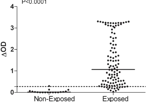
IgG responses to Ae. albopictus SGE were evaluated in individuals from Reunion Island and North of France (Figure 1). In the unexposed group, one individual IgG response was slightly above the cut-off value (DOD = 0.297) (Figure 1). In contrast, a high percentage (88%) of the exposed group from La Reunion responded positively to the anti-SGE IgG Ab. Although
considerable differences in specific Ab level were observed between exposed individuals (DOD from 0.034 to 3.308), a significant difference in specific IgG level was observed between the exposed group (median = 1.067) and the unexposed group (median = 0.015) (p,0.0001, Mann-Whitney test).
Evaluation of IgG cross-reactivity with Ae. aegypti SGE
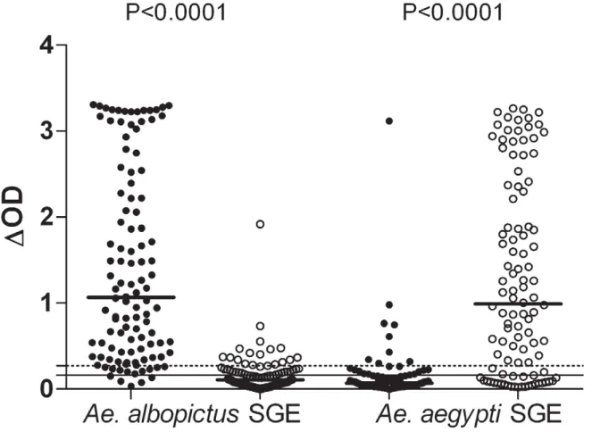
Cross-reactivity between Ae. albopictus and Ae. aegypti SGEs was evaluated by two complementary approaches. First, the specific IgG response to both SGEs (Figure 2) was evaluated in individuals only exposed to Ae. albopictus (from Reunion Island); in parallel, IgG responses to both SGEs were assessed in individuals only exposed to Ae. aegypti (from Bolivia). In the Bolivian group, 16% showed a positive IgG response against Ae. albopictus SGE with only one strong response (DOD.1). The median (0.107) level of IgG against Ae. albopictus SGE was significantly lower in the Bolivians than in the Reunion Island group (1.067) (P,0.0001, Mann-Whitney test). The IgG response to Ae. aegypti SGE was also evaluated in both groups of subjects. As expected, the Bolivian group (exposed only to Ae. aegypti) presented high levels of IgG against Ae. aegypti SGE with 76% of immune responders. In contrast, only 19% of the subjects from Reunion Island were responders to Ae. aegypti SGE and the median (0.068) IgG level was very low compared with the Bolivian group (0.991) (p,0.0001, Mann Whitney test). Only one individual from Reunion presented very high level of IgG to Ae. aegytpi SGE (DOD.3). In addition, in 58% of double immune responders from Reunion Island, the level of IgG against Ae. albopictus SGE was above the 75% percentile value (DOD = 2.426; data not shown). In the Bolivian double immune responders, the corresponding figure for Ae. aegypti SGE was 50% (DOD value of 75% percentile = 2.341). These results
Figure 1. Individual IgG response toAe. albopictusSGE in Reunion Island. Exposed (n = 110, Reunion Island) and unexposed individuals (n = 18, North France). Individual IgG Ab responses are represented by DOD. Bars indicate median value in each group and the dotted line represents the threshold of specific Ab response to Ae. Albopictus SGE (DOD = 0.271).
indicate that IgG to Ae. albopictus and Ae. aegypti SGE are cross reactive, particularly in individuals presenting a very high level of IgG.
Secondly, the level of specific IgG Ab against both SGEs was compared by a statistical correlation analysis (Figure 3). High positive correlation between IgG against Ae. albopictus SGE and Ae. aegypti SGE was observed for both the Reunion Island group (r = +0.445; P,0.0001, Spearman test) and the Bolivian group (r = +0.617; P,0.0001, Spearman test). For each population, IgG cross-reactivity was low with few individuals responding to both Ae. albopictus and Ae. aegypti SGEs. For the Reunion Island group, only 5 individuals showed a strong IgG response to Ae. aegypti SGE. These correlations indicate that cross-reactivity between the two Aedes species may depend on IgG level. In the Bolivian group, the same trend is observed with only 8 individuals showing a strong IgG response to Ae. albopictus SGE.
Discussion
In this study, we investigated IgG responses to Ae. albopictus SGE in exposed adults from Reunion Island where Ae. albopictus—the only anthopophilic Aedes species—transmits chikungunya. First, specific IgG responses were high in the exposed group and significantly different to those observed in an unexposed population from Europe: 88% of exposed individuals developed IgG against Ae. albopictus SGE. In addition, specific IgG Ab levels showed considerable inter-individual variations. Since the intensity
of exposure in a given population living in the same area can obviously vary between individuals, these results suggest that the anti-SGE IgG response may be a reliable biomarker for exposure to Ae. albopictus bites. Furthermore, the significant difference in Ab levels between unexposed and exposed individuals shows that this biomarker could distinguish individuals exposed to Ae. albopictus bites. A useful biomarker for Ae. albopictus bites needs to be highly specific and devoid of immune cross-reactivity with other Aedes species. We evaluated the cross-reactivity between two species using complementary approaches, i.e. in individuals only ever exposed to Ae. aegypti and by analysing IgG levels against both SGEs. In the Bolivian group only exposed to Ae. aegypti, 16% of individuals responded to Ae. albopictus SGE (heterologous ELISA). The DOD values for these ‘‘cross-reactive’’ individuals are characterised by very low IgG levels whereas a high percentage (88%) and high IgG levels (homologous ELISA) were observed in subjects from Reunion Island. In addition, IgG responses against Ae. aegypti SGE are significantly different: 76% of immune responders in Bolivia compared with 19% in Reunion Island. Interestingly, we observed that IgG cross-reactivity was mainly detected in high immune responders. In Reunion Island, in 58% of double immune responders, the level of IgG against Ae. albopictus SGE was above the third quartile. In parallel, in Bolivia, the level of IgG against Ae. aegypti SGE of 50% of double immune responders was above the third quartile. Taken together, these results suggest that there is cross-reactivity between Ae. albopictus SGE and Ae. aegypti SGE, especially in high immune responders.
Figure 2. Individual IgG response toAe. albopictusorAe. aegyptiSGE in Reunion Island and Bolivia. Individual IgG responses against Ae. albopictus SGE and Ae. aegypti SGE are presented in individuals from Reunion Island (black circle) and from Bolivia (white circle). The percentage of positive responders is indicated for each group. Bars indicate median value in each group. The dotted and solid lines represent the threshold of specific Ab response to Ae. albopictus (DOD = 0.271) and Ae. aegypti SGE (DOD = 0.161), respectively.
doi:10.1371/journal.pntd.0001487.g002
Further investigations would be required to identify species-specific salivary antigens. IgG response to Ae. albopictus SGE has been detected in individuals exposed to the bites of this mosquito [27]. It can be hypothesised that, in Reunion Island, the observed specific IgG responses were elicited as a result of antigenic stimulation following biting by Ae. albopictus. In the urban area of Reunion Island, Ae. albopictus is highly antropophilic [28] and characterised by numerous ‘‘artificial’’ breeding sites [25] which could explain the high percentage (88%) of specific responders to Ae. albopictus SGE. These results point up the relevance of this approach to developing a specific biomarker for exposure to Ae. albopictus. However epidemiological factors—history of exposure, genetic background, immune tolerance, etc.—have to be taken into account when explaining variations in responsiveness. Further longitudinal studies could focus on this. To our knowledge, measuring IgGs against Ae. albopictus SGE represents the first direct method for evaluating human exposure to Ae. albopictus and this parameter probably represents the first genuine biomarker for man-vector contact. This method could help overcome the shortcomings of the current methods which only give indirect measurements of exposure to Ae. albopictus and therefore have considerable limitations for evaluating the risk of arbovirus transmission [7]. The current standard methods—immature stage counting and trapping techniques—are both ‘‘static’’ and mainly target ‘‘household exposure’’, ignoring all the anthropogenic factors that can affect exposure (e.g. water storage practices). Ae. albopictus and Ae. aegypti are diurnal mosquitoes, biting both indoors and outdoors, and these characteristics may complicate the
assessment of exposure using conventional methods. Using anti-SGE IgG responses to evaluate exposure to Ae. albopictus, highly heterogeneous Ab levels were observed between exposed individ-uals, as previously reported for another vector [26]. It could be hypothesized that different levels could reflect the intensity of exposure to biting vectors, e.g. the experimental results indicate that a high Ab response is the result of high exposure and the ame association is also observed for low Ab levels [29]. This has also been observed in human populations in the field where arthropods vectors are endemic. In exposed individuals from endemic areas, it has been shown that the level of anti-saliva Ab was closely associated with the intensity of exposure to vector bites [12], [23], [26], [30]. Therefore, this could be a useful tool for comparing the exposure to Ae. albopictus at different sites in a given study area or between different areas, and could be useful for evaluating the efficacy of vector control. In addition, recent findings in the malaria field have shown that Ab responses to saliva antigens are useful in the assessment of low-level exposure to Anopheles bites [31]. Detection of low level exposure in newly colonized areas is of particular interest for Ae. albopictus due to its ongoing worldwide spread, especially in urban contexts. Moreover, a clear correlation between larval indices and pathogen transmission is difficult to establish when the level of exposure is low [32], [33]. In both cases, evaluation of the anti-saliva IgG response could complement entomological methods. However, several validation steps (e.g. seasonal variation and correlation with entomological measurements) will have to be checked.
Figure 3. Individual cross-reactivity IgG response betweenAe. albopictusandAe. aegyptiSGE. Correlations between IgG against Ae. albopictus SGE and Ae. aegypti SGE in Reunion Island (black circle) and Bolivia (white circle). The coefficient of correlation (r) and significance are indicated for each species SGE, black lines represent linear Spearman correlation.
In this study, we measured the Ab response to whole SGE. As long as any salivary proteins are shared with other species or genera, the degree of cross-reactivity with major other Aedes vectors will have to be assessed. Thus, cross-reactivity was investigated between Ae. albopictus and Ae. aegypti, closely related species with shared salivary proteins [34], [35]. Only weak cross-reactivity was detected between Ae. albopictus and Ae. aegypti SGEs, mainly observed in high immune responders. This may suggest that species-specific proteins are more immunogenic than genus-shared proteins. Species-specificity has been already reported with several Aedes species [20], [23], [36] and for a broad range of vectors [12], [29], [37]. In contrast, western-blot analysis reveals extensive cross-reactivity and shows that some antigens are common to all Aedes species [21], [38], [39]. This low level of cross-reactivity also raises the roles of intensity and history of exposure in determining the acquired IgG response against SGE. Individuals from Reunion Island are unlikely to have been exposed to Ae. aegypti because this mosquito is not found in town and its breeding sites are restricted to natural habitats [24]. Cross-reactivity between salivary proteins common to all members of the Aedes genus seems therefore to be the most likely explanation of the observed IgG responsiveness to Ae. aegypti SGE in Reunion Island. Travelling could also lead to contact with Ae. aegypti which is present in most of the islands of the Indian Ocean.
To enhance the usefulness of this biomarker for large-scale applications and to exclude cross-reactivity, an Ae. albopictus-specific salivary antigen needs to be identified. In malaria, only one peptide in whole Anopheles salivary antigen is an efficient biomarker for exposure to Anopheles bites [30]. To improve the sensitivity and the specificity of detection, an immuno-proteomic
study is currently underway to identify Ae. albopictus-specific proteins and peptides.
The study described here represents the first step for estimating human exposure to Ae. albopictus by quantifying the IgG response to vector salivary antigens. In an area of chikungunya transmis-sion, it was shown that the level of Ab against Ae. albopictus SGE can be used to identify individuals who have been exposed to the bites of this important vector. Low level cross-reactivity was observed with Ae. aegypti SGE suggesting that it will be possible to develop a specific biomarker for human exposure to biting Ae. albopictus. By combining the use of such a biomarker with classical entomological and epidemiological methods, it could enhance the assessment of human exposure to Ae. albopictus and therefore contribute to both accurate prediction of the risk of arbovirus transmission and evaluation of the efficacy of vector control.
Acknowledgments
The authors gratefully acknowledge the population of La Reunion and Santa Cruz de la Sierra for their participation in this study. The authors thank also Sirilakassana Patramool, Severine Liccardi, Dr Jean-Jacques Hoarau and La Re´union DRASS-LAV team for their technical support. We thank A. Molloy for reviewing the draft of the manuscript and reviewing the English in the paper.
Author Contributions
Conceived and designed the experiments: SD FM SC FF PG FR. Performed the experiments: SD FM. Analyzed the data: SD FM SC FR. Contributed reagents/materials/analysis tools: SD FM SC JSD AHR YR AW JPH DM FF PG FR. Wrote the paper: SD SC FR.
References
1. Weaver SC, Reisen WK (2010) Present and future arboviral threats. Antiviral Res 85: 328–345.
2. WHO (2009) Dengue: guidelines for diagnosis, treatment, prevention and control. Geneva: World Health Organization. WHO/HTM/NTD/DEN/ 2009.1.
3. Staples JE, Breiman RF, Powers AM (2009) Chikungunya fever: an epidemiological review of a re-emerging infectious disease. Clin Infect Dis 49: 942–948.
4. Paupy C, Delatte H, Bagny L, Corbel V, Fontenille D (2009) Aedes albopictus, an arbovirus vector: from the darkness to the light. Microbes Infect 11: 1177–1185.
5. Rezza G, Nicoletti L, Angelini R, Romi R, Finarelli AC, et al. (2007) Infection with chikungunya virus in Italy: an outbreak in a temperate region. Lancet 370: 1840–1846.
6. Gould EA, Gallian P, De Lamballerie X, Charrel RN (2010) First cases of autochthonous dengue fever and chikungunya fever in France: from bad dream to reality! Clin Microbiol Infect 16: 1702–1704.
7. Focks D (2003) A Review of Entomological Sampling Methods and Indicators for Dengue Vectors. World Health Organization TDR/IDE/Den/03.1. 8. Billingsley PF, Baird J, Mitchell JA, Drakeley C (2006) Immune interactions
between mosquitoes and their hosts. Parasite Immunol 28: 143–153. 9. Ribeiro JM, Francischetti IM (2003) Role of arthropod saliva in blood feeding:
sialome and post-sialome perspectives. Annu Rev Entomol 48: 73–88. 10. Nascimento R, Santana J, Lozzi S, Araujo C, Teixeira A (2001) Human IgG1
and IgG4: the main antibodies against Triatoma infestans (Hemiptera: Reduviidae) salivary gland proteins. Am J Trop Med Hyg 65: 219–226. 11. Rohousova I, Ozensoy S, Ozbel Y, Volf P (2005) Detection of species-specific
antibody response of humans and mice bitten by sand flies. Parasitology 130: 493–499.
12. Poinsignon A, Remoue F, Rossignol M, Cornelie S, Courtin D, et al. (2008) Human IgG antibody response to Glossina saliva: an epidemiologic marker of exposure to Glossina bites. Am J Trop Med Hyg 78: 750–753.
13. Das MK, Mishra A, Beuria MK, Dash AP (1991) Human natural antibodies to Culex quinquefasciatus: age-dependent occurrence. J Am Mosq Control Assoc 7: 319–321.
14. Remoue F, Cisse B, Ba F, Sokhna C, Herve JP, et al. (2006) Evaluation of the antibody response to Anopheles salivary antigens as a potential marker of risk of malaria. Trans R Soc Trop Med Hyg 100: 363–370.
15. Waitayakul A, Somsri S, Sattabongkot J, Looareesuwan S, Cui L, et al. (2006) Natural human humoral response to salivary gland proteins of Anopheles mosquitoes in Thailand. Acta Trop 98: 66–73.
16. Andrade BB, Rocha BC, Reis-Filho A, Camargo LM, Tadei WP, et al. (2009) Anti-Anopheles darlingi saliva antibodies as marker of Plasmodium vivax infection and clinical immunity in the Brazilian Amazon. Malar J 8: 121. 17. Drame PM, Poinsignon A, Besnard P, Le Mire J, Dos-Santos MA, et al. (2010)
Human antibody response to Anopheles gambiae saliva: an immuno-epidemiological biomarker to evaluate the efficacy of insecticide-treated nets in malaria vector control. Am J Trop Med Hyg 83: 115–121.
18. Peng Z, Simons FE (2004) Mosquito allergy: immune mechanisms and recombinant salivary allergens. Int Arch Allergy Immunol 133: 198–209. 19. Wasinpiyamongkol L, Patramool S, Luplertlop N, Surasombatpattana P,
Doucoure S, et al. (2009) Blood-feeding and immunogenic Aedes aegypti saliva proteins. Proteomics 10: 1906–1916.
20. Brummer-Korvenkontio H, Palosuo K, Palosuo T, Brummer-Korvenkontio M, Leinikki P, et al. (1997) Detection of mosquito saliva-specific IgE antibodies by capture ELISA. Allergy 52: 342–345.
21. Reunala T, Brummer-Korvenkontio H, Palosuo K, Miyanij M, Ruiz-Maldonado R, et al. (1994) Frequent occurrence of IgE and IgG4 antibodies against saliva of Aedes communis and Aedes aegypti mosquitoes in children. Int Arch Allergy Immunol 104: 366–371.
22. Orlandi-Pradines E, Almeras L, Denis de Senneville L, Barbe S, Remoue F, et al. (2007) Antibody response against saliva antigens of Anopheles gambiae and Aedes aegypti in travellers in tropical Africa. Microbes Infect 9: 1454–1462. 23. Remoue F, Alix E, Cornelie S, Sokhna C, Cisse B, et al. (2007) IgE and IgG4
antibody responses to Aedes saliva in African children. Acta Trop 104: 108–115. 24. Bagny L, Delatte H, Quilici S, Fontenille D (2009) Progressive decrease in Aedes aegypti distribution in Reunion Island since the 1900s. J Med Entomol 46: 1541–1545.
25. Delatte H, Dehecq JS, Thiria J, Domerg C, Paupy C, et al. (2008) Geographic distribution and developmental sites of Aedes albopictus (Diptera: Culicidae) during a Chikungunya epidemic event. Vector Borne Zoonotic Dis 8: 25–34. 26. Poinsignon A, Cornelie S, Mestres-Simon M, Lanfrancotti A, Rossignol M, et al.
(2008) Novel peptide marker corresponding to salivary protein gSG6 potentially identifies exposure to Anopheles bites. PLoS One 3: e2472.
27. Konishi E (1990) Distribution of immunoglobulin G and E antibody levels to salivary gland extracts of Aedes albopictus (Diptera: Culicidae) in several age groups of a Japanese population. J Med Entomol 27: 519–522.
28. Delatte H, Desvars A, Bouetard A, Bord S, Gimonneau G, et al. (2010) Blood-feeding behavior of Aedes albopictus, a vector of Chikungunya on La Reunion. Vector Borne Zoonotic Dis 10: 249–258.
29. Schwarz A, Sternberg JM, Johnston V, Medrano-Mercado N, Anderson JM, et al. (2009) Antibody responses of domestic animals to salivary antigens of
Triatoma infestans as biomarkers for low-level infestation of triatomines. Int J Parasitol 39: 1021–1029.
30. Poinsignon A, Samb B, Doucoure S, Drame PM, Sarr JB, et al. (2010) First attempt to validate the gSG6-P1 salivary peptide as an immuno-epidemiological tool for evaluating human exposure to Anopheles funestus bites. Trop Med Int Health 15: 1198–1203.
31. Poinsignon A, Cornelie S, Ba F, Boulanger D, Sow C, et al. (2009) Human IgG response to a salivary peptide, gSG6-P1, as a new immuno-epidemiological tool for evaluating low-level exposure to Anopheles bites. Malar J 8: 198. 32. Sanchez L, Cortinas J, Pelaez O, Gutierrez H, Concepcion D, et al. (2010)
Breteau Index threshold levels indicating risk for dengue transmission in areas with low Aedes infestation. Trop Med Int Health 15: 173–175.
33. Sulaiman S, Pawanchee ZA, Arifin Z, Wahab A (1996) Relationship between Breteau and House indices and cases of dengue/dengue hemorrhagic fever in Kuala Lumpur, Malaysia. J Am Mosq Control Assoc 12: 494–496. 34. Arca B, Lombardo F, Francischetti IM, Pham VM, Mestres-Simon M, et al.
(2007) An insight into the sialome of the adult female mosquito Aedes albopictus. Insect Biochem Mol Biol 37: 107–127.
35. Ribeiro JM, Arca B, Lombardo F, Calvo E, Phan VM, et al. (2007) An annotated catalogue of salivary gland transcripts in the adult female mosquito, Aedes aegypti. BMC Genomics 8: 6.
36. Peng Z, Yang M, Simons FE (1995) Measurement of mosquito Aedes vexans salivary gland-specific IgE and IgG antibodies and the distribution of these antibodies in human sera. Ann Allergy Asthma Immunol 74: 259–264. 37. Clements M, Gidwani K, Kumar R, Hostomska J, Dinesh D, et al. (2010)
Measurement of recent exposure to Phlebotomus argentipes, the vector of Indian visceral Leishmaniasis, by using human antibody responses to sand fly saliva. Am J Trop Med Hyg 82: 801–807.
38. Brummer-Korvenkontio H, Lappalainen P, Reunala T, Palosuo T (1994) Detection of mosquito saliva-specific IgE and IgG4antibodies by
immunoblot-ting. Allergy and Clinical Immunology 93: 551–555.
39. Peng Z, Xu W, Lam H, Cheng L, James AA, et al. (2006) A new recombinant mosquito salivary allergen, rAed a 2: allergenicity, clinical relevance, and cross-reactivity. Allergy 61: 485–490.