HAL Id: inserm-00286454
https://www.hal.inserm.fr/inserm-00286454
Submitted on 9 Jun 2008HAL is a multi-disciplinary open access archive for the deposit and dissemination of sci-entific research documents, whether they are pub-lished or not. The documents may come from teaching and research institutions in France or abroad, or from public or private research centers.
L’archive ouverte pluridisciplinaire HAL, est destinée au dépôt et à la diffusion de documents scientifiques de niveau recherche, publiés ou non, émanant des établissements d’enseignement et de recherche français ou étrangers, des laboratoires publics ou privés.
Endothelial cell protein C receptor and the risk of
venous thrombosis.
Sophie Gandrille
To cite this version:
Sophie Gandrille. Endothelial cell protein C receptor and the risk of venous thrombosis.. Haematolog-ica, Ferrata Storti Foundation, 2008, 93 (6), pp.812-6. �10.3324/haematol.13243�. �inserm-00286454�
Endothelial cell activated protein C receptor (EPCR) and the risk of thrombosis
Sophie GANDRILLE§
§
Inserm, Unité de Recherche 765, Paris, and Université Paris Descartes, Unité de Formation et de Recherche (UFR) des Sciences Pharmaceutiques et Biologiques, Paris, France
Dr S. GANDRILLE, PhD INSERM U 765
UR des Sciences Pharmaceutiques et Biologiques 4 Avenue de l'Observatoire 75006 Paris sophie.gandrille@univ-paris5.fr Tel +33 1 53 73 96 19 Fax +33 1 44 07 17 72 Acknowledgements
The author’s work on EPCR is supported by a Transatlantic Network for Excellence in Cardiovascular Research grant from Fondation Leducq, France
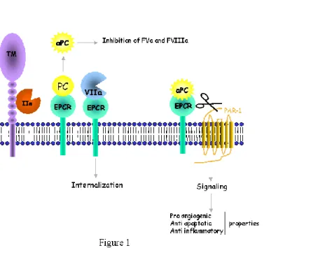
The natural anticoagulant protein C (PC) pathway plays a major role in regulating coagulation and inflammation (reviews 1-3). PC is activated at the endothelial surface when thrombin binds to thrombomodulin (TM), a transmembrane glycoprotein, that transforms thrombin into a potent activator of PC. In the presence of its cofactor protein S, activated protein C (APC) inactivates factors Va and VIIIa, thereby downregulating the thrombin feedback loop. Thrombomodulin is a vascular endothelial cell receptor present in many cells and tissues. Its density is higher in small vessels and capillaries, however, and this is why PC activation was believed to occur mainly in these vessels. However, thrombosis in subjects with hereditary PC or PS deficiency is not restricted to the microcirculation but can also affect larger vessels, suggesting that the PC pathway is also active in zones of lower TM density. This apparent paradox was resolved by the discovery of an endothelial cell receptor specific for PC -- the endothelial cell protein C receptor (EPCR)-- which increases the PC activation rate by thrombin-TM complexes (4) and is most abundant in large vessels (5). Functional studies performed in vitro showed a 20-fold increase in the PC activation rate by membrane thrombin-thrombomodulin complexes when PC was bound to its receptor (6). This increase results from a significant effect of EPCR on the Michaelis-Menten constant (Km) for PC activation by the thrombin-thrombomodulin complex. Indeed, without EPCR intervention, this Km is significantly higher (1 µM) than the circulating concentration of PC (60-70 nM). By presenting PC to the thrombin-TM complex (Figure 1), EPCR reduces the Km and allows the interaction to occur.
Compared to the effect of thrombin binding to TM, which results in a >1000-fold increase in the PC activation rate (7), the 20-fold increase in the PC activation rate after PC binding to EPCR might seem rather inconsequential. Yet, EPCR is even more important for PC activation than suggested by in vitro studies. Indeed, in a baboon model, APC generation induced by thrombin infusion fell by 88% when the animals were pretreated with anti-EPCR antibodies that blocked the PC/EPCR interaction (8). The crucial role of EPCR was further confirmed by studies of PROCR knock-out mice (9): complete invalidation of the gene led to intrauterine death by fibrin deposition in embryonic trophoblast giant cells, leading to thrombosis at the maternal-embryonic interface.
EPCR is a 46-Kda type 1 transmembrane glycoprotein homologous to major histocompatibility complex (MHC) class I/CD1 family proteins (review in 10). This 221-amino-acid (aa) protein comprises an extracellular domain, a 25-aa transmembrane domain, and a very short (3-aa) intracytoplasmic sequence. The PROCR gene is located on chromosome 20, spans 8 kilobases, and possesses 4 exons (11). The first exon encodes the 5'
untranslated region and the signal peptide, exons 2 and 3 encode most of the extracellular domain, and exon 4 encodes the remaining parts of the protein and the 3' untranslated region.
Very recently, EPCR has been shown to serve as a cellular binding site for FVII/FVIIa (12), the affinity of the interaction being similar to that for PC/APC. FVIIa-EPCR complexes do not activate coagulation on unstimulated endothelial cells. FVIIa-EPCR binding reduces FVIIa coagulant activity (13) (Figure 1). The complex does not modulate TF-FVIIa activation of factor X on stimulated endothelial cells, but FVIIa binding to EPCR facilitates FVIIa endocytosis, thus clearing FVIIa from the vascular bed.
In addition to its role in regulating coagulation, EPCR is crucial for the antiinflammatory, antiapoptotic and proangiogenic effects of APC. APC prevents inflammation by downregulating tissue-factor and NFB expression, and by reducing the expression of subunits of the transcription factor NFB and, thus, the expression of NF B-controlled genes (14). Antiapoptotic protein synthesis is promoted, while the production of proapoptotic proteins is reduced. APC inhibits cytokine signaling and TNF induction of cell-surface adhesion molecule expression. Most of these effects are due to PAR-1 cleavage by APC bound to EPCR (15). Inflammation contributes to propagating coagulation, and this effect is thus countered by the anti-inflammatory effect of APC.
A soluble form of EPCR (sEPCR) present in normal human plasma lacks the transmembrane and cytoplasmic domains. Like the membrane-associated form, sEPCR binds PC and APC with similar affinity. However, its binding to APC inhibits the anticoagulant activity of APC by abrogating its ability to inactivate factor Va, and its binding to PC precludes PC activation by thrombin-TM complexes (16) (Figure 2). Soluble EPCR has mainly procoagulant properties. Regarding its effects on inflammation, sEPCR has been shown to bind to activated neutrophils (17). Since it competes with membrane-associated form for APC binding, this could decrease PAR-1 signaling by APC, and thus limit the APC anti-inflammatory properties.
Several situations can interfere with the antithrombotic and antiinflammatory properties of EPCR and thereby promote thrombosis. EPCR dysfunction, decreased membrane EPCR expression, and increased plasma sEPCR levels can theoretically increase the thrombotic risk. It must be kept in mind that studies designed to evaluate the influence of EPCR on the thrombotic risk come up against from two main hurdles. First, EPCR being a membrane protein, its deficiency cannot be measured with a plasma-based assay (contrary to PC and PS deficiencies, for example), and we do not yet know whether sEPCR levels reflect membrane EPCR expression. Second, while circulating concentrations of PC/APC and sEPCR seem too
low to induce complete APC inhibition, local concentrations of sEPCR might be sufficiently high to impair APC functions.
Circumstances leading to decreased membrane EPCR expression and/or increased sEPCR levels
As with other key proteins of the PC pathway, mutations were expected to diminish EPCR expression and thus to be found in subjects with unexplained thrombosis. The first reported abnormality was a 23-base-pair insertion in PROCR exon 3 (17), leading to the synthesis of a truncated protein that is not expressed on endothelial surfaces. Although initially identified in subjects with deep venous thrombosis and myocardial infarction, the precise impact of this mutation on thrombosis is difficult to assess because its allelic frequency is low. Point mutations were then described within the promoter region, but their involvement in gene regulation could not be clearly demonstrated.
Another possible mechanism leading to dysfunction of EPCR-mediated coagulation-regulating mechanisms consists of mutations (or polymorphisms) leading to increased levels of sEPCR. Approximately 15% to 20% of the general population (19) have plasma sEPCR levels between 200 and 800 ng/mL, whereas the remainder have levels below 180 ng/mL. We and others have shown that 50-80% of plasma sEPCR variations are under genetic control (20-22), and that most subjects with elevated sEPCR levels carry the A3 haplotype -- one of the four most frequent PROCR haplotypes in Caucasians. One of the four haplotype-tagging single nucleotide polymorphisms (Ht-SNP) characterizing the A3 haplotype is a g.A6036G substitution leading to a p.Ser219 to Gly substitution in the transmembrane domain of the protein. It was recently shown that sEPCR levels are higher in A3 carriers because the Gly219 receptor isoform is more sensitive than the Ser219 isoform to sheddases such as the metalloprotease ADAM17 (23). While sEPCR was thought to be generated exclusively by shedding, we very recently identified an alternative mRNA splicing (24) that is particularly efficient in A3-carrying subject’s cells and generates a truncated mRNA lacking the sequence encoding the transmembrane and intracytoplasmic domains. The resulting protein is not able to anchor to the membrane; it is thus secreted and can be detected in the plasma of A3-carrying subjects.
Three case-control studies, each involving 350 to 450 thrombophilic patients, examined the A3 haplotype as an independent risk factor for venous thrombosis but gave contradictory results (20-22). While our case-control study pointed to a role of the A3 haplotype in venous thrombosis (20), the other two studies showed no such effect. One of
these studies, however, showed an increased risk (odds ratio) of thrombosis in subjects with increased sEPCR levels, together with a protective effect of the A1 haplotype on venous thrombosis (21). Whether or not the A3 haplotype itself is a risk factor for venous thrombosis remains to be determined in larger studies of thrombophilic subjects.
The A3 haplotype has been examined in two studies as an associated risk factor for venous thrombosis. In the first, the authors observed more severe clinical expression of PC deficiency in A3-carrying subjects than in non-carriers (25). In this issue of the Journal, Navarro et al (26) describes the influence of PROCR haplotypes on the risk of thrombosis in FII g.20210A carriers. Using c.4600G and c.4678C as A3- and A1-Ht-SNPs, respectively, they observed a higher frequency of the A3 haplotype in propositi than in asymptomatic relatives, as well as younger age at disease onset in A3-carrying propositi than in A3-non carriers, and a lower probability of being free of thrombosis at age 40 years. Very interestingly, they observed that symptomatic carriers of the A3 haplotype had higher sEPCR levels than asymptomatic carriers. In addition, they found that the odds ratio for thrombosis increased with the sEPCR quartile in A3 carriers. This finding -- that only higher sEPCR levels are associated with an increased thrombotic risk -- might explain the conflicting results of the three above-mentioned studies examining the A3 haplotype as an independent risk factor for thrombosis. The A1 haplotype was found to have a non significant tendency to protect from thrombosis, confirming previous results obtained by this research group. As underlined by Navarro et al, it is surprising that the effect of the A3 haplotype is seen in subjects with FII g.20210A and not in those with FV Leiden (26). This points to a difference in the mechanisms underlying the prothrombotic effects of these two mutations.
The possible involvement of EPCR Gly219 (A3 haplotype) in arterial thrombosis has been evaluated in a prospective cohort of more than 2000 patients (28). A significantly higher frequency of coronary heart disease was observed in A3 homozygotes, and the A3 haplotype was also associated with an increased risk of coronary heart disease in type-2 diabetic patients.
Acquired EPCR abnormalities and increased sEPCR levels
Quantitative or qualitative modifications of EPCR have been implicated in human systemic lupus erythematosus (SLE), a potentially fatal autoimmune disease affecting multiple organ systems. Immune complexes are believed to induce microvasculature injury, associated with thrombotic manifestations, inflammation, and widespread activation of the vascular endothelium. A recent study strongly suggests that the vascular dysfunction
characterizing SLE may be related to abnormal distribution of both soluble and membrane-associated EPCR (29). In addition to significantly increased plasma sEPCR levels reported in SLE patients by Kurosawa et al (30), increased soluble EPCR levels are observed in cytokine-stimulated endothelial cells in vitro, leading to reduced expression of membrane-bound EPCR. A3-carrying subjects were also more frequent in the SLE group than in the control group (29). These increased sEPCR levels may simply reflect endothelial dysfunction, but they might also contribute to a procoagulant phenotype and thus to the thrombosis observed in SLE. Moreover, among patients with SLE, A3 haplotype carriers are more prone to thrombosis than non-carriers.
Disseminated intravascular coagulation with purpura fulminans due to microvascular thrombosis is a frequent complication of sepsis and is particularly severe during meningococcal sepsis (31). This disorder results from complex dysregulation of hemostatic mechanisms, with activation of procoagulant pathways and impairment of the fibrinolytic system and natural anticoagulant pathways, especially the protein C pathway. Reduced PC levels are found in most patients with sepsis. PC infusion does not always increase APC levels, showing that PC activation is dysregulated in this setting. Histological studies of skin-biopsy specimens (31) revealed evidence of thrombosis, together with consistently diminished TM staining and EPCR staining decreased in 80% of cases.
Thrombosis is a frequent complication of cancer. The thrombophilic phenotype of cancer cells is due to both a cystein protease and to expression of procoagulant factors such as TF. EPCR is expressed on the outer membrane of various cancer cell lines (32,33). APC binding to cancer cell EPCR reduces TF expression while inducing antiapoptotic genes. This cell-protective effect is normally beneficial but, in subjects with cancer, APC binding to EPCR stimulates PAR-1, and PAR-1-associated cellular responses contribute to tumor progression (34), possibly via IL-8 production, which stimulates tumor cell proliferation and metastasis.
In this issue of the Journal, Molina and coworkers (35) report the production of a truncated EPCR mRNA by HUVECs, similar to the isoform we described. More interestingly, Molina and coworkers showed that the corresponding protein was produced and secreted by cultured lung cancer cells. The sEPCR generated by this truncated mRNA can bind PC and APC, and these cells are therefore able to produce a soluble receptor that might compete with membrane-associated EPCR for APC binding. This would tend to limit cell proliferation. In addition, this sEPCR is able to bind FVIIa and to abolish its factor X-activating activity, that counteracts the procoagulant effect of APC binding to sEPCR. The description of this
secreted EPCR isoform opens the way to assessing the thrombotic risk in cancer patients according to the type of malignancy.
Circumstances leading to inhibition of APC–membrane interaction: the case of anti-EPCR antibodies
A recent study from Hurtado et al showed that approximately 20% of patients with the antiphospholipid syndrome (APS) had anti-EPCR antibodies exceeding the amount observed in control subjects (36). Women with APS and a history of thrombosis had the highest anti-EPCR antibody titers, and women who had anti-anti-EPCR IgM had had multiple miscarriages. IgM fractions isolated from these patients potently inhibited PC activation on endothelial cells. These autoantibodies that block PC activation might lead to skin necrosis (37) by reducing APC levels. Low APC levels are a risk factor for thrombosis, and this might explain the multiple episodes of venous thrombosis experienced by APS patients with PC-activation-blocking antibodies. EPCR is also crucial for preventing placental thrombosis and thus for maintaining pregnancy. By undermining both anticoagulant and antiinflammatory mechanisms, these antibodies could precipitate fetal death. Indeed, anti-EPCR antibodies have been identified in 80% of women having experienced a first episode of fetal death, the odds ratio for this outcome depending on the isotype of anti-EPCR antibodies (38).
As EPCR is present on arterial walls, anti-EPCR antibodies could theoretically impair coagulation in these vessels and thus play a role in arterial thrombosis. Among 165 young women who survived a first acute myocardial infarction, Montes et al found that IgA anti-EPCR titers exceeded the cut-off value determined in this study in 22% of cases, compared to only 10% in a control group, while the OR increased with the titer (39). This strongly suggests that anti-EPCR antibodies may be a risk factor for acute myocardial infarction in young women.
EPCR levels vary in a wide variety of pathophysiologic conditions, and the consequences depend partly on whether it is the membrane-associated or soluble form that is affected. While the role of membrane-associated EPCR is clearly antithrombotic and anti-inflammatory in physiological circumstances, the function of the circulating protein (if any) is unclear. The thrombotic risk associated with the A3 haplotype is controversial, and further studies involving larger numbers of patients are necessary. Any deleterious effect of this haplotype could be related to the consistently elevated sEPCR levels seen in A3 carriers.
Many circumstances can lead to increased plasma sEPCR levels, but it is not always easy to tell whether the increase is the cause or an effect of the associated disorder. Given the systemic concentrations of PC and sEPCR, it is unlikely that sEPCR could impair PC activation or activity and thus express its procoagulant properties observed in vitro. And yet evidence continues to accumulate that sEPCR has a noteworthy role in controlling coagulation and, thus, thrombosis.
REFERENCES
1. Esmon CT. The protein C pathway. Chest 2003, 124, 36S-32S
2. Esmon CT. Interactions between the innate immune and blood coagulation systems. Trends Immunol 2004; 25: 536-42
3. Van de Wouwer M, Collen D, Conway EM. Thrombomodulin-protein C-EPCR system. Integrated to regulated coagulation and inflammation. Arterioscler Thromb Vasc Biol 2004; 24: 1374-83
4. Fukudome K, Esmon CT. Identification, cloning, and regulation of a novel endothelial cell protein C/activated protein C receptor. J Biol Chem 1994; 269: 26486-91
5. Laszik Z, Mitro A, Taylor FB Jr, Ferrell G, Esmon CT. Human protein C receptor is present primarily on endothelium of large blood vessels: implications for the control of the protein C pathway. Circulation 1997; 96: 3633-40
6. Stearns-Kurosawa DJ, Kurosawa S, Mollica JS, Ferrell GL, Esmon CT. The endothelial cell protein C receptor augments protein C activation by the thrombin-thrombomodulin complex. Proc Natl Acad Sci U S A 1996 ; 93:10212-6
7. Esmon NL, Owen WG, Esmon CT. Isolation of a membrane-bound cofactor for thrombin-catalyzed activation of protein C. J Biol Chem 1982; 257: 859-64
8. Taylor FB Jr, Peer GT, Lockhart MS, Ferrell G, Esmon CT. Endothelial cell protein C receptor plays an important role in protein C activation in vivo. Blood 2001 ; 15;97: 1685-8 9. Gu JM, Crawley JT, Ferrell G, Zhang F, Li W, Esmon NL, et al. Disruption of the endothelial cell protein C receptor gene in mice causes placental thrombosis and early embryonic lethality. J Biol Chem 2002; 277: 43335-43
10. Esmon CT. The endothelial protein C receptor. Curr Opin Hematol 2006; 13: 382-5 11. Simmonds RE, Lane DA. Structural and functional implications of the intron/exon organization of the human endothelial cell protein C/activated protein C receptor (EPCR) gene: comparison with the structure of CD1/major histocompatibility complex alpha1 and alpha2 domains. Blood. 1999; 94: 632-41
12. Ghosh S, Pendurthi UR, Steinoe A, Esmon CT, Rao LV. Endothelial cell protein C receptor acts as a cellular receptor for factor VIIa on endothelium. J Biol Chem 2007; 282: 11849-57
13. López-Sagaseta J, Montes R, Puy C, Díez N, Fukudome K, Hermida J. Binding of factor VIIa to the endothelial cell protein C receptor reduces its coagulant activity. J Thromb Haemost. 2007; 5: 1817-24
14. Joyce DE, Gelbert L, Ciaccia A, DeHoff B, Grinnell BW. Gene expression profile of antithrombotic protein C defines new mechanisms modulating inflammation and apoptosis. J Biol Chem 2001; 276:11199-203
15. Ludeman MJ, Kataoka H, Srinivasan Y, Esmon NL, Esmon CT, Coughlin SR. PAR1 cleavage and signaling in response to activated protein C and thrombin. J Biol Chem 2005; 280: 13122-8.
16. Liaw PC, Neuenschwander PF, Smirnov MD, Esmon CT. Mechanisms by which soluble endothelial cell protein C receptor modulates protein C and activated protein C function. J Biol Chem 2000;275 :5447-52
17. Kurosawa S, Esmon CT, Stearns-Kurosawa DJ. The soluble endothelial protein C receptor binds to activated neutrophils: involvement of proteinase-3 and CD11b/CD18. J Immunol 2000; 165: 4697-703
18. Biguzzi E, Merati G, Liaw PC, Bucciarelli P, Oganesyan N, Qu D, Gu JM, Fetiveau R, Esmon CT, Mannucci PM, Faioni EM. A 23bp insertion in the endothelial protein C receptor (EPCR) gene impairs EPCR function. Thromb Haemost 2001; 86: 945-8
19. Stearns-Kurosawa DJ, Burgin C, Parker D, Comp P, Kurosawa S. Bimodal distribution of soluble endothelial protein C receptor levels in healthy populations. Thromb Haemost 2003;1: 855-6
20. Saposnik B, Reny JL, Gaussem P, Emmerich J, Aiach M, Gandrille S. A haplotype of the EPCR gene is associated with increased plasma levels of sEPCR and is a candidate risk factor for thrombosis. Blood 2004; 103: 1311-8
21. Medina P, Navarro S, Estellés A, Vayá A, Woodhams B, Mira Y, et al. Contribution of polymorphisms in the endothelial protein C receptor gene to soluble endothelial protein C receptor and circulating activated protein C levels, and thrombotic risk. Thromb Haemost 2004; 91: 905-11
22. Uitte de Willige S, Van Marion V, Rosendaal FR, Vos HL, de Visser MC, Bertina RM..Haplotypes of the EPCR gene, plasma sEPCR levels and the risk of deep venous thrombosis. J Thromb Haemost 2004; 2: 1305-10
23. Qu D, Wang Y, Song Y, Esmon NL, Esmon CT. The Ser219-->Gly dimorphism of the endothelial protein C receptor contributes to the higher soluble protein levels observed in individuals with the A3 haplotype. J Thromb Haemost 2006; 4: 229-35
24. Saposnik B, Lesteven E, Lokajczyk A, Esmon CT, Aiach M, Gandrille S. Alternative mRNA is favored by the A3 haplotype of the EPCR gene PROCR and generates a novel soluble form of EPCR in plasma. Blood 2008; 111: 3442-51
25. Simioni P, Morboeuf O, Tognin G, Gavasso S, Tormene D, Woodhams B, et al. Soluble endothelial protein C receptor (sEPCR) levels and venous thromboembolism in carriers of two dysfunctional protein C variants. Thromb Res 2006; 117: 523-8
26. Navarro S, Medina P, Mira Y, Estellés A, Villa P, Ferrando F, et al. Haplotypes of the EPCR gene, prothrombin levels, and the risk of venous thrombosis in carriers of the prothrombin G20210A mutation. Haematologica 2008,
27. Medina P, Navarro S, Estellés A, Vayá A, Bertina RM, España F. Influence of the 4600A/G and 4678G/C polymorphisms in the endothelial protein C receptor (EPCR) gene on the risk of venous thromboembolism in carriers of factor V Leiden. Thromb Haemost 2005; 94: 389-94
28. Ireland H, Konstantoulas CJ, Cooper JA, Hawe E, Humphries SE, Mather H, et al. EPCR Ser219Gly: elevated sEPCR, prothrombin F1+2, risk for coronary heart disease, and increased sEPCR shedding in vitro. Atherosclerosis, 2005, 183, 283-292
29. Sesin CA, Yin X, Esmon CT, Buyon JP, Clancy RM. Shedding of endothelial protein C receptor contributes to vasculopathy and renal injury in lupus: in vivo and in vitro evidence. Kidney Int 2005; 68: 110-20
30. Kurosawa S, Stearns-Kurosawa D, Carson CW, D’Angelo A, Della Valle P, Esmon CT. Plasma levels of endothelial cell protein C receptor are elevated in patients with sepsis and systemic lupus erythematosus: lack of correlation with thrombomodulin suggests involvement of different pathological processes. Blood 1998; 91: 725-7
31. Faust SN, Levin M, Harrison OB, Goldin RD, Lockhart MS, Kondaveeti S, Laszik Z, Esmon CT, Heyderman RS. Dysfunction of endothelial protein C activation in severe meningococcal sepsis. N Engl J Med 2001; 345: 408-16
32. Tsuneyoshi N, Fukudome K, Horiguchi SI, Ye X, Matsuzaki M, Toi M, et al. Expression and anticoagulant function of the endothelial cell protein C receptor (EPCR) in cancer cell lines. Thromb Haemost 2001; 85: 356-61
33. Scheffer GL, Flens MJ, Hageman S, Izquierdo MA, Shoemaker RH, Scheper RJ. Expression of the vascular endothelial cell protein C receptor in epithelial tumour cells. Eur J
Cancer 2002; 38: 1535-42
34. Beaulieu LM, Church FC. Activated protein C promotes breast cancer cell migration through interactions with EPCR and PAR-1. Exp Cell Res 2007; 313: 677-87
35. Molina E, Hermida J, lopez-Sagaseta , Puy C, Montes R. A truncated form of endothelial cell protein C receptor (EPCR) generated by alternative splicing is functionally similar to soluble EPCR. Haematologica 2008;
36. Hurtado V, Montes R, Gris JC, Bertolaccini ML, Alonso A, Martínez-González MA, et al. Autoantibodies against EPCR are found in antiphospholipid syndrome and are a risk factor for fetal death. Blood 2004; 104: 1369-74
37. Lavigne-Lissalde G, Cochery-Nouvellon E, Granier G, Quere I, Gris JC. Diffuse skin necrosis in a patient with an anti-endothelial cell protein C receptor autoantibody which blocks protein C activation. J Thromb Haemost 2005; 3: 413-5
38. Lavigne-Lissalde G, Cochery-Nouvellon E, Mercier E, Marès P, Gris JC. High plasma levels of endothelial protein C receptor are associated with the risk of unexplained fetal death. J Thromb Haemost 2005; 3: 393-5
39. Montes R, Hurtado V, Alonso A, Foco L, Zonzin P, Mannucci PM, et al. Autoantibodies against the endothelial receptor of protein C are associated with acute myocardial infarction in young women. J Thromb Haemost 2005; 3: 1454-8
Figure 1: Schematic drawing of the anticoagulant and anti-inflammatory properties of membrane associated EPCR
Figure 2: cartoon of the known properties of soluble EPCR. Plasma soluble EPCR
originates from the membrane form by shedding, or from synthesis of a soluble isoform issued from an alternative splicing.