The Toxoplasma gondii Plastid replication and Repair
Enzyme Complex, PREX
A. MUKHOPADHYAY1, C-Y. CHEN2, C. DOERIG3, F. L. HENRIQUEZ4, C. W. ROBERTS4
and M. P. BARRETT1*
1Institute of Biomedical and Life Sciences, Division of Infection and Immunity, Glasgow Biomedical Research Centre, University of Glasgow, Glasgow G12 8TA, UK
2
Joseph Gottstein Memorial Cancer Research Laboratory, Department of Pathology, University of Washington, Seattle, WA 98195-7705, USA
3
INSERM U609, Wellcome Centre for Molecular Parasitology, University of Glasgow, 120 University Place, Glasgow, G12 8TA, Scotland, UK. INSERM U609, Global Health Institute, Ecole Polytechnique Fe´de´rale de Lausanne, CH-1015 Lausnne, Switzerland
4
Strathclyde Institute of Pharmacy and Biomedical Sciences, 27 Taylor Street, University of Strathclyde, Glasgow G4 0NR, UK
(Received 4 December 2008; revised 13 February and 4 March 2009; accepted 4 March 2009; first published online 30 April 2009)
S U M M A R Y
A plastid-like organelle, the apicoplast, is essential to the majority of medically and veterinary important apicomplexan protozoa including Toxoplasma gondii and Plasmodium. The apicoplast contains multiple copies of a 35 kb genome, the replication of which is dependent upon nuclear-encoded proteins that are imported into the organelle. In P. falciparum an unusual multi-functional gene, pfprex, was previously identified and inferred to encode a protein with DNA primase, DNA helicase and DNA polymerase activities. Herein, we report the presence of a prex orthologue in T. gondii. The protein is predicted to have a bi-partite apicoplast targeting sequence similar to that demonstrated on the PfPREX polypeptide, capable of delivering marker proteins to the apicoplast. Unlike the P. falciparum gene that is devoid of introns, the T. gondii prex gene carries 19 introns, which are spliced to produce a contiguous mRNA. Bacterial expression of the polymerase domain reveals the protein to be active. Consistent with the reported absence of a plastid in Cryptosporidium species, in silico analysis of their genomes failed to demonstrate an orthologue of prex. These studies indicate that prex is conserved across the plastid-bearing apicomplexans and may play an important role in the replication of the plastid genome.
Key words : Apicomplexa, DNA replication, Plasmodium falciparum, Toxoplasma gondii, exon, intron, DNA polymerase, DNA primase, DNA helicase.
I N T R O D U C T I O N
The parasite Toxoplasma gondii is a member of the eukaryotic phylum Apicomplexa. The phylum, which includes Plasmodium falciparum, a causative agent of malaria, as well as multiple other parasitic genera, is of immense medical and veterinary im-portance. Members of this phylum share a number of structural attributes. With the likely exception of Cryptosporidium, one such shared characteristic is the apicoplast, a relict plastid believed to have been acquired through secondary endosymbiosis (Wilson et al. 1994 ; McFadden et al. 1996). The T. gondii apicoplast contains its own y35 kb genome that is replicated and segregated with the organelle during parasite proliferation. With the exception of the microgamete stage, all T. gondii life-cycle stages
have been demonstrated to possess a single apico-plast, which is faithfully segregated into the daughter cells (Striepen et al. 2000 ; Ferguson et al. 2007). Like many endosymbiont-derived organelles, the apico-plast has a reduced genome containing genes in-volved principally in its own replication. A variety of the genes originally associated with the prokaryotic endosymbiont have been incorporated into the nu-clear genome and their products are targeted back to the apicoplast using a bi-partite plastid targeting peptide. Consequently, many nuclear-encoded en-zymes associated with the apicoplast are of prokar-yotic ancestry. The organelle is essential as it is the site for numerous biochemical pathways including heme and fatty acid biosynthesis and steroid pro-duction (Roos, 1999 ; Soldati, 1999 ; McLeod et al. 2001). The apicoplast has therefore proven to be a valuable target for drugs active against apicomplexan parasites (Fichera and Roos, 1997).
Genes encoding proteins involved in DNA repli-cation or repair are absent from the apicoplast genome. Certain genes encoding plastid DNA rep-lication enzymes are, however, present in the nuclear
* Corresponding author : Institute of Biomedical and Life Sciences, Division of Infection and Immunity, Glasgow Biomedical Research Centre, University of Glasgow, Glasgow G12 8TA, UK.
genome, with the protein being imported into the plastid post-translationally. Inhibition of compo-nents of the apicoplast DNA replication machin-ery (e.g. DNA gyrase) by ciprofloxacin (Dar et al. 2007) can cause cessation of apicoplast replication and subsequent parasite death (Dar et al. 2007 ; Raghu Ram et al. 2007). Thus, the nuclear encoded apicoplast replication machinery is an established drug target in apicoplexan parasites (Ralph et al. 2001).
The various enzymatic activities required for rep-lication of the apicoplast genome in P. falciparum may be derived in large part from a single gene, pfprex, which encodes a multifunctional protein (Seow et al. 2005) comprising a T7 bacteriophage homologous primase-helicase domain and a prokar-yotic DNA polymerase domain separated by a spacer domain. An N-terminal sequence of PfPREX tar-geted Green Fluorescent Protein (GFP) to the api-coplast, strongly suggesting that this protein may be involved in DNA replication or repair of the organ-elle genome. Thus, this single gene provides multiple functional properties required for apicoplast repli-cation and/or repair (Seow et al. 2005). Herein, we demonstrate the presence of a single prex gene (Tgprex) in T. gondii. Unlike prex in P. falciparum, the gene in T. gondii is interrupted by multiple introns. The polymerase domain expressed as a recombinant protein in E. coli displays DNA poly-merase activity. The conservation of unusual pro-karyotic-like DNA replication enzymes across the Apicomplexa indicates that PREX could be a po-tential target for chemotherapy against this class of organisms.
M A T E R I A L S A N D M E T H O D S
T. gondii tachyzoite preparation and preparation of cDNA
T. gondii tachyzoites (RH strain) were grown in the peritoneum of BALB/c mice as previously de-scribed (Roberts and Alexander, 1992). Tachyzoites were harvested and washed in sterile phosphate-buffered saline (PBS ; 0.1MNaCl, 2.7 mMKCl, 10 mM
Na2HPO4, 1.76 mM KH2PO4, pH 7.4) by
centri-fugation at 500 g for 5 min at 4 xC and stored as pellets atx70 xC. RNA isolation from the tachyzoites was performed using Trizol1(Invitrogen, UK) following a protocol, based on a single-step acid guanidinium thiocyanate-phenol-chloroform for RNA isolation (Chomczynski and Sacchi, 1987).
Complementary DNA (cDNA) was produced from total RNA as previously described (Campbell et al. 2004). In a 13ml volume, 1 mg of RNA was added to 1ml of random hexamer mix (Promega, Southampton, UK) and 1ml of a 10 mM
deoxy-nucleoside triphosphate mixture, incubated at 65 xC for 5 min, and chilled on ice for 1 min. Then 80 units
of RNAsin ribonuclease inhibitor (Promega, UK), 4ml of 5r first strand buffer (250 mMTris-HCl, pH
8.3, 375 mMKCl, 15 mMMgCl2), 1ml of 0.1MDTT
and 200 units of Superscript III reverse transcriptase RNAse Hx(Invitrogen, UK) were added to the re-action and incubated for 5 min at 25 xC followed by 55 xC for 60 min. Inactivation of the reverse tran-scriptase was performed by heating at 70 xC for 10 min.
Identification and sequencing of the prex gene in T. gondii
Initially the PfPREX homologues were identified in the T. gondii genome project by BLASTP analy-sis. As the P. falciparum gene was AT rich, the protein sequence was used as a query and the protein match was extrapolated to its cognate gene in the genome. The two protein coding genes are situated on chromosome VIIb in the T. gondii genome pro-ject (www.toxodb.org release 4) (Gajria et al. 2008). Oligonucleotide primers were designed to confirm the prex gene structure by PCR. The primer lo-cations are listed in relation to the actual gene model (Fig. 1A) determined in this study and are listed in order of their positions on the gene (Table 1). Primer pairs for each PCR are designated in Fig. 1A. The primers situated within the consensus exons predicted by different gene prediction algorithms (GLEAN, GlimmerHMM, TwinScan, TwinScan/ Eimeria and TigrScan) are in bold font (Table 1). After confirmation of predicted sequences these primers were coupled with primers from other pre-dicted exons to confirm the coding sequence.
PCR reactions were performed on both genomic and cDNA from T. gondii (RH strain) tachyzoites for comparison. One ml of the cDNA or 500 ng of the genomic DNA template was amplified by Ex-pand Hi-Fidelity polymerase enzyme (Roche) using 0.2 mMdNTP mix, 0.4mMof sequence specific
for-ward and reverse primers with 1.5mM/2.0mM of
Mg2+ in enzyme-specific buffer. Amplified PCR products from the cDNA were cloned into the pGEM-T Easy vector (Promega) and sequenced to verify the ORF. The cDNA contig was assembled using the Contig Express programme of the Vector NTI suite 9 and 10.
To clarify the 5k end of the coding sequence in the prex cDNA, Rapid Amplification of the cDNA end (RACE) was performed on T. gondii total RNA. The cDNA was amplified (Superscript III, Invitrogen) using a gene-specific reverse primer (RACE 3) situated in the first exon sequenced (Table 1). A poly-cytosine tail was added to the transcribed cDNA using recombinant terminal transferase (Roche). Nested PCR was performed using a poly-guanosine forward primer and 2 gene-specific re-verse primers (RACE 2 and RACE 1 respectively) as described in Table 1.
TgPREX polymerase domain cloning and expression From the alignment of the predicted PREX pro-teins in different apicomplexan organisms (data not shown), the exonuclease-polymerase domain of the T. gondii PREX was identified and a 3060 bp re-gion encompassing the entire domain was amplified from T. gondii tachyzoite cDNA using primers Tg_ polymeraseF and R20.2 (Table 1). The fragment was cloned into the pGEM-T Easy vector and digested with NheI (shown in the Tg_polymeraseF primer in underlined italics in Table 1) and SacI (restriction enzyme site in the pGEM-T Easy plasmid) enzymes, and subsequently ligated into the pET28a+ vector. In the resulting construct, TgPREX is fused to an
N-terminal hexa-histidine tag and expressed under the control of the T7 promoter.
The pET28a-TgPREX polymerase plasmid was used for transformation of the E. coli BL21-AI (Invitrogen) and cells were grown at 37 xC in 3 L of LB medium containing 0.1 % glucose and 50mg/ml carbenicillin ; L-arabinose (0.2 % final concentration) was added at OD600=y0.6. After further incu-bation for 4 h at 37 xC, cells were harvested by centrifugation and resuspended in 60 ml of buffer A [50 mM Tris-HCl (pH 8.0), 50 mM NaCl, 1 mM
EDTA, 1 mM PMSF, 2 mM B-mercaptoethanol,
2 mM benzamidine and 10 % (w/v) glycerol], and
adjusted to 500mg/ml lysozyme prior to storage at x80 xC.
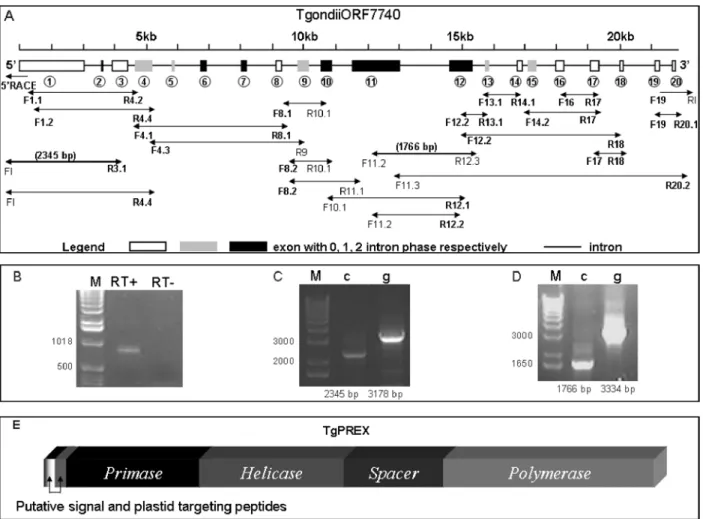
Fig. 1. The exon–intron structure (Guo et al. 2007) and a diagrammatic representation of the domain structure of the Toxoplasma gondii ORF 7740 present on chromosome VIIb of Toxoplasma gondii as elucidated by PCR. The structure of the T. gondii ORF 7740 was confirmed by the sequencing of cDNA from the RH strain of the organism, using primers listed in Table 1 (A). The 7740 bp long ORF was composed of 20 exons separated by 19 introns and different reading frames for the introns are designated. The exon intron structure of the gene was created using GSDS (http://gsds.cbi.pku.edu.cn/) (Guo et al. 2007). Primer pairs used for PCR are delineated in relation to the exon structure of the Tgprex gene. The primers designed on the consensus exon sequences are in bold font. (B) 5kRACE was performed to confirm the initiation codon where RT+ and RTx refers to reactions with or without reverse transcriptase, respectively. (C) The 5k end of the ORF was confirmed by comparing PCR on cDNA (c) and genomic DNA (g) using primers F.I and R3.1 (Table 1) generating 2345 bp and 3178 bp long PCR products, respectively. (D) The continuity of the single gene was confirmed by PCR using primers F11.2 and R12.3 (Table 1) showing a 1766 bp long PCR product from cDNA (c) compared to a 3334 bp long product from genomic DNA (g). The DNA marker is designated by M. (E) The domain structure of the TgPREX putative protein is diagrammatically represented.
The cell suspension was thawed, immediately supplemented with 0.1r protease inhibitor cocktail (EMD Biosciences) and sonicated on ice. The fol-lowing procedures were carried out at 4 xC. After centrifugation at 20 000 g for 30 min, the supernatant was preserved, and the cell pellet resuspended in 15 ml of buffer A ; the sonification and centrifugation
steps were repeated and the supernatant fractions were combined (Fraction I). Fraction I was loaded onto a DEAE cellulose (Sigma) column (4.9 cm2r
4 cm) equilibrated in buffer A and the column was washed with 100 ml of buffer A. Proteins were eluted by using a 120 ml linear gradient of 50–500 mM
NaCl in buffer A. Fractions (5 ml) containing DNA Table 1. List of primers used for confirmation of the Tgprex gene coding sequence on chromosome VIIb of Toxoplasma gondii and expression of recombinant polymerase domain of TgPREX protein
(The table shows the primer name, nucleotide sequence and the position of each primer according to the sequenced gene model presented in Fig. 1A. Primer pairs used for PCR amplification are delineated in Fig. 1A. The reverse primers are highlighted in the grey background. Primers situated in consensus exons predicted by ToxoDB are in bold font. The 5kRACE primers were used for the clarification of the initiation codon of the gene that was confirmed by PCR amplification of the 5k region using the 5kUTR primer. The cDNA primers were used to establish the coding sequence of the gene. The 3kUTR primer was used to ensure the 3k end of the Tgprex gene. Polymerase domain of the TgPREX protein was cloned using forward primer Tg_polymeraseF in conjunction with R20.2 reverse primer. The Nhe I restriction site on Tg_polymeraseF primer is shown in italicized font.)
Primer Name 5kp3k Nucleotide sequence Location
5kUTR primers
FI GCCTCTCCTTGTTCCTGCTTC 175 bp 5k
start codon 5kRACE primers
RACE.1 CCAGAGGAGAAAGAACTGTCAC Exon 1
RACE.2 AACGAAGAAAACCGCAGGG Exon 1
RACE.3 CGAGAAGGAAAAGACGACG Exon 1
cDNA primers F1.1 ATGCGTCCGGTTGAGTACCGG 5kATG F1.2 TACTGCCACCGCTGCGGGTGG Exon 1 R3.1 CAGCGAGTTCTCTGTCTTCAC Exon 3 F4.1 GAAACTCGGAATCGGGAGAT Exon 4 R4.2 TGTCGGGTGTCGGCTCTTCT Exon 4 F4.3 AAGGACGCGAACGAGGCGCTC Exon 4 R4.4 CTCAGATCCCTGAAGGTGAGGATTTG Exon 4 R8.1 CAACATGAACTGCAAGTTGTC Exon 8 F8.1 GACGTAGGCCATGTTGTTCTCGAC Exon 8 F8.2 GTTCAGGCGATTCGCCACAGTC Exon 8 R9 GAAGTCCGCGACGAGGCAGAG Exon 9 R10.1 CCGCTGGTGGGTGCAGCGAGGC Exon 10 F10.1 GCCTCGCTGCACCCACCAGCG Exon 10 R11.1 CGAAGTGACCTGGAACGCGG Exon 11 F11.2 TTTCGTCTCCTCAGCA Exon 11 F11.3 ATGCCTCTCCTGCGAGAACTG Exon 11 R12.1 GGTCGAATTGTCCGTTATGG Exon 12 R12.2 ATCGCTCTGCAAGTCGCCGA Exon 12 F12.2 TCGGCGACTTGCAGAGCGAT Exon 12 R12.3 CATGACTCCAGCTTCCACGAC Exon 12 R13.1 GTTTCTGTTGAAGGCGTTGC Exon 13 F13.1 GCAACGCCTTCAACAGAAAC Exon 13 R14.1 CTCTTTCCATCGTGCGTGGTCG Exon 14 F14.2 CTCCACCCGATCGTCCTGAA Exon 14 F16 CAGCTGCGATTCTCCGAA Exon 16 R17 CCGCACTCATTCCATAAATC Exon 17 F17 GATTTATGGAATGAGTGCGG Exon 17 R18 AAAGTGCTCTCCGTCCTGCG Exon 18 F19 CGGCGGTCGGCTGGTCATGTG Exon 19 R20.1 CAACACACGGGACAAAGCGAAG Exon 20 R20.2 CTACGGCTTGTCTGCCCAGCTGTCGGC Exon 20 3kUTR primer R.I CGACTACGCCGGCCGG 32 bp 3k stop codon TgPREX polymerase cloning
polymerase activity were pooled (Fraction II) and dialysed against buffer B [50 mMTris-HCl (pH 8.0),
50 mMNaCl, 1 mMEDTA, 0.1 mMPMSF, 2 mM
b-mercaptoethanol, 2 mMbenzamidine and 10 % (w/v)
glycerol]. Following dialysis, Fraction II was applied to a P-11 (GE Healthcare, Piscataway, NJ) column (1.8 cm2r7 cm) equilibrated in buffer B. The
col-umn was washed with 60 ml of buffer B, followed by elution with a 60 ml linear gradient of 50–500 mM
NaCl in buffer B. The fractions (2.5 ml) were ana-lysed in 10 % SDS-polyacrylamide gels, and fractions containing DNA polymerase activity were pooled (Fraction III) and dialysed against buffer C [50 mM
sodium phosphate (pH 8.0), 250 mM NaCl, 10 mM
imidazole, 1 mM b-mercatoethanol, 2 mM
benz-amidine and 5 % (w/v) glycerol]. Dialysed Fraction III was loaded onto a Qiagen nickel-nitrilotriacetic acid (Ni-NTA) affinity column (1.77 cm 2r3 cm) equilibrated in buffer C. The column was washed with 25 ml of buffer C and then with a 30 ml linear gradient of 10–300 mMimidazole (pH 8.0) in buffer
C. Fractions (2.5 ml) containing TgPREX poly-merase proteins, identified by electrophoresis in 10 % SDS-polyacrylamide gels, were pooled (fraction IV). After dialysis against storage buffer [50 mM
Tris-HCl (pH 7.4), 1 mMDTT, 30 mMNaCl and 10 % (w/
v) glycerol], the preparation was concentrated using an Amicon filter unit (MW cut-off 50 000). Aliquots were made and stored at x80 xC. The purified TgPREX polymerase protein (Fraction IV) ap-peared homogenous, as assessed in a 10 % SDS-polyacrylamide gel followed by the Commassie Blue G-250 staining. Protein concentration was deter-mined by the Bradford reaction (Bradford, 1976) using the Bio-Rad Protein Assay with bovine serum albumin as a standard.
Mass spectrometric analysis of the expressed protein The purified protein was used for trypsin digestion (Bridges et al. 2008). Tryptic peptide samples were separated on an LC system (Famos/Switchos/Ult-imate, LC Packings) before being analysed by elec-trospray ionisation (ESI) Mass Spectrometry (MS) on a Q-STAR1Pulsar i hybrid LC/MS/MS System. Peptide separation was performed on a Pepmap C18 reversed phase column (LC Packings), using a 5–85 % v/v acetonitrile gradient (in 0.5 % v/v formic acid) run over 45 min. The flow rate was maintained at 0.2ml/min. Mass spectrometric analysis was per-formed using a 3-sec survey MS scan followed by up to 4 MS/MS analyses of the most abundant peptides (3 sec per peak) in Information Dependent Acqui-sition (IDA) mode, choosing 2+ to 4+ ions above threshold of 30 counts, with dynamic exclusion for 120 sec.
Data generated from the Q-STAR1 Pulsar i hy-brid mass spectrometer was analysed using Applied Biosystems Analyst QS (v1.1) software and the
automated Matrix Science Mascot Daemon server (v2.1.06). Protein identifications were assigned using the Mascot search engine, which gives each protein a probability-based MOWSE score. In all cases vari-able methionine oxidation was allowed in searches. An MS tolerance of 1.2 Da for MS and 0.4 Da for MS/MS analysis was used.
DNA polymerase activity assay
DNA polymerase activity was assayed as described (Glick et al. 2002). Briefly, the activity was measured at 37 xC for 15 min in 10ml reaction mixtures con-taining 1mg of activated calf thymus DNA, 25 mM
each dNTP, 1mCi a[32P] dTTP (3000 Ci/mmol)
(PerkinElmer, Waltham, MA, USA), and 1ml of enzyme in 30 mMTris-HCl, pH 7.0, 10 mMMgCl2,
1 mM DTT, 250mg BSA, and 2.5 % glycerol. The
reaction was terminated by the addition of 100ml of 0.1M sodium pyrophosphate/0.05M EDTA. An
aliquot (100ml) of the reaction mixture was trans-ferred to a well in a 96-microwell1plate (Biodyne1 B ; NUNCTM) mounted on a 96-vacuum manifold
(Beckman Coulter, Fullerton, CA, USA). The plates were washed 3 times with 250ml of 0.1M sodium
pyrophosphate. The filter was removed from the plate, dried, and the amount of radioactivity as-sociated with the filter was quantified by phospho-rimager analysis using ImageQuant1software (GE Healthcare, Piscataway, NJ).
Software
Vector NTI AdvanceTM suite 9 and 10 (Infomax
2003 from Invitrogen) was used for sequence analy-sis. The exon-intron structure of the Tgprex gene was created using GSDS, a gene structure display software (http://gsds.cbi.pku.edu.cn/) (Guo et al. 2007). SignalP 3.0 Server (http://www.cbs.dtu.dk/ services/SignalP/) (Bendtsen et al. 2004 ; Nielsen et al. 1997 ; Nielsen and Krogh, 1998), SIG-Pred (http://www.bioinformatics.leeds.ac.uk/prot_analysis/ Signal.html), SIGFIND (http://139.91.72.10/sigfind/ sigfind.html), PrediSi (Hiller et al. 2004) and ChloroP 1.1 Server (http://www.cbs.dtu.dk/services/ChloroP/ #submission) (Emanuelsson et al. 1999) were used for signal and transit peptide identification of TgPREX protein.
R E S U L T S
Identification of a PREX homologue in ToxoDB Two genes (55.m04962/TGME49_061920 and 55.m04960/TGME49_061800) were identified in ToxoDB (release 4 and 5 respectively) on chromo-some VIIb of T. gondii genomic sequence on contigs 994 270 and 994 314 in the antisense strand (Kissinger et al. 2003). The coding sequence of this region
(1 488 133 to 1 515 551 bp) on chromosome VIIb was clarified by PCR amplification from T. gondii cDNA using primer pairs delineated in Fig. 1A.
Following sequencing, it appeared that a single gene homologous to Pfprex is present in T. gondii cDNA ; thereafter, we call this gene Tgprex. The Tgprex gene comprises of 20 exons as shown in Fig. 1A. The 5k end of the ORF was elucidated by 5k RACE (Fig. 1B). The initiation codon of the ORF (Fig. 1C) was confirmed by comparing the 2345 bp long sequenced PCR product from the cDNA with that of genomic DNA (Fig. 1C). Sequencing of the 1766 bp long cDNA PCR product in comparison to genomic DNA PCR (Fig. 1D) confirmed the con-nection between the putative primase-helicase and the polymerase region of the Tgprex gene. The putative domain structure of the translated TgPREX protein is represented in Fig. 1E.
In summary, a 7740 bp long ORF, (Tgondii-ORF7740/Tgprex, GenBank Accession no. FJ665392) was identified from the sequenced region of chro-mosome VIIb (between 1,491,400 and 1,512,118). A sequencing gap between contigs 994 270 and 994 314 in the database was sequenced from both the genomic DNA and the cDNA of the RH strain of the organism and a 146 bp long sequence was subse-quently deposited in the database.
Prediction of localization of TgPREX in silico The TgPREX translated protein sequence possessed a primase, helicase and polymerase homologous do-main structure (Fig. 1E) similar to that of the PREX protein of Plasmodium. The peptide region of the TgPREX protein that separated the primase-helicase and the polymerase domains has no similarity to the spacer region occupying the same region of the PfPREX predicted peptide sequence.
T. gondii uses the same general mechanism to mediate transport of proteins into the apicoplast as Plasmodium (He et al. 2001). A bipartite leader pep-tide, including a primary secretory domain followed by a secondary plastid transit domain, can be pre-dicted by specialized tools including PlasmoAP (Foth et al. 2003) and PATS (Waller et al. 1998 ; Zuegge et al. 2001) in PlasmoDB. These programmes, how-ever, failed in systemically identifying the apicoplast targeted proteins in T. gondii (Harb et al. 2004). For example, known T. gondii nuclear encoded apico-plast proteins (e.g. acyl carrier protein [AAC63956], beta-hydroxyacyl-ACP dehydratase [AAC72191], small ribosomal protein S9 [AAC63957], large ri-bosomal protein L28 [AAC63958] (Waller et al. 1998) and a putative ferredoxin NADP+ oxidore-ductase [CAC15394] (Vollmer et al. 2001) were all
Fig. 2. SDS-PAGE and functional analysis of the purified TgPREX polymerase protein. The SDS-PAGE of purified TgPREX revealed a 114 kDa protein band in purified (2) followed by concentrated (1) protein samples. Functional properties of the purified TgPREX protein. (B) For TgPREX DNA polymerase activity Mg2+ions are required. In the absence of Mg2+ions, the amount of incorporated [a]32
P-dTTP by the TgPREX to the activated calf thymus DNA is similar to that without the enzyme addition (Lanes 3 and 2, respectively). The amounts of incorporated [a]32P-dTTP to the activated calf thymus DNA increases as the concentrations (3.1, 6.3, 12.5, 25, 50 and 100 nm, respectively) of TgPREX increases in the reaction (Lanes 4–9, respectively). The DNA polymerase activity of E. coli Klenow (1 unit) is also shown as a control (Lane 1). (C) TgPREX polymerase activity measured as a function of MgCl2concentration ; (D) TgPREX polymerase activity measured as a function of pH ; (E) TgPREX polymerase activity measured as a function of temperature. Error bars represent the standard deviation of 3 independent experimental determinations.
unrecognized. For each of these proteins, however, the signal peptide component was recognized by SignalP 3.0 and the plastid targeting peptide fol-lowing the signal peptide was recognized by ChloroP 1.1.
The N-terminal end of the TgPREX protein was analysed sequentially for the presence of a bipartite leader peptide. A 70 amino acid long N-terminal sequence of TgPREX was used as a query in PrediSi and SIGFIND software. Amino acid residues from 30 to 70 were used as a query in SignalP 3.0 as the maximum length of sequence input is restricted to 30 in this programme. All the analyses predicted TgPREX as an apicoplast-localized protein similar to its homologous counterpart PfPREX whose N-terminus was shown to direct GFP to the apico-plast (Seow et al. 2005). TgPREX appears to possess a putative, comparatively long N-terminal signal peptide, 60 to 61 amino acids in length. The cleavage sequence has been identified within the multiple serine residues (VLS-SS or VLSS-SS). The signal peptide was followed by a 61-amino acid long, putative plastid transit peptide as identified by the ChloroP1.1 programme.
Analysis of the PREX polymerase
The recombinant polymerase domain of the TgPREX, with a predicted mass of 114 kDa was expressed in E. coli. The histidine-tagged protein was purified by Ni2+affinity chromatography. The protein was analysed by SDS-PAGE and the identity was confimed by mass spectrometry. The identified peptides are detailed in the Supplementary Data 1 (Online version only).
The purified TgPREX polymerase recombinant protein (Fraction IV) (Fig. 2A) was tested for its DNA polymerase activity. Activity was optimal at a temperature of 50 xC (Fig. 1C) and at pH 7.0 (Fig. 1D). The enzyme required Mg2+ ions for activity (Fig. 1B), which reached the optimum at 5 mMMgCl2(Fig. 1E).
D I S C U S S I O N
PREX, the plastid replication and repair enzyme complex of apicomplexan parasites was first ident-ified in Plasmodium. Homology to DNA primase, helicase and polymerase in other systems indicated that the gene codes for all of these key functions. The presence of a predicted plastid targeting sequence and the fact that a similar sequence in P. falciparum guides marker proteins to the apicoplast suggest that PREX is instrumental in the replication of apicoplast DNA. Among the annotated genes related to Tgprex in ToxoDB, TGME49_061800 expression is linked to one EST (Expressed Sequence Tag) and 7.6 normalized SAGE (serial analysis of gene ex-pression) tag counts (Gajria et al. 2008). There are no such data related to the TGME_061920 gene. Mass spectrometry analysis has identified 2 pep-tide sequences (ARPLSPEHSALNESAGCAR and LFLESATPVPHAQILTFR) (Xia et al. 2008) which correspond to peptides in exon 4 and 5 of the sequenced Tgprex gene respectively. The lack of good EST coverage for this gene probably corresponds to a low level expression as documented in ToxoDB at the 32.2 and 25.2 percentile for TGME49_061800 and TGME_061920, respectively. A similarly low level of expression was also observed in P. falciparum (Le Roch et al. 2003).
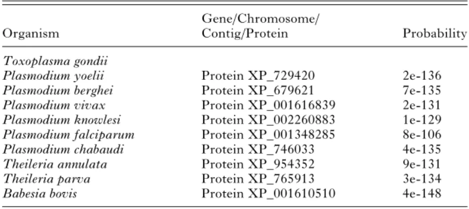
BLAST searches identified full length PREX or-thologues in other Plasmodium species, in Theileria parva [XP_765913] and Theileria annulata [XP_ 954352] and in Babesia bovis (XP_001610510) (Table 2) (Alignment in Supplementary Data 2 – Online version only). Homologues representing fragments of the total predicted protein were also identified in Babesia bigemina, Eimeria tenella, Neospora caninum and Sarcocystis neurona where respective genome sequencing is incomplete. Inter-estingly, no homologous protein was found in the Cryptosporidium species. Genome sequencing also suggests that Cryptosporidium hominis (Xu et al. 2004) and Cryptosporidium parvum (Abrahamsen et al. 2004) lack an apicoplast.
Table 2. PREX homologous genes in other apicomplexa
Organism
Gene/Chromosome/
Contig/Protein Probability
Toxoplasma gondii
Plasmodium yoelii Protein XP_729420 2e-136
Plasmodium berghei Protein XP_679621 7e-135
Plasmodium vivax Protein XP_001616839 2e-131
Plasmodium knowlesi Protein XP_002260883 1e-129
Plasmodium falciparum Protein XP_001348285 8e-106
Plasmodium chabaudi Protein XP_746033 4e-135
Theileria annulata Protein XP_954352 9e-131
Theileria parva Protein XP_765913 3e-134
Interestingly, the prex gene of T. gondii differs from that of P. falciparum in that it is interrupted by 20 introns. The intron footprint of eukaryotic, nuclear-encoded genes with possible prokaryotic cyanobacterial ancestry has made a significant con-tribution to our understanding of gene evolution. The presence of introns in such genes suggests the insertion of introns into pre-assembled genes of eu-karyotes late in evolution (‘ intron late ’) as the an-cestral cyanobacterial genes were devoid of introns. Similar comparison of ribosomal protein S9, L28 and acyl carrier protein genes between P. falciparum and T. gondii suggests a process of continuous intron insertion during evolution even after the divergence of Plasmodium and T. gondii from their common ancestor (Schaap et al. 2001).
The polymerase function of this putative protein, identified only in apicoplast-bearing apicomplexans, was confirmed in T. gondii. The nuclear encoded PREX appears to serve as a replicative enzyme for the 35 kb genome of the apicoplast. The participation of nuclear-encoded proteins in apicoplast replication has also been suggested for DNA gyrase subunits A and B (Dar et al. 2007 ; Raghu Ram et al. 2007), a DNA ligase and 2 hypothetical proteins bearing similarities with DNA-repair proteins (Dahl and Rosenthal, 2008).
Given that inhibitors of other enzymes involved in plastid replication (e.g. inhibitors of DNA gyrase) are toxic to apicomplexan parasites the PREX pro-tein complex may be considered a target for chemo-therapy. This may apply particularly to the primase and polymerase domains since the helicase domain has substantial homology to the Twinkle helicase that is active in mitochondria of most eukaryotic species.
We thank Dr Richard J. S. Burchmore of Sir Henry Wel-lcome Functional Genomics Facility, Institute of Bio-medical and Life Sciences, University of Glasgow, for performing the MS analysis of protein in our study. A.M. was funded by the Wellcome Trust as part of their ‘‘ Molecular Functions in Disease ’’ programme.
R E F E R E N C E S
Abrahamsen, M. S., Templeton, T. J., Enomoto, S., Abrahante, J. E., Zhu, G., Lancto, C. A., Deng, M., Liu, C., Widmer, G., Tzipori, S., Buck, G. A., Xu, P., Bankier, A. T., Dear, P. H., Konfortov, B. A., Spriggs, H. F., Iyer, L., Anantharaman, V., Aravind, L. and Kapur, V. (2004). Complete genome sequence of the apicomplexan, Cryptosporidium parvum. Science304, 441–445.
Bendtsen, J. D., Nielsen, H., von Heijne, G. and Brunak, S. (2004). Improved prediction of signal peptides : SignalP 3.0. Journal of Molecular Biology340, 783–795.
Bradford, M. M. (1976). A rapid and sensitive method for the quantitation of microgram quantities of protein
utilizing the principle of protein-dye binding. Analytical Biochemistry72, 248–254.
Bridges, D. J., Pitt, A. R., Hanrahan, O., Brennan, K., Voorheis, H. P., Herzyk, P., de Koning, H. P. and Burchmore, R. J. (2008). Characterisation of the plasma membrane subproteome of bloodstream form Trypanosoma brucei. Proteomics8, 83–99.
Campbell, S. A., Richards, T. A., Mui, E. J., Samuel, B. U., Coggins, J. R., McLeod, R. and Roberts, C. W. (2004). A complete shikimate pathway in Toxoplasma gondii : an ancient eukaryotic innovation. International Journal for Parasitology34, 5–13.
Chomczynski, P. and Sacchi, N. (1987). Single-step method of RNA isolation by acid guanidinium thiocyanate-phenol-chloroform extraction. Analytical Biochemistry162, 156–159.
Dahl, E. L. and Rosenthal, P. J. (2008). Apicoplast translation, transcription and genome replication : targets for antimalarial antibiotics. Trends in Parasitology24, 279–284.
Dar, M. A., Sharma, A., Mondal, N. and Dhar, S. K. (2007). Molecular cloning of apicoplast-targeted Plasmodium falciparum DNA gyrase genes : unique intrinsic ATPase activity and ATP-independent dimerization of PfGyrB subunit. Eukaryotic Cell6, 398–412.
Emanuelsson, O., Nielsen, H. and von Heijne, G. (1999). ChloroP, a neural network-based method for predicting chloroplast transit peptides and their cleavage sites. Protein Science8, 978–984.
Ferguson, D. J., Campbell, S. A., Henriquez, F. L., Phan, L., Mui, E., Richards, T. A., Muench, S. P., Allary, M., Lu, J. Z., Prigge, S. T., Tomley, F., Shirley, M. W., Rice, D. W., McLeod, R. and Roberts, C. W. (2007). Enzymes of type II fatty acid synthesis and apicoplast differentiation and division in Eimeria tenella. International Journal for Parasitology37, 33–51.
Fichera, M. E. and Roos, D. S. (1997). A plastid organelle as a drug target in apicomplexan parasites. Nature, London390, 407–409.
Foth, B. J., Ralph, S. A., Tonkin, C. J., Struck, N. S., Fraunholz, M., Roos, D. S., Cowman, A. F. and McFadden, G. I. (2003). Dissecting apicoplast targeting in the malaria parasite Plasmodium falciparum. Science299, 705–708.
Gajria, B., Bahl, A., Brestelli, J., Dommer, J., Fischer, S., Gao, X., Heiges, M., Iodice, J., Kissinger, J. C., Mackey, A. J., Pinney, D. F., Roos, D. S., Stoeckert, C. J., Jr., Wang, H. and Brunk, B. P. (2008). ToxoDB : an integrated Toxoplasma gondii database resource, Nucleic Acids Research,36, D553–D556.
Glick, E., Anderson, J. P. and Loeb, L. A. (2002). In vitro production and screening of DNA polymerase eta mutants for catalytic diversity. Biotechniques33, 1136–1142, 1144.
Guo, A. Y., Zhu, Q. H., Chen, X. and Luo, J. C. (2007). [GSDS : a gene structure display server]. Yi. Chuan 29, 1023–1026.
Harb, O. S., Chatterjee, B., Fraunholz, M. J., Crawford, M. J., Nishi, M. and Roos, D. S. (2004). Multiple functionally redundant signals mediate
targeting to the apicoplast in the apicomplexan parasite Toxoplasma gondii. Eukaryotic Cell3, 663–674. He, C. Y., Striepen, B., Pletcher, C. H., Murray, J. M.
and Roos, D. S. (2001). Targeting and processing of nuclear-encoded apicoplast proteins in plastid segregation mutants of Toxoplasma gondii. Journal of Biological Chemistry276, 28436–28442.
Hiller, K., Grote, A., Scheer, M., Munch, R. and Jahn, D. (2004). PrediSi : prediction of signal peptides and their cleavage positions. Nucleic Acids Research32, W375–W379.
Kissinger, J. C., Gajria, B., Li, L., Paulsen, I. T. and Roos, D. S. (2003). ToxoDB : accessing the Toxoplasma gondii genome. Nucleic Acids Research31, 234–236. Le Roch, K. G., Zhou, Y., Blair, P. L., Grainger, M.,
Moch, J. K., Haynes, J. D., De, L. V., Holder, A. A., Batalov, S., Carucci, D. J. and Winzeler, E. A. (2003). Discovery of gene function by expression profiling of the malaria parasite life cycle, Science301, 1503–1508.
McFadden, G. I., Reith, M. E., Munholland, J. and Lang-Unnasch, N. (1996). Plastid in human parasites. Nature, London381, 482.
McLeod, R., Muench, S. P., Rafferty, J. B., Kyle, D. E., Mui, E. J., Kirisits, M. J., Mack, D. G., Roberts, C. W., Samuel, B. U., Lyons, R. E., Dorris, M., Milhous, W. K. and Rice, D. W. (2001). Triclosan inhibits the growth of Plasmodium falciparum and Toxoplasma gondii by inhibition of apicomplexan Fab I. International Journal for Parasitology31, 109–113. Nielsen, H., Engelbrecht, J., Brunak, S. and von Heijne, G. (1997). Identification of prokaryotic and eukaryotic signal peptides and prediction of their cleavage sites. Protein Engineering Design and Selection 10, 1–6.
Nielsen, H. and Krogh, A. (1998). Prediction of signal peptides and signal anchors by a hidden Markov model. Proceedings of the International Conference on Intelligent Systems for Molecular Biology6, 122–130.
Raghu Ram, E. V., Kumar, A., Biswas, S., Kumar, A., Chaubey, S., Siddiqi, M. I. and Habib, S. (2007). Nuclear gyrB encodes a functional subunit of the Plasmodium falciparum gyrase that is involved in apicoplast DNA replication. Molecular and Biochemical Parasitology154, 30–39.
Ralph, S. A., D’Ombrain, M. C. and McFadden, G. I. (2001). The apicoplast as an antimalarial drug target. Drug Resistance Updates4, 145–151.
Roberts, C. W. and Alexander, J. (1992). Studies on a murine model of congenital toxoplasmosis : vertical disease transmission only occurs in BALB/c mice infected for the first time during pregnancy. Parasitology 104, 19–23.
Roos, D. S. (1999). The apicoplast as a potential therapeutic target in Toxoplasma and other
apicomplexan parasites : some additional thoughts. Parasitology Today15, 41.
Schaap, D., van Poppel, N. F. and Vermeulen, A. N. (2001). Intron invasion in protozoal nuclear encoded plastid genes. Molecular and Biochemical Parasitology 115, 119–121.
Seow, F., Sato, S., Janssen, C. S., Riehle, M. O., Mukhopadhyay, A., Phillips, R. S., Wilson, R. J. and Barrett, M. P. (2005). The plastidic DNA replication enzyme complex of Plasmodium falciparum. Molecular and Biochemical Parasitology 141, 145–153.
Soldati, D. (1999). The apicoplast as a potential
therapeutic target in and other apicomplexan parasites. Parasitology Today15, 5–7.
Striepen, B., Crawford, M. J., Shaw, M. K., Tilney, L. G., Seeber, F. and Roos, D. S. (2000). The plastid of Toxoplasma gondii is divided by association with the centrosomes. Journal of Cell Biology151, 1423–1434.
Vollmer, M., Thomsen, N., Wiek, S. and Seeber, F. (2001). Apicomplexan parasites possess distinct nuclear-encoded, but apicoplast-localized, plant-type ferredoxin-NADP+ reductase and ferredoxin. Journal of Biological Chemistry276, 5483–5490.
Waller, R. F., Keeling, P. J., Donald, R. G., Striepen, B., Handman, E., Lang-Unnasch, N., Cowman, A. F., Besra, G. S., Roos, D. S. and McFadden, G. I. (1998). Nuclear-encoded proteins target to the plastid in Toxoplasma gondii and Plasmodium falciparum. Proceedings of the National Academy of Sciences, USA95, 12352–12357. Wilson, R. J., Williamson, D. H. and Preiser, P.
(1994). Malaria and other Apicomplexans : the ‘‘ plant ’’ connection. Infectious Agents and Disease3, 29–37. Xia, D., Sanderson, S. J., Jones, A. R., Prieto, J. H.,
Yates, J. R., Bromley, E., Tomley, F. M., Lal, K., Sinden, R. E., Brunk, B. P., Roos, D. S. and Wastling, J. M. (2008). The proteome of Toxoplasma gondii : integration with the genome provides novel insights into gene expression and annotation, Genome Biology9, R116.
Xu, P., Widmer, G., Wang, Y., Ozaki, L. S.,
Alves, J. M., Serrano, M. G., Puiu, D., Manque, P., Akiyoshi, D., Mackey, A. J., Pearson, W. R., Dear, P. H., Bankier, A. T., Peterson, D. L., Abrahamsen, M. S., Kapur, V., Tzipori, S. and Buck, G. A. (2004). The genome of Cryptosporidium hominis. Nature, London431, 1107–1112.
Zuegge, J., Ralph, S., Schmuker, M., McFadden, G. I. and Schneider, G. (2001). Deciphering apicoplast targeting signals–feature extraction from nuclear-encoded precursors of Plasmodium falciparum apicoplast proteins. Gene280, 19–26.