www.elsevier.com / locate / cardiores www.elsevier.nl / locate / cardiores
Effects of verapamil on atrial fibrillation and its electrophysiological
determinants in dogs
a,c a a,b ,
*
`
´
Agnes Benardeau
, Samir Fareh , Stanley Nattel
a
Department of Medicine and Research Center, Montreal Heart Institute and University of Montreal, Montreal, Quebec, Canada
b
Department of Pharmacology and Therapeutics, McGill University, Montreal, Quebec, Canada
c
Hoffman-La Roche Pharmaceuticals, Basel, Switzerland Received 22 June 2000; accepted 18 December 2000
Abstract
Background: Atrial tachycardia-induced remodeling promotes the occurrence and maintenance of atrial fibrillation (AF) and decreases
21 2
L-type Ca current. There is also a clinical suggestion that acute L-type Ca channel blockade can promote AF, consistent with an AF
21 21
promoting effect of Ca channel inhibition. Methods: To evaluate the potential mechanisms of AF promotion by Ca channel blockers, we administered verapamil to morphine–chloralose anesthetized dogs. Diltiazem was used as a comparison drug and autonomic blockade with atropine and nadolol was applied in some experiments. Epicardial mapping with 240 epicardial electrodes was used to evaluate activation during AF. Results: Verapamil caused AF promotion in six dogs, increasing mean duration of AF induced by burst pacing, from 864 s (mean6S.E.) to 95639 s (P,0.01 vs. control) at a loading dose of 0.1 mg / kg and 2286101 s (P,0.0005 vs. control) at a dose of 0.2 mg / kg. Underlying electrophysiological mechanisms were studied in detail in five additional dogs under control conditions and in the presence of the higher dose of verapamil. In these experiments, verapamil shortened mean effective refractory period (ERP) from 12265 to 11464 ms (P,0.02) at a cycle length of 300 ms, decreased ERP heterogeneity (from 1561 to 1061%, P,0.05), heterogeneously accelerated atrial conduction and decreased the cycle length of AF (9464 to 8463 ms, P,0.005). Diltiazem did not affect ERP, AF cycle length or AF duration, but produced conduction acceleration similar to that caused by verapamil (n55). In the presence of autonomic blockade, verapamil failed to promote AF and increased, rather than decreasing, refractoriness. Neither verapamil nor diltiazem affected atrial conduction in the presence of autonomic blockade. Epicardial mapping suggested that verapamil promoted AF by increasing the number of simultaneous wavefronts reflected by separate zones of reactivation in each cycle. Conclusions: Verapamil promotes AF in normal dogs by promoting multiple circuit reentry, an effect dependent on intact autonomic tone and not shared by diltiazem. 2001 Elsevier Science B.V. All rights reserved.
Keywords: Arrhythmia (mechanisms); Ion channels; Mapping; Remodeling
1. Introduction prevent thromboembolic complications with oral
antico-agulants [3]. Among the drugs used to control the
ventricu-21
Atrial fibrillation (AF) is presently the most common lar response rate are inhibitors of L-type Ca current such sustained cardiac arrhythmia in clinical practice, and its as diltiazem and verapamil. There is, however, clinical
21
treatment is less than optimal in many cases [1]. Antiar- evidence that Ca channel blockers such as verapamil and rhythmic drugs improve sinus rhythm maintenance, but at diltiazem may promote AF maintenance [4]. Two recent
21
the expense of a variety of potential adverse effects, of experimental studies also suggest that the Ca channel
which one of the most worrisome is ventricular proar- blocker verapamil may promote experimental AF. Lee et
rhythmia [2]. An alternative approach is to leave patients al. showed that verapamil administration during a 7–42
in AF, but to control the ventricular response and to day period of atrial tachycardia and at the time of a
subsequent electrophysiological study fails to prevent tachycardia-induced remodeling and promotes AF
mainte-*Corresponding author. Tel.: 514-376-3330, ext. 3990; fax: 11-514-376-1355.
E-mail address: [email protected] (S. Nattel). Time for primary review 26 days.
0008-6363 / 01 / $ – see front matter 2001 Elsevier Science B.V. All rights reserved. P I I : S 0 0 0 8 - 6 3 6 3 ( 0 1 ) 0 0 2 0 1 - 2
nance during the electrophysiological study [5]. Friedman et al. showed that verapamil promotes AF maintenance in anesthetized dogs, an effect that can be prevented by
b-adrenoceptor blockade [6]. Neither study evaluated the
electrophysiological mechanisms potentially underlying verapamil’s apparent AF promoting action. The present study was designed to (1) determine whether verapamil increases AF duration in the anesthetized dog in a dose related fashion; (2) evaluate potential underlying mecha-nisms by measuring atrial effective refractory period (ERP) and conduction velocity, as well as by studying changes in activation during AF with the use of atrial
21
epicardial mapping; and (3) determine whether the Ca channel blocker diltiazem shares verapamil’s AF promot-ing potential.
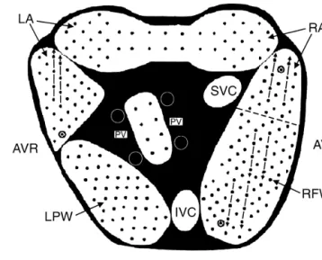
Fig. 1. Schematic of electrode arrays and bipolar electrode sites (dots).
2. Methods
Stimulation sites for conduction measurements are indicated by haloed dots. Arrows indicate the series of electrodes used for conduction velocity
2.1. Animal preparation
analysis in each zone. AVR, atrio-ventricular ring; RFW, right free wall; IVC, SVC, inferior, superior vena cava; PV, pulmonary vein; LPW, left
Twenty-five mongrel dogs (2761 kg) were studied in posterior wall.
four groups: (1) Group 1: a dose–response group (n56), in which the effects of verapamil on AF duration were
studied at two dose levels (0.1 and 0.2 mg / kg loading previously described in detail [7,8]. A mapping system was doses, followed by maintenance infusions of 0.5 and 1.5 connected to five arrays covering the atrial epicardial
mg / kg / min infusions, respectively); (2) Group 2: a de- surfaces with 240 bipolar electrodes (Fig. 1) as previously tailed electrophysiology group (n55), in which the electro- described. Signals were filtered (bandwidth of 10–900 Hz), physiological effects of verapamil were studied at a single, digitized (12-bit resolution and 2-kHz sampling rate), and high loading dose (0.2 mg / kg, followed by 1.5 mg / kg / transmitted into a Silicon Graphics computer for analysis. min); (3) Group 3: a diltiazem group (n55), in which the Activation data were analyzed off line with computer effects of diltiazem (0.8 mg / kg, followed by 15 mg / kg / determined peak-amplitude criteria for activation, and data
min) were studied; and (4) Group 4: an autonomic for each electrode were reviewed manually. Atrial ERP
blockade group, in which the effects of verapamil (n55) (longest coupling interval at which premature electrical
and diltiazem (n54) were studied in the presence of extrastimuli failed to capture) and conduction velocity
pharmacological blockade of cardiac b-adrenergic and were measured during stimulation at sites in various atrial
muscarinic cholinergic receptors. regions as in previous work [7,8]. Activation maps for
Animal handling procedures followed the guidelines of conduction measurement were obtained after 60 s at a the Canadian Council on Animal Care, and were approved variety of basic cycle lengths. Conduction velocity was by the institutional animal research ethics committee. On measured with the use of two parallel sets of four the study day, dogs were anesthetized with morphine (2 electrodes during local pacing in each of four regions: the mg / kg s.c.) and a-chloralose (120 mg / kg i.v. load, 29.3 left atrial appendage, the right atrial appendage, the right
21
mg / kg h ) and ventilated to maintain physiological superior free wall and the right inferior free wall (Fig. 1). arterial blood gases (pH 7.38–7.45, SaO .90%). Body2 Because of variable contact in the left atrium, complete temperature was maintained at 378C and a femoral artery conduction data were only available at right atrial sites. To and both femoral veins were cannulated for arterial blood measure ERP, a 15 stimulus basic train (2-ms twice
pressure monitoring and drug administration. A median threshold current pulses) was followed by a premature
sternotomy was performed and bipolar PTFE coated extrastimulus at a progressively increasing coupling inter-stainless electrodes hooked into right and left atrial appen- val and a 2-s pause to observe the response between trains. dages for stimulation and recording. The surface ECG was The premature coupling interval was increased by 10-ms monitored and epicardial mapping arrays sewn to the increments to obtain an initial estimate of the ERP. The
epicardial surfaces of both atria. measurement was then repeated with 5-ms increments and
the resulting value taken as the ERP. In the case of a
2.2. Electrophysiological study .10-ms difference between the two measurements, a third
measurement with 5-ms steps was obtained and the mean The study preparation and instrumentation were as of all three ERP values was used. This protocol was used
to measure ERPs at cycle lengths of 150, 200, 250, 300 2.4. Data analysis and 350 ms at the right atrial appendage, and to measure
ERPs during stimulation at a single pacing cycle length The conduction velocity was determined in each region
(300 ms) at 2362 right and left atrial sites. as previously described. The coefficient of variance in ERP
AF was induced by stimulating the right atrial appen- was calculated as S.D. / mean3100% and used as an index
dage with 10 Hz, 2 ms stimuli at four times threshold of ERP heterogeneity. The number of sites for ERP
current for 2–10 s. To calculate mean AF duration, AF determination in each region was equivalent across dogs was induced with burst pacing ten times for AF duration and between groups, to prevent any selection bias.
Statisti-,10 min and twice for AF duration .10 min. AF that cal comparisons between two groups only were performed lasted .20 min, requiring electrical cardioversion for by Student’s t test. Analysis of variance with a range test termination, was considered persistent. A 20-min rest was used for multiple group comparisons. Average results
period was allowed before continuing the experiment. are given as the mean6S.E.M., and a two-tailed P,0.05
was considered statistically significant. 2.3. Pharmacological study
3. Results
Verapamil and diltiazem were dissolved in 0.9% saline
the day of the experiment and were kept at 48C until 15 3.1. Dose related effects of verapamil min before use. In the first part of the study (Group 1), we
tested two intravenous doses of verapamil hydrochloride The dose related effects of verapamil observed in Group (Sigma): first dose: a loading infusion of 0.10 mg / kg over 1 dogs are summarized in Table 1. Verapamil slightly, but 10 min, followed by a maintenance dose of 0.5 mg / kg / min non-significantly, increased sinus cycle length, while caus-for the time needed to complete electrophysiological ing clear, dose related decreases in arterial pressure. The
measurements (|30–45 min); second dose: load of 0.20 PR interval was also increased by the drug, with other
mg / kg, i.v.) followed by a maintenance infusion of 1.5 ECG intervals unaffected. AF duration was clearly
in-mg / kg / min. In each case, measurements were begun 10 creased in a dose related fashion. min after the completion of the loading dose. These doses
were based on previously published data showing signifi- 3.2. Electrophysiological mechanisms of AF promotion cant and stable pharmacological actions and plasma con- by verapamil
centrations [9]. We then pursued more detailed
electro-physiological evaluations with the higher dose only (Group In order to assess the mechanism by which verapamil
2 dogs). We then studied a group of dogs receiving promotes AF, we performed epicardial mapping during AF
diltiazem (Sigma) at the highest dose shown in a previous under control conditions and in the presence of the higher multiple dose study [9] to be tolerated and produce stable dose of verapamil (Group 2 dogs). The general effects of electrophysiological effects in the dog (loading dose 0.8 verapamil in Group 2 dogs are shown in Table 2. As in mg / kg, followed by 15 mg / kg / min maintenance infusion). Group 1 dogs, 0.2 mg / kg verapamil slightly (but nonsig-In a final group (Group 4), the effects of the higher dose of nificantly) prolonged sinus cycle length, reduced arterial verapamil and of diltiazem were studied in the presence of pressure, and significantly increased both PR interval and autonomic blockade produced with nadolol (0.5 mg / kg, AF duration. The results of mapping studies are illustrated followed by an additional 0.25 mg / kg every 2 h) and in Fig. 2, which shows activation during three consecutive
atropine (1 mg every 2 h). cycles under control conditions (Fig. 2A) and in the
Table 1
a
Dose–response effects of verapamil (Group 1 dogs)
Control Verapamil Verapamil P
c c
(lower dose) (higher dose)
b n 6 6 6 Sinus CL (ms) 39365 424646 472645 0.480 Mean arterial BP (mmHg) 10469 8669 7668 0.0004 PR interval (ms) 10566 124611 158612 0.006 QRS duration (ms) 4362 4362 4361 0.990 QTc interval 30264 29369 28567 0.260 AF duration (s) 864 95639 2286101 0.0056 a
Values are mean6S.E.M. CL, cycle length; BP, blood pressure; AF, atrial fibrillation; NS, not significant.
b
Each dog was studied under both control and all drug conditions.
c
Table 2
Comparison between effects of verapamil (Group 2 dogs) and diltiazem (Group 3)
Control Verapamil P Control Diltiazem P
(0.2 mg / kg, followed (0.8 mg / kg, followed by 1.5 mg / kg / min) by 15 mg / kg / min) a a n 5 5 – 5 5 – Sinus CL (ms) 373621 451641 0.113 376631 393611 0.656 Mean arterial BP (mmHg) 10169 8667 0.0081 8969 69611 0.025 PR interval (ms) 9264 12864 0.00014 9264 164621 0.0101 AF duration (s) 562 3996351 0.043 865 26615 0.318 a
Each dog was studied under control and all drug conditions.
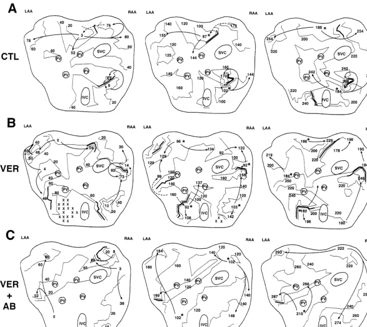
Fig. 2. Activation maps during three consecutive cycles of AF under control conditions (A), in the presence of verapamil, 0.2 mg / kg followed by 1.5
mg / kg / min (B) and in the presence of the same dose of verapamil and autonomic blockade (C). Lines are 20-ms isochrones, arrows are zones of wavefront
propagation during a cycle, dashed arrows are possible propagation to reactivate atrial tissue at the beginning of the next cycle and asterisks are zones of reactivation at the beginning of the cycle. Under control conditions, only one or two zones of reactivation are seen per cycle, whereas in the presence of verapamil (autonomic transmission intact) multiple reactivation zones occur (four per cycle in the example shown) and the activation pattern is clearly more complex. In the presence of autonomic blockade, activation patterns in the presence of verapamil resemble those under control conditions.
presence of verapamil (Fig. 2B). Isochrone maps (20-ms) are shown, with the activation times indicated all refer-enced to the earliest activation in the first cycle. Solid arrows represent the direction of impulse propagation and solid lines, zones of functional block (.40 ms activation time differences between adjacent electrodes). Regions that are not activated within a cycle are designated by 3s. The dashed lines indicate probable paths of reactivation initiat-ing the next cycle. Under control conditions (Fig. 2A), activation is relatively homogeneous, with approximately two discrete zones of reactivation (indicated by the aster-isks) per cycle. In the presence of verapamil, activation is more heterogeneous. There are more zones of functional block, more apparent reentry circuits and more zones of reactivation at the beginning of each cycle. Fig. 3A shows mean data for the number of reactivation zones per cycle, based on measurements for three consecutive cycles under each condition in each dog. Verapamil clearly increased the number of reactivation zones considerably, from just over one to almost three / cycle. Thus, verapamil’s ability to promote AF is likely related to the promotion of multiple wavelet reentry by decreasing the size and increasing the number of functional reentry circuits during the arrhyth-mia.
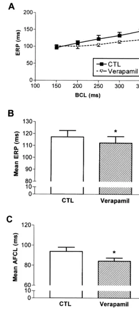
In order to explore further the electrophysiological mechanisms underlying verapamil’s AF promoting action, we assessed drug-induced changes in atrial refractoriness and conduction properties. ERP at the right atrial appendix is shown as a function of cycle length in Fig. 4A. Verapamil tended to decrease ERP slightly but not sig-nificantly at long cycle lengths, with no change at short cycle lengths. Fig. 4B shows mean ERP data from all sites at a cycle length of 300 ms, and indicates that verapamil slightly but significantly decreased ERP. Verapamil-induced changes in AF cycle length are shown in Fig. 4C, which indicates that the drug significantly decreased the mean cycle length during AF, consistent with the ERP changes.
Fig. 4. Effects of verapamil (0.2 mg / kg followed by 1.5 mg / kg / min) in
The drug’s effects on ERP at a cycle length of 300 ms are the presence of intact autonomic tone on (A) ERP in the right atrial further analyzed in Fig. 5. Verapamil did not alter sig- appendage; (B) mean ERP at all sites tested; and (C) AF cycle length.
Fig. 3. Effects of verapamil (0.2 mg / kg followed by 1.5 mg / kg / min) on the number of reactivation zones calculated on the basis of activation maps during AF in the absence (A) or presence (B) of autonomic blockade.
ERP and conduction, mean reentrant wavelength was unaffected by the drug (11.060.6 cm before and 11.360.5 cm after verapamil administration).
3.3. Effects of diltiazem on atrial electrophysiology
and AF
To determine whether diltiazem shares verapamil’s actions on atrial electrophysiology and arrhythmias, we studied the effects of the drug in Group 3 dogs. Like high dose verapamil, diltiazem decreased arterial pressure and increased the PR interval (Table 2). Unlike verapamil, diltiazem did not significantly alter AF duration. Diltiazem did not alter atrial ERP, which averaged 11262 ms at all sites (cycle length 300 ms) before diltiazem and 11663 ms after the drug. Similarly, AF cycle length was not affected by diltiazem (9463 ms control, 9263 ms drug, P5NS). Diltiazem did produce significant increases in conduction velocity, with a spatial distribution similar to those caused by verapamil (Fig. 6, right). Thus, despite similar effects on blood pressure, PR interval and intra-atrial conduction, diltiazem did not share verapamil’s ability to decrease atrial ERP or AF cycle length and did not promote AF. 3.4. Effects of verapamil and diltiazem in the presence
of autonomic blockade
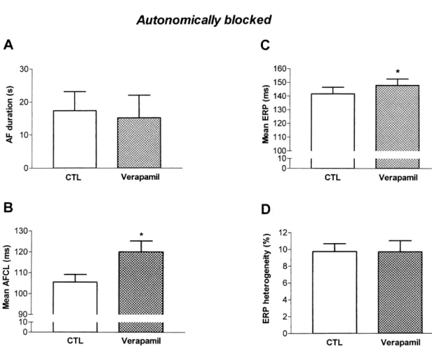
In order to evaluate effects of verapamil and diltiazem in the absence of autonomic reflexes, we studied Group 4 dogs in the presence of b-adrenergic and muscarinic receptor blockade. In the presence of autonomic blockade, verapamil reduced arterial pressure and increased the PR interval, but did not affect AF duration (Table 3, Fig. 7A). Typical activation maps for AF in the presence of ver-apamil and autonomic blockade are shown in Fig. 2C, and indicate that under these conditions activation was rela-tively homogeneous and there were few reactivation zones
Fig. 5. Effects of verapamil (0.2 mg / kg followed by 1.5 mg / kg / min) in
the presence of intact autonomic tone on (A) minimal ERP value at a per cycle. A quantitative analysis is shown in Fig. 3B, and cycle length of 300 ms in each dog; (B) maximal ERP in each dog; and indicates that in the presence of autonomic blockade (C) ERP heterogeneity calculated for each condition in each dog as the
verapamil did not alter the number of reactivation zones
standard deviation of all ERP values divided by the mean ERP value
per cycle of AF. In contrast to its effects in the presence of
times 100%.
intact autonomic reflexes, verapamil significantly increased AF cycle length in the presence of autonomic blockade nificantly the shortest ERP value for all sites in each dog (Fig. 7B) and slightly but significantly increased ERP (Fig. (Fig. 5A), but substantially decreased the maximum value 7C) without affecting ERP heterogeneity (Fig. 7D). Unlike (Fig. 5B). Since verapamil decreased longer ERP values the effects of both verapamil and diltiazem in the presence without significantly affecting short ERPs, the drug sig- of intact autonomic reflexes, conduction velocity was
nificantly reduced ERP heterogeneity (Fig. 5C). unaffected by verapamil administration in the presence of
Verapamil produced spatially variable changes in con- autonomic blockade (Fig. 8left). These results indicate an duction. The drug resulted in faster conduction in the right important role for autonomic reflexes in mediating the atrial superior and posterior free walls, but did not change electrophysiological changes observed after verapamil conduction speed in the right atrial appendage (Fig. 6left). administration. Diltiazem produced qualitatively similar When all observations in the right atrium were combined, changes to verapamil in sinus cycle length, arterial
pres-verapamil reduced ERP from 12265 to 11464 ms and sure and PR interval in the presence of autonomic
bloc-increased conduction velocity from 9062 to 9963 cm / s kade (Table 3), and similarly failed to alter AF duration. (P,0.02 for each). Because of the offsetting changes in Changes in conduction velocity after diltiazem
administra-Fig. 6. Effect of verapamil (0.2 mg / kg followed by 1.5 mg / kg / min, left panels) and diltiazem (0.8 mg / kg followed by 15 mg / kg / min, right panels) in the presence of intact autonomic tone on conduction velocity in each of three atrial regions.
tion in the presence of autonomic blockade are shown in 4. Discussion
Fig. 8 (right panels). As for verapamil, no significant
changes in conduction were seen after diltiazem adminis- We have shown that verapamil promotes the
mainte-tration when the experiment was conducted in the presence nance of AF in normal dogs. The underlying mechanism
Table 3
Comparison between effects of verapamil and diltiazem in presence of autonomic blockade (Group 4)
Control Verapamil P Control Diltiazem P
(0.2 mg / kg, followed (0.8 mg / kg, followed by 1.5 mg / kg / min) by 15 mg / kg / min) a a n 5 5 – 4 4 – Sinus CL (ms) 448640 590646 0.048 585657 750623 0.113 Mean arterial BP (mmHg) 9268 68610 0.048 9163 6064 0.041 PR interval (ms) 10868 149615 0.011 11568 156622 0.123 AF duration (s) 1766 1567 0.80 65632 33616 0.54 a
Each dog was studied under continuous autonomic blockade for both control and drug conditions.
requiring intact autonomic function. Diltiazem did not dependent ERP adaptation being hallmark features [12].
21
share the AF promoting action of verapamil in this model. Decreases in L-type Ca current seem to be central to these electrophysiological effects of tachycardia-induced
21 21
4.1. Role of L-type Ca currents and their blockade remodeling [13]. Ca overload due to an increased rate of
in AF action potential generation may be important in initiating
tachycardia-related remodeling, which may in part be a
21
L-type Ca current plays an important role in maintain- protective response of cardiomyocytes to prevent damage
21
ing the plateau of the atrial action potential and changes in caused by Ca loading [14]. Consistent with this concept,
21
Ca current are an important contributor to rate-depen- verapamil prevents some of the changes caused by short dent adaptations in atrial action potential duration [10,11]. term (,24 h) atrial tachycardias [15–18]; however, L-type
21
Atrial tachycardias, including AF, modify atrial properties Ca channel blockers do not prevent remodeling by atrial to promote AF maintenance, with reduced ERP and rate- tachycardias lasting a week or longer [5,19]. Furthermore,
Fig. 7. Effects of verapamil (0.2 mg / kg followed by 1.5 mg / kg / min) in the presence of autonomic blockade on (A) AF duration; (B) AF cycle length; (C) mean ERP at all atrial sites; and (D) ERP heterogeneity.
Fig. 8. Effect of verapamil (0.2 mg / kg followed by 1.5 mg / kg / min, left panels) and diltiazem (0.8 mg / kg followed by 15 mg / kg / min, right panels) in the presence of autonomic blockade on conduction velocity in each of three atrial regions.
dogs maintained on verapamil up to and during electro- changes caused by atrial tachycardia-induced remodeling
21
physiological study after a 7-day or greater period of atrial [13], it would not be surprising for Ca channel blockers tachycardia have more sustained AF than control dogs [5]. like verapamil to promote AF maintenance. The results of
21
Given the fact that reduced Ca current seems to be the present study do show that verapamil promotes AF;
notion that this action of verapamil is due to a mimicking AF. Alternatively, verapamil’s actions on atrial activation of the changes caused by atrial tachycardia. First, atrial during AF may have resulted from presently unrecognized tachycardia-induced remodeling causes substantial de- actions on impulse propagation during the arrhythmia, or creases in atrial ERP [12,13], whereas the decreases caused from other at present unrecognized actions.
by verapamil in the present paper were relatively modest. Verapamil has also been noted to have effects on
Second, increased atrial ERP heterogeneity is a prominent ventricular fibrillation (VF). In isolated hearts, which by AF-promoting feature of tachycardia-induced remodeling definition lack reflex autonomic responses, verapamil in the dog [20,21], whereas ERP heterogeneity was decreases the complexity of fibrillatory activity [24,25]. On reduced by verapamil. Third, if verapamil’s AF-promoting the other hand, verapamil increases the frequency of
21
effects were due to L-type Ca channel blockade alone, ventricular fibrillatory activity in the in situ heart [26].
one would have expected them to be enhanced by b- These results are consistent with AF activation changes
21
adrenergic blockade, since the latter reduces L-type Ca seen respectively in the absence and presence of autonomic current; however, the opposite result (an elimination of AF blockade in the present study. They suggest that
ver-promotion) was seen in autonomically blocked dogs. apamil’s effects on AF and VF have some interesting
21
Finally, if Ca channel blockade was the primary mecha- similarities, which warrant more detailed analysis in
nism of verapamil’s AF-promoting properties, one might subsequent work.
have expected to have observed similar phenomena with Although intact autonomic reflexes were required for
diltiazem, particularly at doses (like the ones we used) that verapamil’s AF-promoting actions, consistent with previ-cause comparable degrees of PR interval prolongation. ous observations [6], autonomic interactions alone were insufficient to explain the effects of verapamil. Vagal 4.2. Mechanism of verapamil’s AF promoting properties enhancement is unlikely to have played any significant role, since increasing vagal tone substantially increases Verapamil promoted AF in the present study by increas- atrial ERP heterogeneity [27], the opposite of the changes ing the apparent number of reentry circuits during AF and seen with verapamil. Friedman et al. showed that the thereby stabilizing multiple circuit reentry. Precisely how AF-promoting effects of verapamil are prevented by b-verapamil achieved this remains unclear. Although ver- adrenergic receptor blockade, pointing to an important role apamil did decrease atrial ERP, this effect was relatively for sympathetic stimulation [6]. Sympathetic enhancement small during 1:1 atrial pacing, and seems insufficient to is likely to have occurred during verapamil infusion, but explain the significant changes in atrial activation that increased sympathetic outflow itself has not been found to occurred during AF. Furthermore, verapamil accelerated have a significant AF-promoting effect in the normal dog conduction in several atrial regions, which counteracts the heart [27]. It is therefore likely that intact sympathetic
effect of ERP reductions on wavelength, leaving the responses are necessary for verapamil’s AF-promoting
minimal reentrant circuit size apparently unchanged. It is action, but that in themselves sympathetic effects are possible that the regional variability of the conduction insufficient to account fully for this property of the drug. speeding effect of verapamil contributed to heterogeneous The discrepancy between verapamil and diltiazem in activation during AF; however, this is unlikely to have AF-promoting properties is an interesting observation that been of prime importance, since diltiazem had similar requires explanation. Unlike verapamil, diltiazem did not effects on atrial conduction but did not promote AF. The decrease ERP or AF cycle length. Of note, diltiazem’s abolition of conduction-speeding by both drugs with effects on atrial conduction were very similar to those of autonomic blockade suggests that this action was indirect, verapamil in both the presence and absence of autonomic
quite possibly due to adrenergic enhancement. tone (Figs. 6 and 8). Diltiazem’s lack of AF-promoting
We measured atrial ERP and conduction velocity during potential could be due to a different time and voltage
21
1:1 atrial pacing, and it is possible that these do not dependent profile of Ca channel blockade or to different necessarily reflect the drug’s electrophysiological actions collateral properties. Further studies to clarify this issue, in
at the very rapid rates of AF. Verapamil has complex both experimental and clinical settings, might be of
1
effects on a variety of K channels [22], as well as rate considerable interest.
21
and voltage dependent blocking actions on L-type Ca 1
channels [23]. The K -channel inhibiting actions of the 4.3. Novel aspects and potential clinical relevance drug may explain its effect to prolong ERP in the present
study when adrenergic reflexes were absent in au- Verapamil’s AF promoting action has been reported
tonomically-blocked dogs. The rate dependent effects of clinically [4] and in an experimental model [6]. Neither
1 21
verapamil on various K and Ca channels, the varying publication addressed potential underlying electrophysio-importance of each channel during the action potential logical mechanisms. Friedman et al. did show that beta-plateau at different rates, and varying degrees of adrenergic adrenoceptor blockade prevents AF promotion by ver-reflex activity at different heart rates may have resulted in apamil, but did not examine electrophysiological changes. much smaller ERP changes at slower 1:1 rates than during In the present study, we show that verapamil promotes AF
[3] Reiffel JA. Drug choices in the treatment of atrial fibrillation. Am J
by increasing the heterogeneity of activation and the
Cardiol 2000;85(Suppl 1):12–19.
number of apparent reentry zones during the arrhythmia.
[4] Shenasa M, Kus T, Fromer M et al. Effect of intravenous and oral
This observation is consistent with the multiple wavelet calcium antagonists (diltiazem and verapamil) on sustenance of mechanism of AF maintenance [28]. On the other hand, atrial fibrillation. Am J Cardiol 1988;62:403–407.
despite obtaining careful measurements of properties (like [5] Lee SH, Yu WC, Cheng JJ et al. Effect of verapamil on long-term tachycardia-induced atrial electrical remodeling. Circulation
ERP and ERP heterogeneity) usually associated with the
2000;101:200–206.
stability of multiple circuit reentry, the precise
electro-[6] Friedman HS, Rodney E, Sinha B et al. Verapamil prolongs atrial
physiological mechanism by which multiple wavelet reen- fibrillation by evoking an intense sympathetic neurohumoral effect. J try was promoted remains obscure. It is possible that Investig Med 1999;47:293–303.
21 changes in ERP, ERP heterogeneity, conduction velocity [7] Fareh S, Benardeau A, Thibault B et al. The T-type Ca channel
blocker mibefradil prevents the development of a substrate for atrial
and wavelength during 1:1 pacing do not, in the presence
fibrillation by tachycardia-induced atrial remodeling in dogs.
Circu-of verapamil, reflect well changes during AF.
Alternative-lation 1999;100:2191–2197.
ly, multiple wavelet reentry may have been promoted by
[8] Li D, Fareh S, Leung TK et al. Promotion of atrial fibrillation by
changes in variables that we were unable to study. For heart failure in dogs: atrial remodeling of a different sort. Circula-example, heterogeneous changes in connexin 40 distribu- tion 1999;100:87–95.
[9] Talajic M, Nattel S. Frequency-dependent effects of calcium
antago-tion may be important in tachycardia-remodeling related
nists on atrioventricular conduction and refractoriness:
demonstra-AF [29]. Although the number of connexins is very
tion and characterization in anesthetized dogs. Circulation
unlikely to have changed over the time course of the
1986;74:1156–1167.
present experiments, changes in connexin function (or [10] Li GR, Nattel S. Properties of human atrial I at physiological Ca
some other aspect of cellular coupling) could have been temperatures and relevance to action potential. Am J Physiol 1997;272:H227–H235.
induced by verapamil during AF and would not have been
[11] Courtemanche M, Ramirez RJ, Nattel S. Ionic mechanisms
underly-detected by the methods we used.
21 ing human atrial action potential properties: insights from a
mathe-Patients with AF are often treated with Ca channel
matical model. Am J Physiol 1998;275:H301–H321.
blockers to control the ventricular response rate. Our [12] Wijffels MC, Kirchhof CJ, Dorland R et al. Atrial fibrillation begets observations raise the question of whether diltiazem might atrial fibrillation. A study in awake chronically instrumented goats.
be a better choice for rate control among patients in whom Circulation 1995;92:1954–1968.
[13] Yue L, Feng J, Gaspo R et al. Ionic remodeling underlying action
a subsequent cardioversion is considered, since diltiazem
potential changes in a canine model of atrial fibrillation. Circ Res
did not promote AF maintenance in our dogs. Shenasa et
1997;81:512–525.
al. did observe AF promoting effects of diltiazem, as well [14] Nattel S. Atrial electrophysiological remodeling caused by rapid as verapamil, administration in man [4]. Further clinical atrial activation: underlying mechanisms and clinical relevance to
studies of the effects of verapamil and diltiazem on AF atrial fibrillation. Cardiovasc Res 1999;42:298–308.
[15] Goette A, Honeycutt C, Langberg JJ. Electrical remodeling in atrial
maintenance are warranted, to determine the relative
fibrillation. Time course and mechanisms. Circulation
actions of these agents on the tendency of AF to sustain
1996;94:2968–2974.
itself. Should our findings be substantiated by such clinical [16] Daoud EG, Knight BP, Weiss R et al. Effect of verapamil and studies, our observations could contribute to providing procainamide on atrial fibrillation-induced electrical remodeling in
more effective drug therapy for patients with AF. humans. Circulation 1997;96:1542–1550.
[17] Yu WC, Chen SA, Lee SH et al. Tachycardia-induced change of atrial refractory period in humans: rate dependency and effects of antiarrhythmic drugs. Circulation 1998;97:2331–2337.
Acknowledgements [18] Tieleman RG, De Langen C, Van Gelder IC et al. Verapamil reduces
tachycardia-induced electrical remodeling of the atria. Circulation 1997;95:1945–1953.
This work was supported by the Medical Research
[19] Fareh S, Benardeau A, Nattel S. Comparative study of the efficacy
Council of Canada, the Heart and Stroke Foundation of of T- and L-type calcium channel blockers against atrial remodeling Quebec, the Fonds de Recherche de l’Institut de Car- due to sustained atrial tachycardia (abstract). Circulation
´ ´
diologie de Montreal. Dr. Benardeau was supported by 1999;100(Suppl I):11.
[20] Gaspo R, Bosch RF, Talajic M et al. Functional mechanisms
Hoffmann-La Roche, Basel, Switzerland. The authors wish
underlying tachycardia-induced sustained atrial fibrillation in a
to thank Emma De Blasio, Mirie Levi and Chantal Maltais
chronic dog model. Circulation 1997;96:4027–4035.
for their skilled technical assistance, and Diane Campeau [21] Fareh S, Villemaire C, Nattel S. Importance of refractoriness for secretarial help with the manuscript. heterogeneity in the enhanced vulnerability to atrial fibrillation
induction caused by tachycardia-induced atrial electrical remodeling. Circulation 1998;98:2202–2209.
[22] Waldegger S, Niemeyer G, Morike K et al. Effect of verapamil
References 1
enantiomers and metabolites on cardiac K channels expressed in Xenopus oocytes. Cell Physiol Biochem 1999;9:81–89.
[1] Nattel S. Newer developments in the management of atrial fibrilla- [23] Nawrath H, Wegener JW. Kinetics and state-dependent effects of tion. Am Heart J 1995;130:1094–1106. verapamil on cardiac L-type calcium channels. Naunyn [2] Nattel S. Experimental evidence for proarrhythmic mechanisms of Schmiedebergs Arch Pharmacol 1997;355:79–86.
fibrillation by flecainide, verapamil, and sotalol: an experimental [27] Liu L, Nattel S. Differing sympathetic and vagal effects on atrial study. Circulation 2000;101:1606–1615. fibrillation in dogs: role of refractoriness heterogeneity. Am J [25] Samie FH, Mandapati R, Gray RA et al. A mechanism of transition Physiol 1997;273:H805–H816.
from ventricular fibrillation to tachycardia: effect of calcium channel [28] Moe GK. On the multiple wavelet hypothesis of atrial fibrillation. blockade on the dynamics of rotating waves. Circ Res 2000;86:684– Arch Int Pharmacodyn Ther 1962;140:183–188.
691. [29] van der Velden HMW, Ausma J, Rook MB et al. Gap junctional
[26] Stewart AJ, Allen JD, Devine AB et al. Effects of blockade of fast remodeling in relation to stabilization of atrial fibrillation in the and slow inward current channels on ventricular fibrillation in the goat. Cardiovasc Res 2000;46:476–486.