Received: May 23, 2014; Revised: July 29, 2014; Accepted: October 2, 2014
© The Author 2014. Published by Oxford University Press. All rights reserved. For Permissions, please e-mail: [email protected].
doi:10.1093/jnci/dju364
First published online November 24, 2014 Article
1 of 8
ar
ticle
article
Selective Bispecific T Cell Recruiting Antibody and
Antitumor Activity of Adoptive T Cell Transfer
Sebastian Kobold, Julius Steffen, Michael Chaloupka, Simon Grassmann,
Jonas Henkel, Raffaella Castoldi, Yi Zeng, Markus Chmielewski,
Jan C. Schmollinger, Max Schnurr, Simon Rothenfußer, Dolores J. Schendel,
Hinrich Abken, Claudio Sustmann, Gerhard Niederfellner, Christian Klein,
Carole Bourquin, Stefan Endres
Affiliations of authors: Center of Integrated Protein Science Munich (CIPS-M) and Division of Clinical Pharmacology, Department of Internal Medicine IV, Ludwig-Maximilians-Universität München, Munich, Germany (SK, JS, MiC, SG, JH, YZ, JCS, MS, SR, CB, SE); Roche Pharmaceutical Research and Early Development, Oncology Discovery and Translational Area, Roche Innovation Center Penzberg, Penzberg, Germany (RC, CS, GN); Center for Molecular Medicine Cologne and Department I for Internal Medicine, University Hospital Cologne, Cologne, Germany (MaC, HA); Institute of Molecular Immunology, Helmholtz Zentrum München and Clinical Cooperation Group Immune Monitoring, Helmholtz Zentrum München, German Research Center for Environmental Health, Munich, Germany (DJS); Roche Pharmaceutical Research and Early Development, Oncology Discovery and Translational Area, Roche Innovation Center Zurich, Switzerland (CL); Chair of Pharmacology, Department of Medicine, University of Fribourg, Fribourg, Switzerland (CB). Correspondence to: Sebastian Kobold, MD, Division of Clinical Pharmacology, Ludwig-Maximilians-Universität München, Ziemssenstraße 1, 80336 München, Germany (e-mail: [email protected]).
Abstract
Background: One bottleneck for adoptive T cell therapy (ACT) is recruitment of T cells into tumors. We hypothesized that combining tumor-specific T cells, modified with a marker antigen and a bispecific antibody (BiAb) that selectively recognizes transduced T cells and tumor cells would improve T cell recruitment to tumors and enhance therapeutic efficacy. Methods: SV40 T antigen–specific T cells from T cell receptor (TCR)-I–transgenic mice were transduced with a truncated human epidermal growth factor receptor (EGFR) as a marker protein. Targeting and killing by combined ACT and anti-EGFR–anti-EpCAM BiAb therapy was analyzed in C57Bl/6 mice (n = six to 12 per group) carrying subcutaneous tumors of the murine gastric cancer cell line GC8 (SV40+ and EpCAM+). Anti-EGFR x anti-c-Met BiAb was used for targeting of human tumor-specific T cells to c-Met+ human
tumor cell lines. Differences between experimental conditions were analyzed using the Student’s t test, and differences in tumor growth with two-way analysis of variance. Overall survival was analyzed by log-rank test. All statistical tests were two-sided. Results: The BiAb linked EGFR-transduced T cells to tumor cells and enhanced tumor cell lysis. In vivo, the combination of ACT and Biab produced increased T cell infiltration of tumors, retarded tumor growth, and prolonged survival compared with ACT with a control antibody (median survival 95 vs 75 days, P < .001). In human cells, this strategy enhanced recruitment of human EGFR–transduced T cells to immobilized c-Met and recognition of tyrosinase+ melanoma cells by
TCR-, as well as of CEA+ colon cancer cells by chimeric antigen receptor (CAR)–modified T cells.
Conclusions: BiAb recruitment of tumor-specific T cells transduced with a marker antigen to tumor cells may enhance efficacy of ACT.
ar
ticle
Tumor antigen–specific T cells are attractive tools for cancer therapy, because they are highly specific (1), can actively infiltrate tumor tissue where their target antigen is expressed (2), and are capable of serial-target cell killing (3). However, efficacy of T cell therapy against solid tumors without host conditioning is limited, and this treatment only leads to responses in a small subset of patients (two out of 11) (4).
Host conditioning with total body irradiation or T cell– depleting chemotherapy has been investigated to improve the therapeutic efficacy of adoptive T cell therapy (ACT) (5). However, these treatments are accompanied by frequent high-grade (3 or 4) toxicities, limiting their use to restricted patient populations (6). Thus, there is a need for strategies addressing the inherent limitations of ACT without causing severe side effects.
Bispecific antibodies (BiAbs) with specificity against a tumor-associated antigen and against a T cell marker are cur-rently in clinical development to recruit T cells to cancer cells (7,8). Most T cell–recruiting BiAb address T cells by the pan-T cell marker CD3. They have proven their efficacy in clinical phase II (7) and III trials (9). The combination of ACT with BiAb application is an attractive strategy to enhance the efficacy of either approach alone, but so far have yielded disappointing results because of the induction of a cytokine storm upon T cell activation (10).
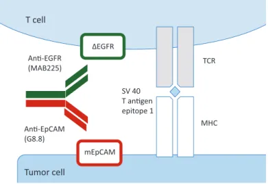
Because of the ability of BiAbs to link T cells with tumor cells, we hypothesized that a nonsignaling (inert) antigen introduced into the tumor-reactive T cell could be used as the target for one binding arm of a BiAb (Figure 1). In contrast to using an anti-CD3 BiAb, T cell activation in this setting can only result from T cell receptor (TCR) engagement. Thus T cell degranulation can only occur at the tumor site and not systemically in other organs or in the circulation. Combining ACT of tumor-specific T cells expressing an inert marker antigen with a cognate BiAb recognizing both the transduced T cell and a tumor antigen has the potential to facilitate tumor-specific T cell engagement in the tumor tissue while avoiding systemic T cell activation by functionally separating the linking of T cells to tumors and T cell activation.
Methods
Animal Experiments
Wild-type C57BL/6RJ mice were purchased from Janvier (St. Berthevin, France). Mice transgenic for a T cell receptor specific for the immunodominant epitope I of the simian virus 40 (SV40) T antigen (TCR-I) were purchased from the Jackson Laboratory (stock number 005236). mGC8 tumors were induced subcutaneously with 3 x 10⁶ cells. Prior to treatment, mice were randomized to the
treat-ment groups (n = six to 12 per group), and tumors were measured every other day in a nonblinded fashion. Tumor volume was esti-mated using the following formula: V = 4/3 x π x L12 x L
2 (with L1
defined as maximal diameter and L2, the diameter perpendicular to L1). T cells and antibodies were given intravenous (i.v.). All animal studies were approved by the local regulatory agency (Regierung von Oberbayern). Power calculations were done prior to study approval.
T Cell Transduction
Truncated human epithelial growth factor receptor (referred to as ΔEGFR), comprising amino acids 1–667, was cloned into the retroviral vector pMP71 (kindly provided by C. Baum, Hannover, Germany) and into the retroviral bicistronic pBULLET vector containing BW431/26-scFv-Fc-CD28-CD3 CAR specific for CEA (11). pMP71-ΔEGFR was used for transduction of human and murine wild-type T cells, the murine T cell line B3Z, the human tyrosinase specific T cell clone IVSB and the human T cell line Jurkat. For human CEA–CAR T cell transduction, the pBULLET vector was used. Transduction protocols have been described in detail (11–13). In brief, Plat-E cells were transfected and the produced retrovirus was used to transduce murine T cells pres-timulated by anti-CD3 and anti-CD28 antibodies (eBioscience, Frankfurt, Germany, clones 145-2C11 and 37.51, respectively) and IL-2 (10 IU/mL). In subsequent cultures, CD3, anti-CD28 beads (Life Technologies, Carlsbad, CA), and IL-15 (50 ng/ mL, Peprotech, Hamburg, Germany) were applied. For human T cell transduction, 293T cells were transfected with the respec-tive retroviral vector together with the plasmids pcDNA3.1-MLVg/p and pALF10A1 (both kindly provided by W. Uckert, Max Delbruck Centre, Berlin). Retrovirus was used to transduce
Figure 1. Schematic diagram of bispecific antibody-enhanced binding of tumor antigen–specific T cells to tumor cells. The anti–epidermal growth factor receptor (EGFR) x anti-EpCAM bispecific antibody (ER-Ep BiAb) in a two-plus-two format contains two binding sites for EpCAM (rat antimurine EpCAM – disulphide stabilized ScFv – VH – (G4S)3-VL), which is expressed on the tumor cell surface, and two binding sites for a truncated form of EGFR (ΔEGFR) which is expressed by the transduced T cells. The T cell receptor of the transduced T cells is specific for the immunodominant epitope-1 of the SV40 large T antigen, which is presented in the context of major histocompatibility complex (MHC) class I by the tumor cell. Cross-linking by anti-EGFR x anti-EpCAM bispecific antibody may increase or stabilize T cell–tumor cell contact established by the T cell receptor–MHC class I interaction.
ar
ticle
primary human T cells or the IVSB clone. Primary human T cells were stimulated using anti-CD3 and anti-CD28 antibod-ies (ebioscience, clones HIT3a and CD28.2, respectively) and IL-2 (200 U/mL).
Antibody Cross-Linking Assay
Epithelial cell adhesion molecule (EpCAM+) 4T1 were seeded
and grown to confluence. ΔEGFR-transduced B3Z T cells were labeled with calcein (Life Technologies, Carlsbad, CA) and loaded with EGFR–EpCAM BiAb (ER-Ep BiAb) or control anti-EGFR–anti-digoxigenin BiAb (ER-Dig BiAb). For human T cells, ΔEGFR-transduced, calcein-labeled Jurkat T cells preloaded with anti-EGFR–anti-c-Met BiAb (ER-Met BiAb) were incu-bated on recombinant c-Met (R & D, Wiesbaden-Nordenstadt, Germany)–coated plates. The plate was washed and cells were visualized by fluorescence microscopy. Calcein retention was measured using a Mithras multilabel reader (Berthold, Bad Wildbad, Germany). Percentage of retained cells was normal-ized to total cell input.
Killing Assay
mGC8 murine gastric cancer cells were labeled with calcein. ΔEGFR-transduced T cells were preloaded with ER-Ep BiAb or control ER-Dig BiAb and cocultured with tumor cells. Calcein mean fluorescence intensity (cMFI) in the supernatant was quantified. Specific lysis (%) was calculated as (cMFIof interest -
cMFIbackground)/(cMFItotal lysis - cMFIbackground) x 100%.
Interferon-γ Release Assay
transduced IVSB T cell clone and ΔEGFR-carcinoembryonic antigen (CEA)–chimeric antigen receptor (CAR)–transduced T cells were cocultivated with mel624.38 and MelA375 or LS174T for 48 hours at a ratio of 10:1 or 1:2.5, respectively. IFN-γ release was quantified by enzyme-linked immunosorbent assay (ELISA; R & D). For IVSB T cell clone experiments, the coculture was preceded by cell adhesion of transduced and untransduced T cells using anti-EGFR antibody AY13 (Biolegend, London, UK) labeled with APC and anti-APC magnetic cell sorting (Miltenyi, Bergisch-Gladbach, Germany); retained cells were used for the assay. For CEA-CAR experi-ments, T cells were preloaded with ER-Met BiAb for 30 minutes prior to coculture.
Statistical Analysis
For statistics, GraphPad Prism software version 5.0b was used. All variables reported are continuous. For in vitro analysis, dif-ferences between experimental conditions were analyzed using the unpaired two-sided Student’s t test; P values under .05 were considered statistically significant. For in vivo data, dif-ferences between groups were analyzed using two-way analy-sis of variance (ANOVA) with correction for multiple testing by the Bonferroni method. For tumor growth analysis, D’Agostino Pearson omnibus normality analysis was performed on 21 tumor-bearing but untreated mice (620 ± 619 mm2, P = .021); thus,
normality of tumor growth distribution was assumed. Overall survival was analyzed by log-rank test. Survival is defined in days from tumor induction until natural death or until mice were killed because one of the following criteria was reached: tumor size greater than 225 mm2, weight loss greater than 15%
or prolonged distress, as defined by the German Society for Laboratory Animal Science. Data are shown as mean values of a minimum of three biological replicates or independent experi-ments ± standard deviation.
Results
Murine EpCAM and Human EGFR as Model Targets in a Syngeneic Murine Tumor Model
To test our hypothesis of enhancing T cell to tumor binding by BiAbs, we used mGC8 gastric cancer cells as target cells or for inducing subcutaneous tumors. mGC8 cells are posi-tive for murine EpCAM on the cell surface (Supplementary Figure 1A, available online) and present the immunodominant epitope of the SV40 T antigen in the context of H-2 Kd (14). To assess whether EpCAM would be amenable to antibody-based therapy, we injected tumor-bearing mice with the monoclo-nal rat anti-EpCAM antibody G8.8. Deposition of this antibody was detected by direct immunofluorescence in the tumor and in all organs found positive for EpCAM by expression analy-sis (Supplementary Figure 1, B and C). Human EGFR was used because of lack of cross-reactivity of the anti-EGFR antibody cetuximab (Mab225) with murine tissue (15). Thus, Mab225 did not detect the cell line mGC8 and did not react to any of the tested organs (tumor, spleen, liver, lung, kidney, and bowel, data not shown).
Generation of a Novel Anti-EGFR x Anti-EpCAM (ER-Ep) BiAb
We generated a novel BiAb-binding EGFR and murine EpCAM ectodomains with similar affinity as compared with the mono-clonal parental antibodies (Figure 1 and Table 1). The ER-Ep BiAb is not cross-reactive to murine EGFR and thus does not bind to the cell line mGC8 used in this study. Surface plasmon reso-nance analysis confirmed that the BiAb was able to bind both antigens simultaneously (Table 1; Supplementary Figure 2A, available online). The ER-Ep BiAb did not induce statistically significant antibody-dependent cellular cytotoxicity or comple-ment dependent cytotoxicity (data not shown).
Cross-Linking of ΔEGFR+ T Cells With EpCAM+ Tumor
Cells and Directed Killing of Tumor Cells
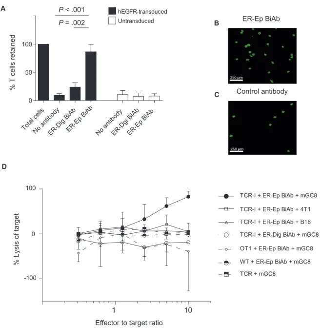
The ΔEGFR construct was used to stably transduce T cells (for representative transduction result see Supplementary Figure 2B, available online). The ability of the ER-Ep BiAb to bring ΔEGFR-transduced T cells and EpCAM+ tumor cells into
physical proximity was addressed in vitro. The ER-Ep BiAb retained more ΔEGFR+ T cells in proximity to the tumor cells
compared with control ER-Dig BiAb, which binds only to EGFR (mean retention ± SD 87 % ± 13 % vs 24% ± 8 %, P = .002) and to incubation without antibody (87 % ± 13 % vs 9 ± 3 %, P < .001) (Figures 2, A, B, and C). Remarkably, this ability to link T cells to tumor cells resulted in lysis of the target cells, with a maxi-mal lysis at a E:T ratio of 10:1 (TCR-I + ER-Ep BiAb + mGC8 vs TCR-I + ER-Dig BiAb + mGC8, 83 ± 12 % vs -18.5 ± 1%, P < .001) (Figure 2D). Lysis was dependent on the simultaneous presence of the four interacting components: ΔEGFR and TCR-I targeting SV40 T antigen on the T cells; EpCAM and SV40 T antigen on the tumor cells (Figure 2D; Supplementary Table 1 for P values, available online).
ar
ticle
Impact of ER-Ep BiAb and ΔEGFR+ TCR Transgenic
T Cells on mGC8 Tumor Growth
Therapeutic efficacy of the combination of the ER-Ep BiAb and ACT was investigated in mice carrying subcutaneous mGC8 tumors. The combined treatment ER-Ep BiAb plus ΔEGFR+ TCR-I
T cells of mice with established tumors (mean size at onset 133 mm3) delayed tumor growth as compared with treatment
with ΔEGFR+ TCR-I T cells in conjunction with the murinized
monospecific anti-EpCAM antibody (44 ± 52 mm3 vs 346 ±
113 mm3, P = .014 from day 55) (see Supplementary Table 2,
available online, for a summary of P values) or with the anti-body alone (44 ± 52 mm3 vs 464 ± 233 mm3, P = .004 at day 44)
(Figure 3A). The in vivo efficacy of the combined T cell and BiAb treatment was confirmed in a second in vivo study by treatment of mice with the ER-Ep BiAb alone, as well as with two different doses of the ER-Ep BiAb together with ACT (Figure 3B). The ER-Ep BiAb alone did not confer therapeutic efficacy, whereas ER-Ep BiAb together with ACT showed a dose-dependent effect with 10 mg/kg of BiAb being more effective than 2 mg/kg (10 ± 7 mm3
vs 205 ± 83 mm3, P = .038 at day 54) (see supplementary Table 3,
available online, for a summary of P values; Figure 3B). Both low and high doses of ER-Ep BiAb plus ACT prolonged survival of tumor-bearing mice compared with ACT with a murinized monospecific anti-EpCAM antibody (median survival high dose BiAb 95 vs control 75 days, P < .001) (Figure 3C). Mice tolerated the treatment well with no obvious signs of toxicity. In addition, non–tumor-bearing animals treated with the combination regi-men showed no diarrhea or weight loss (data not shown) during an observation period of 100 days. In line with these results, no increase in T cell infiltration was found in organs lacking the SV40 T antigen (Supplementary Figure 3A, available online).
Influence of the Combination Therapy on T Cell Infiltration Into the Tumor
To investigate the mechanisms contributing to tumor control or to escape from therapy, we analyzed by immunohistochemistry the tumors depicted in Figures 3, A and B, for the presence of infiltrating T cells (from mice which had to be killed because of a prespecified tumor-size threshold). Tumors from mice treated with ER-Ep BiAb plus ΔEGFR-transduced T cells contained more CD3+ T cells than the group treated with ΔEGFR-transduced T
cells without BiAb (0.40 ± 0.42 vs 0.09 ± 0.14 per high power field,
P = .021) (Supplementary Figure 3, B-D, available online).
Bispecific Antibody Enhances Recognition of Tumor Cells by TCR- and CAR-Transduced Human T Cells
To test if our strategy can also be applied to target human tumors, we used the previously described EGFR x c-Met bispecific antibody (ER-Met BiAb) (16) together with either ΔEGFR-transduced human
T cells, the HLA-A2–tyrosinase-specific human T cell clone IVSB (17) or human CEA-CAR–specific T cells (Figure 4A) (11). We chose c-Met because of its wide expression on human tumor cells (18). The ER-Met BiAb retained ΔEGFR+ T cells to immobilized c-Met (57
± 12 % vs 7 ± 2 %, P = .002) (Figure 4B), without affecting ΔEGFR- T
cells (6 ± 2 % vs 5 ± 0.4 %, P = .280). Remarkably, retention of ΔEGFR-transduced tyrosinase-specific IVSB T cells strongly enhanced rec-ognition of the tyrosinase+ HLA-A2+ mel624 melanoma cells but not
of the tyrosinase- HLA-A2+ melA375 melanoma cells, as measured
by IFN-γ release by the T cells (877 ± 63 ng/mL vs 166 ± 27 ng/mL, P < .001) (Figure 4C). Similarly, coculture of ΔEGFR- and CEA-CAR–trans-duced T cells with CEA+ LS174T colon cancer cells in the presence
of ER-Met BiAb increased CAR-mediated T cell activation indicated by antigen-specific IFN-γ release; the effect was most prominent when using suboptimal numbers of anti–CEA-CAR T cells (IFN-γ release 330 ± 39 ng/mL vs 0 ± 0 ng/mL, P = .001) (Figure 4D). Thus, the ER-Met BiAb is able to enhance the TCR- or CAR-mediated acti-vation of T cells by targeting T cells to tumor cells.
Discussion
Our study shows that, in mice, treatment with ER-Ep BiAb in conjunction with ACT of marker antigen–transduced tumor-spe-cific T cells resulted in enhanced T cell–mediated tumor killing and in T cell accumulation in subcutaneous mGC8 tumors. This novel treatment approach also enhanced tumor recognition by human T cells via a tumor-specific TCR or CAR, suggesting the amenability of the strategy against human tumors. This is the first report demonstrating that a nonactivating BiAb is able to specifically accumulate tumor-specific T cells in the tumor with-out the need of polyclonal T cell activation. The strategy has the potential to enhance safety of ACT by avoiding triggering of T cells of other specificities at sites distant to the tumor.
The principal amenability of targeting EpCAM+ murine and
human tumors for recruitment and activation of T cells has previously been shown (19). This provided the basis for select-ing EpCAM as a model antigen for BiAb therapy. Similarly, we used c-Met as a BiAb target because of its broad expression in human malignancies and because of the already proven employability of a c-Met–bispecific antibody (16,18). A chal-lenge in translating the combined ACT plus BiAb strategy into clinical application is the choice of an appropriate marker antigen for human T cell modification. Because ΔEGFR, as employed in the present study, has been described to be of low spontaneous immunogenicity (20), we selected it as a suitable human target in both murine and human tumor models. Given the favorable safety profile of EGFR-targeted antibody therapy, EGFR may be a suitable antigen for clinical application. In fact, ΔEGFR-transduced T cells are currently used as a safety switch in an adoptive T cell trial (NCT01815749) to permit T cell deple-tion through EGFR-targeted antibodies if required. However, the ubiquitous EGFR expression and thus the targeting of T Table 1. Surface plasmon resonance analysis of antibody affinities to murine EpCAM and human EGFR ectodomains*
Antibody Analyte Ka, M-1s-1 kd, s-1 t ½, min KD, M
ER-Ep BiAb EpCAM 2.0 x 10⁵ 2.5 x 10⁻³ 4.6 1.2 x 10⁻⁸
ER-Ep BiAb EGFR 1.1 x 10⁶ 5.7 x 10⁻³ 2.0 5.5 x 10⁻⁹
Murinized antibody G8.8 EpCAM 2.2 x 10⁵ 1.3 x 10⁻² 0.9 6.1 x 10⁻⁸
Murinized antibody Mab225 EGFR 1.3 x 10⁶ 5.7 x 10⁻³ 2.0 4.3 x 10⁻⁹
* Ka = association constant; t ½ = half life; EGFR = epithelial growth factor receptor; EpCAM = epithelial cell adhesion molecule; ER-Ep Biab = anti-EGFR x anti-EpCAM bispecific antibody; kd = dissociation constant; KD = dissociation equilibrium constant.
ar
ticle
cells to nontumor tissue may limit therapeutic benefit. Other T cell marker proteins will be needed to be explored before clini-cal testing, such as EGFR variant 3, which is a tumor-specific mutation in EGFR, targetable by antibodies and found only in certain tumor entities such as glioblastoma (21).
The choice of tumor antigen specificity for the BiAb design is also challenging, because most tumor-associated antigens are not tumor specific (22). In the absence of ideal targets, both the TCR of the adoptively transferred T cells and the BiAb-targeted tumor antigen should be selected on the basis of a restricted expression pattern. However, even targeting less abundant
antigens may result in severe side effects (23). In our strategy, the absence of an activating component in the BiAb design, such as a CD3 binding domain, and the specificity of the T cells for a distinct tumor antigen presented in the context of major histocompatibility complex (MHC) class I are likely to enhance therapeutic safety. Avoiding unwanted, undirected T cell acti-vation remains a major challenge with respect to the T cell recruiting BiAb.
Previous clinical studies have used BiAb to recruit T cells to tumor cells in patients (10,24,25). A major difference to our study is that all previous approaches used the activating
B
C
D
To
tal
ce
lls
No
an
tib
od
y
ER-Dig
BiA
b
ER
-E
p B
iAb
No
an
tib
od
y
ER-Dgi
BiA
b
hEGFR-transducedUntransduced
ER-Ep BiAb
Control antibody
%L
ys
is of target
Effector to target ratio
TCR-I + ER-Ep BiAb + mGC8 TCR-I + ER-Ep BiAb + 4T1 TCR-I + ER-Ep BiAb + B16 TCR-I + ER-Dig BiAb + mGC8 OT1 + ER-Ep BiAb + mGC8 WT + ER-Ep BiAb + mGC8 TCR + mGC8
ER
-E
p B
iAb
P < .001
P = .002
1
10
A
0
50
100
100
0
-100
%T
cells retained
Figure 2. Linking of T cells to tumor cells and induction of specific lysis by anti-epidermal growth factor receptor (EGFR) x anti-EpCAM bispecific antibody. A) Confluent EpCAM-positive 4T1 cells were incubated with ΔEGFR-transduced calcein-labelled B3Z T cells in the presence of the anti-EGFR x anti-EpCAM bispecific antibody (ER-Ep BiAb, 10 µg/mL) or the control anti-EGFR x anti-digoxigenin bispecific antibody (ER-Dig BiAb, 10 µg/mL). The number of labeled B3Z T cells retained after washing was quantified by the amount of remaining fluorescent calcein. Mean values of three independent experiments performed each in triplicates are shown (n = 3). Error bars are standard devia-tion; P values are calculated using two-sided, unpaired Student’s t test. B and C) Representative fluorescence microscopy images depicting fluorescently (green) labelled T cells retained in proximity to the tumor cells when the anti-EGFR x anti-EpCAM bispecific antibody is applied (B) vs a control antibody (C) at 10 x magnitude; scale bar represents 250 μm. D) Lysis mediated by cross-linking of the T cell to the tumor cell by the anti-EGFR x anti-EpCAM bispecific antibody measured by calcein release. T cells with a trans-genic TCR specific for SV40 T antigen or with the OT1-transtrans-genic TCR specific for ovalbumin or wild type (WT) T cells were incubated overnight with or without the anti-EGFR x anti-EpCAM bispecific antibody or anti-EGFR x anti-digoxigenin bispecific antibody and with tumor cells expressing EpCAM (cell lines 4T1 and mGC8), SV40 T antigen (mGC8), or neither (B16). Mean results of four independent experiments are shown. Error bars represent standard deviation. P values were calculated by the two-sided Student’s t test.
ar ticle A B C 0 20 40 0 20 40 60 80 100 60 80 100 PBS Anti-EpCAM antibody ER-Ep BiAb
EGFR+ CD8+ T cells + anti-EpCAM +anti-EGFR antibodies
EGFR+ CD8+ T cells + low-dose ER-Ep BiAb
EGFR+ CD8+ T cells + ER-Ep BiAb Non-tumor bearing mice
Time [days] P er ce nt s ur vi va l PBS Anti-EpCAM antibody EGFR+ CD8+ T cells + anti-EpCAM antibody EGFR+ CD8+ T cells + ER-Ep BiAb Tu mo r v ol um e [m m 3 ] PBS Anti-EpCAM antibody ER-Ep BiAb EGFR+ CD8+ T cells +
anti-EpCAM + anti-EGFR antibodies EGFR+ CD8+ T cells
+ low dose ER-Ep BiAb
EGFR+ CD8+ T cells + ER-Ep BiAb
Tu mo r v olume [mm 3 ] PBS Anti-EpCAM antibody ER-Ep BiAb EGFR+ CD8+ T cells + anti-EpCAM +anti-EGFR antibodies EGFR+ CD8+ T cells + low-dose ER-Ep BiAb EGFR+ CD8+T cells + ER-Ep BiAb
Non-tumor bearing mice 8-6-5-3 7- 5-4-1 6 5-3-1 7 6-1 6 5-4 12 11-10-9 8-7-6 3 3 Numbers at risk 20 40 60 80 0 500 1000 1500 2000 2500 20 40 60 80 100 0 200 400 600 800 1000 Time [days] Time [days]
Figure 3. In vivo efficacy of the anti-EGFR x anti-EpCAM BiAb combined with adoptive transfer of ΔEGFR-transduced T cells. A) Anti–epidermal growth factor recep-tor (EGFR) x anti-EpCAM bispecific antibody (ER-Ep BiAb) combined with transfer of ΔEGFR-transduced T cells retards growth of subcutaneous mGC8 tumors. Mice received either PBS i.v. (group 1, n = 6), anti-EpCAM antibody 300 µg i.v. (group 2, n = 6,), 107 ΔEGFR-transduced TCR-I expressing T cells i.v. together with 300 µg anti-EpCAM antibody i.v. (group 3, n = 6), or 107 ΔEGFR-transduced TCR-I expressing T cells i.v. together with 300 µg of anti-EGFR x anti-EpCAM bispecific antibody i.v. (group 4, n = 11) on days 27 and 34 (represented by the arrows) from tumor induction. Error bars represent standard deviation. Statistics were calculated using two-way analysis of variance (ANOVA) with correction for multiple testing by the Bonferroni method. B) Therapeutic efficacy of combined application of anti-EGFR x anti-EpCAM bispecific antibody and ΔEGFR-transduced T cells is dose dependent. Mice received either PBS i.v. (group 1, n = 7), anti-EpCAM antibody i.v. (group 2, 300 µg, n = 7), anti-EGFR x anti-EpCAM bispecific antibody i.v. (group 3, 300 µg, n = 6), anti-EpCAM antibody plus anti-EGFR antibody i.v. together with 5 x 10⁶ ΔEGFR-transduced T cells (group 4, 300 µg, n = 7), a low dose (60 µg per injection) of anti-EGFR x anti-EpCAM bispecific antibody together with 5 x10⁶ ΔEGFR-transduced T cells (group 5, n = 6), or a high dose (300 µg per injection, split in 100 µg together with the T cells i.v. and 200 µg i.p. given simultaneously) of anti-EGFR x anti-EpCAM bispecific antibody together with 5 x 10⁶ ΔEGFR-transduced T cells (group 6, each n = 12). Treatment was applied twice, on days 27 and 35 after tumor induction (represented by the arrows). Error bars represent standard deviation. Statistics were calculated using two-way ANOVA with correction for multiple testing by the Bonferroni method. C) Survival curves for the treatment groups in experiment (B). Survival of the groups treated with the combination of anti-EGFR x anti-EpCAM bispecific antibody and transduced T cells is longer than survival in any of the control groups (P < .001 for comparison of median survival by log-rank test with the groups treated with anti-EGFR x anti-EpCAM bispecific antibody, anti-EpCAM antibody, as well as anti-EpCAM and anti-EGFR antibodies with ΔEGFR-transduced T cells; no statistically significant difference between high and low anti-EGFR x anti-EpCAM bispecific antibody dose plus ΔEGFR-transduced T cells groups). All statistical tests were two-sided.
ar
ticle
component of the BiAb, eg, an activating anti-CD3 antibody. In contrast, the present study is the first to show that a non-activating BiAb physically linking T cells to tumor cells can enhance the efficacy of adoptively transferred tumor-specific T cells and favor T cell recruitment and/or retention in the tumor. However, additional studies using primary human samples in xenograft models are needed to move the concept towards clinical application.
An important advantage of the present approach, com-pared with conventional T cell recruiting BiAb, is the selective recruitment of adoptively transferred tumor antigen-specific T cells in comparison with the recruitment of all CD3-positive T cells. Our strategy is potentially adaptable to a variety of T cells, such as tumor-infiltrating lymphocytes, or, as shown in the pre-sent study, to TCR- and CAR-modified T cells. Such modified T cells are currently under investigation in clinical trials and show promising efficacy (26,27). Thus potential combination
candidates for clinical development of the therapeutic principle may include T cells transduced with a TCR, eg, NY-ESO-1, or with a CAR, eg, recognizing CEA or CD19.
Funding
This work was supported by the Bayerisches Immunther- apienetzwerk (BayImmunet, project D2-F5121.7.1.1/6/15 to SK, SE, and CB), the “Programm zur Förderung von Forschung und Lehre (FöFoLe)”, Medical Faculty of the Ludwig-Maximilians-Universität München (projects 714/2010, 29/2010, 08/2011, and 10/2012 to SK and SE), the Bayerische Forschungsstiftung (project PIZ-199-13 to SK), the Graduiertenkolleg 1202 “Oligonucleotides in cell biology and therapy,” funded by the Deutsche Forschungsgemeinschaft (to SG, JH, SK, MS, SE, and CB), the international doctoral program “i-Target: Immunotargeting of Cancer,” funded by the Elite Network of Bavaria (to SK, MS, and Figure 4. Linking of T cells and tumor cells and enhancement of tumor cell recognition by anti–epidermal growth factor receptor (EGFR) x anti-c-Met bispecific antibody. A) Schematic diagram of bispecific antibody-enhanced binding of tumor antigen–specific human T cells to tumor cells. The anti-EGFR x anti-c-Met bispecific antibody (ER-Met BiAb) in a two-plus-one format contains one binding site for c-Met (antihuman c-Met 5D5 antibody), which is expressed on the tumor cell surface, and two binding sites for a truncated form of EGFR, which is expressed by the transduced human T cells. The T cell receptor of the transduced T cells is specific for tyrosinase in the context of HLA-A2, which is presented in the context of MHC class I by the tumor cell. Alternatively, the chimeric antigen receptor (CAR) specific for CEA expressed on the tumor cell is targeted. Cross-linking by anti-EGFR x anti-c-Met bispecific antibody may increase or stabilize T cell–tumor cell contact established by the T cell receptor–MHC class I or the CAR-CEA interaction. B) T cell adhesion to c-Met. Human T cell line (Jurkat) was transduced with EGFR, labelled with calcein and preincubated with or without anti-EGFR x anti-c-Met bispecific antibody prior to incubation on a plate coated with recombinant c-Met. Untransduced human T cell line with or without anti-EGFR x anti-c-Met bispecific antibody were used as controls. After washing, calcein was quantified in each well. Fluorescence was normalized to unwashed wells (100% fluorescence). Results are the mean of three independent experiments. Error bars represent standard deviation; P values were calculated by the two-sided unpaired Student’s t test. C) ΔEGFR-transduced or untransduced clonally-derived tyrosinase-specific IVSB T cells were incubated with EGFR-specific antibody and adhesion was performed by magnetic cell separation (MACS). Retained cells were cocultured for 48 hours at a 10:1 ratio with tyrosinase+, HLA-A2+ mel624.38 cells or with tyrosinase-, HLA-A2+ MelA375 cells. T cell activation was assessed by IFN-γ release. Results shown are the mean of triplicates and are representative of three independent experiments. Error bars represent standard deviation; P values were calculated by two-sided unpaired Student’s t test. D) ΔEGFR- and CEA-CAR–transduced primary human T cells or untransduced T cells were preincubated with or without anti-EGFR x anti-c-Met bispecific antibody (10 μg/mL) and cocultured for 48 hours with CEA+ LS174T cells. IFN-γ was quantified by ELISA. Results shown are the mean of triplicates and are representative of two independent experiments. Error bars represent standard deviation; P values were calculated by two-sided unpaired Student’s t test.
ar
ticle
SE), the Melanoma Research Alliance (grant number N269626 to SK and SE), the Deutsche Krebshilfe (to SK and HA), the European Union (European Regional Development Fund – Investing in your future) and the German federal state North Rhine-Westphalia (NRW) (to HA), the Swiss National Research Foundation (project 138284 to CB), and Krebsforschung Schweiz (2910-02-2012 to CB). Parts of this work have been performed for the doctoral theses of JS, MC, SG, JH, and RC.
Notes
CS, GN, and CK are employees of Roche. All other authors declare no potential conflict of interest.
References
1. Restifo NP, Dudley ME, Rosenberg SA. Adoptive immunother-apy for cancer: harnessing the T cell response. Nat Rev Immu-nol. 2012;12(4):269–281.
2. Chinnasamy D, Yu Z, Theoret MR, et al. Gene therapy using genetically modified lymphocytes targeting VEGFR-2 inhibits the growth of vascularized syngenic tumors in mice. J Clin Invest. 2010;120(11):3953–3968.
3. Isaaz S, Baetz K, Olsen K, Podack E, Griffiths GM. Serial kill-ing by cytotoxic T lymphocytes: T cell receptor triggers degranulation, re-filling of the lytic granules and secretion of lytic proteins via a non-granule pathway. Eur J Immunol. 1995;25(4):1071–1079.
4. Dudley ME, Wunderlich J, Nishimura MI, et al. Adoptive trans-fer of cloned melanoma-reactive T lymphocytes for the treat-ment of patients with metastatic melanoma. J Immunother. 2001;24(4):363–373.
5. Rosenberg SA, Yang JC, Sherry RM, et al. Durable complete responses in heavily pretreated patients with metastatic melanoma using T-cell transfer immunotherapy. Clin Cancer Res. 2011;17(13):4550–4557.
6. Dudley ME, Yang JC, Sherry R, et al. Adoptive cell therapy for patients with metastatic melanoma: evaluation of intensive myeloablative chemoradiation preparative regimens. J Clin Oncol. 2008;26(32):5233–5239.
7. Topp MS, Kufer P, Gokbuget N, et al. Targeted therapy with the T-cell-engaging antibody blinatumomab of chemother-apy-refractory minimal residual disease in B-lineage acute lymphoblastic leukemia patients results in high response rate and prolonged leukemia-free survival. J Clin Oncol. 2011;29(18):2493–2498.
8. Bargou R, Leo E, Zugmaier G, et al. Tumor regression in cancer patients by very low doses of a T cell-engaging antibody. Sci-ence. 2008;321(5891):974–977.
9. Heiss MM, Murawa P, Koralewski P, et al. The trifunctional antibody catumaxomab for the treatment of malignant ascites due to epithelial cancer: Results of a prospective ran-domized phase II/III trial. Int J Cancer. 2010;127(9):2209–2221. 10. Riechelmann H, Wiesneth M, Schauwecker P, et al. Adoptive
therapy of head and neck squamous cell carcinoma with antibody coated immune cells: a pilot clinical trial. Cancer Immunol Immunother. 2007;56(9):1397–1406.
11. Hombach A, Wieczarkowiecz A, Marquardt T, et al. Tumor-specific T cell activation by recombinant immunoreceptors: CD3 zeta signaling and CD28 costimulation are simultane-ously required for efficient IL-2 secretion and can be inte-grated into one combined CD28/CD3 zeta signaling receptor molecule. J Immunol. 2001;167(11):6123–6131.
12. Leisegang M, Engels B, Meyerhuber P, et al. Enhanced func-tionality of T cell receptor-redirected T cells is defined by the transgene cassette. J Mol Med. 2008;86(5):573–583.
13. Mueller K, von Massenhausen A, Aichele U, et al. Protective capacity of virus-specific T cell receptor-transduced CD8 T cells in vivo. J Virol. 2012;86(19):10866–10869.
14. Bourquin C, von der Borch P, Zoglmeier C, et al. Efficient eradication of subcutaneous but not of autochthonous gas-tric tumors by adoptive T cell transfer in an SV40 T antigen mouse model. J Immunol. 2010;185(4):2580–2588.
15. Luo FR, Yang Z, Dong H, et al. Correlation of pharmacokinet-ics with the antitumor activity of Cetuximab in nude mice bearing the GEO human colon carcinoma xenograft. Cancer Chemother Pharmacol. 2005;56(5):455–464.
16. Castoldi R, Ecker V, Wiehle L, et al. A novel bispecific EGFR/ Met antibody blocks tumor-promoting phenotypic effects induced by resistance to EGFR inhibition and has potent anti-tumor activity. Oncogene. 2013;32(50):5593–5601.
17. Wolfel T, Van Pel A, Brichard V, et al. Two tyrosinase nonapep-tides recognized on HLA-A2 melanomas by autologous cyto-lytic T lymphocytes. Eur J Immunol. 1994;24(3):759–764. 18. Christensen JG, Burrows J, Salgia R. c-Met as a target for
human cancer and characterization of inhibitors for thera-peutic intervention. Cancer Lett. 2005;225(1):1–26.
19. Amann M, Brischwein K, Lutterbuese P, et al. Therapeu-tic window of MuS110, a single-chain antibody construct bispecific for murine EpCAM and murine CD3. Cancer Res. 2008;68(1):143–151.
20. Sampson JH, Crotty LE, Lee S, et al. Unarmed, tumor-specific monoclonal antibody effectively treats brain tumors. Proc Natl Acad Sci U S A. 2000;97(13):7503–7508.
21. Gan HK, Cvrljevic AN, Johns TG. The epidermal growth factor receptor variant III (EGFRvIII): where wild things are altered. FEBS J. 2013;280(21):5350–5370.
22. Seidman A, Hudis C, Pierri MK, et al. Cardiac dysfunction in the trastuzumab clinical trials experience. J Clin Oncol. 2002;20(5):1215–1221.
23. Keefe DL. Trastuzumab-associated cardiotoxicity. Cancer. 2002;95(7):1592–1600.
24. Nitta T, Sato K, Yagita H, Okumura K, Ishii S. Preliminary trial of specific targeting therapy against malignant glioma. Lan-cet. 1990;335(8686):368–371.
25. Buhmann R, Simoes B, Stanglmaier M, et al. Immunotherapy of recurrent B-cell malignancies after allo-SCT with Bi20 (FBTA05), a trifunctional anti-CD3 x anti-CD20 antibody and donor lym-phocyte infusion. Bone Marrow Transplant. 2009;43(5):383–397. 26. Kalos M, Levine BL, Porter DL, et al. T cells with chimeric
anti-gen receptors have potent antitumor effects and can estab-lish memory in patients with advanced leukemia. Sci Transl Med. 2011;3(95):95ra73.
27. Robbins PF, Morgan RA, Feldman SA, et al. Tumor regression in patients with metastatic synovial cell sarcoma and mela-noma using genetically engineered lymphocytes reactive with NY-ESO-1. J Clin Oncol. 2011;29(7):917–924.
28. Goldstein N, Giorgio NA, Jones ST, Saldanha JW. Antibody and antibody fragments for inhibiting the growth of tumors. In: Organization WIP, ed. 1996.
29. Castoldi R, Jucknischke U, Pradel LP, et al. Molecular char-acterization of novel trispecific ErbB-cMet-IGF1R antibod-ies and their antigen-binding propertantibod-ies. Protein Eng Des Sel. 2012;25(10):551–559.
30. Panke C, Weininger D, Haas A, et al. Quantification of cell sur-face proteins with bispecific antibodies. Protein Eng Des Sel. 2013;26(10):645–654.