Images in cardio-thoracic surgery
Aortic dissection limited to the ascending aorta mimicking
intramural hematoma
P.A. Berdat, T. Carrel*
Clinic for Thoracic and Cardiovascular Surgery, University Hospital, CH-3010 Berne, Switzerland
Received 6 October 1998; accepted 12 October 1998
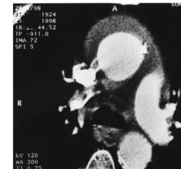
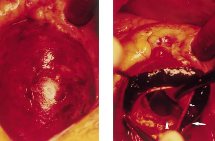
Intramural hematoma of the thoracic aorta has emerged as a diagnosis of exclusion in recent years. Significant over-lap with acute aortic dissection exists despite the increased sensitivity of preoperative CT-scan, transesophageal echo-cardiography (TEE) or magnetic resonance imaging. Surgi-cal treatment has been recommended when the ascending aorta is involved. This 74-year-old patient was admitted with acute chest pain and chest X-ray showed an enlarged mediastinum. CT scan and TEE demonstrated a large intra-mural hematoma of the ascending aorta but did not show any intimal flap (Fig. 1). However, an acute dissection lim-ited to the ascending aorta was found intraoperatively with the intimal tear located above the left coronary ostium (Figs. 2 and 3). Supracoronary graft replacement of the ascending aorta was performed immediately, using deep hypothermic cardiopulmonary bypass and a brief period of circulatory arrest for the confection of the distal anastomosis. The patient recovered well and was discharged 2 weeks follow-ing surgery.
European Journal of Cardio-thoracic Surgery 15 (1999) 108–109
1010-7940/99/$ - see front matter 1999 Elsevier Science B.V. All rights reserved. P I I : S 1 0 1 0 - 7 9 4 0 ( 9 8 ) 0 0 2 5 9 - 0
* Corresponding author. Tel.: +41-31-632-2376; fax: +41-31-382-0279; e-mail: [email protected]
Fig. 1. CT scan showing the large intramural hematoma of the ascending aorta.
Fig. 2. Intraoperative view of the ascending aorta after sternotomy.
Fig. 3. Intimal tear located above the left coronary ostium (arrowhead) and typical finding of a thrombosed false channel (small arrow: intima-media layers, large arrow: aortic adventitia).
109 P.A. Berdat, T. Carrel / European Journal of Cardio-thoracic Surgery 15 (1999) 108–109