Quarterly Journal ofMedicine, New Series L, No. 197, pp. 1-30, Winter, 1981
Renal Disease in Essential Mixed Cryoglobulinaemia
LONG-TERM FOLLOW-UP OF 4 4 PATIENTS
A. TARANTINO*, A. DE VECCHI*, G. MONTAGNINO*, E. IMBASCIATI*, M. J. MIHATSCHt, H. U. ZOLLINGERt, G. BARBIANO DI BELGIOJOSO*, G. BUSNACHt AND C. PONTICELLI*
From the Divisione di Nefrologia, Ospedale Maggiore Policlinico* and NiguardaX, Milano, Italia, and Institutfur Pathologie, Basel Universitdt, Switzerland^
Accepted 21 June 1980
SUMMARY
The mode of presentation of renal disease in 44 patients with essential mixed cryoglobulinaemia (EMC) was: acute renal failure (two patients), acute nephritic syndrome (six patients), nephrotic syndrome (eight patients), proteinuria and/or haematuria (28 patients). Renal biopsy, performed in 35 patients, showed proliferative lesions in 33, while only minimal glomerular changes were seen in the remaining two. Immunofluorescence studies showed: IgG (85 per cent), IgA (36 per cent), IgM (90 per cent), C3 (90 per cent), Clq (47 per cent), and C4 (33 per cent) deposits, mainly located in subendothelial position. On electron microscopy, crystalloid structure of deposits and monocy te infiltration of capillary loops were the outstanding feature. The survival rate was 75 per cent at 10 years from the onset of clinical symptoms. Thirty-nine patients were followed for three to 146 months (mean 53-8). Twelve patients died, cardiovascular disease and infection being the commonest cause of death. Thirteen patients showed acute renal failure or acute nephritic syndrome: nine recovered completely, whereas the remaining four died during the acute renal episode. Three patients developed chronic renal failure, but only one required chronic dialysis. The ominous significance of renal impairment in EMC should therefore be revaluated. The high prevalence of hypertension (28/44 patients) which was refractory to treatment in six, may be important to the clinical outcome.
INTRODUCTION
Wintrobe and Buell (1933) first defined cryoglobulins as cold precipitating or geling plasma proteins. Although several plasma proteins have been found in the cryoprecipitate (Putnam, 1960; Ritzmann, Levin, and Galvestone, 1961; Varriale, Ginsberg, and Sass, 1962; Allen, 1966; Alper, 1966; Lewis, Van Ommen, and Page, 1966; Hanauer and Christian, 1967; Liss, Fudenberg, and Kritzman, 1967; Correspondence to: Dr. Antonio Tarantino, Divisione di Nefrologia, Ospedale Policlinico-Pad. CrofT, Via Commenda 15, 20122-1 Milano, Italia.
2 A. Tarantino and others
Grupe, 1968; Zinneman, Levi, and Seal, 1968; Cuprak, Stollar, Kritzraan, and Liss, 1970), immunoglobulins (Igs) are the proteins more frequently detected so that the term of'cryoimmunoglobulinaemia' has been proposed (Grey and Kohler, 1973). Immunochemical analysis of cryo-Igs identified three distinct groups: Type I are characterized by the presence of isolated monoclonal Igs or Bence-Jones proteins. In Type II mixed cryo-Igs with monoclonal component possessing antibody activity against polyclonal Igs can be detected. Type III is characterized by a mixture of polyclonal Igs composed of one or more heavy chain classes. The presence of cryo-Igs has been described in multiple myeloma and Waldenstrom's macroglobulinaemia (Meltzer and Franklin, 1966; Barnett, Bluestone, Cracchiolo, Goldberg, Kantor, and Mclntosh, 1970; Bonomo, Dammacco, Tursi, and Trizio, 1970), in other haematopoietic malignancies (Alexanian, 1975), in tropical splenomegaly (Fakunle, Onyewotu, Greenwood, Mohammed, and Holborow, 1978), in acute and chronic infections (Mullinax, James, Mullinax, and Himrod, 1966; Mustakallio, Lassus, Putkonen, and Wager, 1967; Kaplan and Parker, 1966; Wager, Rasanen, Hagman, and Klenola, 1968; Kantor, Goldberg, Johnson, Derechin, and Barnett, 1970; Bonomo and Dammacco, 1971; Kaufman and Mclntosh, 1971; Hurwitz, Quismorio, and Friou, 1975), in autoimmune diseases (Christian, Hatfield, and Chase, 1963; Block, Buchanan, Wohl, and Bunin, 1965; Mustakallio, Lassus, and Wager, 1967; Talal, Zisman, and Schur, 1968; Statsny and Ziff, 1969; Weisraan and Zvaifler, 1976; McPhaul, 1978), in human acute and chronic glomerulonephritis (Mclntosh, Kaufman, Kulvinskas, and Grossman, 1970; Adam, Morel-Maroger, and Richet, 1973; Drouet, Leutounturier, Contet, and Mandet, 1973; Conte, Blanc, Mignon-Conte, Abbal, and Orfila, 1974; Mclntosh, Griswold, Chernack, Williams, Strauss, Kaufman, Koss, Mclntosh, Cohen, and Weil, 1975), in experimental glomerulonephritis (Griswold and Brady, 1978), in acute and chronic liver disease (Florin-Christensen, Roux, and Arana, 1974; Mclntosh, Koss, and Gocke, 1975; Jori, Buonanno, D'Onofrio, Tirelli, Gonnella, and Gentile, 1977), and in renal allotransplantation (Andrejak, Bariety, Bedrossian, Callard, Duret, Duboust, Idatte, and Kuhn, 1978). Cryoimmunoglobulinaemia has also been reported without evidence of any underlying disease. In 1966, Meltzer, Franklin, Elias, McCluskey, and Cooper described mixed essential cryoimmunoglobulinaemia, a syndrome consisting of purpura, arthralgia, and a high prevalence of renal damage. However the description of cryoglobulinaemic nephropathy is generally based upon individual cases and follow-up data are very sparse.
In this paper 44 patients with essential mixed cryoglobulinaemia (EMC) and nephropathy are reported. Consideration is given to (a) the clinical and biological symptoms, (b) light-, electron- and immunofluorescence microscopy findings, and (c) long-term evolution and pathogenetic mechanisms responsible for renal damage.
METHODS
The methods used for the detection of serum cryoglobulins, complement component concentrations and rheumatoid factor activity have been described
Renal Disease in Cryoglobulinaemia 3
previously (Tarantino, Anelli, Costantino, De Vecchi, Monti, and Massaro, 1978). HBS antigen (Ag) was measured in whole serum kept in 37 °C by radio-immunoassay using commercially available Abbott kits.
A, G, and M immunoglobulin concentrations in whole serum were determined by radial immunodifFusion utilizing commercially available immunoplates (Behring). C3 and C3PA (Proactivator) breakdown products were measured by immunofixation and/or crossed immunoelectrophoresis (Tarantino et ai, 1978).
Renal histology
Percutaneous renal biopsies were performed in the majority of patients. Three (micrometre) sections of Dubosq-Brazil-fixed tissue were stained with haematoxylin and eosin, periodic acid Schiff, Masson's thrichrome and silver methenamine for light microscopy. The slides were evaluated independently by two observers without previous knowledge of clinical and laboratory data.
Glomerular lesions were semiquantitatively scored for the following items: intracapillary proliferation, epithelial proliferation, polymorphonuclear cells, mononuclear cells, increase of mesangial matrix, 'double contour' appearance of glomerular basement membranes, subendothelial deposits, intraluminal thrombi, glomerular scarring. Tubular and vascular lesions were scored for tubular vacuolization, tubular atrophy, interstitial infiltration, interstitial fibrosis, vessel sclerosis, and vasculitis. Each item was rated negative (—) and + to + + +.
Immunofluorescence
Immunohistological studies with fluorescein isothiocyanate labelled antisera to human IgA, IgG, IgM, C3, Clq, and fibrinogen were performed on 4 /urn frozen sections of renal specimens. Staining for IgD, IgE, and C4 was obtained by indirect immunofluorescence method.
Electron microscopy
Renal biopsy fragments (1 mm3) and 15 in-vitro cryoprecipitates were fixed in 3 per cent glutaraldehyde and embedded in epon. Ultrathin sections—which were stained with uranyl acetate and lead citrate—were examined with a Philips EM 200 (Zollinger and Mihatsch, 1978). At least two glomeruli were studied, and tubules, interstitial tissue and vessels if available. The glomerular lesions were semi-quantitatively scored as described under light microscopy for the following items: cells in glomerular capillaries (polymorphonuclear leukocytes, lymphocytes, monocytes, monocytes with droplets), osmiophilic deposits with and without crystalloid structure, glomerular and vascular thrombi as well as further parameters mentioned under light microscopy.
Patients
Criteria for inclusion in this study were: (a) longstanding hystory of purpura and arthralgia, (b) absence of myelomatosis, Waldenstrom's macroglobulinaemia or patent infection, (c) presence of more than 80 //g/ml of cryoprecipitate, and (d) rheumatoid factor activity. Clinical history was carefully taken to establish the
4 A. Tarantino and others
onset of symptoms. The beginning of the period of observation was considered as the time of the first admission.
Definition and classification of renal manifestations
Proteinuria: protein excretion exceeding 0-2 g/24 hours.
Haematuria was classified as follows: - (0 to 3 RBC/HPF), + (4 to 10 RBC/HPF), + + (11 to 20 RBC/HPF), + + + (>20 RBC/HPF).
Nephrotic syndrome: proteinuria greater than 3 g/24 hours, serum albumin concentration lower than 3 g/dl, and oedema.
Acute nephritic syndrome: a combination of haematuria and proteinuria with a sudden rise of serum creatinine up to 600 /miol/1.
T A B L E 1. Extrarenal symptoms at the time of first admission
Number Sex Age Cryoprectpruue Cryocrit Rheumatoid Weakness Purpura ArtimJjU Fever Hcptto- Spfcno-of (yean) immunoiWxiUm (%> •cavrty ™*»ly pulem tilre , G, G,-G, 3 4 5 6 7 8 9 10 II 12 13 F F F F F F M M M F M 46 52 33 47 53 68 44 45 36 49 31 G-M G-M G M G-M G-M G-M G-M G-M G-M G-M G-M 2 4 3 4 5 3 27 3 3 1 19 640 1280 80 80 320 160 2560 320 320 320 320 16 17 18 19 20 21 22 23 24 25 26 27 28 29 30 31 32 33 34 35 3* 37 38 39 40 F F M F M M F F F F M M F M M F F M F M F F F M M 43 44 51 55 43 33 50 64 21 36 52 38 51 64 32 48 60 50 53 48 IS 51 36 55 34 G-M G-M G-M G-M C M C M G-M G-M G-M O-M G-M G M O-M G M G-M G-M G-M G-M G-M G-M G M G-M G M G-M G-M 2 2 30 21 3 35 3 13 8 5 40 9 15 25 5 11 5 16 5 5 3 2 3 4 80 320 2560 1380 80 640 640 640 320 320 320 160 320 320 160 640 320 640 160 320 1280 640 1280 to Nccrotlzini ikin lesions NecroUzui| dun kskxu Abdominal pain NearotorpcaJ symptoms Abdominal pun Chronic persistent hepatitis Chronic active hepawn Lympboadenoptlhy. GOT 48 U. GPT29U Chronic peruitenl hepathn Raynand Neorologo] symptoms Etacoid lopcj Liver drrbotb SjjofTta jyodrome Raynaad phenomenon Riictnnatoio aitlu itis«
Chjooic pemxteBt bepukis, Neuro-F 37 M 52 A G-M A-G-M I 17 SO 640 L j v e r <i' u i *H?CT
Renal Disease in Cryoglobulinaemia 5
Acute renal failure: a rapid increase of serum creatinine to levels higher than 800 //mol/1 with or without a reduction of urinary output.
Hypertension: diastolic blood pressure persistently greater than 95 mm.Hg.
Statistical studies
Students' / and corrected chi-square test were used. The results were considered statistically significant only when/? was at least <0-01.
RESULTS
(a) Initial Data
Among the 44 patients included, 17 were males and 27 females. The mean age was 47-2 ± 11-5 years (range 21-80). The main presenting features are shown in Table 1. Purpura, arthralgia and hepatomegaly were the commonest manifest-ation (50 per cent of patients), whereas weakness, fever and splenomegaly were observed less frequently. Necrotizing skin lesions occurred in two patients (Cases
1 and 2), abdominal pain in two (Cases 4 and 12) and Raynaud's phenomenon
in three (Cases 29, 40 and 41). Liver damage was found in seven (Cases 13 to 16,
35, 42 and 43), in six liver biopsy was performed showing cirrhosis (Cases 35 and 43), chronic active hepatitis (Case 14) and chronic persistent hepatitis (Cases 13, 16 and 42). The remaining patient had abnormally high levels of serum
trans-aminases at repeated determinations.
In whole serum HB viral antigen showed a 7 per cent incident (two out of 28 patients).
(b) Renal abnormalities at the time of diagnosis
The onset of renal manifestations was coincident to the extrarenal signs in 13 patients (29 per cent). In the others the delay between the onset of systemic and renal symptoms ranged from 2 to 447 months (mean 77 months). Clinical and biochemical features of renal disease at the time of diagnosis are summarized in Table 2. Acute renal failure was the presenting syndrome in two patients (4-5 per cent) (Cases 1 and 39); nephrotic syndrome in eight (18 per cent) (Cases 3,
7, 12, 13, 17, 29, 42 and 44); acute nephritic syndrome in six (13-5 per cent) (Cases 8,10, 25, 27, 32 and 34); isolated proteinuria in six (13-5 per cent) (Cases 5, 9, 19, 24, 31 and 40) and isolated microscopic haematuria in 10 (23 per cent) (Cases 14 to 16, 30, 33, 36 to 38, 41 and 43). In 12 patients (27-3 per cent)
renal abnormalities consisted of microscopic haematuria and proteinuria (Cases 2,
4, 6, 11, 18, 20 to 23, 26, 28 and 35). Hypertension was present in 22 patients
(50 per cent) (Cases 1 to 4, 6 to 10,18, 20, 22, 25, 28, 29, 31, 34, 36, 37, 39, 41 and 44).
(c) Serum complement levels
Serum samples (243) from 30 patients were assessed for Clq, Cls, C4, C3, C5, C9, C3PA, C l INH (Inhibitor) as well as whole serum complement activity (Table 3). Early complement components (Clq, C l s and C4) and CH50 were strongly depressed, while C3 did not differ from normal values; later components (C5 and C9), C3PA and C l INH were higher than normal controls. In 18 patients
T A B L E 2 . Clinical and biochemical data at the time of admission and at the end of the follow-up Dat a a t th e tim e o f admissio n Dat a a t th e en d o f th e follow-u p Numbe r Plasm a Seru m Seru m of creatinin e protei n albumi n patien t (^mol/l ) (g/dl ) (g/dl ) Urin e Hacmaturi a Bloo d Onse t o f Onse t o f protei n pressur e symptom s rena l (g/2 4 (mmHg ) (months ) symptom s hours ) (months ) Follow-u p Plasm a Urin e tim e creatinin e protein s (months ) Oanol/1 ) (g/2 4 hours ) Haematuri a Bloo d pressure * T T R R N R T T N N N R N T N N T T N T N T N N T R T T T N T Survival s A A A A A A A A A A D D A D A A D A D A A A A A A D D A D A A D 1 2 3 4 5 6 7 8 9 10 I I 12 1 3 1 4 1 5 1 6 1 7 1 8 1 9 2 0 2 1 22 2 3 2 4 2 5 2 6 2 7 2 8 2 9 3 0 3 1 32 3 3 3 4 3 5 3 6 3 7 3 8 3 9 4 0 4 1 4 2 4 3 4 4 83 4 9 9 15 0 12 3 7 0 11 2 15 8 25 6 10 5 19 4 13 1 10 5 10 5 9 9 7 9 7 9 9 9 8 7 26 5 11 2 8 7 7 9 9 9 4 6 16 8 7 0 33 5 9 9 12 2 8 7 8 7 26 7 9 9 26 5 8 7 7 9 12 3 9 9 88 4 9 9 7 0 9 9 7 0 15 8 6-8 6-0 3-8 5-4 6-9 6-4 5 1 5 1 6 4 5-2 6 1 5 1 5-9 5-8 9 1 6-9 4-9 6-5 4 1 5-8 6 1 6-4 7-6 7-9 6 0 6-3 6 0 5-3 4-4 7-1 8 0 4-2 7 0 4-2 5-4 5-7 7 0 6-9 6-5 6-2 7-8 5-7 7-4 4-4 2-4 6 21 0 2-7 8 2-8 3 3-5 0 2-9 6 2-7 8 20 9 3-2 0 2-5 0 2-9 0 2-6 0 3-4 0 1-8 8 2-9 0 3-5 0 3-7 0 4-1 0 40 0 2-8 0 2-8 0 21 5 2-4 0 4-2 0 3-7 8 20 0 2-1 5 2-5 3 3-4 7 3-9 0 20 5 20 0 3-0 0 2-3 0 10 5 4-5 4 1-8 0 10 8 0-2 3 7-7 0 01 1 4-5 1 4-2 0 2-8 0 3-3 0 40 0 00 4 00 0 00 0 4-7 0 0-4 9 2-6 0 2-8 0 2-0 0 3-1 4 0-2 2 20 5 5-7 4 0-4 3 20 0 1-5 4 6-5 1 00 3 0-2 5 0-9 0 0-0 9 2-5 0 18 0 00 2 00 0 00 8 20 0 0-9 8 00 4 8-3 4 00 4 30 0 160/8 0 165/10 0 180/12 0 200/12 0 130/8 0 170/11 0 220/10 0 170/10 0 160/10 0 190/9 0 135/9 0 130/8 0 130/9 0 150/8 0 140/9 0 130/8 0 130/8 0 200/12 0 120/8 0 140/10 0 120/7 0 180/10 5 135/8 0 130/8 0 180/10 0 140/8 0 140/8 5 120/8 0 140/10 0 135/8 5 200/10 0 165/6 0 120/8 0 160/10 0 140/8 0 170/10 0 190/12 0 120/8 0 190/11 0 150/9 0 150/10 0 140/9 0 140/8 0 200/12 0 44 8 36 36 84 12 24 12 26 4 12 0 27 6 60 12 12 3 6 2 12 7 2 6 48 6 3 9 5 61 36 35 28 96 16 6 4 9 37 24 42 0 48 36 6 24 36 24 84 1 3 6 6 2 12 0 1 1 1 1 1 2 6 12 2 2 4 27 6 1 12 12 3 6 2 0 1 6 12 6 3 5 12 18 2 1 6 3 5 2 1 0 1 0 24 6 0 1 0 78 1 0 6 0 24 7 0 2 4 16 56 14 6 97 61 28 4 3 32 24 60 46 39 34 48 84 22 3 48 6 8 1 48 84 60 6 0 24 78 4 8 91 4 6 10 5 9 9 15 0 7 9 79 70 13 1 11 2 8 7 99 30 2 33 5 73 23 1 79 79 75 6 88 5 3 9 9 88 70 70 6 1 79 44 2 85 1 11 4 10 6 99 87 Die d withi n on e mont h Los t fo r th e follow-u p Die d withi n on e mont h Los t fo r th e follow-u p Los t fo r th e follow-u p 6 2 8 4 5 0 96 6 5 0 76 68 12 3 4 4 54 8 17 6 70 53 0 70 11 4 0-2 5 0-5 0 9-1 4 0-2 7 1-6 6 0-4 0 1-6 0 1-4 5 1-4 8 0-0 4 0-5 9 2-1 4 3-2 8 0-5 0 00 0 00 0 3-0 0 0-3 9 0-2 9 0-4 0 2-4 9 00 3 00 0 0-7 2 00 0 1-2 6 2-7 0 00 1 00 9 00 2 0-0 2 00 0 0-0 5 1-4 0 30 0 00 7 1-9 0 2-2 9 1-8 9 T N R T T T N T A A D A A D A A a" © o 3 54. ©
3-
3
* N = normal ; T = withi n norma l value s wit h pharmacologica l therapy ; R = refractor y t o therapy .Renal Disease in Cryoglobulinaemia 7
T A B L E 3 . Serum complement component and immunoglobulin levels in normal
subjects and patients with essential mixed cryoglobulinaemia
Component Normal controls Patients
No. of Mean ± S.D. No. of Mean ± S.D. samples samples CH50 C l q C l s C4 C3 C5 C9 C3PA 46 47 42 48 48 44 46 48 43-52+ 16-36 98-23 + 16-65 99-29 + 22-13 103-24 + 34-40 99-33 ±26-69 97-14 + 23-45 104-57 + 39-82 95-38 + 20-96 243 243 160 243 243 134 151 243 20-62 + 18-85 53-08 + 39-44 63-34 + 36-95 43-04 + 67-57 92-69 + 42-85 116-44 ±31-13 127-86 + 93-16 110-47 + 49-08 8-51 12-90 8 0 0 9 1 3 1-40 -4-34 -2-43 -3-49 < 0-001 < 0-001 < 0-001 < 0-001 N.S. < 0-001 < 0 0 2 < 0-001 C1INH 30 101-45 ±16-83 71 145-43 ± 33-72 -8-72 < 0-001 IgA IgG IgM 43 43 43 190- 1453- 167-16 37 34 + 98-18 + 226-15 + 57-80 213 213 211 143-54 1143-24 231-28 + 83-+ 591 + 173 17 •76 •44 2 5 - 4 •91 « •85 < •30 < CO CO CO •005 •001 •001
serial measurements of complement components were performed for a period ranging from six to 55 months; 13 out of them (72 per cent) had persistently low C4 and/or C l q concentrations while fluctuation of C3 levels occurred. Five patients with normal values at the time of first determination showed only slight changes from baseline values for all components during the follow-up study. Fifty-two EDTA plasma samples from 26 patients were checked at different times but circulating C3 and C3PA breakdown products were never found.
(d) Immunoglobulin levels
Thirty patients (213 serum samples) were tested for immunoglobulin concentra-tions (Table 3). A significant decrease of serum IgA and IgG was observed, while IgM levels were significantly higher than in normal controls.
(e) Pathology: light microscopy
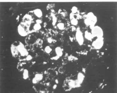
Renal biopsy was performed in 35 patients (47 biopsies), but in two (Cases 6 and 20) one tissue specimen was unavailable for light microscopy. Morphological findings of 45 biopsies are semiquantitatively reported in Table 4. Three biopsies from two patients (Cases 30 and 41) showed normal renal histology. Mesangial proliferative glomerulonephritis was observed in 15 biopsies from 13 patients. In 27 out of 45 biopsies (22 patients) a membranoproliferative glomerulonephritis was present (Fig. 1). Thirty biopsies showed a cell accumulation in glomerular capillary loops. In 12 out of 30 biopsies mononuclear cells were the only or pre-dominant in glomerular capillaries. Sometimes these cells were filled by large pro-tein droplets and occluded capillary lumina (Fig. 2). In 16 biopsies hyaline thrombi were found in the glomeruli (Figs. 1 and 2), in four patients (Cases 1,5,32 and 34) the thrombi were very extensive and occluded most of the glomerular capillaries. Crescents were present in six biopsies (Cases 1, 10, 11, 17, 25 and 29), but they
T A B L E 4 . Semiquantitative analysis of renal biopsy at light microscopy 0 0 Numbe r o f Numbe r o f Obsolescen t paucn t tlomeni h |loroerut i Wopue j Intracaptllar y Crescent s proHferauo n Pdymorptao - Mono - Increueo f Redupbcatior t Sub - Imra - Foca l nuclea r nuctea r tnesanfia l ofbweme m axkxheba j hjmini ] sclerou s leukocyt e cefl i matri i membran e depotu j thromb i Tubula r Tubula r Inlcr -vacuo - atroph y sdlia l Irzauo n Infiltra -tio n later - Vessel s VascuEU s stitia l sclerosi s sclerou s Hototoftca l patter n I I II 3 4 J I II III 6 I 14 10 10 8 13 15 20 1 0 II 12 13 17 18 19 Inadequat e spedme n 0 0 0 0 10 13 10 50 8 6 10 9 10 2 0 I Inadequat e specime n 2 1 22 1 II III 23 24 25 26 27 I II 2 8 29 1 II 3 0 I II 3 1 32 34 35 37 38 4 0 I II 4 1 42 5 0 7 23 45 23 7 60 20 2 0 6 10 10 24 23 5 3 0 8 IS 10 14 20 20 14 12 7 20 Membranoprotircrau\ * G N Mesangia l prouferaliv c G N McmbranoprouTcrativ e G N Membranoprolireratlv e G N Meianpa l prouferairv c G N Mesangia l protiferauv e G N MembranoprouTeratlv e G N Membranoproltfcrativ e G N Membranoproureraliv e G N Membranoprouferativ e G N MembranoproUferatlV e G N MejnbranoproJtferativ c G N Membranoprohferallv c G N MembranoproWenuv e G N Membranoproliferativ e O N MembranoproWerBtlv e O N Meunfia l proWcriliv e G N Mesanila l protUcraUv e C N MembranoprouTeratlv e G N Mesaniia l prouTcratlv e O N MembranoprouTerativ c O N MembranoprouTerativ e G N MembranoproUterativ e G N Mcsanfia l prouTeratlv e G N Mesantia l prouferauv e O N Membranoprohferauv c O N Mesantia l prooferau\ T G N Mesanaia l probferauv e G N Mesanfia l proWerativ e G N MembrtnoproUfcrauv e O N MembranoproflTcraUv x O N Membranoprohferauv T O N Norma l Norma l Mesaniia l protu*eradv e G N MembranoprouTerativ c G N Membranoprohferauv e G N Membranoprotiferativ e O N Mesangta l prohferauv e O N MesanAta l prouTeratlv e O N MembranopeoaTcradv e G N Membranoprorifcratlv t O N Norma l Mesan(la l prouferauv e O N MembranoprouTerativ e G N O Q 3
a.
Renal Disease in Cryoglobulinaemia
FIG. 1. Case No. 9. Membranoproliferative glomerulonephritis with marked increase of mesangial matrix. Fibrinoid thrombi occluding some loops. (Masson thrichrome x 320.)
FIG. 2. Case No. 8. Part of a glomerulus showing massive hyaline thrombi in capillary lumen (*). In other loops the lumina are occluded by mononuclear cells filled by hyaline
10 A. Tarantino and others
were focal and segmental. Tubular necrosis was found in four patients (Cases 1,
17, 32 and 34). Interstitial inflammation was observed in 24 biopsies; in two
patients (Cases 22 and 29) lymphoid follicles were present.
Vasculitis with fibrinoid necrosis of the wall and infiltration of polymorpho-nuclear and monopolymorpho-nuclear cells involved focally the small arteries, arterioles and less frequently interlobular arteries in eight patients (Cases 8, 10, 11, 23, 28, 29, 38 and 44) (Fig. 3). A marked intimal fibrosis of interlobular arteries was observed in ten patients (Cases 11,17, 22, 23, 25,27,38, 40, 41, and 44).
Serial biopsies
Ten out of 35 patients had repeated kidney biopsies, and 12 such biopsies were performed 10 to 50 months after the initial one. Two serial biopsies were carried out in eight patients (Cases 1,6,9, 20, 27, 29, 30 and 40), and three in two patients (Cases 5 and 22). In Cases 6 and 20 renal tissue was not available for histology in the first biopsy. Six patients showed identical morphologic pictures in successive biopsies. In Case 1, membranoproliferative glomerulonephritis in the first biopsy changed to mesangial proliferative glomerulonephritis in the second; and vice versa in Case 5.
An increase in tubular atrophy, interstitial fibrosis and glomerular obsolescence was found in four patients (Cases 22,27,29 and 40).
Immunofluorescence studies
Thirty-three renal biopsies from 29 patients were examined by immuno-fluoresence. Diffuse granular deposits of immunoglobulins and complement
Fio. 3. Case No. 11. Severe necrotizing artheritis involving a small artery. Part of a glomerulus showing typical lesions of membranoproliferative type. (PAS x32O.)
Renal Disease in Cryoglobulinaemia 11
FIG. 4. Immunofluorescence studies of case No. 6. Diffuse granular deposits irregularly distributed on peripheral capillary walls. (Anti-IgM serum x32O.)
T A B L E 5. Semiquantitative evaluation of renal biopsy at immunofluorescence
No. M D Clq C4 C3 Fibrin Thrombi Vessels
1 II 3 4 5 D 6 I II 8 10 12 13 17 19 20 I II 21 22 23 24 25 26 27 28 29 I II 30 I II 31 32 34 38 41 42 44 Total A, G,M M,C3 A, G, M G,M G,M A , G , M , C l q 1 1 1 1 1 1 G, M A, G, M, D, E G . M . C l q M G,C3 M,C3 G M . C l q M G,M M G G,M G M A,G,M,C3 85% 36% 90% 33% 29% 47% 33% 90% 24% G,M,C4,Clq M,C3,Clq
12 A. Tarantino and others
FIG. 5. Immunofluorescence study of case No. 32. Large thrombi in many capillary lumina and scanty deposits along glomerular basement membranes. (Anti-IgM serum x280.)
were found in all but four biopsies (Table 5). IgG, IgM and C3 were found in about 90 per cent of cases, the other immunoglobulins and complement com-ponents in 29 to 47 per cent of cases. Clq and/or C4 deposits were found in 11 out of 22 biopsies in which these components were simultaneously assessed. Deposits were mainly localized along the glomerular basement membranes (Fig. 4). In two biopsies (Cases 30 and 41) the deposits were only in the mesangial region. Fluorescence from immuneglobulin was generally more intense than complement (Figs. 5 and 6). In 10 biopsies intraluminal thrombi were the most characteristic
FIG. 6. Immunofluorescence study of the same case illustrated in Fig. 5. Complement deposits are present only on peripheral capillary loops. (Anti-Clq serum x28O.)
Renal Disease in Cryoglobulinaemia 13
T A B L E 6. Electron microscopy findings
Patient Hmotogic*! Polymorpho- Lymphocytes Mooocytej Mooocytes SubendotheEal No. pattern aucteir mkb targe crysulkcd
teococyta droplet* Deposits with Mesanfk! latraJiumm] crysuSoid 3 8 9 10 13 17 32 37 41 MP MP + MP MP + MP + MP + MP + mp + N
MP - MerobnnoprohTerarive G N , mp - Mesangia] profifenlive G N . N - Norm*] gloraerub or structure, t In flomenilar loopj or mterstitU] vessels.
* Obsolescent loops, t With crysulkxd
feature and again the staining with immunoglobulins always predominant. Complement component staining was slight if present at all. Four different patterns of deposit distribution were recognized: (1) Intraluminal thrombi, alone or in association with scanty parietal deposits. (2) Peripheral subendothelial deposits. (3) Mesangial deposits only. (4) No deposits.
In 14 specimens granular deposits were present in the arterioles or small arteries walls. In nine instances immunoglobulins only were detected, in the remaining
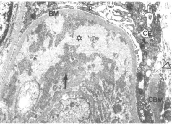
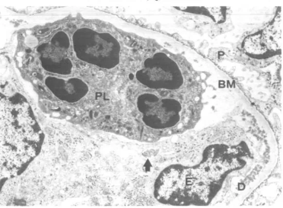
FIG. 7. Glomerular capillary loop with massive subendothelial deposits exhibiting a crystalloid structure (D) and part of a hyaline thrombus (T). E = erythrocytes, P =
14 A. Tarantino and others
specimens C3 was seen four times and Clq two times as well. Fine granular deposits of immunoglobulins were observed rarely in the interstitial tissue.
Electron microscopy
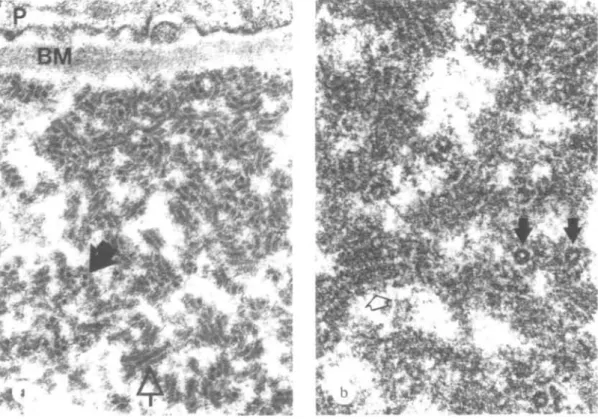
Ten kidney specimens obtained from 10 patients were examined (Table 6). In one patient, showing minimal glomerular lesions at light microscopy, an increase of mesangial matrix with electron dense mesangial deposits was observed. In the other patients in addition to the glomerular abnormalities evaluated by light microscopy, the following ultrastructural characteristics were identified: (1) A variable amount of crystalloid deposits was located in subendothelial position with or without mesangial interposition (Fig. 7). (2) The crystalloid deposits were often surrounded by areas of amorphous, weakly osmiophilic and translucent material, attributed to deposit degradation (Fig. 8). (3) Circulating aggregates of crystalloid material were found in two cases in glomerular and peritubular capillaries (Fig. 9) and arterioles. (4) Hyaline (protein) thrombi, e.g. giant crystalloid aggregates, occluded capillary lumina (Fig. 7). (5) The crystalloid material consisted of straight or slightly curved pairs of cylinders with a faint cross striation. In cross section they appeared as annular bodies (Fig. 10a). In vitro cryprecipitates showed an identical structure (Fig. 10b). (6) Non-crystalloid deposits were found in subendothelial
FIG. 8. Glomerular capillary loop with a large subendothelial deposit The strongly osmiophilic parts of the deposit ( ^ ) exhibit a crystalloid structure, the more translucent central parts (*) are amorphous (sign of a deposit dissolution?). Defect (<=>) in Bowman's capsule basement membrane (CBM). P = podocyte, CE = capsular epithelium, BM =
Renal Disease in Cryoglobulinaemia 13
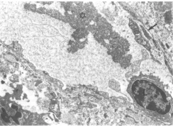
position in all cases, and in the mesangium on three occasions. (7) Monocytes, polymorphonuclear leucocytes and in some cases lymphocytes were present in variable number in glomerular capillaries. They were often found in close contact to crystalloid deposits, and seemed sometimes to be engaged in deposit phago-cytosis and degradation (Figs. 11 and 12). (8) Monocytes containing highly osmiophilic, proteic droplets were present in most cases. These proteic droplets never showed a crystalloid structure (Fig. 12). (9) Virus particles were never seen in glomerular deposits or in the in vitro precipitates.
(/) Follow-up data
Three patients {Cases 33, 35 and 36) were lost to follow-up. The clinical course of 41 patients is summarized in Fig. 13. Two patients {Cases 32 and 34) died within one month after admission during an episode of acute nephritic syndrome. Thirty-nine patients were followed for a period ranging from three to 146 months (mean 53-8). Total follow-up time was 2099 months. Ten patients showed a complete and prolonged remission {Cases 15,16, 22, 23, 25, 29 to 31, 38 and 41). At the time of diagnosis isolated haematuria or proteinuria was present in six out of 10 patients, while the presenting symptoms were proteinuria and haematuria in two other patients, nephrotic or acute nephritic syndrome in two patients. One of these patients {Case 29) died of cerebral haemorrhage 97 months after the onset of extrarenal symptoms.
FIG. 9. Peritubular capillary with a large circulating aggregate with crystalloid structure (*). Scanty subendothelial deposits (••). L = lymphocyte. (EM x 7600.)
A. Tarantino and others
FIG. 10. Crystalloid structure of deposit in the glomemlus (a) and identical structure in the
in vitro cryoprecipitate (b) of the same patient. The deposits consist of straight or slightly
curved paired cylinders (<=» (up to 1 fim long) and annular structures (H)- Between the cylinders a faint cross striation can be discerned. P = podocyte, BM = basement membrane.
(EM x 13500.)
In 16 other patients {Cases 2, 3, 6, 9, 11, 13, 14, 18, 19, 21, 24, 28, 37, 40, 43 and 44) renal function remained normal despite the persistence of urinary symp-toms. Three patients {Cases 11, 14 and 19) in this group died of cerebral hae-morrhage, sepsis and cachexia after 84, 75, and 51 months respectively. The remaining 13 patients experienced several clinical manifestations including acute renal failure (four episodes), acute nephritic syndrome (seven episodes), and nephrotic syndrome (seven episodes), sometimes followed by periods of complete remission. Three of them developed chronic renal failure and died of heart failure
{Cases 12 and 39) or hyperkalaemia {Case 27), 72, 120, 134 months after the
onset of extrarenal symptoms, and 72, 30 and 128 months after the onset of renal manifestations. Patient 27 was the only one who was treated by chronic haemo-dialysis. Three other patients {Cases 17, 26 and 42) died of myocardial infarction, acute B hepatitis, and sepsis (Table 7). The duration of EMC was 156, 88 and 56 months. The remaining seven patients {Cases 1,4, 5, 7, 8,10 and 20) had normal renal function with persistence of urinary abnormalities.
In summary, 19 cumulative episodes of acute renal failure or nephritic syndrome were observed in 14 patients. Four patients {Cases 17, 32, 34 and 42) died during acute renal disease, but the cause of death was not strictly related to renal failure.
Renal Disease in Cryoglobulinaemia 17
FIG. 11. Glomerular capillary loop with numerous subendothelial deposits (D). A poly-morphonuclear leukocyte (PL) lying immediately upon the basement membrane and an activated endothelial (?) cell (E) are possibly engaged in deposit phagocytosis ( ^ ) and degradation. P = podocyte with fusion of foot processes, BM = basement membrane,
E = activated endothelial (?) cell. (EM x97OO.)
In the remainder, normal renal function was achieved. Twenty-two patients showed an increase in blood pressure at the time of diagnosis. During follow-up six other patients (Cases 12, 14, 26, 27, 40 and 42) became hypertensive. All the hypertensive patients required vigorous antihypertensive therapy; in six patients (Cases 3, 4, 6, 12, 26 and 39) poor control was achieved despite adequate therapy.
The actuarial survival rate of 44 patients calculated from the onset of symptoms was 93 and 75 per cent at five and 10 years respectively (Fig. 14). No significant relationship could be detected between mortality rate and some clinical and T A B L E 7. Causes of death
Sepsis
Cerebral haemorrhage Heart failure
Myocardial infarction Acute HbsAg + hepatitis Hyperkalaemia Cachexia Total 4 (Patients no. 14, 32, 34, 42) 2 (Patients no. 11, 29) 2 (Patients no. 12,39) 1 (Patient no. 17) 1 (Patient no. 26) 1 (Patient no. 27) 1 (Patient no. 19) 12 out of 44 patients (28%)
18 A. Tarantino and others
r
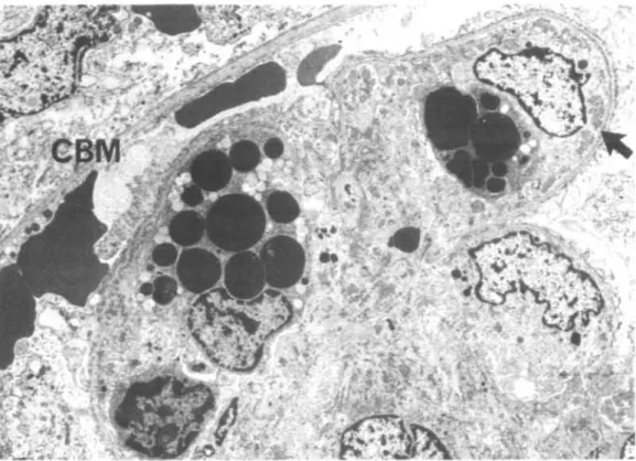
F I G . 12. The glomerular capillary loops are occluded by monocytes (M) stuffed with osmiophilic (protein) droplets. Numerous subendothelial deposits ( ^ ) are in immediate
contact with the monocytes. CBM = capsular basement membrane. (EM x37OO). morphological parameters such as mode of presentation, hypertension, intra-luminal thrombi, membranoproliferative glomerulonephritis, mononuclear cell infiltration.
(g) Treatment
In many patients treatment varied widely during the course of the syndrome. Table 8 shows the initial treatment. Three patients (Cases 15, 18 and 31) were T A B L E 8. Long-term effect of different immunosuppressive schedules in essential
mixed cryoglobulinaemia (a) (*)
to
(d) to Total No. ofpatiemi 3(15,18,31) 9(14, 16, 19,23, 32,39,41,43,44) 3(11,21,40) 11(17,20,22,24, 25, 27-30, 34, 37) 15(1-10, 12, 13, 26, 38, 42) 41 Treatment Untreated Oral corticoiteroidi alone Cytotoxic agents Oral corticosteroids phucytotoxic agents Oral corticosteroids plus
cytotoxic agents and Lv. MRf pulses Improved 1(15) 3(16,23,44) — 5 (20, 22, 25, 28,30) 8 ( 1 , 2 , 4 , 5 , 7 , 8, 10, 38) 17 Unchanged 2(18,31) 2(41,43) 2(21,40) 2 (24, 37) 4 ( 3 , 6 , 9 , 13) 12
Death, with or without chronic renal failure
_ 4(14, 19,32,39') 1(11) 4(17, 27*, 29, 34) 3(12*, 26,42) 12 * Chronic renal failure.
Renal Disease in Cryoglobulinaemia 19
T*wa 40 30 20 10 5 • 1
dl«flno«4t
— I —
11
ONSET OF SYSTEKIC SYCTT0B3 D ONSET OP RHIAL 3TVPT0KS H TREATED HTPERTEH3I0H -REFRACTORT HTPERTERSIOT ^ ~ REIIAL BIOPSY A EECTRAnOPROLIPSRATIVE COT MP LTSANOIAL HiOLIPrRATIVi OT mp HY3T0I0GICAL PATTERJI N
l _ l ROHIIAL PLASVJl CREATIHIIIE AND URinALYSIS H i PROTEimmiA ( > 0 . 2 s / 2 4 h o u r s ) ^ HAEUATURIA
H HASWTURIA AND PROTEINURIA t J NEPHHOTIC SYKDROITE H I ACUTE IHPHRITIC ST1I3R0L3 B ACUTE REITAL FAILURE
• cinonic HSNAL FAILURE
20 A. Tarantino and others 100 90 <70
j
D _i40 30 10 42 42 40 •v[7 2P^A i V 1816V 1 \ JO 7 \o FROM THE ONSET OF SYMPTOMS
. FROM THE TIME OF DIAGNOSIS
1 5 6 7 i
YEARS HT 15 20 25
FIG. 14. Actuarial survival rates from the onset of symptoms and from the diagnosis of EMC. The bars indicate the standard error on the estimated survival, calculated according to Peto, Pike, Armitage, Breslow, Cox, Howard, Mantel, McPherson, Peto, and Smith (1977). The number of patients still being followed at each point are indicated below
and above the survival curves respectively.
hcparin 200 mg/day o leukeran 10-j mg/day 5 j prednisone 10 mg/d»y 5 i.v pulses 0 plasma crcalinine m M o l / l 0.2-0.1 I proteinuna On 0 -Vl \\ +-lhaemaluria 2^ , "ft' + 10+ + + i-1^""; scrum complement 100 i 50-0 FM No 10 " ^ CM OO
FIG. 15. Clinical course in patient No. 10, treated by different drugs including intravenous methylprednisolone pulses.
Renal Disease in Cryoglobulinaemia 21
never treated; nine patients {Cases 14,16,19, 23, 32, 39, 41, 43 and 44) received low doses of oral prednisone only; in three patients {Cases 11, 21 and 40) immunosuppressive drugs, azathioprine or cyclophosphamide, were used; a combination of oral prednisone (0-5-1 mg/kg/day) and immunosuppressive agents was given to 11 patients {Cases 17, 20, 22, 24, 25, 27 to 30, 34 and 37). Fifteen patients {Cases 1 to 10,12, 13, 26, 38 and 42) were given in addition to oral prednisone and immunosuppressants, three intravenous doses of 1 g methyl-prednisolone at the beginning of treatment, repeated in most cases. Eleven patients
{Cases 1, 2, 4 to 8, 10, 12, 13 and 42) also received dipyridamole and heparin.
Four patients underwent plasmapheresis {Cases 1,6,13 and 26). The results with intravenous high dose methylprednisolone have been impressive; the most interesting finding being the dramatic improvement in the extrarenal signs such as fever, purpura, arthralgia and abdominal pain, within 24 hours of administration. Figure 15 illustrates the clinical course in one patient {Case 10) treated with intravenous methylprednisolone.
DISCUSSION (a) Clinical features
The mode of presentation described here, purpura, arthralgia, and weakness, is typical of EMC and similar to those of other series (Jori and Buonanno, 1972; Levo, Gorevic, Kassab, Tobias, and Franklin, 1977). There was a slight pre-ponderance of females, and the onset of symptoms generally occurred in the third or fourth decade of life. There is evidence that EMC is closely related to Waldenstrom's macroglobulinaemia and other haematopoietic malignancies (Invernizzi, Pioltelli, Cattaneo, Gavazzeni, Borzini, Monti, and Zanussi, 1979). Repeat bone marrow examinations did not show any underlying lymphopro-liferative disease in any of our patients, except one, who developed Waldenstrom's macroglobulinaemia three years after the diagnosis of EMC.
Kidney involvement is frequent in EMC and may lead to a wide spectrum of manifestations such as: renal tubular acidosis (LoSpalluto, Dorward, Miller, and Ziff, 1962), nephrotic syndrome (Porush, Grishman, Alter, Mandelbaum, and Churg, 1969; Bengtsson, Larsson, Lindstedt, and Svalander, 1975; Zimmerman, Dreher, Burkholder, Goldfarb, and Weinstein, 1976), acute renal failure (Feizi and Gitlin, 1969; Golde and Epstein, 1968; Lapes and Davies, 1970; Martinez and Kohler, 1971), acute nephritic syndrome (Cordonnier, Vialtel, Martin, Renversez, Chenais, Micouin, and Stoebner, 1977), papillary necrosis (Koelz and Bourke, 1977), and minimal urinary abnormalities only (Klein, Van Rood, Van Furth, and Radema, 1968). In our series the presentation of renal disease was rather heterogeneous. Most of the patients had haematuria and/or proteinuria, eight patients presented with the nephrotic syndrome, six patients with acute nephritis, and two with acute renal failure. In eight out of 44 patients plasma creatinine was above 150 //mol/1 at the first examination. Even if arterial hyper-tension is a frequent finding in EMC, surprisingly this has not been noted in previous reports. In this series 22 patients had diastolic hypertension on admission and six others became hypertensive during the follow-up period.
22 A. Tarantino and others
(b) Aetiology
The aetiology of EMC is unknown. A specific antibody activity against parasitic, bacterial, mycotic, and viral agents has been occasionally found in individual cases (Williams and Kunkel, 1972; Kaplan and Parker, 1966; Wager, Mustakallio, and Rasanen, 1968; Bonomo and Dammacco, 1971; Kaufman and Mclntosh, 1971; Gamble and Ruggles, 1978). Moreover the demonstration of antibodies against denatured or native DNA and the absence of rheumatoid factor activity in some patients suggested that both IgG and IgM could be antibodies directed against unknown antigens (Bluestone, Goldberg, Cracchiolo, and Barnett, 1970; Roberts and Lewis, 1978).
Recently, the high incidence of HBs viral antigen as well as HBs antibodies both in the serum and cryoprecipitate of patients affected by EMC was emphasized (Levo, Gorevic, Kassab, Zucher-Franklin, and Franklin, 1977; Bombardieri, Paoletti, Ferri, Di Munno, Fornai, and Giuntini, 1979). HBsAg particles were also found by electron microscopy in four cryoprecipitates. These data would imply that HBsAg is an aetiological factor of EMC. However several other authors never detected HBsAg in patient sera (Zlotnick, Slavin, and Eliamkim, 1972; Bartlow, Oyama, Jng, Miller, Economou, Rennie, and Lewis, 1975; Cream, 1976; Zimmerman et al., 1976; Cordonnier et al., 1977; Koelz and Bourke, 1977). In 14 renal biopsies, studied by immunofluorescence technique, we found strong staining for anti-HBsAg serum in six cases. However this staining was completely abolished by previous blocking of rheumatoid activity in tissue specimens and no staining was achieved by anti-HBsAg (Fab)2 serum (Maggiore, L'Abbate, Bartolomei, Caccamo, Barbiano di Belgiojoso, Tarantino, and Colasanti, to be published). These results indicate that the staining with HBsAg antiserum was related to the presence of rheumatoid factor bound in renal tissue, therefore the search for antigens in tissue must include rheumatoid activity blocking. Furthermore, virus particles were found neither in the glomerular deposits, nor in in vitro cryoprecipitates, using electron microscopy.
Finally the reports mentioned above do not take into account the possibility that the patients might have been transfused. One of our two patients with HBsAg in serum had received many blood transfusions, and the other one died from acute B hepatitis, after repeated plasmapheresis. Therefore HBsAg may be regarded as a consequence rather than a cause of EMC.
(c) Renal pathology
The light microscopy features were those of a diffuse proliferation of glomeruli in all but three renal specimens; it was surprising that the commonest lesion was a typical picture of membranoproliferative glomerulonephritis (22 out of 35 patients). The majority of renal specimens from EMC patients, reported in the literature, show glomerular changes compatible with this histological pattern (Skrifvars, Tallqvist, and Tornroth, 1973; Morel-Maroger and Mery, 1974; Zlotnick and Rosenmann, 1975; Cordonnier et al., 1977). Other morphological features such as fibrinoid thrombi, the crystalloid structure of deposits, monocytic infiltration and necrotizing vasculitis deserve more detailed discussion.
Renal Disease in Cryoglobulinaemia 23
Fibrinoid or hyaline thrombi in gloraerular capillary loops have been previously considered as a characteristic finding of EMC nephritis (Verroust, Mery, Morel-Maroger, Clauvel, and Richet, 1971; Zimmerman et ah, 1976; Zlotnick and Rosenmann, 1975). Generally their presence paralleled more severe renal impairment Our studies showed that fibrinoid thrombi found in one third of biopsies, contained mostly immuneglobulins, while complement components were less often present and fibrin was absent. In contrast to classic immune complex disease, glomerular deposits of EMC nephropathy had a predominantly crystalloid or fibrillar ultrastructure that may be considered as a unique finding (Cordonnier, Martin, Grosslambert, Micouin, Chenais, and Stoebner, 1975; Feiner and Gallo, 1977; Ogihara, Saruta, Saito, Abe, Ozawa, Kato, and Sakuguchi, 1979). In our cases, deposits with the crystalloid structure were mainly localized in sub-endothelial sites and rarely in the mesangium as well as free in the lumina of glomerular capillary loops, peritubular capillaries, and arterioles. This material is composed of straight or curved cylinders and annular structures in cross sections. We were unable to detect intracellular crystals or crystalloid material previously described in glomerular cells of a few cases of EMC or monoclonal paraprotein-aemia with nephritis (Porush et ai, 1969; Bengtsson et al., 1975; Monga, Mazzucco, Coppo, Piccoli, and Coda, 1976).
It is also noteworthy that in vitro cryoprecipitates and immunoglobulins present in glomeruli shared the same ultrastructural pattern. Moreover the ultrastructural features of both circulating cryoglobulins and glomerular deposits could vary according to the different heavy chain classes of immunoglobulins (Cordonnier et al, 1977; Feiner and Gallo, 1977; Gallo and Feiner, 1978).
In EMC a monocytic infiltration of glomerular capillary loops was first reported by Monga et al., (1976) and Mihatsch, ZoUinger and Imbasciati (1978). Mono-nuclear cells were found in the majority of our renal specimens.
These cells, stuffed with numerous droplets of an homogeneous and electron-dense material, might contribute to the occlusion of capillary loops. Similarly several recent studies pointed out that the presence of macrophages in glomeruli contributes significantly to the glomerular hypercellularity in experimental glomerulonephritides (Kalowski, McKay, Howes, Csavossy, and Wolfson, 1976; Holdsworth, Thompson, Glasgow, Dowling, and Atkins, 1978; Kondo, Shige-matsu, and Kobayashi, 1972; Schreiner, Cotran, Pardo, and Unanue, 1978). The significance and the role of monocytes in EMC will be discussed below.
(d) Pathogenesis of tissue damage
The demonstration of the immune complex nature of mixed cryoglobulins led to the postulate that tissue damage and clinical manifestations were due to the circulating IgG and IgM complexes and complement activation. Evidence for such a mechanism was supported by several observations such as: the demonstration of immunoglobulins and complement components in glomeruli and skin vessels (Miescher, Paronetto, and Koffler, 1965; Douglas, Lahav, and Fudenberg, 1970; Doe, Evans, Hobbs, and Booth, 1972; Nightingale, Solez, and Humphrey, 1977); the presence of complement components in cryoprecipitates (Golberg and Barnett,
24 A. Tarantino and others
1970; Cream, Howard, and Virella, 1972); the anticomplementary activity of cryoglobulinaemic sera or isolated cryoglobulins (Balasz and Frolich, 1966; Rother, Rother, Flad, and Miescher, 1972; Muller, Rother, and Westerhausen, 1976); hypocomplementaemia (Riethmuller, Meltzer, Franklin, and Miescher, 1966; Linscott and Kane 1975); and the production of vasculitis after the injection of isolated cryoglobulins or rheumatoid factor in experimental animals (Baum, Stastny, and Ziff, 1964; Herd, 1973).
We found a strong reduction of mean serum Clq, Cls and C4 levels in 30 patients. C3 levels did not differ from normal controls and mean C5, C9 and C3PA concentrations were higher than normal (see Table 3). In 13 out of 18 patients the lowering of C4 and/or Clq, irrespective of clinical course, persisted during the whole period of the study. This complement profile, already described in EMC, has been related to a complement activation through classical pathway. Further support for this contention arises by observation that the same comple-ment abnormalities were associated with an acquired Cl Inhibitor deficiency in several cases of EMC (Casali, Borzini, Pioltelli, Invernizzi, and Zanussi, 1978). In all our cases C1 INH concentrations were higher than normal, thus excluding the possibility that Clq and C4 reduction could be referred to Cl INH deficiency. Moreover C3 and C3PA breakdown products were never detected.
On the other hand immunofluorescence studies showed C3 deposits in 27 out of 33 biopsies, whereas C l q and/or C4 deposits were only present in 47 and 37 per cent of renal specimens respectively. The staining for complement components was fainter than that of immunoglobulins. Now, if the complement profile was due to complement activation, an immunofluorescence pattern similar to that observed in diffuse proliferative lupus nephritis should be expected: i.e., bright deposits of Clq and C4 in most cases according to the reduction of these serum complement components (Verroust, Wilson, Cooper, Edington, and Dixon, 1974; Wyatt, Me Adams, Forristal, Snyder, and West, 1979).
These data support our previous hypothesis that hyposynthesis rather than activation and consumption is the cause of the marked lowering of early reacting complement components (Tarantino et al., 1978).
Lastly two main immunofluorescence patterns were recognized: (1) deposits along glomerular basement membranes, and (2) intraluminal thrombi with scanty, if any, parietal deposits. On electron microscopy the intraluminal thrombi were constituted by crystalloid material in the lumina of capillary loops as well as in other kidney vessels. These findings suggest the possibility that physicochemical rather than immunological mechanisms may be involved in the trapping of protein aggregates by the kidney.
Though the mechanisms for the in vivo cryoprecipitation has not been yet elucidated, several factors have been proposed (Grey and Kohler, 1973; Abraham, Podel, Wistor, Johnston, and Welch, 1979). Amongst them, cooling seems occasionally to induce vasculitis manifesting as purpura or urticaria (Wiltink, Esseveld, Gerbrandy, and Van Eijk, 1973); however clinical manifestations were seldom related to the cold exposure (Brouet, Clauvel, Danon, Klein, and Seligman, 1974). At any rate, if glomerular thrombi form after a cold challenge on the
Renal Disease in Cryoglobulinaemia 25
extremities, it is unlikely that the protein aggregates reach the kidney or deeper tissues in spite of the higher central blood temperature.
Protein concentration seems to be one of the most important variables which affects cold precipitation. Indeed, the higher the protein concentration, the higher the temperature at which cryoprecipitation may occur (Lockwood, 1979). Since a pronounced increase in serum viscosity after concentration of plasma in glomeruli takes place, physicochemical characteristics of cryoglobulins could account for the presence of intraluminal thrombi in kidney vessels.
In this series a monocytic infiltration was detected in the majority of renal specimens studied by electron microscopy. Although the role of these infiltrating cells is unknown, one might speculate that they have a scavenger function akin to that occurring in wound healing. Such a possibility is confirmed by the observation of homogeneous electron dense droplets in monocytes, similar to macrophages actively phagocyting the debris. In addition these cells have been reported to phagocytose mesangial deposits in experimental immune complex nephritis (Striker, Mannik, and Tang, 1979). Alternatively, monocytes may represent another mediator of tissue damage. In fact, Cardella, Davies, and Allison (1974) stressed that the release of lysosomal enzymes by activated macrophages causes effects similar to the neutrophil mediated tissue damage. Moreover macrophage infiltration may suggest a participation of cell-mediated immune mechanisms other than classic immune complex deposition in the pathogenesis of the glomerular lesions and widespread vasculitis. This would be consistent with the presence of monocytes in and around the vessels in several idiopathic vasculitis (Waksman, 1971; Soter, 1976). A more intriguing possibility is that the accumulation of mononuclear cells may contribute per se to the proliferation of glomerular cells; indeed factors secreted by macrophages induce proliferation of cultured fibroblasts and vascular endothelium (Leibowich and Ross, 1976; Polverini, Cotran, Gimbrone and Unanue, 1977). The almost constant presence of proliferative lesions in cryoglobulinaemic nephritis might be related to monocytes.
Whatever the role of monocytes, the simultaneous presence of intraluminal thrombi and monocytes suggests that tissue damage may be initiated by circulating IgM-IgG complexes. Subsequently their deposition in glomeruli stimulates the arrival of mononuclear cells. During the phagocytosis of the cryoproteins the release of some enzymes by macrophages as well as the raised protein concen-tration and/or other haemodynamic factors in the kidney cortex might trigger a massive cryoprecipitation.
(e) Prognosis
The follow-up of 39 patients extended from three to 146 months (mean 53-8). Ten patients showed a longstanding remission of renal manifestations and 16 patients maintained normal renal function in spite of persistent urinary abnor-malities. Conversely 13 patients experienced several syndromes as acute renal failure, nephrotic and acute nephritic syndrome, followed sometimes by complete remission. Some patients developed more than one of these syndromes during the
26 A. Tarantino and others
period of observation. A similar propensity to acute exacerbations and apparent spontaneous remission of renal disease characterizes cryoglobulinaemic nephritis.
In previous reports progressive scarring of glomeruli was found in serial biopsies (Morel-Maroger et al, 1974) and a high mortality rate has been observed in patients with acute nephritic episodes. Therefore an ominous prognosis has been related to the appearance of renal impairment in EMC. Despite these earlier experiences, some other reports emphasized that progressive renal failure may be reversible (Verroust et al., 1971; Morel-Maroger and Verroust, 1974; Ponticelli, Imbasciati, Tarantino, and Pietrogrande, 1977). Amongst 50 patients, reviewed in the literature, with histologically proven diffuse glomerulonephritis, only two underwent regular dialysis (Bartlow et al., 1975; Cream, 1976), showing, thus a better prognosis than that of primary or systemic proliferative glomerulo-nephritides such as in systemic lupus erythematosus (Cameron, Glasgow, Ogg, and White, 1970; Habib, Kleinknecht, Gubler, and Levy, 1973; Hecht, Siegel, Adler, Kashgarian, and Hayslett, 1976; Baldwin, Gluck, Lowenstein and Gallo,
1977).
In the present series 12 patients died. Renal failure was directly responsible for death only in one patient {Case 2 7). Furthermore, recovery of kidney function was achieved in nine out of 13 patients with acute renal failure or acute nephritic syndrome. Chronic renal failure developed in three patients but only one required regular dialysis. The cumulative survival rate in our patients, all of them showing kidney disease, was 75 per cent at 10 years. No difference was detectable between patients with or without renal manifestations at the onset of the disease.
Most of our patients died of cardiovascular disease. This may be accounted for by the high incidence of arterial hypertension which proved to be refractory to treatment in about one fifth of our patients. Similarly most of the patients reported in the literature with an unfavourable outcome had severe hypertension; although the prognostic role of hypertension has never been stressed before, we think that the prognosis in EMC is determined more by the development of severe hyper-tension, than kidney damage.
( / ) Treatment
Since the course of EMC often extends over several years with unpredictable remissions and exacerbations, therapy remains a puzzling problem. Different approaches have been used such as corticosteroids (Klein et al., 1968; Skrifvars
et al., 1973; Reza, Roth, Pops, and Goldberg, 1974; Gamble and Ruggles, 1978),
cytotoxic drugs (Rigo, Leval-Rutten, and Salmon, 1974), penicillamine (Goldberg and Barnett, 1970), heparin (Martinez and Kohler, 1971), splenectomy (Mathison, Condemi, Leddy, Callerame, Panner, and Vaughan, 1971), plasmapheresis (Clark and Long, 1979), or a combination of these measures (Golde and Epstein, 1968; Zlotnick et al., 1972; Slatopolski, 1976; Ristow, Griner, Abraham, and Shoulson, 1976). The value of these methods of treatment is difficult to assess since a controlled study is lacking; however the clinical impression is that they are not effective.
Renal Disease in Cryoglobulinaemia 27
described in immune complex glomerular diseases (Cathcart, Scheinberg, Idelson, and Couser, 1976; Levinski, Cameron, and Soothill, 1977; Lockwood, Rees, Pinching, Pussel, Sweny, and Peters, 1977; Ponticelli, Tarantino, Pioltelli, and Invernizzi, 1977; Bolton and Couser, 1979) and in one case of monoclonal cryoglobulinaemia with acute renal failure (Ponticelli et al., 1977a) suggesting that intravenous high doses of steroids might be useful in EMC too. In addition to combined immunosuppressive agents and steroids, we repeatedly used intravenous methylprednisolone pulses in 15 patients with severe renal and/or extrarenal symptoms. In 11 of them we added antiplatelet drugs and heparin on the assump-tion that cryoglobulins may activate platelets (Cortellaro, Lambertenghi-Deliliers, Cofrancesco, Pogliani, Pozzoli, Imbasciati, and Praga, 1975). A dramatic remission of extrarenal symptoms such as fever, purpura, arthralgias and abdominal pain was obtained soon after the methylprednisolone infusion. Also in eight out of 15 patients renal manifestations (proteinuria, haematuria, and plasma creatinine) showed some improvement. The mortality rate was lower— even if non-significantly—in patients treated with methyl prednisolone.
B I B L I O G R A P H Y
ABRAHAM, G. N., PODELL, G. N., WISTOR, R., J R . , JOHNSTON, S. L., and WELCH, E. A., 1979.
Clin. exp. Immunol. 36,63.
ADAM, C , MOREL-MAROGER, L., and RICHET, G., 1973. Kidney Int. 3,334.
ALEXANIAN, R., 1975. Arch, intern. Med. 135,62. ALLEN, J. A., 1966. Fed. Proc. 25, 726.
ALPER, C. A., 1966. Ada med. scand. 179 (Suppl. 445), 200.
ANDREJAK, M., BARIETY, S., BEDROSSIAN, J., CALLARD, P., DRUET, P., DUBOUST, A., IDATTE,
J. M., and KUHN, J., 1978. Transplantation 26,446.
BALASZ, V., and FR6HLICH, M. M., 1966. Amer. J. med. Sci. 89, 51.
BALDWIN, D. S., GLUCK, M. C , LOWENSTEIN, J., and GALJLO, G. R., 1977. Amer. J. Med. 62, 12. BARNETT, E. V., BLUESTONE, R., CRACCHIOLO, A., GOLDBERG, L. S., KANTOR, G. L., and
MCINTOSH, R. M., 1970. Ann. intern. Med. 73,95.
BARTLOW, B. G., OYAMA, J. H., JNG, T. S., MILLER, A. W., ECONOMOU, S. G., RENNIE, I. D. B.,
and LEWIS, E. J., 1975. Nephron 14,309.
BAUM, J., STATSNY, P., and ZIFF, M., 1964. / . Immunol. 93,985.
BENGTSSON, V., LARSSON, O., LINDSTEDT, G., and SVALANDER, C , 1975. Quart. J. Med. NS. 175,
491.
BLOCK, K. J., BUCHANAN, W. W., WOHL, H. J., and BUNIN, J. J., 1965. Medicine {Baltimore) 44,187.
BLUESTONE, R., GOLBERG, L. S., CRACCHIOLO, A., and BARNETT, E. V., 1970. Int. Arch. Allergy 39,16.
BOLTON, W. W , and COUSER, W. G., 1979. Amer. J. Med. 66,495.
BOMBARDIERI, A., PAOLETTL P., FERRI, C , D I MUNNO, O., FORNAL, E., and GIUNTWI, C ,
1979. Amer. J. Med. 66, 748.
BONOMO, L., DAMMACCO, F., TURSI, A., and TRIZIO, D., 1970. Clin. exp. Immunol. 6,531.
1971. Clin. exp. Immunol. 9, 175.
BROUET, J. C , CLAUVEL, J. P., DANON, F., KLEIN, M., and SELIGMAN, M., 1974. Amer. J. Med. 57, 775.
CAMERON, J. S., GLASGOW, E. F., OGG, C. S., and WHITE, R. H. R., 1970. Brit. med. J. n, 7.
CARDELLA, C. J., DAVIES, P., and ALLISON, A. C , 1974. Nature 247,46.
CASALI, P., BORZINI, P., PIOLTELLI, P., INVERNIZZI, F., and ZANUSSL C , 1978. Acta hematol. 59,277.
CATHCART, E. S., SCHEINBERG, M. A., IDELSON, B. A., and COUSER, W. G., 1976. Lancet i, 163.
CHRISTIAN, C. L., HATFIELD, W. B., and CHASE, P. A., 1963. J. clin. Invest. 42,823. CLARK, R. A. F., and LONG, J. C , 1979. New Engl. J. Med. 300,610.
28 A. Tarantino and others
CONTE, J., BLANC, M., MIGNON-CONTE, M., ABBAL, M., and ORFILA, C , 1974. J. Urol. Nephrol.
80, 773.
CORDONNIER, D., MARTIN, H., GROSSLAMBERT, P., MICOUIN, C , CHENAIS, F., and STOEBNER, P.,
1975. Amer.J.Med. 59,867.
VIALTEL, P., MARTIN, H., RENVERSEZ, J. C H . , CHENAIS, F., MICOUIN, C , and STOEBNER,
P., 1977. Actualites Nephrologiques de Hopital Necker, p. 349. Flammarion Medicine-Sciences, Paris.
CORTELLARO, M., LAMBERTENGHI-DELILIERS, G., COFRANCESCO, E., POGLIANL, E., POZZOLI, E., IMBASCIATI, E., and PRAGA, C , 1975. Ada haemal. 54, 36.
CREAM, J. J., HOWARD, A., and VIRELLA, G., 1972. Immunology, 23,405.
1976. Quart. J. Med. N.S. 178,255.
CUPRAK, L. J., STOLLAR, B. D., KRITZMAN, J., and Liss, M., 1970. Immunochemistry 7, 199.
D O E , W. F., EVANS, D. J., HOBBS, J. R., and BOOTH, C. C , 1972. Gut 13, 112.
DOUGLAS, S. D., LAHAV, M., and FUDENBERG, H. H., 1970. Amer. J. Med. 49,274.
DROUET, P., LEUTOUNTURIER, P., CONTET, A., and MANDET, C , 1973. Clin. exp. Immunol.
15,483.
FAKUNLB, Y. M., ONYEWOTU, I. I., GREENWOOD, B. M., MOHAMMED, I., and HOLBOROW, E. J.,
1978. Clin. exp. Immunol. 31,55.
FEINER, H., and GALLO, G., 1977. Amer. J. Pathol. 88,145. FEIZI, T., and GraiN, N., 1969. Lancet, ii, 873.
FLORIN-CHRISTENSEN, A., ROUX, M. E. B., and ARANA, R. M., 1974. Clin. exp. Immunol. 16,
599.
GAMBLE, C. N., and RUGGLES, S. W., 1978. New Engl. J. Med. 299,81.
GALLO, G., and FEINER, H., 1978. In Cryoproteins, p. 139. Chenais, Villars de Lans. GOLDBERG, L. S., and BARNETT, E. V., 1970. Arch, intern. Med. 125,145.
G O L D E , D., and EPSTEIN, W., 1968. Ann. intern. Med. 69, 1222.
GREY, H. M., and KOHLER, P. F., 1973. Sent. Haemal. 10, 87. GRISWOLD, W. R., and BRADY, R., 1978. J. Lab. clin. Med. 92,423. GRUPE, W. E., 1968. Pediatrics, 42,474.
HABIB, R., KLEINKNECHT, C , GUBLER, M. C , and LEVY, M., 1973. Clin. Nephrol. 1,194.
HANAUER, L. B., and CHRISTIAN, C. L., 1967. J. clin. Invest. 46,400.
H E C H T , B., SIEGEL, N., ADLER, M., KASHGARIAN, M., and HAYSLETT, J. P., 1976. Medicine
(Baltimore) 55, 163.
HERD, Z. L., 1973. Immunology 25,931.
HOLDSWORTH, S. R., THOMPSON, N. M., GLASGOW, E. F., DOWLING, J. P., and ATKINS, R. C ,
1 9 7 8 , / exp. Med. 147,98.
HURWITZ, D., QUISMORIO, F. P., and FRIOU, G. J., 1975. Clin. exp. Med. 19,131.
INVERNIZZI, F., PIOLTELLI, P., CATTANEO, R., GAVAZZENI, V., BORZINI, P., MONTI, G., and
ZANUSSI, C , 1979. Ada haemal. 61,93.
JORI, G. P., and BUONANNO, G., 1972. Gut 13,610.
D'ONOFRIO, F., TIRELLI, A., GONNELLA, F., and GENTILE, S., 1977. Gut 18, 245. KALOWSKI, S., M C K A Y , D. G., HOWES, E. L., J R . , CSAVOSSY, I., and WOLFSON, M., 1976.
Nephron 16,415.
KANTOR, G. L., GOLDBERG, L. S., JOHNSON, B. L., JR., DERECHIN, M. M., and BARNETT, E. V.,
1970. Ann. intern. Med. 73,553.
KAPLAN, M. E., and PARKER, C. W., 1966. / . Lab. clin. Med. 68,885. 1968. J. Lab. clin. Med. 71, 754.
KAUFMAN, D. B., and MCINTOSH, R., 1971. Amer. J. Med. 50, 262.
KLEIN, F., VAN ROOD, J. J., VAN FURTH, R., and RADEMA, H., 1968. Clin. exp. Immunol. 3, 703.
KOELZ, A. M., and BOURKE, E., 1977. Nephron 19,242.
KONDO, Y., SHIGEMATSU, H., and KOBAYASHI, Y., 1972. Lab. Invest. 27,620.
LAPES, M. J., and DAVIS, J. S., 1970. Arch, intern. Med. 126, 287. LEIBOWICH, S. J., and Ross, R., 1976. Amer. J. Pathol. 84, 501.
LEVINSKY, R. J., CAMERON, J. S., and SOOTHILL, J. F., 1977. Lancet i, 564.
LEVO, Y., GOREVIC, P. D., KASSAB, H. J., TOBIAS, H., and FRANKLIN, E. C , 1977. Ann. intern.
Med. 87,287.
ZUCHER-FRANKLIN, D., and FRANKLIN, E. C , 1977. New Engl. J. Med. 296,