HAL Id: inserm-00108751
https://www.hal.inserm.fr/inserm-00108751
Submitted on 28 Sep 2009HAL is a multi-disciplinary open access archive for the deposit and dissemination of sci-entific research documents, whether they are pub-lished or not. The documents may come from teaching and research institutions in France or abroad, or from public or private research centers.
L’archive ouverte pluridisciplinaire HAL, est destinée au dépôt et à la diffusion de documents scientifiques de niveau recherche, publiés ou non, émanant des établissements d’enseignement et de recherche français ou étrangers, des laboratoires publics ou privés.
an Elderly Population: Results of the EVA Study.
N Tasnime Akbaraly, Henri Faure, Veronique Gourlet, Alain Favier, Claudine
Berr
To cite this version:
N Tasnime Akbaraly, Henri Faure, Veronique Gourlet, Alain Favier, Claudine Berr. Plasma Carotenoid Levels and Cognitive Performance in an Elderly Population: Results of the EVA Study.. Journals of Gerontology, Series A, Oxford University Press (OUP): Policy B - Oxford Open Option D, 2007, 62 (3), pp.308-16. �inserm-00108751�
Plasma carotenoids levels and cognitive performances in an elderly population N.T Akbaralya, Msc; H Faureb, PhD; V Gourletc, PhD; A Favierb, PhD; C Berra, MD, PhD.
(a)
Inserm, E361, Montpellier, 34000 France ; Université Montpellier1, Montpellier, F-34000 France;
(b)
Département de biologie intégrée. CHU de Grenoble, Grenoble, France
(c)
INSERM, U708, Paris, F- 75000 France; Université Paris 6, Paris, F-75000, France Corresponding author
N.Tasnime AKBARALY
INSERM E0361, Hôpital La Colombière, 39 Avenue Charles Flahault, BP 34493, 34093 Montpellier, Cedex 5, France
Tel: 33 (0)4 99 614 569, Fax : 33 (0)4 99 614 579 akbaraly@montp.inserm.fr
Abstract
Background: The hypothesis of carotenoids having a preventive role in cognitive impairment is suggested by their antioxidant properties.
Methods: We examined, in a cross sectional analysis, the relationship between cognitive performances (assessed by the Mini Mental Status Examination: MMSE, Trail Making Test part B: TMTB, Digit Symbol Substitution: DSS, Finger Tapping Test: FTT and Word Fluency Test: WFT) and different plasma carotenoids (lutein, zeaxanthin,
β-cryptoxanthin, lycopene, α-carotene, trans and cisβ-carotene), in a healthy elderly population: the EVA study “Etude du Vieillissement Artériel” (n=589, 73.5 3 years).
Results: Logistic regression showed that subjects with the lowest cognitive functioning (<25th percentile) had a higher probability of having low levels of specific plasma carotenoids
(<1st quartile): lycopene and zeaxanthin. For zeaxanthin ORDSS=1.97 [1.21; 3.20],
ORFTT=1.70 [1.05; 2.74] and ORWFT =1.82 [1.08; 3.07]), for lycopene ORDSS =1.93 [1.20;
3.12] and ORTMTB=1.64 [1.04; 2.59].
Conclusion: Even if it is not possible to affirm if these low levels of carotenoids precede or are the consequence of cognitive impairment, our results suggest that low
carotenoid levels could play a role in cognitive impairment. The biological significance of our findings needs further research.
Keywords:
Plasma carotenoids Cognitive performances Elderly population
1. Introduction
Among the leading causes of cognitive impairment, an increase in brain oxidative stress is well documented (1). In fact, the brain is particularly prone to free radical attacks owing to its relatively low antioxidants content, a considerable amount of polyunsaturated fatty acid chains in the neuronal membrane lipids and its high oxygen consumption rate (2). The hypothesis of carotenoids having a preventive role in cognitive impairment is suggested by their ability to trap peroxyl radicals and their singlet oxygen quenching properties, which enables them to prevent lipid peroxidation (3, 4). Epidemiological studies (5-9) and clinical trials (10, 11) on cognitive impairment and plasma carotenoids mainly concern β-carotene, which is a major carotenoid. But some biological studies showed that antioxidant activity of other carotenoids could be more effective than β-carotene activity (12, 13). This study’s aim is to examine the relationship between cognitive performances and a large variety of
carotenoids including xanthophylls (lutein, zeaxanthin, β-cryptoxanthin) and carotenes (lycopene, α-carotene, transβ-carotene and cisβ-carotene) in a healthy elderly population.
2. Materiel and methods 1. Study population
The EVA study (“Etude du Vieillissement Artériel”) is a nine-year longitudinal study with 6 waves of follow-up (14). During the first two years 1991-1993 (EVA0), 1389
volunteers (574 men and 815 women) born between 1922 and 1932 (mean age = 65) residing in the town of Nantes (Western France) were recruited from electoral rolls, and to a lesser extent, via information campaigns. The sixth and last follow-up of the EVA study (EVA6) was conducted between June 2000 and December 2001. During the 9-year follow-up, one hundred and one deaths occurred. Cancer was the first leading cause of death (n=45, 44.5%), the second one was cardiovascular diseases (n=22, 21.8%). The main factors related to mortality were found to be, as reported in the literature: male gender, smoking (current and ex-), alcohol intake, medication use, obesity, diabetes, hypertension and cardiovascular diseases (15). At EVA6, the blood samplings after a 12-hour fast were obtained for 773 subjects. Subjects who did not completed the whole study (n=616, 44.3%) were significantly more frequently men (those who not completed: 44.3% vs. those who completed: 38.9%, p=0.04)), obese or overweight subjects (57.9% vs. 47.3%, p=0.0004) and those who had hypertension (52.6% vs. 47.2%, p=0.05). They were statistically more frequently subjects in the lowest cognitive performances class (<25th percentile of the distribution) for MMSE
(25.2% vs. 18.2%, p=0.002), DSS (28.3% vs. 20.3%, p=0.0006), TMT B (30.5% vs. 21.4%, p=0.0002) and WFT (25.1% vs. 19.3%, p=0.009), but we did not observe that for FTT (p=0.37).
The present analysis was restricted to the 589 subjects who underwent at EVA6 a cognitive evaluation and blood sampling. The study protocol was approved by the Ethical
Committee of the University Center Hospital of Kremlin-Bicêtre, Paris. Signed informed consent was obtained from all participants at enrolment.
2. Data collection
Cognitive evaluation and depressive symptoms
Trained neuropsychologists evaluated cognition with a neuropsychological battery of tests including a global test, the Mini-Mental State Examination (MMSE) (16), and an
assessment of a range of cognitive domains. Visual conceptual and visuomotor tracking were assessed by the Trail Making Test part A (TMTA) and B (TMTB) (17). Involving motor speed and attention functions, the trail making test is highly vulnerable to the effects of brain injury (18). The A form is considered as exploring motor speed and control and working memory whereas the B form assesses executive function such as set shifting. The variables of interest are the time in seconds (19). These tests (part A and B) were performed with a
maximum allotted time to perform the test of 180 and 240 seconds respectively. When subjects exceeded the time allotted for each Part, the maximum allotted time was imputed. The Digit Symbol Substitution (DSS) from the Wechsler Adult Intelligence Scale-Revised (WAIS-R) measured sustained attention and logical reasoning (20). Manual dexterity and psychomotor speed were evaluated with the Finger Tapping Test (FTT). Verbal fluency was evaluated with the Word fluency Test (WFT). Depression symptoms were assessed by the Centre of the Epidemiological Studies-Depression (CESD) scale using score (score=17 for men and score=23 for women) for high risk of depression to define depressive
Carotenoids measurements
Retinol and carotenoids were measured with a Biotek-Krontron HPLC system (UVK Lab – Trappes France), which consists in a 525 dual pump, a 465 auto-sampler and a 540 diode array detector. Retinol and β-carotene were purchased from Fluka (Sigma-France – L’Isle d’Abeau), and other carotenoids were provided by Hoffman-Laroche (Hoffman-Laroche – Bâle Switzerland) as a generous gift. The LC separation was run with an Alltech
Adsorbosphere C18 column 150 x 4.5 mm ID, 3 µ particle size (Alltech-France), which was thermostated at 28° C with a 402 column oven. Carotenoids and retinol were measured by HPLC after 2 extractions with a hexane/tetrahydrofurane mixture. For the quantization we used the Steghens method (22) with minor modifications. Indeed, we used a single 150 mm long column instead of 2, and we added 10 ppm water in the mobile phase A to improve the separation of retinol, lutein and zeaxanthin. The laboratory participates in the NIST (National Institute for Standards and Technology – New York) external quality assurance program, and ChromSystems internal controls are analyzed in every series of measurements (one control every 10 unknown serums) and in the SFVB quality assurance programmes. The limits of detection were calculated as 5 fold the maximum baseline noise in the region of the peaks. Hence we found limits of detection of 0.05 µM for retinol, and 0.02 µM for carotenoid. All concentrations of retinol, lycopene and β-carotene were above these respective limits, while only 5% of lutein, 8% of zeaxanthin, 2% of β-cryptoxanthin were under.
Total plasma carotenoids levels were obtained by summing levels of lutein, zeaxanthin, β-cryptoxanthin, lycopene, α and β-carotenes.
Questionnaire and medical examination
The general questionnaire allowed us to obtain information on socio-demographic factors such as sex, age, educational achievement, plus consumption habits like tobacco status, alcohol consumption (which was determined from the subject’s estimated average
amount of alcoholic beverages ingested weekly) and medication use. In addition, height and weight were measured. Two independent measures of systolic and diastolic blood pressure were made with a digital electronic tensiometer after a 10-minute rest. Total plasma cholesterol, and plasma glucose levels were also measured using routine methods. The apolipoprotein E genotype of subjects was determined on DNA samples.
3. Statistical Methods
The characteristics of the 589 subjects included in the analysis were described compared to the 184 subjects who had blood sampling but not the cognitive evaluation at EVA6, results were expressed by percentage and means with their standard deviation (SD). To test the differences between these two groups, Chi square test and the Student T test were used. These characteristics comprised sex, age, educational achievement (≤ primary school /
high school), smoking status (current, ex-smokers /non-smokers), alcohol consumption (< 20ml / 20ml per day) and medicine use <3/ 3 per day). Health characteristics were body mass index (BMI) classes (underweight: BMI<21kg/m² (23);”normal weight”: 21BMI<25; overweight: 25BMI<30 (24); obesity30 kg/m² (24)), diabetes (plasma glucose level 7.80 mmol/L or use of anti-diabetic drugs or diabetes medical history), dyslipidemia (total
cholesterol 6.20 mmol/L or use of lipid-lowering drugs or dyslipidemia medical history), hypertension (systolic or diastolic blood pressure 140 or 90 mm Hg respectively, or use of hypertensive drugs or hypertension medical history), history of vascular diseases
(self-reported history of myocardial infarction, angina pectoris, stroke or use of vascular drugs), the depressive symptomatology (yes/no) and apolipoprotein E genotype (ε4 allele +/-).
To calculate the correlation between carotenoids, we calculated Pearson correlation coefficients on log transformed carotenoids.
The graphic representation of the percentage of subjects with low cognitive
functioning for the different carotenoids levels showed that the relation between cognition and these biological variables were not linear. It brought us to consider them as categorical
variables. We compared the characteristic of subjects for the different levels in carotenoids (<25th percentile versus 25th percentile) by using the Student T-Test or Chi square test for both continuous and categorical variables.
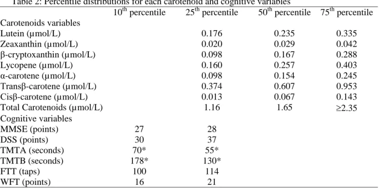
To define subjects with the lowest cognitive performances in this well-educated
cohort, we chose two cut-offs: subjects who had cognitive test scores below the 25th percentile
(and above 75th for TMTB), and subjects who had cognitive test scores below the 10th
percentile (and above 90th for TMTB). Values for these 25th and 10th percentile cut-offs for
each test are presented in Table 2.
Classical multivariate logistic regressions were performed to test associations between probability of subjects to have the lowest cognitive functioning and levels of plasma
carotenoids (<25th percentile vs. >25th percentile) adjusting on all potential confounding
variables. Results were expressed by odds ratios (OR) with their 95% confidence intervals (CI). All interactions between each carotenoid and each consumption habits and health variables were calculated and were not statistically significant. Statistical analyses were performed using SAS software version 9.1 (SAS Institute, Inc. Cary, North Carolina).
Results
Characteristics of the 589 EVA subjects (Table1)
The 589 subjects of EVA study (361 women and 228 men, aged 73.5 2.9), on which, we carried the analysis, were well educated (52.1% had a high school or superior degree). Among them, 39.0% were smokers or ex-smokers, 24% consumed alcohol regularly (2glass/day). For the repartition of subjects between BMI classes, we observed 13.8% of underweight, 34.0% of overweight and 8.5% of obese. Concerning health status, 8.3% showed depressive symptomatology, 8.1% were diabetics, 65.0 % were dyslipidemic, 78.8% had hypertension, 19.7 % had a history of cardiovascular diseases and 20.5% carried at least one allele 4 of the apolipoprotein E. For the different carotenoids and scores of
neuropsychologic tests, concentrations and ranges are described in table 1, and percentile distributions in table 2.
We also compared the characteristics of these 589 subjects included in the analysis to the 184 subjects for which we only obtained a blood sample but not a cognitive evaluation. Results showed that these 184 subjects were significantly older (74.23.1 vs. 73.5 2.9), proportions of subjects with hypertension (p=0.05) or with a history of cardiovascular diseases (p=0.03) were higher. On the other hand, proportions of subjects with low levels in
-cryptoxanthin, trans and cis -carotene were significantly lower in this group (results not showed).
Description of carotenoids: correlation and associated factors
Plasma carotenoids were highly and significantly inter-correlated. The highest correlations were found among carotenes: -carotene/ trans-carotene (r=0.78), trans
(r=0.58). The other correlation coefficients ranged from 0.16 for lycopene/ -cryptoxanthin to 0.50 for trans-carotene/ lycopene.
In Table 3a and 3b, we describe the characteristics of subjects according to their total plasma carotenoid, the different xanthophylls and carotenes levels (<25th percentile vs. 25th percentile). We observe that the profile of factors associated to -carotene, trans-carotene and cis -carotene were identical. Between the other carotenoids, however, the associated factors can differ. Gender was associated to all carotenoids, which reach higher levels in women with the exception of zeaxanthin. Tobacco status and alcohol consumption were significantly associated to lower levels for total plasma carotenoids, -cryptoxanthin, -carotene, trans and cis -carotene. Diabetes was associated to low levels of all carotenoids, while hypertension were significantly associated to low levels of lutein, -carotene, trans and cis -carotene. Obesity and overweight were associated to low levels of all carotenoids excepted for zeaxanthin and -cryptoxanthin. Age, education, depressive symptomatology, dylipidemia, history of cardio-vascular diseases and apolipoprotein E genotype were not associated with any plasma carotenoids.
Association between cognition and carotenoids
Table 4 showed results of crude association between cognitive performances and carotenoid levels, obtained by univariate logistic regression analyses. Subjects with the lowest cognitive performances (neuropsychologic tests scores <25th percentile) had a higher
probability of having low levels of some carotenoids (level < 1st quartile). Significant
associations were observed between zeaxanthin and all cognitive tests excepted MMSE (for TMTA OR= 1.66 [1.08; 2.55], for TMTB OR=1.60 [1.04; 2.44], for DSS OR=1.87 [1.21; 2.89], for FTT OR=1.70 [1.10; 2.62] and for the WFT OR=1.87 [1.16; 3.00]). Low levels of
(OR=2.02 [1.32; 3.11]). A significant association was found between low performances in TMTB and low levels of total plasma carotenoids and trans-carotene (OR=1.57 [1.03; 2.40] and OR=1.58 [1.04; 2.41]). After taking into account socio-demographic factors (sex, age, education), consumption habits (tobacco, alcohol), diabetes, hypertension and BMI classes (Table5), associations between zeaxanthin and cognitive tests remained statistically
significant for TMTA (OR=1.67 [1.06; 2.66]), DSS (OR=1.92 [1.18; 3.14]), FTT (OR= .69 [1.05; 2.72]) and WFT (OR=1.80 [1.07; 3.05]) but not for TMTB (OR=1.51 [0.95; 2.39], p=0.08). Lycopene remained associated to TMTB (OR=1.54 [0.97; 2.43], p=0.06) and DSS (OR=1.85 [1.14; 2.98]). The other associations between carotenoids and cognitive
performances observed in the crude analyses did not remained statistically significant after adjustment. Total plasma carotenoids, -carotene and -carotene (trans or cis) levels were not statistically associated to low cognitive performances, neither were lutein and
-cryptoxanthin.
Sensitivity analysis
The same analyses were first performed, by removing subjects with depressive symptomatology (n=49), then by removing subjects who were underweight (BMI<21kg/m²) (n=81) and thirdly by adjusting on levels of plasma retinol. In the three situations, we obtained similar results. We also performed analyses on subjects who had a MMSE score 25 (n=570), to exclude subjects with potential clinically significant cognitive impairment (n=19). Results were identical (data not showed).
On the other hand, we performed models which explain the probability to have cognitive tests scores <10th percentile of the distribution scores for the different plasma
carotenoids levels. Only associations between zeaxanthin and FTT (p=0.003) and TMTB (p=0.06), and lycopene and DSS (p=0.07) remained marginally statistically significant (data
not showed). In these analyses, the number of subjects, which had the lowest cognitive performances was 2.2 to 2.8 smaller than for the precedent analyses (n=54 for MMSE, 49 for DSS, 55 for TMTA, 53 for TMTB, 52 for FTT and 38 for the TEL). It comes from a lack of statistical power, which could explain the drop in statistically significant results.
All our analyses were conducted with dichotomized variables for both the plasma carotenoids and the cognitive outcomes; in supplemental analyses, we tested if similar findings were observed when a continuous measure for cognition was used. We calculated Spearman correlation coefficients between cognitive variables and log-transformed
carotenoids. Results showed a significant association for zeaxanthin and lycopene and DSS (r=0.08, p=0.01 and r=0.11, p=0.006 for zeaxanthin and lycopene respectively), TMTA (r=-0.11, p=0.01 and r=-0.08, p=0.05), TMTB (r=-0.08, p=0.05 and r=-0.12, p=0.004) and FTT (r=0.09 p=0.04 and r=0.09 p=0.04). These correlations confirm that our findings were not driven by the chosen dichotomous classifications for cognition and carotenoids levels.
Discussion
To our knowledge, this study is the first one, which investigated, in a healthy elderly population, the relation between cognitive performances measured by five neuropsychological tests and the different plasma carotenoids: xanthophylls (lutein, zeaxanthin, β-cryptoxanthin) and carotenes (lycopene, α-carotene, trans β-carotene and cis β-carotene).
The EVA study included volunteers with higher educational status, higher incomes and greater cognitive function than the average elderly French population. Despite this selection, plasma carotenoids concentrations in the EVA study population were in the same ranges as those in different European or American populations (4). In this present study low levels of specific plasma carotenoids - lycopene and zeaxanthin- were associated to poor cognitive functioning in a highly educated free-living elderly community.
Carotenoids are found in coloured fruits and vegetables. Some studies showed that plasma carotenoids levels could be related to dietary fruits and vegetables intake (25, 26). More specifically, it seems that mango, papaya, peaches, prune, squash, orange, green fruits and vegetables were source of zeaxanthin, while tomatoes, pink grapefruit, and watermelon are sources of lycopene (3). Two cross sectional studies (27, 28) showed an association between greater intake of fruits and vegetables and better cognitive performances. In a large prospective study of older women (n=13388) (29), the authors reported relation between low vegetables intake and cognitive decline but no relation with fruits, the strongest association being with greater intake of green leafy vegetables and cruciferous vegetables. Even if some studies show that intakes of foods with high carotenoid contents were correlated with their corresponding plasma concentrations (26), intake of fruits and vegetables are sources of many other nutrients which have been associated to cognitive functions : vitamin E (30), folates (31, 32), flavonoids (33). It is now impossible to know if the association between carotenoids and
cognitive functions is the results of a specific effect of carotenoids or if it is the resultant of combined effects of the different fruits and vegetables compounds.
For each cognitive test, we tested eight-association hypothesis (Ho) between cognitive functions and carotenoids. In order to ensure that significant observed associations were not hazard related (alpha risk=5%), specific multiple test (Bonferroni, Sidak) corrections were applied to data. Considering, however, that the tested hypothesis are not independent, submitting them to these corrections entails an overcorrection of threshold significance thus not necessarily leading to rejection of the Ho hypothesis, which we know to be false. In this exploratory analysis, the fact that one specific carotenoid was associated to more than one cognitive function, and that these associations remained statistically significant after
controlling on potential confounding factors, or after removing some subjects (subjects with depressive symptomatology, subjects with a MMSE score <25 or undernourished subjects) seems to us more likely to ensure that our significant observed associations were not hazard related.
Although there were important correlations among all carotenoids, we observed significant associations of low cognitive performances with some but not all carotenoids. More specifically, we found no associations between β-carotenes (trans or cis) or α-carotene, which are the most studied carotenoids in epidemiological literature on cognitive impairment (5-11). These studies give conflicting results. Previously, in the EVA study, we showed that a low level of baseline total plasma β-carotene (<25th percentile) was not significantly
associated to a 4-year cognitive decline (5). In a cross-sectional study, Perrig et al. (9) showed that a higher β-carotene plasma level was associated to better memory performances (free recall, recognition and vocabulary) in 442 healthy persons aged 65 to 94. Studying dietary intake of β-carotene, Morris et al. (8) found no association between carotene intake and cognitive decline, while the Rotterdam Study showed that a lower intake of β-carotene was
associated with impaired cognitive function measured by MMSE (7). Three studies
investigated the link between cognitive performances and supplementation of antioxidants including β-carotene (6, 10, 11). All, except the work of the Age-Related Eye Diseases Study Research Group (11), found that use of these supplements reduced the risk of cognitive decline. In these studies with multi-antioxidant supplementation it is, however, impossible to isolate the specific effect of β-carotene on cognitive impairment. Only one study, concerning 1769 subjects, focused on the association of a large spectrum of carotenoids and cognitive performances in elderly subjects without neuropsychiatric disease, but it found no association (34). Discrepancies can be explained by methodological differences (neuropsychological tests to assess cognitive performances, number of tests used, choice of modelling of
carotenoids…). Finally we found two clinical epidemiological studies (35, 36) which focused on the comparison of plasma carotenoids and retinol levels in elderly patients with or without Alzheimer disease (AD). One showed that levels of vitamin A, lutein, zeaxanthin,
β-cryptoxanthin and α-carotene were lower in AD patients (n=63) than controls (n=56). However, they found no difference for lycopene and β-carotene (36). In the second study, levels of carotenoids were lower in AD patients (n=40) than in controls (n=39) for zeaxanthin, β-cryptoxanthin, lycopene and β-carotene but not for lutein and α-carotene (35). These
conflicting results could be explained by the limited sample size of these studies. The major problem for interpreting these two studies is that they are studying AD cases for which nutritional habits, and consequently, biological status, can be modified, as a consequence of the disease progression. The different measurement methods and the bioavailability of
carotenoids, which is influenced by several factors such as characteristics of the food sources, interactions with other dietary factors and various subject characteristics (3) could also explain the differences between studies.
Many epidemiological studies have associated high carotenoid status with a decrease in the incidence of chronic diseases (heart diseases and cancer), the biological mechanism for such protection is, however, currently unclear (3). Multiple possibilities exist, amongst them, certain carotenoids can be converted to retinoid and have a pro-vitamin A activity. To assess if the relation we showed could be explained partly by this hypothesis, we adjusted our models on retinol concentration, but results remained unchanged.
Our results are supported by those of a biological study on oxidation of carotenoids by free radicals led by Woodall et al (37), which found that lycopene, lutein and zeaxanthin all reacted rapidly with oxidising agents and must also be considered as potential dietary
antioxidants. A possible explanation for low levels of plasma carotenoids in AD or cognitive impairment is that they might be consumed because of higher rate of free radical production in the brain.
Our results on the relation between carotenoids and cognitive performances were not modified when we excluded the subjects with BMI <21 kg/m² for which the hypothesis of under nutrition is probable. This allows us to say that this relation could not be limited to the influence of undernourished subjects. However, in this cross sectional frame-work, it is not possible to affirm if these low levels of carotenoids preceded or were the consequence of cognitive impairment in a context where poor cognitive status may be a risk factor for poor nutrition. In this study, we used various tests to explore cognitive performances and this approach is more powerful than using only MMSE, which is not a very sensitive measurement of cognitive impairment. With the other tests, the psychometric scores ranges are large and more powerful to study cognitive impairment. Beside, because the scores were not normally distributed, a percentual cut-off is appropriate. The observed association will probably have no functional significance yet, because participants had only a subtle impairment. Moreover, we have no basis to expect specific association between carotenoids and psychometric
evaluation. However, it is well known, that low plasma lutein and zeaxanthin concentrations were implicated in the age-related macular degeneration (38). While retina is a puzzle whose ultimate solution lies on the other side of the optic nerve in its connection with the brain, a highly specific accumulation of lutein and zeaxanthin in the retina and in the macula is described (39). Could other area of the brain have the same affinity for some specific carotenoids? The biological significance of our finding needs further research by biological studies, longitudinal epidemiological studies and by specific clinical trials with carotenoid supplementation.
Acknowledgements
N. Tasnime Akbaraly was supported by a grant from the French Alzheimer’s disease Association.
The EVA study was carried out under an agreement between INSERM and the Merck, Sharp and Dohme-Chibret Laboratories (WestPoint, PA) and was supported by EISAI laboratory, France.
Corresponding author N.Tasnime AKBARALY
INSERM E0361, Hôpital La Colombière, 39 Avenue Charles Flahault, BP 34493, 34093 Montpellier, Cedex 5, France
Tel: 33 (0)4 99 614 569, Fax : 33 (0)4 99 614 579 akbaraly@montp.inserm.fr
References
1. Finkel T, Holbrook NJ. Oxidants, oxidative stress and the biology of ageing. Nature 2000;408:239-47.
2. Coyle JT, Puttfarcken P. Oxidative stress, glutamate, and neurodegenerative disorders. Science 1993;262:689-95.
3. Paiva SA, Russell RM. Beta-carotene and other carotenoids as antioxidants. J Am Coll Nutr 1999;18:426-33.
4. Stahl W, Sies H. Lycopene: a biologically important carotenoid for humans? Arch Biochem Biophys 1996;336:1-9.
5. Berr C, Balansard B, Arnaud J, Roussel AM, Alperovitch A. Cognitive decline is associated with systemic oxidative stress: the EVA study. Etude du Vieillissement Arteriel. J Am Geriatr Soc 2000;48:1285-91.
6. Gray SL, Hanlon JT, Landerman LR, Artz M, Schmader KE, Fillenbaum GG. Is antioxidant use protective of cognitive function in the community-dwelling elderly? Am J Geriatr Pharmacother 2003;1:3-10.
7. Jama JW, Launer LJ, Witteman JC, den Breeijen JH, Breteler MM, Grobbee DE, Hofman A. Dietary antioxidants and cognitive function in a population-based sample of older persons. The Rotterdam Study. Am J Epidemiol 1996;144:275-80.
8. Morris MC, Evans DA, Bienias JL, Tangney CC, Wilson RS. Vitamin E and cognitive decline in older persons. Arch Neurol 2002;59:1125-32.
9. Perrig WJ, Perrig P, Stahelin HB. The relation between antioxidants and memory performance in the old and very old. J Am Geriatr Soc 1997;45:718-24.
10. Smith A, Clark R, Nutt D, Haller J, Hayward S, Perry K. Anti-oxidant Vitamins and Mental Performance of the Elderly. Hum Psdycopharmacol Clin Exp 1999:459-71.
11. Yaffe K, Clemons TE, McBee WL, Lindblad AS. Impact of antioxidants, zinc, and copper on cognition in the elderly: a randomized, controlled trial. Neurology 2004;63:1705-7.
12. Stahl W, Junghans A, de Boer B, Driomina ES, Briviba K, Sies H. Carotenoid mixtures protect multilamellar liposomes against oxidative damage: synergistic effects of lycopene and lutein. FEBS Lett 1998;427:305-8.
13. Woodall AA, Britton G, Jackson MJ. Carotenoids and protection of phospholipids in solution or in liposomes against oxidation by peroxyl radicals: relationship between carotenoid structure and protective ability. Biochim Biophys Acta 1997;1336:575-86.
14. Berr C, Coudray C, Bonithon-Kopp C, Roussel AM, Mainard F, Alperovitch A. Demographic and cardiovascular risk factors in relation to antioxidant status: the EVA Study. Int J Vitam Nutr Res 1998;68:26-35.
15. Akbaraly NT, Arnaud J, Hininger-Favier I, Gourlet V, Roussel AM, Berr C. Selenium and mortality in the elderly: results from the EVA study. Clin Chem 2005;51:2117-23.
16. Folstein M, Anthony JC, Parhad I, Duffy B, Gruenberg EM. The meaning of cognitive impairment in the elderly. J Am Geriatr Soc 1985;33:228-35.
17. Robins Wahlin TB, Backman L, Wahlin A, Winblad B. Trail Making Test performance in a community-based sample of healthy very old adults: effects of age on completion time, but not on accuracy. Arch Gerontol Geriatr 1996;22:87-102.
18. Deutsch Lezak M, ed. Neuropsychological Assessment. New York, 1983:p768pp.
19. Reitan R. Validity of the Trail Making test as an indicator of organic brain damage. Perceptual and Motor Skills 1958:271 – 6.
20. Wechsler D. The Wechsler Adult Intelligence Scale-Revised. New-York, 1981. 21. Fuhrer R, Rouillon F. La version française de l'échelle CES-D. Psychiat & Psychobiol
1989;4:163-6.
22. Steghens JP, van Kappel AL, Riboli E, Collombel C. Simultaneous measurement of seven carotenoids, retinol and alpha-tocopherol in serum by high-performance liquid
23. ANAES. Évaluation diagnostique de la dénutrition protéino-énergétique des adultes hospitalisés. In: professionnelles Sdr, ed., Vol.: ANAES, 2003.
24. Obesity: preventing and managing the global epidemic. Report of a WHO consultation. World Health Organ Tech Rep Ser 2000;894:i-xii, 1-253.
25. Al-Delaimy WK, Ferrari P, Slimani N, Pala V, Johansson I, Nilsson S, et al. Plasma
carotenoids as biomarkers of intake of fruits and vegetables: individual-level correlations in the European Prospective Investigation into Cancer and Nutrition (EPIC). Eur J Clin Nutr 2005;59:1387-96.
26. Campbell DR, Gross MD, Martini MC, Grandits GA, Slavin JL, Potter JD. Plasma
carotenoids as biomarkers of vegetable and fruit intake. Cancer Epidemiol Biomarkers Prev 1994;3:493-500.
27. Lee L, Kang SA, Lee HO, Lee BH, Park JS, Kim JH, et al. Relationships between dietary intake and cognitive function level in Korean elderly people. Public Health 2001;115:133-8. 28. Ortega RM, Requejo AM, Andres P, Lopez-Sobaler AM, Quintas ME, Redondo MR, et al.
Dietary intake and cognitive function in a group of elderly people. Am J Clin Nutr 1997;66:803-9.
29. Kang JH, Ascherio A, Grodstein F. Fruit and vegetable consumption and cognitive decline in aging women. Ann Neurol 2005;57:713-20.
30. Masaki KH, Losonczy KG, Izmirlian G, Foley DJ, Ross GW, Petrovitch H, et al. Association of vitamin E and C supplement use with cognitive function and dementia in elderly men. Neurology 2000;54:1265-72.
31. Ramos MI, Allen LH, Mungas DM, Jagust WJ, Haan MN, Green R, Miller JW. Low folate status is associated with impaired cognitive function and dementia in the Sacramento Area Latino Study on Aging. Am J Clin Nutr 2005;82:1346-52.
32. Ravaglia G, Forti P, Maioli F, Martelli M, Servadei L, Brunetti N, et al. Homocysteine and folate as risk factors for dementia and Alzheimer disease. Am J Clin Nutr 2005;82:636-43. 33. Commenges D, Scotet V, Renaud S, Jacqmin-Gadda H, Barberger-Gateau P, Dartigues JF.
Intake of flavonoids and risk of dementia. Eur J Epidemiol 2000;16:357-63.
34. Schmidt R, Hayn M, Reinhart B, Roob G, Schmidt H, Schumacher M, et al. Plasma antioxidants and cognitive performance in middle-aged and older adults: results of the Austrian Stroke Prevention Study. J Am Geriatr Soc 1998;46:1407-10.
35. Mecocci P, Polidori MC, Cherubini A, Ingegni T, Mattioli P, Catani M, et al. Lymphocyte oxidative DNA damage and plasma antioxidants in Alzheimer disease. Arch Neurol 2002;59:794-8.
36. Rinaldi P, Polidori MC, Metastasio A, Mariani E, Mattioli P, Cherubini A, et al. Plasma antioxidants are similarly depleted in mild cognitive impairment and in Alzheimer's disease. Neurobiol Aging 2003;24:915-9.
37. Woodall AA, Lee SW, Weesie RJ, Jackson MJ, Britton G. Oxidation of carotenoids by free radicals: relationship between structure and reactivity. Biochim Biophys Acta 1997;1336:33-42.
38. Delcourt C, Carrière I, Delage M, Barberger-Gateau P, Schalch W, Group PS. Plasma lutein and zeaxanthin and other carotenoids as modifiable risk factors for Age-related maculopathy and cataract; The POLA study. Investigative Ophthalmology & Visual Science 2006;In press. 39. Bone RA, Landrum JT, Tarsis SL. Preliminary identification of the human macular pigment.
Tables
Table1: Demographic and clinical characteristics of the 589 subjects studied
% or mean ( SD)* Range
Sex (women) 61.3
Age 73.55 2.93 68-79
Education (High school) 52.1 Smoking Status (S, Ex-S) 39.9 Alcohol consumption (2 glasses/day) * 24.0 Medicine consumption (3/day)* 58.8 BMI classes * (kg/m²) Underweight (<21) 13.8 Overweight (25-<30) 34.0 Obese (30) 8.5 Diabetes 8.1 Dyslipidemia 65.0 Hypertension 78.8 Cardio-vascular diseases 19.7 APOE ε 4 carrier* 20.5 Depressive Symptomatology 8.3 Lutein (µmol/L) 0.27 0.15 0.05-1.29 Zeaxanthin (µmol/L) 0.032 0.021 0.01-0.34 β-cryptoxanthin (µmol/L) 0.23 0.21 0.01-2.03 Lycopene (µmol/L) 0.31 0.20 0.06-1.24 α-carotene (µmol/L) 0.19 0.15 0.02-0.93 Transβ-carotene (µmol/L) 0.73 0.52 0.04; 5.48 Cisβ-carotene (µmol/L) 0.10 0.12 0.01-1.19 Total Carotenoids (µmol/L) 1.86 0.99 0.15-8.35
MMSE (points) 28 2 5-30 DSS* (points) 45 11 12-85 TMTA* (seconds) 48.9 19.6 18-180 TMTB* (seconds) 110.4 46.3 30-240 FTT* (taps) 127 21 62-185 WFT* (points) 27 8 5-50
Table 2: Percentile distributions for each carotenoid and cognitive variables
10th percentile 25th percentile 50th percentile 75th percentile Carotenoids variables Lutein (µmol/L) 0.176 0.235 0.335 Zeaxanthin (µmol/L) 0.020 0.029 0.042 β-cryptoxanthin (µmol/L) 0.098 0.167 0.288 Lycopene (µmol/L) 0.160 0.257 0.403 α-carotene (µmol/L) 0.098 0.154 0.245 Transβ-carotene (µmol/L) 0.374 0.607 0.953 Cisβ-carotene (µmol/L) 0.013 0.067 0.143
Total Carotenoids (µmol/L) 1.16 1.65 2.35 Cognitive variables MMSE (points) 27 28 DSS (points) 30 37 TMTA (seconds) 70* 55* TMTB (seconds) 178* 130* FTT (taps) 100 114 WFT (points) 16 21
Table 3a: Characteristics of subjects according to their level of plasma total carotenoids and xanthophylls (<25th perc. versus 25th percentile)
Total carotenoids Lutein Zeaxanthin Beta-cryptoxanthin <25thperc. 25thperc. <25thperc. 25thperc. <25thperc. 25thperc. <25thperc. 25thperc.
n %* n %* p n %* n %* p n %* n %* p n %* N %* p Age* 144 73.5 2.9 445 73.6 2.9 0.71 174 73.6 3.0 442 73.5 2.9 0.92 143 73.8 3.0 446 74.5 2.9 0.32 146 73.7 2.8 443 73.5 3.0 0.46 Sex Men 80 35.1 148 64.9 <10-4 71 31.1 157 68.9 0.005 59 25.9 169 74.1 0.47 89 39.0 139 61.0 <10-4 Women 64 17.7 297 82.3 76 21.0 285 79.0 84 23.3 277 76.7 57 15.8 304 84.2 Education Primary Sch. 71 25.2 211 74.8 0.69 68 24.1 214 75.9 0.65 69 24.5 213 75.5 0.43 74 26.2 208 73.8 0.43 High school 73 23.8 235 76.2 79 25.7 228 74.3 74 24.1 233 75.9 72 23.4 235 76.5 Tobacco status No Smoker 67 18.7 292 81.3 <10-4 81 22.6 278 77.4 0.09 89 24.8 270 75.2 0.72 68 18.9 291 81.1 <10-4 Smoker or ex 77 33.5 153 66.5 66 28.7 164 71.3 54 23.5 176 76.5 78 33.9 152 66.1 Alcohol <20 ml /day 97 21.8 347 78.1 0.008 105 23.6 339 76.3 0.18 108 24.3 336 75.7 0.86 99 22.3 345 77.7 0.01 ≥20 ml /day 46 32.9 94 67.1 41 29.3 99 70.7 33 23.6 107 76.4 46 32.9 94 67.1
Medicine use <3 / day 43 17.8 199 82.2 0.001 62 25.6 180 74.3 0.71 52 21.5 190 78.5 0.21 53 21.9 189 78.1 0.19 ≥3 / day 161 29.2 245 70.81 84 24.3 262 75.7 96 26.0 256 74.0 92 26.6 254 73.4 BMI classes Kg/m² Normal 46 18.5 202 81.4 <10-4 50 20.2 198 79.8 <10-4 51 20.6 197 79.4 0.26 60 24.2 188 75.8 0.13 Underweight 9 11.5 69 88.5 9 11.5 69 88.5 19 24.4 59 75.6 12 15.4 66 84.6 Overweight 60 31.1 133 68.9 56 29.0 137 71.0 48 24.9 145 75.1 56 29.0 137 71.0 Obese 21 43.7 27 56.2 21 43.7 27 56.2 16 33.3 32 66.7 12 25.0 36 75.0 Depressive symptoms No 125 24.1 394 78.1 0.82 126 24.3 393 75.7 0.64 124 23.9 395 76.1 0.94 128 24.7 391 75.3 0.89 Yes 12 25.5 35 43.7 10 21.3 37 78.7 11 23.4 36 76.6 12 25.5 35 74.5 Diabetes No 117 21.6 424 78.4 <10-4 127 23.5 414 76.5 0.005 123 22.7 418 77.3 0.003 124 22.9 417 77.1 <10-3 Yes 27 56.2 21 43.7 20 41.7 28 58.3 20 41.7 28 58.3 22 45.8 26 54.2 Hypertension No 15 12.0 110 88.0 0.003 20 16.0 105 84.0 0.009 29 23.2 96 76.8 0.75 31 24.8 94 75.2 0.99 Yes 129 27.8 335 72.2 127 27.4 337 72.6 114 24.6 350 75.4 115 24.8 349 75.2 History of CVD No 111 23.5 362 76.5 0.26 115 24.3 358 75.7 0.46 111 23.5 362 76.5 0.35 116 24.5 357 75.5 0.76 Yes 33 28.5 83 71.5 32 27.6 84 72.4 32 27.6 84 72.4 30 25.9 86 74.1 Dyslipidemia No 44 21.4 162 78.6 0.21 53 25.7 153 74.3 0.75 52 25.2 154 74.8 0.69 57 27.7 149 72.3 0.23 Yes 100 26.1 283 73.9 94 24.5 289 75.5 91 23.8 292 76.2 89 23.2 294 76.8 Apoe4 No 107 24.5 330 75.5 0.90 103 23.6 334 76.4 0.30 100 22.9 337 77.1 0.31 110 25.2 327 74.8 0.93 Yes 27 23.9 86 76.1 32 28.3 81 71.7 31 27.4 82 72.6 28 24.8 85 75.2
Table3b : Characteristics of subjects according to their level of plasma carotenes (<25th percentile versus 25th percentile)
Lycopene -carotene Trans-carotene Cis-carotene <25thperc. 25thperc. <25thperc. 25thperc. <25thperc. 25thperc. <25thperc. 25thperc.
n %* n %* p n %* n %* p n %* n %* p n %* N %* p Age* 145 73.7 2.9 444 73.5 2.9 0.34 144 73.7 3.0 445 73.5 2.9 0.58 147 73.5 2.9 442 73.5 2.9 0.99 144 73.3 3.0 445 73.6 2.9 0.28 Sex Men 66 28.9 162 71.1 0.05 72 31.6 156 68.4 0.001 82 36.0 146 64.0 <10-4 79 34.6 149 65.3 <10-4 Women 79 21.9 282 78.1 72 19.9 289 80.1 65 18.0 296 82.0 65 18.0 296 92.0 Education Primary Sch. 73 25.9 209 74.1 0.49 63 22.3 219 77.7 0.25 70 24.8 212 75.2 0.94 67 23.8 215 76.2 0.71 High school 72 23.4 235 76.5 81 26.4 226 73.6 77 25.1 230 74.9 77 25.1 230 74.9 Tobacco status No Smoker 83 23.1 276 76.9 0.29 75 20.9 284 79.1 0.01 68 18.9 291 81.1 <10-4 67 18.7 292 81.3 <10-4 Smoker or ex 62 27.0 168 73.0 69 30.0 161 70.0 79 34.3 151 65.6 77 33.5 153 66.5 Alcohol <20 ml /day 106 23.9 338 76.1 0.43 96 21.6 348 78.4 0.002 100 22.5 344 77.5 0.009 99 22.3 345 77.7 0.02 ≥20 ml /day 38 27.1 102 72.9 48 34.3 92 65.7 47 33.6 93 66.4 45 32.1 95 64.9
Medicine use <3 / day 50 20.7 192 72.3 0.06 53 21.9 189 78.1 0.22 54 22.3 188 77.7 0.21 45 18.6 197 81.4 0.007 ≥3 / day 95 27.5 251 72.5 91 26.3 255 73.7 93 26.9 253 73.1 98 28.3 248 71.7 BMI classes Kg/m² Normal 49 19.8 199 80.2 0.03 48 19.3 200 80.7 <10-4 40 16.1 208 83.9 <10-4 51 20.6 197 79.4 0.03 Underweight 20 25.6 58 74.4 7 9.0 71 91.0 12 15.4 66 84.6 12 15.4 66 84.6 Overweight 49 25.4 144 74.6 61 31.6 132 68.4 66 34.2 127 65.8 58 30.0 135 69.9 Obese 19 39.6 29 60.4 19 39.6 29 60.4 21 43.7 27 56.2 13 27.1 35 72.9 Depressive symptoms No 128 24.7 391 75.3 0.40 130 25.0 389 75.0 0.57 131 25.2 388 74.8 0.35 124 23.9 395 76.1 0.69 Yes 9 19.1 38 80.8 10 21.3 37 78.7 9 19.1 38 80.8 10 21.3 37 78.7 Diabetes No 123 22.7 418 77.3 0.0004 121 22.4 420 77.6 <10-4 122 22.5 419 77.5 <10-4 122 22.5 419 77.4 0.000 3 Yes 22 45.8 26 54.2 23 47.9 25 52.1 25 52.1 23 47.9 22 45.8 26 54.2 Hypertension No 27 21.6 98 78.4 0.38 20 16.0 105 84.0 0.01 14 11.2 111 88.8 <10-4 17 13.6 108 86.4 0.001 Yes 118 25.4 346 74.6 124 26.7 340 73.3 133 28.7 331 71.3 127 27.4 337 72.6 History of CVD No 114 24.1 359 75.9 0.55 111 23.5 362 76.5 0.26 117 24.7 356 75.3 0.80 114 24.1 359 75.9 0.69 Yes 31 26.7 85 73.3 33 28.4 83 71.5 30 25.9 86 74.1 30 25.9 86 74.1 Dyslipidemia No 51 24.8 155 75.2 0.95 50 24.3 156 75.7 0.94 42 20.4 164 79.6 0.06 45 21.8 161 78.2 0.28 Yes 94 24.5 289 75.5 94 24.5 289 75.5 105 27.4 278 72.6 99 25.8 284 74.1 Apoe4 No 105 24.0 332 76.0 0.26 115 26.3 322 73.7 0.27 111 25.4 326 74.6 0.60 109 24.9 328 75.1 0.97
Table 4: Crude risk of low cognitive functioning (score <25th) associated to plasma carotenoids for each cognitive test.
Plasma antioxidant <25th vs. 25 th
MMSE TMTA TMTB
OR* [95%] CI* p OR* [95%] CI* p OR* [95%] CI* p Total carotenoids 0.88 0.55 ; 1.40 0.59 1.13 0.73 ; 1.76 0.58 1.57 1.03 ; 2.40 0.04 Lutein 1.00 0.63 ; 1.58 0.99 1.01 0.65 ; 1.57 0.96 1.50 0.98 ; 2.30 0.06 Zeaxanthin 1.37 0.88 ; 2.13 0.17 1.66 1.08 ; 2.55 0.02 1.60 1.04 ; 2.44 0.03 β-cryptoxanthin 1.25 0.80 ; 1.96 0.32 0.95 0.61 ; 1.47 0.81 1.07 0.69 ; 1.65 0.77 Lycopene 1.03 0.65 ; 1.62 0.91 1.37 0.89 ; 2.10 0.15 1.76 1.16 ; 2.67 0.008 α-carotene 0.83 0.51 ; 1.33 0.44 0.84 0.53 ; 1.32 0.45 1.21 0.78 ; 1.86 0.39 transβ-carotene 0.71 0.44 ; 1.15 0.17 0.98 0.63 ; 1.53 0.94 1.58 1.04 ; 2.41 0.03 cisβ-carotene 0.61 0.37 ; 1.00 0.05 1.17 0.76 ; 1.82 0.47 1.02 0.65 ; 1.58 0.94 DSS FTT TEL Total carotenoids 1.12 0.71 ; 1.76 0.62 1.22 0.78 ; 1.89 0.38 0.67 0.40 ; 1.13 0.13 Lutein 1.24 0.79 ; 1.95 0.34 1.39 0.90 ; 2.15 0.13 0.89 0.54 ; 1.47 0.65 Zeaxanthin 1.87 1.21 ; 2.89 0.005 1.70 1.10 ; 2.62 0.02 1.87 1.16; 3.00 0.01 β-cryptoxanthin 1.25 0.80 ; 1.94 0.33 0.79 0.50 ; 1.25 0.31 0.83 0.50 ; 1.36 0.46 Lycopene 2.02 1.32 ; 3.11 0.01 1.43 0.93 ; 2.21 0.10 1.16 0.72 ; 1.11 0.54 α-carotene 0.73 0.45 ; 1.18 0.20 1.11 0.71 ; 1.73 0.64 0.66 0.39 ; 1.11 0.12 transβ-carotene 0.93 0.58 ; 1.47 0.75 1.08 0.70 ; 1.68 0.72 0.72 0.43 ; 1.10 0.21 cisβ-carotene 1.24 0.79 ; 1.95 0.34 0.94 0.59 ; 1.48 0.78 0.74 0.45 ; 1.23 0.25
Table 5: Adjusted risk of low cognitive functioning (score <25th) associated to plasma carotenoids for each cognitive test.
Plasma antioxidant <25th vs. 25th
MMSE TMTA TMTB
OR* [95%] CI* p OR* [95%] CI* p OR* [95%] CI* p Total carotenoids 0.93 0.55 ; 1.58 0.80 1.27 0.77 ; 2.09 0.34 1.43 0.88 ;2.32 0.14 Lutein 1.03 0.62 ; 1.71 0.89 1.15 0.71 ; 1.88 0.57 1.51 0.94 ; 2.42 0.09 Zeaxanthin 1.24 0.77 ; 2.00 0.38 1.67 1.06 ; 2.66 0.03 1.51 0.95 ; 2.39 0.08 β-cryptoxanthin 1.39 0.85 ; 2.28 0.19 1.05 0.64 ; 1.71 0.84 1.06 0.65 ;1.737 0.82 Lycopene 0.91 0.55 ; 1.50 0.71 1.39 0.88 ; 2.21 0.16 1.54 0.97 ; 2.43 0.06 α-carotene 0.89 0.53 ; 1.49 0.66 0.96 0.59 ; 1.58 0.88 1.16 0.72 ; 1.86 0.55 transβ-carotene 0.74 0.43 ; 1.27 0.27 1.11 0.68 ; 1.83 0.67 1.48 0.91 ; 2.37 0.11 cisβ-carotene 0.56 0.32 ; 0.98 0.04 1.41 0.87 ; 2.29 0.16 0.96 0.58 ; 1.57 0.86 DSS FTT TEL Total carotenoids 0.95 0.56 ; 1.63 0.87 1.53 0.91 ; 2.55 0.11 0.66 0.36 ; 1.21 0.18 Lutein 1.25 0.75 ; 2.10 0.39 1.48 0.90 ; 2.42 0.12 0.94 0.54 ; 1.65 0.84 Zeaxanthin 1.92 1.18 ; 3.14 0.008 1.69 1.05 ; 2.72 0.03 1.80 1.07 ; 3.05 0.03 β-cryptoxanthin 1.01 0.61 ; 1.68 0.97 1.01 0.61 ; 1.69 0.95 0.72 0.41 ; 1.27 0.26 Lycopene 1.85 1.14 ; 2.98 0.01 1.44 0.89 ; 2.32 0.13 1.05 0.61 ; 1.79 0.87 α-carotene 0.64 0.37 ; 1.12 0.12 1.24 0.76 ; 2.03 0.39 0.78 0.43 ; 1.41 0.41 transβ-carotene 0.75 0.43 ; 1.29 0.30 1.36 0.82 ; 2.25 0.23 0.75 0.41 ; 1.37 0.35 cisβ-carotene 1.24 0.74 ; 2.09 0.41 1.27 0.76 ; 2.13 0.37 0.80 0.45 ; 1.41 0.44
Captions for tables
Table1: Demographic and clinical characteristics of the 589 subjects studied
MMSE= Mini Mental Status Examination, TMTA = Trail Making Test part A, TMTB = Trail Making Test part B; DSS = Digit Symbol Substitution; FTT = Finger Tapping Test; WFT = Word fluency Test, APOE = apolipoprotein E
* For these variables, there are missing values. Alcohol consumption: n=10, medicine consumption: n=1, BMI classes: n=22,APOE ε4: n=39, depressive symptomatology: n=23, DSS :n=30, TMTA :n= 45, TMTB: n=31, FTT: n=43 and TEL: n=128.
Table 2: Percentile distributions for each carotenoid and cognitive variables
*For TMTB and TMTA, we presented values of 75th and 90th percentile of distribution of time.
Table3a: Characteristics of subjects according to their level of total plasma carotenoids and xanthophylls (<25th percentile versus 25th percentile)
*Mean SD for age and BMI
Table3b: Characteristics of subjects according to their level of plasma carotenes (<25th percentile versus 25th percentile)
*Mean SD for age and BMI
Table 4: Crude risk of low cognitive functioning (score <25th) associated to plasma carotenoids for each cognitive test.
* Odds Ratio, with their confident interval at 95 %, of probability of subject to have the lowest cognitive functioning associated to each plasma carotenoid level.
Table 5: Adjusted risk of low cognitive functioning (score <25th) associated to plasma carotenoids for each cognitive test.
* Odds Ratio, with their confident interval at 95 %, of probability of subject to have the lowest cognitive functioning associated to each plasma carotenoid level, adjusted on socio-demographic factors (sex, age and education, consumption habits (tobacco status, alcohol consumption, medicine use) and health variables (BMI classes, diabetes and hypertension).