Development of a Microfluidic Droplet System for
Immune Cell Multiplexing Experimentation
by
Samantha Michelle Leff
S.B. Biological Engineering
Massachusetts Institute of Technology, 2019
Submitted to the Department of Biological Engineering
in partial fulfillment of the requirements for the degree of
Master of Engineering in Biomedical Engineering
at the
MASSACHUSETTS INSTITUTE OF TECHNOLOGY
September 2019
c
Massachusetts Institute of Technology 2019. All rights reserved.
Author . . . .
Department of Biological Engineering
August 8, 2019
Certified by . . . .
Paul Blainey
Associate Professor of Biological Engineering
Thesis Supervisor
Accepted by . . . .
Scott Manalis
Professor of Biological and Mechanical Engineering,
Associate Department Head
Development of a Microfluidic Droplet System for Immune
Cell Multiplexing Experimentation
by
Samantha Michelle Leff
Submitted to the Department of Biological Engineering on August 8, 2019, in partial fulfillment of the
requirements for the degree of
Master of Engineering in Biomedical Engineering
Abstract
An incredibly intricate system composed of many different cell types, tissues, and molecules, the immune system is integral to a body’s health and well-being. Yet, with the multitude of components present within the system comes equal opportu-nity for malfunction and defect. As a leading cause of hospital readmittance and often fatal, sepsis is a dangerous and commonplace immune disorder. Often initially camouflaged by the ubiquity of its symptoms across many diseases, sepsis is challeng-ing to diagnose and treat in a timely fashion. Recent work has indicated potential in blood-based immune signatures – the unique response of immune cells of peripheral blood mononuclear cell (PBMC) samples to antagonistic molecules and cytokines.
However, unearthing signatures relevant to immune disorders necessitates an enor-mous amount of experimentation and resources and remains an infeasible task us-ing traditional plate-based assay techniques. This thesis presents the adaption and application of a polydimethylsiloxane (PDMS) microwell array and droplet system followed by single cell RNA sequencing as a means of ascertaining immune signa-tures through parallel microfluidic droplet multiplexing. The PBMCs display viabil-ity within droplets of over 90% over long timescales and within the chip. Additionally, stimulation, antibody tagging, and antibody-based barcoding were successful within the PDMS microwell array. A direct comparison of PBMCs’ behavior in microwell arrays and plate stimulation experiments confirms that PBMC response is repre-sentative of plate-based response and reveals a microwell array signature as average Pearson coefficients between the two conditions were consistenly over 0.90 for various stimulants. In doing so, this thesis evaluates and validates a more practical method for uncovering immune signatures. This method can be applied to discover differences between septic and non-septic immune signatures as well as be expanded to compare additional immune condition signatures against controls.
Thesis Supervisor: Paul Blainey
Acknowledgments
This research would not have been possible without the gracious support I’ve re-ceived throughout my time working on this thesis. I would first like to thank my thesis advisor Professor Paul Blainey whose continued assistance guided me through-out my work. Both as a teacher and as an advisor, Professor Blainey’s willingness to help me learn and to answer any questions I had was invaluable. I am also deeply indebted to MIT’s Biological Engineering department and staff for my education; the opportunities that have been made available to me throughout my time at MIT have changed my life forever.
Thank you to everyone in the Blainey Lab, especially Miguel Reyes, Kianna Bill-man, and the members of the droplets team. Without Miguel’s tutelage, this thesis would have been impossible and I would not be half the researcher that I am today. When I joined the lab, I had no idea that I would be gaining such an influential mentor who would not only teach me about microfluidics, creative problem solving, and perseverance in research, but who would also provide career guidance and per-sonal support. I’m extremely grateful to Kianna, who taught me the ins and outs of the Blainey Lab. Whenever I needed help with a protocol or someone to empathize with about experiments, Kianna was always there. And, of course, I want to express appreciation to the members of the droplets team, whose expertise and openness to troubleshooting my work with me made this thesis possible.
Above all, I am thankful for my family and friends – to paraphrase one of my favorite plays, Wicked, because I knew you all, I have been changed for good. To my parents, I could never say enough thank you’s to truly express the magnitude of my appreciation of you both for always being there to catch me when I fall and pushing me to be the best possible version of myself. To my sisters, thank you for being exemplary role models for me to look up to and always being there for me, no matter what. Lastly, I am extremely grateful to the friends I’ve made throughout my time at MIT. Sharing this once in a lifetime experience with you all has been the best, and I will always cherish the support that each and every one of you provided and the memories we share together.
My work was funded in part through a research assistant position supported by the Blainey lab and the Biological Engineering department.
Contents
1 Introduction 9
1.1 Motivation . . . 9
1.2 Immune system composition and mechanics . . . 10
1.3 Failures in identifying and treating immune disorders . . . 11
1.4 New technologies increase experimentation options . . . 13
1.4.1 Massively parallel testing possible through droplets . . . 13
1.4.2 Producing high resolution transcriptomic reads . . . 14
1.5 Thesis objectives and outcome . . . 15
1.6 Figures . . . 17
2 Methods 20 2.1 Generating of droplet emulsions . . . 20
2.2 Thawing PBMCs . . . 20
2.3 Testing the viability of dropletized cells . . . 21
2.4 Fabricating, preparing, loading, and sealing of PDMS microwell array device . . . 22
2.5 Determining the relationship between initial cell concentration and dis-tribution of cells per droplet . . . 23
2.6 Optimizing cell extraction yield from the chip . . . 23
2.6.1 Fluorescent tagging . . . 23
2.6.2 Altering the extraction protocol washes . . . 23
2.6.3 Ficoll filtering . . . 25
2.7 Testing in-chip antibody tagging and stimulation efficiency . . . 25
2.8 Comparing plate-based versus chip-based assay performance . . . 26
2.8.1 Plate stimulation . . . 27
2.8.2 Chip stimulation . . . 27
2.9 Data analysis . . . 27
2.10 Figures . . . 29
3 Results 30 3.1 Cell viability confirmed through live/dead cell assay . . . 30
3.2 Distribution of cells per droplet dependent on initial solution cell con-centration . . . 30
3.4 Antibody tagging and stimulation testing . . . 32
3.5 Plate versus chip platform comparison . . . 33
3.5.1 Analysis of scRNA-seq data . . . 33
3.5.2 Filtering the data set . . . 34
3.5.3 Gene expression data analysis . . . 35
3.5.4 Uncovering the chip signature . . . 37
3.6 Figures . . . 38
4 Discussion 50
List of Figures
1-1 Lineages of cells composing the immune system. The graphic displays a variety of cells that play important roles in the immune system and their ancestral origins. . . 17 1-2 Layout of PDMS microwell array and acrylic loader. The
de-sign of the PDMS microwell array used in experimentation is displayed above. . . 18 1-3 Droplet generation and loading workflow. This figure portrays
the general progression of an immune cell multiplexing experiment in the chip. . . 19 2-1 Emulsion breaking protocol used during viability testing. The
protocol portrayed was used to break emulsions containing cell droplets during viability estimation experiments. . . 29 2-2 Cell counting enhanced by ImageJ functionality. Example
im-ages representing the image analysis used to quantify cell per droplet distributions. . . 29 3-1 High viability of PBMCs following droplet conditions. The
affects of incubating cells in droplets and merging droplets on PBMC viability was quantified using a live/dead cell assay; the results are visualized in this figure. . . 38 3-2 Distribution of cells per droplet depends on initial cell
con-centration. The distribution visualizes the final data and resembles a Poisson distribution. . . 39
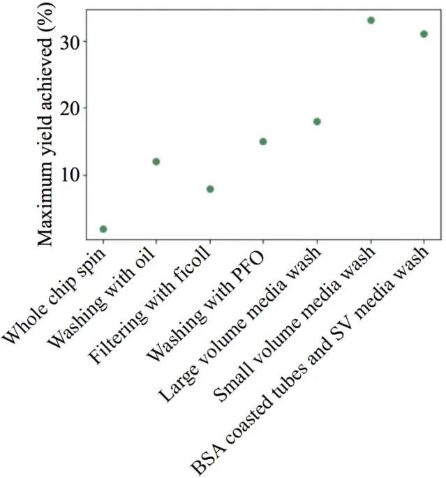
3-3 Cell extraction yield was contigent on method used. When optimizing removing cells from chips, a wide array of methodologies was investigated. This plot displays the maximum yield achieved with each protocol. . . 40 3-4 Optimized protocol for extracting cells from chip. The most
efficient cell removal protocol is displayed. . . 41 3-5 Antibody tagging successful in chip protocol. This figure
dis-plays the results of testing the efficacy of antibody tagging when using chip based stimulation. . . 42 3-6 PBMC response triggered in chip based stimulation. After
incubating a mixture of merged droplets containing cells and antag-onistic chemicals in chips and labelling the cells for activation with fluorescent antibodies, the resulting FACS plots confirm the success of cell activation. . . 43 3-7 Preliminary measures and controls validate data quality.
Be-fore performing in-depth analysis, the scRNA-seq data was initially checked for baseline irregularities. . . 44 3-8 Distribution of cell types by origin used in experimentation.
The originating donor and condition (either plate or chip) for each cell used in analysis was examined to investigate potential bias. . . 45 3-9 t-SNE visualization of all data. For all data collected, principal
component analysis and t-SNE visualization was used to envision initial clustering in the data. . . 46 3-10 Resampling data and comparing mean expression of various
genes. Contrasting plate and chip originating cells’ responses to the cytokines affirms correlation between the two. . . 47 3-11 Cell type specific expression dominates platform specific
in-fluence. The ap displays a visualization of the correlation matrix comparing chip and plate specific responses. . . 48
3-12 Chip specific expression signature. This violin plot contrasts the expression profiles of PMBCs for the 23 genes consistently identified as significantly differentially expressed between chip and plate assays. 49 5-1 Visualizing gene counts and mean expression. These scatter
plots display the distributions of number of genes by counts and percent mitochondrial genes by count for all of the scRNA-seq data. . . 53
Chapter 1
Introduction
1.1
Motivation
The immune system is as complex as it is critical for healthy living, employing a variety of cells and biological agents to protect the body against constant pathogen invasion. However, proper functioning of this intricate biological powerhouse is not guaranteed. Failed cooperation and faulty interactions between its components can result in multitudes of immune system disorders. While some immune system mal-functions and autoimmune conditions can be subdued, though rarely cured, with medication, the vast majority of treatments for these diseases fall short of expecta-tions and can have dire, even deadly, consequences. [3] Take, for example, one of the most rampant afflictions resulting from immune disfunction: sepsis.
A leading cause for hospital readmittance, sepsis is an incredibly dangerous and enigmatic affliction. Because many of its early symptoms are indistinguishable from those of other conditions, early detection, which is necessary for successful treatment, is nearly impossible. [13] Recent studies have attempted to identify “immune sig-natures” of sepsis – these signatures are irregular immune system responses to the known antagonists that are unique to the septic condition. But, testing septic pa-tients’ immune system responses to multitudes of antagonistic cytokines individually is inefficient and costly. Therefore, there is a necessity to develop a means of assessing immune cell response to an assortment of cytokines in a massively parallel system. In this thesis, a microfluidic droplet-based system is tested and proposed for effectively
evaluating immune system signatures against many cytokines in one trial.
1.2
Immune system composition and mechanics
Despite being inundated with waves of potentially harmful microbes from sur-rounding environments, our bodies are typically void of active infections and rampant disease. This is because the immune system combats constant onslaughts of micro-bial invaders using an assortment of tissues, cells, and molecules as host defensive mechanisms. For example, the breadth of cells deriving from the common lymphoid progenitor and the myeloid progenitor lineages, shown in Figure 1-1, play primary roles in the immune system’s defenses. The functionality of the immune system can be broken into two high-level categories: innate and adaptive immunity.
Innate immunity refers to the body’s nonspecific defense mechanisms; examples of innate immunity components span from the macroscopic physical barrier of the skin to the microscopic recruitment of immune cells to an infection via chemical signaling. [5] Signaling cascades critical to the identification and destruction of foreign pathogens are functions of innate immunity that can also induce adaptive immunity. [5] For instance, bacteria display surface receptors that can be recognized by key innate immune system cells, such as macrophages. Once the presence of a bacterium within the body has been detected, a macrophage will consume the intruding organism and release signaling molecules, known as cytokines and chemokines, in order to attract other members of the immune system such as lymphocytes and neutrophils to the site. [5] This release of chemicals typically leads to inflammation (heat, pain, redness, and swelling) in the surrounding area, a conspicuous signal of an innate immune response occurring within the body. [5] Further, inflammation and residual fragments of foreign invaders, such as those produced during the aforementioned bacterium’s destruction, activate the second branch of the immune system, adaptive immunity.
While the innate immune system is sufficient to protect against a wide range of pathogens, it relies on the presence of germline encoded surface markers for its efficacy. [5] In that, there is a limited set of pathogenic receptors that can trigger an innate
immune response. However, a critical difference between the innate and the adaptive immune system’s functionality is that the adaptive immune system is not bound by this constraint. The first step in adaptive immunity’s activation is the ingestion of pathogen fragments by immature dendritic cells. [5] In doing so, the dendritic cells evolve into antigen-presenting cells (APCs) and are able to uptake the pathogenic receptors and display them to other immune cells. The receptors are now “learned” by the adaptive immune system as belonging to a pathogen, and this new knowledge is then used to fight infection. The APCs migrate to the lymph nodes and trigger a lymphocytic response while secreting additional cytokines to further manipulate both innate and adaptive immune responses. [5] However, this description of the immune system’s two branches only superficially analyzes the initial activation of both the innate and adaptive immune systems. In actuality, there are many more cells, signals, and factors critical to the proper activity of both branches.
The complexity and multitudes of components within the innate and adaptive responses heavily contributes to the difficulty of studying the immune system. There-fore, for the sake of processing data and performing experimentation, many studies tend to focus on specific elements of the immune system when attempting to charac-terize behavior patterns. In this thesis, five main cellular components of the immune system were the focal points of analysis. These five cell types were all isolated periph-eral blood mononuclear cells (PBMCs), and PBMC samples are considered represen-tative of cells that are regularly circulating throughout the donor’s body. Specifically, the bulk of analysis focused on four lymphocytic cell types (B cells, CD4+ T cells, CD8+ T cells, and Natural killer (NK) cells) and monocytes.
1.3
Failures in identifying and treating immune
disorders
The immune system requires the proper functioning of all of its components to protect effectively. However, this does not always occur; a malfunctioning immune
system leads to painful, and even fatal, conditions. From auto-immune inflammatory psoriasis to deadly severe combined immunodeficiency (SCID), there is a breadth of diseases associated with faulty immune system behavior. Each of these conditions correlates with its own unique blend of irregularities in the immune system’s response, making causation identification and treatment development for these diseases chal-lenging. [2] For instance, Lupus is an autoimmune disease resulting from immune system cells attacking host tissues, while immune system caused inflammation in joints is the cornerstone symptom of Rheumatoid arthritis, a chronic condition heav-ily present in the United States and across the globe. While symptoms of immune and autoimmune disorders can be alleviated through costly hospital visits, prescriptions, and avoiding environmental triggers, a wide array of immune system conditions are lacking cure, or even treatment, options.
One such example, sepsis, is a particularly devastating immune disease. As one of the main causes for hospital readmission in the United States, sepsis results from the immune system’s improper response to infection and most commonly plagues infants, the elderly, and people suffering from serious medical problems. [11] The disease affects upwards of a million Americans annually, and, in recent years, the number of sepsis cases has been rising. [11] But, because many of the early symptoms of sepsis are indistinguishable from those of other disease conditions, sepsis is extremely difficult to diagnose and treat in a timely manner. [11] The efficacy of sepsis treatment is dependent on early detection, and, therefore, countless septic patients are suffering due to improper identification of the condition rather than the lack of an available treatment. Although it is well established that sepsis correlates to abnormal immune system behavior, a blood test cannot reliably reveal whether a patient is septic. One issue is that immune cells circulating in the blood do not consistently demonstrate the expression patterns of immune cells localized in tissues; for example, PMBCs are thought to be more dormant than their tissue inhabiting counterparts. [12]
Moreover, even when sepsis is properly identified and treatment is pursued, recov-ery is not guaranteed. The underlying mechanics of the disease are still unclear; while previous studies have linked sepsis to immune system hyperactivity, recent research
has indicated that immunosuppression also plays a critical role in the disease’s rapid progression. [1] In response, attempts to treat sepsis have revolved around boosting immunity through antibiotics, but these efforts have largely failed. [15, 10] Although immunosuppression is connected to sepsis, the immune pathways which are disrupted and responsible for this suppression have yet to be determined. [10]
However, recent studies have shown that whole blood stimulations can potentially remediate this discrepancy. The activated response of immune system PBMCs to vari-ous cytokines can provide functional information regarding the state of an individual’s immune system. In that, each immune cell’s unique transcriptomic response to an-tagonistic cytokines and other triggering molecules – its immune expression signature – holds potential as a means for discerning whether the cell’s origin was a septic or non-septic donor. [4] By comparing healthy patients’ activated PBMC immune signatures with those of immunosuppressed septic patients, fundamental differences between the two immune states may be brought to light.
1.4
New technologies increase experimentation
options
1.4.1
Massively parallel testing possible through droplets
Performing the multitudes of experiments required to test the innumerable com-binations of stimuli against healthy controls and immunosuppressed septic samples would be material and time intensive. Characterizing the specific abnormalities in im-munosuppressed septic patients’ immune systems would provide key information that could be leveraged to improve the condition’s current treatment plan and may provide insight into biological markers of the disease. An analogous issue exists within the drug discovery arena as well. High-throughput screening of small molecules and their effects on different cells and conditions is a popular means of identifying potential novel drug molecules and combinations. [8] Yet, testing innumerable molecules alone, in countless combinations, and in various concentrations can transform a manageable
library of molecules into an insurmountable list of permutations and respective ex-periments. To overcome this challenge, Kulesa et al., propose a nanoliter droplets and polydimethylsiloxane (PDMS) synthetic polymer microwell array system capable of combinatorial drug discovery; this PDMS microwell array is also referred to as a “chip”. [8]
Kulesa et al.’s protocol involves creating an emulsion of droplets containing cells, different titrations and various combinations of molecules, and discernable tags. Var-ious microfluidic architecture such as T-junctions can be used to make the emulsions. [7] This droplet mixture is then loaded into the chip, the droplets pair into random combinations, and droplets merging is induced. The versatility of the protocol and system designed by Kulesa et al. is of monumental importance – not only can this tool be used to aid in combinatorial drug discovery, but its applicability also extends into other fields.
For instance, in this thesis, Kulesa et al.’s protocol was adapted to allow for multiplexing immune cells against a variety of cytokine triggers to generate robust immune signature readouts. Figure 1-2 displays the design and layout of the PDMS microwell array used in experimentation. When properly loaded, each well within the device holds, at maximum, two droplets. Each chip contains approximately 50,000 wells, allowing for a multitude of immune cells’ responses against many cytokines to be tested in one chip, thereby reducing the experimental burden of measuring immune signatures.
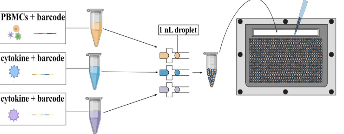
In the device, cell viability is maintained through the use of a biocompatible combination of oil and surfactant. [7] Once loaded, as demonstrated in Figure 1-3, the droplets randomly pair within wells and produce different combinations of PBMC and cytokine solutions. After loading, sealing the device isolates pairs from any other droplet pairings, and droplet pairs can then be merged and incubated.
1.4.2
Producing high resolution transcriptomic reads
Recently, there has been an influx of genome and transcriptome sequencing and single cell sequencing techniques. These methods allow for detailed readings of cellular
genome expression and behavior. While there are a variety of different methods to assess available genomic and transcriptomic data, for the purpose of this project, single cell RNA sequencing (scRNA-seq) was employed. The scRNA-seq technique provides a wealth of in-depth reads regarding individual cell’s gene expression profiles through next generation sequencing of a cell’s RNA.
The scRNA-seq data can not only unveil underlying mechanisms in the immune system and immune signatures, but, because of the high resolution of this data, can also elucidate subsets within the cell samples and irregularities that may be lost in bulk cell analysis. [14] This technique relies on the use of unique RNA barcode libraries to keep track of the origin of RNA reads – each barcode exclusively correlates with one cell or cell sample depending on which barcoding procedures are followed. Barcode reads can be used during analysis to connect data to its ancestral cell. In this thesis, antibody bound scRNA-seq barcodes were added to preliminary PBMC and cytokine solutions both to connect PBMCs to their original donor and to link PBMCs to whichever cytokine they were paired with in the wells of the device (Figure 1-3). When a cell-containing droplet is merged with a cytokine-containing droplet, the barcode correlating to the cytokine, as well as the barcode included in the PBMC solution, will associate with the cells and will aid in future data analysis.
1.5
Thesis objectives and outcome
The primary purpose of this work was to adapt the existing PDMS microwell array methodology such that it was usable to measure immune expression response to various cytokines while testing PBMC/cytokine pairings in parallel. To do so, the specific aims of this thesis were to:
1. Confirm the viability of PBMCs within droplets over various time spans and verify the sustained viability of cells following dropletizing PBMCs and merging said droplets within the chip.
solution and the distribution of cells among droplets generated.
3. Develop an extraction protocol for removing PBMCs from the chip while main-taining cell viability and optimize cell yield.
4. Establish the optimal concentration of antibody and cytokine stimulant to en-sure cell tagging and activation in the chip while minimizing noise.
5. Validate that PBMC response to cytokine triggers in plate-based and chip-based assays are analogous and comparable.
The final product of my thesis is a microfluidic droplet protocol that allows for parallel immune cell stimulation by various cytokines and takes advantage of scRNA-seq techniques to reveal each cell’s distinct expression profile against its par-ticular antagonist cytokine cohort. In doing so, this work opens the door for testing a broad spectrum of cytokines and triggering molecules against septic and healthy patient’s PBMCs which may lead to improved diagnosis techniques; furthermore, be-cause of the nature of the high-resolution data, this multiplexing of immune cells may uncover subsets within the sepsis condition with distinct immune expression responses. Moreover, this methodology can be expanded and applied to contrast additional immune conditions’ PBMC signatures against a healthy control.
1.6
Figures
Pluripotent hematopoietic stem cell common lymphoid progenitor common myeloid progenitor granulocyte/macro-phage progenitorB cell T cell neutrophil eosinophil basophil unknown monocyte immature precursor dendritic cell granulocytes (or polymorphonuclear leukocytes)
plasma cell
activated
T cell mast cell macrophage immaturedendritic cell dendritic cellmature
Figure 1-1: Lineages of cells composing the immune system. The above graphic displays a variety of cells that play important roles in the immune system and their ancestral origins. Adapted from [5].
B)
C)
A)
D)
Figure 1-2: Layout of PDMS microwell array and acrylic loader. The design of the PDMS microwell array used in experimentation is displayed above. Panel A demonstrates an overhead and side view of the chip and the upper level of the acrylic loader. The loading and sealed orientation of the chip are contrasted in panels B and C. Lastly, actual images of the chip design and example loading using colored dye are shown in panel D. Adapted from [8].
1 nL droplet
cytokine + barcode cytokine + barcode PBMCs + barcode
Figure 1-3: Droplet generation and loading workflow. This figure portrays the general protocol for setting up an immune cell multiplexing experiment in the chip. Firstly, solutions containing cells or cytokines with respective barcodes are dropletized and aggregated. The emulsion is mixed well and inserted into the chip through the loading slot. Generated using BioRender images.
Chapter 2
Methods
2.1
Generating of droplet emulsions
The Bio-Rad QX200 instrument was used to generate 1 nL droplet emulsions for experimentation. To create droplets, 20 µL of each sample was pipetted into respec-tive inputs on a Bio-rad QX200 cartridge and was emulsified using 70 µL of fluoro-carbon oil 3M Novec 7500 and RAN Biotech 008-FluoroSurfactant. The surfactant was incorporated in varying concentrations from 0.1% to 2.0% wt/wt depending on the trial. For viability testing, cell distribution evaluation, antibody tagging, stimu-lation confirmation, and chip versus plate comparison experiments, 0.5% wt/wt RAN Biotech 008-FluoroSurfactant was used. During iterations of cell yield optimization, a variety of wt/wt values were utilized.
2.2
Thawing PBMCs
Clinical samples of PBMCs from healthy donors processed in the Blainey Lab by Miguel Reyes and Kianna Billman were used for experimentation. During the time period between processing and thawing, PBMCs were kept frozen in a Thermo-Fischer Scientific liquid nitrogen tank. To thaw, samples were incubated at 37 degrees Celsius for 2 minutes. During this time, media (89% RPMI-1640, 1% Pennicillin-Streptomycin, and 10% Fetal Bovine Serum; Thermo-Fischer) was prepared. Then, 1 mL of media was added to the sample and repeatedly pipetted until there was no remaining frozen pellet. The final volume was added to 9 mL of media and spun
down at 300 rcf for 5 minutes. Cells were resuspended in 10 mL of media, counted using a hemocytometer, and spun down once again at 300 rcf for 5 minutes. Lastly, cells were resuspended with media to the appropriate concentration.
2.3
Testing the viability of dropletized cells
PBMC samples were thawed and suspended to a final concentration of 1 million cells per mL. The viability of the initial cells was calculated using Trypan Blue Solu-tion, 0.4% (Thermo-Fischer) for a live/dead cell assay. A PBMC solution containing cells at a concentration of 1 million cells/mL was used to generate 14 aliquots of droplets. Pairs of these aliquots were incubated at 37 degrees Celsius for one, two, three, four, five, six, and twenty-four hour intervals. Droplet emulsions were broken by adding 1mL of media to the emulsion, repeated pipetting, adding 1 mL of fluo-rocarbon oil 3M Novec 7500 (0% wt/wt surfactant), and spinning down the solution at 70 rcf for 3 minutes at 4 degrees Celsius. The resulting layer of supernatant was collected and the washing process was repeated a total of three times (Figure 2-1).
After three repetitions, the collected supernatant was spun down at 300 rcf for 5 minutes to pellet the cells. A control solution of PBMCs at the same concentration was maintained in a cell culture container and incubated for twenty-four hours as well. Lastly, two emulsions of PBMC-containing droplets were loaded into chips, merged, and immediately extracted. The viability of cells in each of the described conditions and in the initial solution was determined used a Trypan Blue live/dead cell staining assay. Final pellets were resuspended in fresh media and equal parts of Trypan Blue Solution, 0.4% (Thermo-Fischer), were added to the aliquots. Lastly, 10 µL of each aliquot was pipetted into a hemocytometer; viable and dead cells, indicated by blue Trypan dyeing, were counted. The percent viability of each solution was determined by comparing the result cells’ viability to the initial viability.
2.4
Fabricating, preparing, loading, and sealing of
PDMS microwell array device
Chips were fabricated using inherited molds. Two designs were used: one chip (6.2 x 7.2 x 0.62 cm) had 43,000 wells with diameters of 148.6 µm and heights of 100-120 µm and the second chip (6.2 x 7.2 x 0.31 cm) had approximately 45,000 wells with the same diameters and heights. [7] Each microwell array, made using PDMS (Dow Corning Sylgard), was mixed in a 9:1 ratio with a crosslinking reagent (Dow Corning Sylgard) via standard soft lithography techniques. All chips were coated with 1.5 µm of parylene C (Paratronix) to ensure hydrophobicity and reduce stickiness. [7]
To prepare a chip for experimentation, the microwell array was adhered to an acrylic loader. When inside the loader, the chip was suspended above an Aquapel treated, hydrophobic glass slide (1.2 mm thickness, Brain Research Laboratories) via magnetic repulsion (Figure 1-2). By adjusting the force exerted by the mechanical clamps on the upper level of the acrylic loader, the height of the gap between the chip and the glass slide could be adjusted to the optimal height of 0.31 cm. The gap between the two was then flooded with fluorocarbon oil 3M Novec 7500 (0% wt/wt surfactant), and the oil was maintained in the space for a minimum of five minutes. The oil was then drained via a slot in the acrylic loader, and droplets were loaded into the wells by injection in the loading slot. Following droplet loading, residual drops were washed out from the loader by fluorocarbon oil 3M Novec 7500 (0% wt/wt surfactant). To seal the chip, the gap was once again filled with oil (0% wt/wt surfactant), and the mechanical clasps were tightened until the chip surface was firmly pressed against the glass. Merging was induced via mechanical force applied to the glass surface and confirmed by light microscopy.
To remove the cells from the chip, media was pipetted directly onto the surface of the chip followed by an oil wash. All runoff was collected and underwent the breaking protocol proposed in Figure 2-1 followed by counting. This manner of extraction proved to be rather inefficient, and the protocol of cell removal from the chip was optimized during later experimentation.
2.5
Determining the relationship between initial
cell concentration and distribution of cells per
droplet
Various aliquots of PBMC suspension samples with cell concentrations of between 150,000 cells/mL and 10,000,000 cells/mL were used to generate emulsions of droplets. Subsequently, 10 µL of each sample was pipetted from these mixtures and loaded into hemocytometers. Using an electronic light microscope, images of the cell-containing droplets were acquired. For each concentration evaluated, a minimum of five images were taken from random locations on the hemocytometer. On average, each image contained 30 complete droplets, and, therefore, for each concentration tested, there were upwards of 150 droplets considered. Following collection, the number of cells contained within each complete droplet was counted and recorded. ImageJ was used to aid in image analysis and to keep track of counting, as demonstrated in the example analysis displayed in Figure 2-2.
2.6
Optimizing cell extraction yield from the chip
2.6.1
Fluorescent tagging
When counting cells successfully removed from the chip, the high amount of noise due to the presence of oil in the final resuspension made it difficult to accurately count the number of cells present. To combat this effect, PBMCs were stained prior to dropletization. To do so, pelleted PBMCs were suspended in a 5 µg/mL CD45 PE fluorescent antibody media solution (BioLegend) for 30 minutes on ice. Following incubation, cells were washed with 10 mL of media and ready for use in experiments.
2.6.2
Altering the extraction protocol washes
Initially, to remove the cells from the microwell array, media was pipetted directly onto the surface of the chip followed by an oil (0% wt/wt surfactant) wash. All
runoff was collected in a petri dish below the chip and used in the breaking protocol proposed in Figure 2-1. The first adaptation to the emulsion breaking protocol was to replace the oil between spins and increase the number of washes performed. After spinning down the mixed solution of media, droplets, and oil, the bottom layer of oil visible in Figure 2-1 was removed. After the supernatant was taken out, 1 mL of media and 1 mL of oil (0% wt/wt surfactant) was added to the solution. Up to fifteen washes were tested, and each wash’s supernatant was tested individually for yield by cell counting (e.g. yield per wash for each wash leading up to fifteen washes was determined). As FBS is itself a surfactant and aids in stabilizing the droplets, using RPMI-1640 for washes in the place of media was another permutation on the original extraction protocol that was tested.
Since previous studies showed 1H,1H,2H,2Hperfluoro-1-octanol (PFO; Sigma-Aldrich) is successful as an emulsion breaking chemical, another set of trials incorporated oil enhanced with PFO (1-2% wt/wt) during washes. Additionally, various centrifuge speeds ranging from 70 to 500 rcf for time spans between 1 and 8 minutes were tested in an attempt to increase droplet coalescing. Moreover, a recent publication indicates that hand-held antistatic guns are capable of chemical-free emulsion breaking. [6] Therefore, a sample of droplets was produced and targeted by an electrostatic gun between fifteen and thirty times though no noticeable difference was observed.
Lastly, in another trial, the protocol depicted in Figure 2-1 was adapted by remov-ing all oil addremov-ing steps and usremov-ing either media or RPMI-1640 for all washes. In this case, the chip was repeatedly washed directly with either only media or RPMI-1640, and the collected runoff was immediately spun without additional washes to pellet cells. In an effort to decrease the probability of cells sticking to the collection tube walls, a 2% Bovine serum albumin (BSA; Thermo-Fischer) phosphate-buffered saline (PBS; Thermo-Fischer) solution was used to coat collection tubes. To do so, collection tubes were filled with 2% BSA solution and incubated in refrigeration overnight. Prior to use, the solution was removed and the tubes were used immediately thereafter.
2.6.3
Ficoll filtering
Endeavoring to reduce the amount of oil present in the final sample, Ficoll (Stem Cell Technologies) and SepMate PBMC Isolation tubes (Stem Cell Technologies) were incorporated into the extraction protocol at two different time points in two separate experiments. For the first time point, Ficoll filtering was used in lieu of the spin following the addition of media and oil. Ficoll filtering was done by adding room temperature Ficoll into a SepMate PBMC Isolation tube until slightly above the tapered barrier. The droplet solution was slowly pipetted down the side of the tube such that it rested above the Ficoll layer. The tube was spun at room temperature at 70 rcf for 3 minutes. In a second round of experimentation, a Ficoll filtering step was added just before pelleting the supernatant; to do so, after all the washes, the final supernatant volume was suspended over a Ficoll layer and spun at 70 rcf for 3 minutes.
2.7
Testing in-chip antibody tagging and
stimulation efficiency
The optimal experimental antibody and stimulant concentration was determined by testing the performance of various concentrations of CD298 APC and CD298 PE antibodies in tagging cells, as well as using LPS and CD3/CD28 beads as stimuli to trigger cell activation (BioLegends). Two distinct solutions of equal volume containing either CD298 PE or APC in concentrations between 1 and 5 µg/mL with either 1 µL/20 µL of beads or 1 µL/20 µL of diluted LPS (stock was 5 mg/mL, diluted 1:100 then 2:25) respectively were used to generate droplets. A volume of cell containing droplets was also created and aggregated with the antibody/stimuli droplets such that the final volume had a 1:1 ratio of cell containing droplets to total antibody-/stimuli-containing droplets. The well-mixed emulsions were loaded into chips. Once the droplets were merged and the chip sealed, the device was incubated at 37 degrees Celsius for four hours. Following incubation, the cells were extracted, washed, and
stained with secondary antibodies: CD69-APC/Cy7, CD40-PE/Cy7, CD3-AF700, CD14-FITC. During staining, appropriate compensation beads were prepared as well. Cells were washed once again following staining and, along with the compensation beads, used in Fluorescence-activated cell sorting (FACS) flow cytometry to measure activation and tagging through flourescence.
2.8
Comparing plate-based versus chip-based
as-say performance
To ensure that chip-based assay results were comparable to those of plate-based assay techniques, a plate versus chip response experiment was developed. To do so, plate and chip assays using shared PBMC samples and the same cytokine solutions were run in parallel. The cytokines IFNB, IL1B, and TNF were used in experimen-tation. Cells were antibody barcoded via the 10X Genomics protocol according to originating patient donor. As many variables as possible were maintained constant between the two platforms; thus, droplets were merged simultaneously as cytokines were added to plated cells. Cytokine solutions also contained antibody barcodes unique to each stimulant. Following stimulation and a four hour incubation, cells were extracted from the chip, and removed from plates. The 10X Genomics proto-col for scRNA-seq was used to process plate and chip samples and obtain expression reads.
For this experiment, two samples of PBMCs were thawed; these samples originated from two different donors and each sample was tagged with a donor specific barcode. Both samples were used in plate and chip assays. Four conditions, TNF, IFNB, and IL1B stimulation (each diluted to 1 µg/mL with 0.1% BSA) and no stimulation, were tested during experimentation.
2.8.1
Plate stimulation
A total of 50,000 PBMCs per condition were plated in 100 µL of complete media with monesin (1000X, 1 µL/1 mL media) in separate wells of a 96 well plate. PBMCs from separate patients were kept in different wells. For the TNF condition, 25 µL of TNF stimulant and 950 µL of media were added to the plated cells. For the IFNB and IL1B condition, 50 µL of stimulant and 950 µL of media were added to the plated cells. For the no stimulation condition, only media was added to the plate. Plates were incubated for four hours at 37 degrees Celsius. Following incubation, each condition was stained using a unique HTO Barcode (10X Genomics), washed, and resuspended in media.
2.8.2
Chip stimulation
Cells were suspended in media at a concentration of 5 million cells/mL and drople-tized. Patient specific emulsions were kept separate. Solutions of stimuli and unique barcodes were created such that the final merged concentration of the stimuli would be 50 ng/mL for IFNB and IL1B and 25 ng/mL for TNF. These solutions were used to make stimulation droplets. For the no stimulation condition, droplets contained only media and no stimulation specific barcodes. Emulsions containing equal volumes of PBMC and condition droplets were mixed well and then loaded into chips. Three chips were used per patient for a total of six chips. Chips were sealed, droplets merged, and the microwell array was incubated for four hours. Cells were released from chips, and PBMCs from the same donor were pooled together. Cells were pelleted and resuspended in media.
2.9
Data analysis
The majority of data analysis was performed using Python and a variety of open source platforms, namely scanpy, numpy, and scikit learn Python packages. A minor-ity of data analysis, specifically a spreadsheet used to keep track of cell per droplet
counter, occurred in Microsoft Excel. When ranking genes and determining differen-tial expression, a t-test was used to compare gene expression profiles. A p-value less than 0.05 was considered the cutoff for statistical significance.
2.10
Figures
droplets oil
1 mL of media 1 mL of oil spin
supernatant residual droplets oil
repeat 2x
Figure 2-1: Emulsion breaking protocol used during viability testing. The protocol portrayed above was used to break emulsions containing cell droplets during viability estimation experiments. Following in-chip merging, droplets were extracted from chips by flowing oil (0% wt/wt surfactant) over the surface of the chip and the above protocol was used to remove cells from droplets. Generated using BioRender images.
Figure 2-2: Cell counting enhanced by ImageJ functionality. Images of droplets, which were generated using various initial cell concentrations, were captured using 10X light microscopy. ImageJ was then used to tag cells in each droplet, which made counting cells per droplet considerably easier. An example of this tagging is shown above.
Chapter 3
Results
3.1
Cell viability confirmed through live/dead cell
assay
The first checkpoint in developing this methodology was to evaluate the general viability of PBMCs within oil droplets. To do this, PBMC solutions containing cells were dropletized and incubated over various time periods. Additionally, PBMC-containing droplets were merged in the chip. The viability of the cells following these actions was evaluated using a live/dead cell assay. Each condition displayed a viability over 90%. Although there was a slight downward trend in viability as incubation time increased, this decrease was consistent with the 24 hour control sample. Overall, this result was promising as PBMCs displayed continued viability once dropletized over long time spans, as well as once loaded into a chip, merged, and extracted.
3.2
Distribution of cells per droplet dependent on
initial solution cell concentration
The next step in protocol development was to determine the optimal concentration of cells for making PBMC droplets. Therefore, a variety of cell concentrations, ranging from 125,000 cells/mL to 10,000,000 cells/mL, were dropletized and the cells per droplet for each condition were counted. A total of over 1,000 droplets were examined. When counting cells per droplet, droplets that contained more than ten cells were
difficult to analyze and were considered ten-cell droplets.
The resulting relationship between initial solution cell concentration and distribu-tion of cells per droplet is visualized in Figure 3-2. The lowest concentradistribu-tion tested, 125,000 cells/mL, demonstrated the highest frequency of empty droplets followed by a steep drop for increasing cells per droplet incidence. Additionally, 125,000 cells/mL and 250,000 cells/mL showed the highest frequencies for single cell droplets. The distribution resulting from an initial solution concentration of 5,000,000 cells/mL was the first instance of no empty droplets and had peak frequencies ranging over three, four, and five cell droplets. The overall distribution generated was reminis-cent of a Poisson distribution, which was expected given the nature of dropletizing cells. In order to avoid loading empty droplets into the chip and to maximize data yield, 5,000,000 cells/mL was selected as the initial solution PBMC concentration for experimentation. However, the distribution data illuminates that other cell concen-trations may hold potential for future experimentation. For instance, for single cell pertubations, an initial solution concentration of 250,000 or 125,000 cells/mL would be more applicable since these concentrations displayed the maximum frequency of single cell droplets.
3.3
Optimization of cell extraction from chips
Washing loaded chip surfaces with oil (0% wt/wt surfactant) and following the extraction protocol proposed in Figure 2-1 proved to have an incredibly low cell yield of approximately 1.875%. Therefore, a wide array of alternative removal procedures was pursued in an effort to maximize yield. A variety of steps in the extraction protocol were adapted and amended: fluorescent tagging was implemented to increase counting accuracy, the number of oil washes was altered, chemicals such as PFO and Ficoll were used to aid in emulsion breaking, and BSA coated collection tubes were added to the protocol in an attempt to reduce cell stickiness. Figure 3-3 displays the maximum cell yields achieved using the different extraction protocols. In practice, it was very difficult to achieve consistent yield with any of the extraction protocols.
For instance, the most effective protocol, washing the chip with a small volume of media, collecting the runoff, and pelleting cells without any washes would typically achieve a yield of between 15% and 30%, varying greatly from test to test. This protocol, shown in Figure 3-4, was still consistently more effective than any of the devised extraction methods and is therefore recommended for use during microwell array immune cell mutliplexing.
3.4
Antibody tagging and stimulation testing
The optimal concentration of antibody for cell tagging was determined. This con-centration was representative of the concon-centration of barcoding antibodies needed to label cells in the chip protocol. To test this, solutions containing various concentra-tions between 5 µg/mL and 1 µg/mL of two antibodies, CD298 APC and CD298 PE, were used in lieu of cytokines in the general workflow.
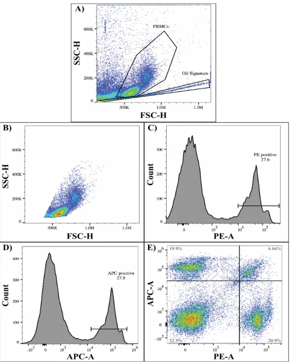
Fluorescence-activated cell sorting (FACS) flow cytometry was used to measure the efficiency of cell antibody tagging within the chip. On all forward versus side scatter plots, which were used to identify PBMC populations during FACS, a unique, narrow array of points was present just below the cells. This line of points is visible in panel A of Figure 3-5. Based on the visible presence of oil observed via light microscopy in cell samples following chip extraction, this array was deemed to result from oil in the sample and was considered an oil signature in the data.
Histograms displaying FACS reads for PBMC PE and APC fluorescence are shown in panels C and D of Figure 3-5 respectively. In both histograms, there is a distinct divot between two peaks. The first peak in both plots, which contains all data below approximately 103, represents unlabeled cells, whereas the second peak contains cells
that have been tagged by fluorescent antibodies. Both peaks contain tagged cells that account for just over 27% of the cell population analyzed. Given that the probability of pairing a cell-containing droplet with either antibody-containing droplet in the chip was 25%, these proportions in the FACS data were expected and reassuring. Lastly, comparing PE fluorescence against APC fluorescence, as displayed in panel E
of Figure 3-5, quantifies the amount of cross-talk present in the protocol. This noise is population in the upper right quadrant of the plot which accounts for 6.66% of the PBMCs. Using a concentration of 1 µg/mL of antibody, which is what was used to generate the FACS plots in Figure 3-5, proved to be the most effective in cell tagging while minimizing the amount of noise in the resulting data.
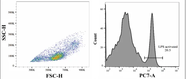
Furthermore, the efficacy of stimulating PBMCs in the chip was also confirmed through FACS. Instead of droplets filled with antibodies, droplets were generated that contained LPS and CD3/CD8 beads, known antagonists of PBMCs. Following extrac-tion, cells were incubated with fluorescent antibodies that targeted either activated LPS or CD3/CD8 receptors. Neither the control (tube treated) nor chip PBMCs showed elevated response to CD3/CD8 beads in FACS. However, as shown in Figure 3-6, through heightened PC7 fluorescence, over 20% of the chip-originating PBMCs displayed LPS activation. These two experiments confirmed that both cell tagging and stimulation were possible within the microwell array with minimal cross-talk.
3.5
Plate versus chip platform comparison
With each component of the proposed chip platform developed, the full protocol for stimulating PBMCs with distinct cytokines in parallel through a droplet microwell array system was functional and applicable. In order to confirm the validity of this platform as a means of uncovering immune cell expression signatures, the similarity between chip and plate based assay performances needed to be quantified, as did the overall transcriptomic effect of the protocol on PBMCs.
3.5.1
Analysis of scRNA-seq data
Produced scRNA-seq data was used to compare and contrast chip and plated cells’ responses to the stimuli. Following a preliminary check of the sequencing data, the scRNA-seq data was filtered to include only cells of interest and subjected to PCA and t-SNE in order to quantify the chip and plate assay performance. Significant differences between the two populations would indicate a discrepancy between PBMC
behavior in a microwell array chip versus in a plate.
Standard quality measures for scRNA-seq data type include visualizing distribu-tions for the number of genes detected in each cell, the number of counts per gene, and frequency of mitochondrial genes identified in each cell. Each of these factors can be used to maintain the integrity of the data and illuminate low quality data. [9] These quality metrics are shown in Figure 3-7 alongside histograms displaying barcode to cell incidence rates. For the chip barcode reads, the majority of unique barcode reads were either linked to no cell or one cell with a minority of barcodes overlapping between two and three cells. This result was expected given the noise observed during antibody tagging experimentation.
There was an almost even representation of barcodes that linked to either no cell’s data or one cells data; this phenomenon was also an anticipated result. Given that the probability of a cell to cytokine/barcode droplet pairing was 50% based on the emulsion loaded into the chip, this result was expected. Within the plate specific data, barcodes were mostly linked to one cell, though a small portion of the data connected barcodes to no, two, and even three cells. In this way, barcode tagging efficiency was consistent across the plate and chip platforms. Moreover, given that the quality metrics did not highlight any issues with the quality of the single cell data even though samples extracted from chips tended to contain residual oil, this evidence shows that cells stimulated through the chip platform are suitable for scRNA-seq techniques. Further quality metrics assuring the success of scRNA-sequencing can be found in Figure 5-1 in the Appendix.
3.5.2
Filtering the data set
As means of refining data analysis, the data was filtered to contain only cells of interest. For all analysis, the five most populace immune cell types in the PBMC samples were considered: B, CD4 T, CD8 T, monocyte (mono), and natural killer (NK) cells. Although cells were not labelled by type before experimentation, through principal component analysis (PCA), louvain clustering, and using known marker genes, the cells could be labelled by type in analysis. In addition to applying the
standard scanpy filtering function to the data set, cells that contained too many mitochondrial genes (greater than 0.05% of genes) or had gene counts greater than 2500 were excluded from the data. Both of these factors have been linked to poor quality cells in scRNA-seq analysis. [9] Subsequent to the filtering, the final data set contained a total of 17,228 individual cells; 8,064 of those cells originated from one donor, and the other 9,164 cells came from the second PBMC donor. Of the cells analyzed, 3,805 cells derived from the microwell array platform and 13,423 stemmed from plate experimentation. The overall distribution of cell types for each donor and experimental platform are shown in Figure 3-8. The composition of cells stemming from each donor and from each platform in the data is fairly comparable. This demonstrates that it is unlikely that donor bias or cell type distribution bias affected analysis.
3.5.3
Gene expression data analysis
In order to compare the multitudes of genes and their expression levels between the plate and chip cells, PCA was performed on the total data set. To further reduce the dimensionality of and visualize the data, t-distributed stochastic neighbor embed-ding (t-SNE) using 10 principal components was used. The t-SNE plots, shown in Figure 3-9, are labelled by cytokine trigger, originating platform, cell type, and donor. Labelling by cell type most strongly correlates with clusters produced through t-SNE, though patterns in stimulation, platform, and donor are also apparent in the plot. Both in plate and chip stimulation, IFNB most strongly influenced and activated PBMCs, and this facet is visible in the t-SNE plot. Cells originating from the chip platform tend to cluster together, but this clustering does not overpower the cell type clustering. Still, this group indicates that a transcriptomic response is most likely triggered following dropletizing PBMCs and working through the chip protocol. This chip signature is investigated further later in this thesis. Lastly, cells also appear to cluster according to originating donor, though this grouping together is not as strong as it was for chip versus plate cells. Overall, because cells mostly strongly aligned by cell type, the chip versus plate effects appeared negligible in the t-SNE analysis.
To delve further into the differences and similarities between plate and chip de-rived cells’ behavior, analysis focused on cell type and stimuli specific transcriptomic responses. Because honing in on niches of the data produced samples of limited sizes, resampling was introduced to increase the robustness of the analysis. For instance, Figure 3-10 displays the average Pearson coefficient, a statistical metric which nears 1 as two data sets are more closely correlated, which was calculated for each cell subtype for each stimulus. This average value was found by resampling subsets of the data to produce a new, analogous data set and calculating the Pearson coefficient be-tween the relevant plate and chip samples. After resampling the data for each subset 100 times, all Pearson coefficients were averaged, and the overall standard deviation across all 100 coefficients was used to generate an error margin. Because IFNB trig-gered a strong response in the PBMCs tested, chip and plate cells typically shared over 200 differentially expressed genes for this stimulus. Therefore, only the mean expression of these shared genes was used in calculating the Pearson coefficient for all cell types treated by IFNB. However, the other cytokines did not trigger equally as strong responses from the PBMCs, so the set of shared differentially expressed genes was much less robust. Therefore, the mean expressions of all genes were used in calcu-lating the Pearson coefficients for IL1B and TNF treated cells. Across all stimulants, the plate and chip responses had very high average Pearson coefficients; the average Pearson coefficient was above 0.90, indicating a strong correlation between chip and plate responses across all cytokines tested.
Furthermore, to confirm the inclination that cell type expression profiles corre-sponded more strongly than platform specific expression ties, a correlation matrix and dendrogram was calculated for the entirety of the data set. This matrix was visualized in a heatmap which is presented in Figure 3-11. Lighter segments in the heatmap indicate lower correlation while darker, deeper orange coloring signifies stronger corre-lation. Outside of the heatmap, rows and columns are labelled by stimulus, cell type, and platform respectively. The pattern of dark orange squares down the diagonal of the heatmap aligns with the cell type labels, highlighting a consistency in expression profiles across cell types regardless of originating platform. The checkerboard pattern
in the heatmap affirms that the chip platform does not drastically and irreparably alter cell expression; however, it does not guarantee that the chip has no affect on cellular gene expression.
3.5.4
Uncovering the chip signature
To explore the presence of a “chip signature” on the cells’ gene expression profiles, an approach similar to the average Pearson coefficient calculation was taken. The data was divided into stimuli and cell type specific subsets, and all genes that were differentially expressed between plate and chip cells and their adjusted p value was recorded. A total of 23 genes with p values less than 0.05 recurred in all five cell types. These genes were considered the chip signature. These genes’ mean expressions in chip and plate derived cells are contrasted in Figure 3-12. Although the profiles appear similar overall, the chip expression tends to be broader and the genes seem to be more strongly expressed. However, the majority of these genes are housekeeping genes and none appear to have substantial influence over immune cell signature. Therefore, the chip signature does not to interfere with the immune signatures measured in response to the cytokines.
3.6
Figures
Post-mer
ging 1 hr 2 hr
3 hr
3 hr
4 hr
4 hr
5 hr
5 hr
6 hr
6 hr
24 hr
24 hr
24 hr (control)
24 hr (control)
100.0
97.5
95.0
92.5
90.0
87.5
85.0
Cell
V
iability
(%)
Figure 3-1: High viability of PBMCs following droplet conditions. The affects of incubating cells in droplets and merging droplets on PBMC viability was quantified using a live/dead cell assay. The above bar graph displays the maintained viability of PBMCs after various test conditions in respect to the initial viability of the cells. The final bar in purple is the viability of PBMCs incubated for 24 hours in a tube rather than in the droplets. For all conditions tested, the viability of the PBMCs was well above 90%.
Frequency (%)
125,000 cells/mL 250,000 cells/mL 500,000 cells/mL 1,000,000 cells/mL 5,000,000 cells/mL 7,000,000 cells/mL 10,000,000 cells/mL 0 1 2 3 4 5 6 7 8 9 10Cells per droplets
Figure 3-2: Distribution of cells per droplet depends on initial cell concen-tration. Aliquots with various concentrations of cells were dropletized and imaged using light microscopy. Analyzing an average of 150 droplets per condition, the fre-quencies of incidence of cells per drop ranging from 0 cells to 10 cells were calculated. The above distribution visualizes the final data and, as expected, resembles a Poisson distribution.
Figure 3-3: Cell extraction yield was contigent on method used. When optimizing removing cells from chips, a wide array of methodologies was investigated. The above plot displays the maximum yield achieved with each protocol, however, there was a large amount of variability in yield between trials for each one.
Total volume: 3 mL Collect all runoff and mix
Spin
Cell pellet
Figure 3-4: Optimized protocol for extracting cells from chip. The most efficient cell removal protocol is displayed above. This methodology entails washing a chip’s surface directly with 3 mL of media, mixing it well, and pelleting the cells from the sample.
FSC-H FSC-H SSC-H SSC-H Count APC-A Count APC-A PE-A PE-A A) B) C) D) E)
Figure 3-5: Antibody tagging successful in chip protocol. In order to test the efficacy of antibody tagging when using the chip, PBMC and antibody droplets were used in chip experimentation. All samples extracted from chips contained lingering oil that formed an “oil signature” in FACS is visible in panel A. The selection of data representing the PBMCs extracted from the chip is shown in panel B. Panels C and D display the distinction in fluorescence observed between successfully tagged and untagged cells. Lastly, panel E compares the tagging of PE antibody against the tagging of APC antibody. The upper right quadrant of this graph contains cells that have been tagged by both antibodies, possible only through cross talk during breaking and droplet contamination during droplet aggregation.
FSC-H
SSC-H
Count
PC7-A
Figure 3-6: PBMC response triggered in chip based stimulation. After incubating a mixture of merged droplets containing cells and antagonistic chemicals in chips and labelling the cells for activation with fluorescent antibodies, the resulting FACS plots confirm the success of cell activation. As seen in the right panel above, a fraction of the cells extracted from the chip demonstrate heightened PC7 fluorescence, which indicates LPS activation.
Figure 3-7: Preliminary measures and controls validate data quality. Before performing in-depth analysis, the scRNA-seq data was initially checked for baseline irregularities. The first violin plot displays the distribution of total genes available per cell in the data, and the second plot demonstrates the counts, or overall incidence measured in the total data, for each gene. A gene detected in only three cells will have a count of three. The last violin plot displays the mitochondrial genes detected. The histograms on the second row of the figure display the distribution of distinct barcodes detected per cell from both plate and chip platforms. Barcode demultiplexing was performed by Miguel Reyes.
Figure 3-8: Distribution of cell types by origin used in experimentation. The originating donor and condition (either plate or chip) for each cell used in analysis was examined to investigate potential bias. The above graph displays the percent composition of cell types from each donor integrated into data analysis, as well as the distribution of cell types evaluated from the chip and plate platforms.
Figure 3-9: t-SNE visualization of all data. For all data collected, principal component analysis and t-SNE visualization was used to envision initial clustering in the data. Cell type appears to dominate clustering patterns, although stimulation, condition (chip versus plate), and patient donor also appear to affect the distribution of data. NS signifies no stimulation control cells.
Figure 3-10: Resampling data and comparing mean expression of various genes. Contrasting plate and chip originating cells’ gene expression responses to the cytokines affirms correlation between the two. For each population evaluated, the data available was randomly resampled to generate a new dataset of equal size one hundred times. The Pearson coefficient between the plate and chip expression profiles for each condition was calculated and the average expression over one hundred trials is shown above in the bar plot.
Figure 3-11: Cell type specific expression dominates platform specific influence. The heatmap displays a visual correlation matrix comparing chip and plate specific responses. External labels highlight relevant cytokine, cell type, and platform information for each data point. The pattern of heightened correlation down the diagonal of the plot signifies the strong correlation of cell type specific expression.
Figure 3-12: Chip specific expression signature. This violin plot contrasts the expression profiles of PMBCs for the 23 genes consistently identified as significantly differentially expressed between chip and plate assays. For the majority of the genes, the chip expression profiles tend to be wider and slightly stronger in expression.
Chapter 4
Discussion
The immune system is an intricate and convoluted system, a mosaic of cell, tissues, and molecules. When functioning properly, this system is a sound defense against the many pathogens and dangerous molecules invading the body at any given moment. However, when it fails, treating and repairing the immune system is incredibly diffi-cult. Honing in on specific mechanisms and interactions within the immune system proves to be challenging because of the multitudes of components at work.
Studies have indicated that elevated immune responses, specifically in activated peripheral blood mononuclear cells (PBMCs), can indicate the state of an immune system and illuminate underlying immune system structures. [4] These immune re-sponse signatures are triggered via antagonistic cytokines, but it is infeasible to eval-uate PBMCs’ responses to individual cytokines at the scale that would be required to uncover condition specific immune signatures. A similar issue was present in the small molecule drug discovery field and was recently tackled by Kulesa et al. by the devising of a polydimethylsiloxane (PDMS) microwell array capable of parallel, combinatorial small molecule testing. [7] Though originally limited to in-chip experi-mentation, this device held potential as a means of discovering immune cell signatures through multiplexing PBMCs and cytokines.
This thesis aimed to adapt and tailor the microwell array, also referred to as a chip, and its usage protocol such that it could be used for parallel PBMC stimula-tion experimentastimula-tion. To do so, PBMC viability within droplets and throughout the entirety of the chip workflow had to first be confirmed. Additionally, refinement of
the appropriate loading solution cell concentration was necessary such that data yield would be maximized without overcrowding the droplets with cells. Once the prelim-inary conditions were evaluated, testing within the chip began. Since the original protocol did not intend for droplets to be extracted and products released once the chip had been loaded, these aspects of the new methodology had to be designed from the outset. This process necessitated a lot of optimization, however, a final extraction and emulsion-breaking protocol was achieved with a maximum yield of over 30%.
Once the mechanics of the methodology were constructed and tested, a preliminary test to determine the feasibility of antibody cell tagging and antagonist mediated cell activation was conducted. The resulting FACS plots and flourescence measurements confirmed the success of cell tagging and activation in the chip platform, affirming the functionality of the protocol design, and aided in determining the optimal concentra-tions of antibody and antagonists for experimentation. Since the chip protocol was apt for measuring immune cell expression response, the final step in the technology development was to conduct an experiment comparing parallel plate and chip stimu-lation. This test confirmed that chip-based PBMC stimulation produced comparable results to plate-based PBMC stimulation and affirmed the success of the technology. One of the most unique capabilities of the chip-based PBMC stimulation method-ology is that the amount of stimulants multiplexed against the cells within the chip is almost limitless. For instance, although the chip versus plate experiments only utilized three distinct cytokines, simply increasing the number of cytokines used to make droplets and loaded into the chip would expand the number of cytokines tested in actuality. Moreover, because cell samples are kept separate by droplets in this system, more than one condition can be loaded into the chip during one trial. As a means of detailing and exploring the immune signatures specific to particular con-ditions, PBMCs from healthy and unhealthy donors can be multiplexed against a variety of cytokines within the chip.
Recall that sepsis is a particularly prominent and dangerous immune condition across the globe – yet, because of the commonality of its symptoms, the disease is incredibly difficult to diagnose and treat properly. Potentially, this microwell array


