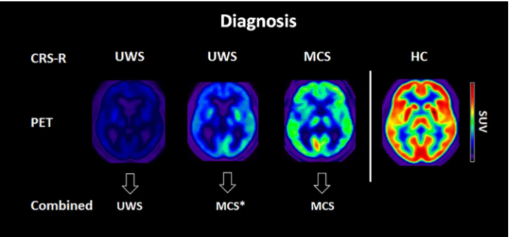
Preservation of Brain Activity in Unresponsive Patients Identifies MCS Star
13
0
0
Texte intégral
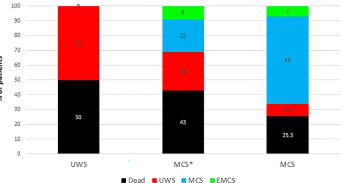
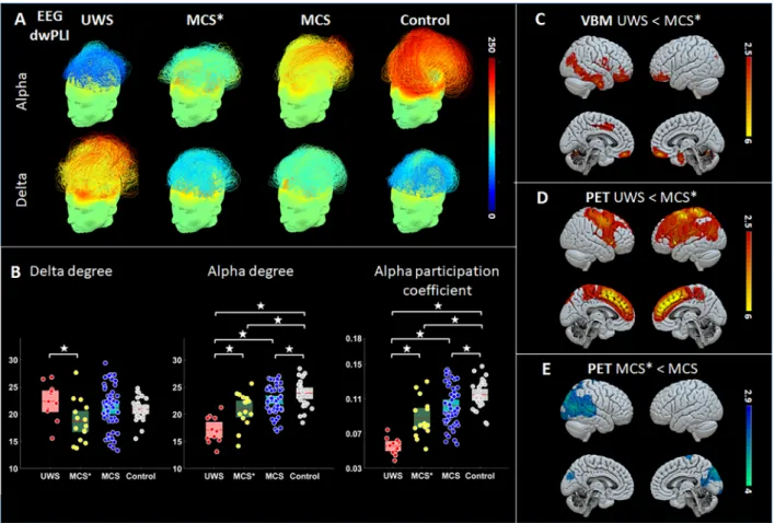
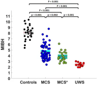
Figure

+2
Documents relatifs