Development of Lanthanide-Binding Tags (LBTs) as Powerful and Versatile Peptides For Use in Studies of Proteins and Protein Interactions
by
Langdon James Martin B.A. Chemistry Kalamazoo College, 2003
Submitted to the Department of Chemistry in Partial Fulfillment of the Requirements for the
Degree of Doctor of Philosophy at the
Massachusetts Institute of Technology May 2008
© 2008 Massachusetts Institute of Technology All rights reserved
Signature of Author: ____________________________________________________________ Department of Chemistry
May 14th, 2006
Certified by: ___________________________________________________________________ Barbara Imperiali Class of 1922 Professor of Chemistry and Professor of Biology Thesis Supervisor
Accepted by: __________________________________________________________________ Robert W. Field Haslam and Dewey Professor of Chemistry Chairman, Departmental Committee on Graduate Students
This doctoral thesis has been examined by a committee of the Department of Chemistry as follows:
______________________________________________________________________________ Timothy M. Swager
Chairman John D. MacArthur Professor of Chemistry Massachusetts Institute of Technology
______________________________________________________________________________ Barbara Imperiali Thesis Supervisor Class of 1922 Professor of Chemistry and Professor of Biology Massachusetts Institute of Technology
______________________________________________________________________________ Stuart S. Licht Assistant Professor of Chemistry Massachusetts Institute of Technology
______________________________________________________________________________ Karen N. Allen Professor of Physiology and Biophysics Boston University School of Medicine
Development of Lanthanide-Binding Tags (LBTs) as Powerful and Versatile Peptides
For Use in Studies of Proteins and Protein Interactions
by
Langdon James Martin
Submitted to the Department of Chemistry on May 14th, 2008 in partial fulfillment of the requirements for the Degree of Doctor of Philosophy in
Organic Chemistry
ABSTRACT
To determine the function of proteins of interest, chemical biologists employ their full panoply of techniques, including X-ray crystallography and NMR spectroscopy for structural information, and luminescence spectroscopy to determine cellular localization and binding interactions. These techniques generally require a spectroscopic handle, and trivalent lanthanide ions (Ln3+) are protean in this regard: an ordered Ln3+ can have many uses. Paramagnetic lanthanide ions can be exploited to align biomolecules in a magnetic field, and the anomalous signal of any lanthanide ion may be used to obtain phase information from X-ray diffraction data. Most lanthanide ions are luminescent upon sensitization by an organic fluorophore; for example, Tb3+ may be sensitized by the side chain of the amino acid tryptophan. Ln3+ emission profiles are distinct and long lived, and therefore ideal for imaging and resonance energy transfer experiments.
Lanthanide-binding tags (LBTs) are short peptide sequences developed to tightly and selectively chelate lanthanide ions. LBTs contain an appropriately placed tryptophan residue for sensitizing Tb3+ luminescence, and are composed entirely of encoded amino acids; incorporation at the genetic level into any protein of interest is thus facilitated. Subsequent expression of the tagged protein may be done using standard biochemical techniques, and the resultant protein contains a site for introducing an ordered lanthanide ion. Within this thesis is discussed the further optimization of LBTs for lanthanide affinity and structural stability. A combination of combinatorial peptide libraries and computational studies has resulted in the discovery of peptides that bind Tb3+ with dissociation constants of better than 20 nM. Furthermore, the concatenation of two LBT motifs has enabled the generation of so-called “double lanthanide-binding tags” (dLBTs). These slightly larger tags have additional advantages including the ability to bind two lanthanide ions, reduced mobility with respect to the tagged protein, and comparable or improved affinity for Ln3+ ions. Furthermore, since the lanthanide Gd3+ is a common handle for magnetic resonance imaging, progress has commenced to expand the utility of LBTs to include this type of experiment. Finally, LBT technology has been used to study the protein Calcineurin by uniquely modifying one calcium-binding loop to selectively bind and sensitize Tb3+.
Thesis Supervisor: Barbara Imperiali
ACKNOWLEDGEMENTS
Intelligence is like four-wheel drive. It simply enables you to get stuck in more-remote locations.
-Garrison Keillor
I would be remiss if I did not first acknowledge my advisor, Barbara Imperiali. I cannot honestly say whether I myself would have had the audacity to accept a first-year graduate student who didn’t know his twenty amino acids into my chemical biology lab, much less sell him on the LBT project, but Barbara did, and here I am. So, thank-you, Barbara, for promoting my development as a researcher—and giving advice when I did get stuck in more-remote locations, and for teaching me all sorts of chemistry and biology, for improving my ability to write like a scientist, for writing the grants that provided my funding, for providing a great environment for me to work in, and for putting up with my facetiousness through many a long minimeeting.
I also need to thank my fellow Imperiali lab rats (or “lablings,” as I am wont to call them). They helped make my days interesting, and I dare say I have learned something from all of them. Thank you all for your conversations, friendships, and for laughing at my jokes.
First and foremost, I thank my fellow LBT-ers for lots of helpful discussions, advice, and the occasional co-authorship. I am indebted to Mark Nitz, who really helped get my feet on the ground when I joined the lab, tolerated many inane babblings of a first-year grad student, provided loads of ideas of things to work on (many of which wound up in this thesis), and was the only person who also sported an “LBT-goatee”. Ryu Yoshida was unmatched in his motivation, and was an outstanding undergrad researcher. I thank Bianca Sculimbrene for her patience with me when she joined the lab, and for her panoply of insights, advice, and encouragement on chemistry, lab-work, and the world; also, she was the first to fully embrace the idea of LBT subgroup color-coordination at minimeetings. It has been a tremendous pleasure to work with Anne Reynolds, who is perpetually tenacious and unfailingly helpful, willing to lend an ear (or a car) when needed, and deserves credit for considerably improving my understanding of d-orbitals and expanding my enological palate. I have been glad to get to know Katja Barthelmes these past few months, and thank her for her explanations of protein NMR and her enthusiasm. Although I did not get to overlap with Kathy Franz, she initiated the LBT project, and therefore deserves accolades here as well.
As for the rest of you, I am going to run out of adjectives such as “friendly”, “kind”, “insightful”, “helpful”, “brilliant,” and “grad student/post-doc extraordinaire”; furthermore, the introduction “I am so glad I got to know and work with…” is hard to vary twenty-odd times, so suffice it to say that I could use these to describe any of you as both scientists and coworkers. Thank-you to Guofeng Zhang, Eranthie Weerapana, Beth Vogel Taylor, Eugenio Vazquez, Jay Troutman, Melissa Shults, Emi Sei, Matthieu “Chewie” Sainlos, Debbie Rothman, Debby Pheasant, Mary O’Reilly, Nelson Olivier, Elvedin Luković, Galen Loving, Angelyn Larkin, Wendy Iskenderian, Meredith Hartley, Christian Hackenberger, Susana Gordo, Juan Antonio “Nono” Gonzalez-Vera, Brenda Goguen, K. Jebrell Glover, Seungjib Choi, Mark Chen, Christina Carrigan, Dora Carrico-Moniz, Melanie Bonnekessel, Mayssam Ali, and Andreas Aemissegger. Good luck also to our rotator, Marcie Jaffee, and upcoming post-docs James Morrison and Cliff Stains.
I also owe some specific thank-yous. First, to Jebrell, Beth, and Guofeng, and also to Christina, Nelson, Mary, Eranthie, Bianca, both Marks, Angelyn, and Meredith for various
Melissa for teaching me how to use the fluorometer. Thank-you to my editors, who are mentioned after each chapter; and to paraphrase Seungjib, I defer to them all responsibility for any typographical errors. A special thank-you, too, to my podmates (and pod-annex) Guofeng, Christina, Mark, Meredith, Wendy, and Brenda, who have made sitting at my computer fun and enjoyable. Thank you to those who have done their research in A-Lab hoods: Mark, Mayssam, Debbie, Seungjib, Eranthie, Galen, Bianca, Anne, Angelyn, Andreas, and Melanie, who have made doing experiments there more exciting. Thank-you to the Barbarians volleyball team members: Chieftans Mary and Meredith, and Eranthie, Jebrell, Melissa, Debbie, Christina, Christian, Guofeng, Matthieu, Emi, Mark, Andreas, Jay, and Galen, for glorious victories and enjoyable defeats. Thank-you to my outstanding grad-student classmates, Galen and Elvedin, for going through tribulations such as Orals and Proposal Writing with me, and for bonding experiences such as trips to the Muddy, dinner parties, and to Galen especially for organizing a sailing…adventure. Thank-you to Wendy and Meredith for an awesome baby shower. And thank-you to my fellow Red Sox fans Debbie, Beth, Melissa, Bianca, Seungjib, and Anne for going to games at Fenway, and for going nuts with me in 2004 and 2007.
I would also be remiss if I neglected to thank Elizabeth Fong, who does so much behind the scenes and keeps the lab running so very smoothly that we don’t appreciate her enough.
Of course, my academic coworkers and mentors are in no way limited to my lab. I should first say thank-you to my thesis chair, Tim Swager, for helping to advise me the past five years. And I thank the rest of my committee, Stuart Licht and Karen Allen, as well.
The LBT project would have been nowhere near as exciting without our collaborators, who have been outstanding at filling in holes in my knowledge and skill set. Although I will say this in over half of my Thesis chapters, I am grateful for our collaboration with Karen Allen’s lab at Boston University. Karen Allen herself has been a great mentor, and has put together an outstanding lab, including Nick Silvaggi, Manashi Sherawat, Kelly Daughtry, and Ezra Peisach, who are unceasing in their perseverance of LBT-crystallography and MRI. I thank Harald Schwalbe and his lab at Frankfurt University, especially Martin Hähnke and Jens Wöhnert. I have enjoyed collaborating with Bracken King in Bruce Tidor’s lab at MIT, and learned a lot about in silico work from him. And it has been a fruitful collaboration with Patrick Hogan’s lab at Harvard Medical School; he has given me a lot of helpful advice about the Calcineurin project, and Alina Iuga did a lot of the initial cloning and expression and control experiments, and Huiming Li will continue the project. I thank Becky Sommers in the lab of Dan Nocera at MIT for working with me to try and get their old fluorometer up and running. And I thank the lab of JoAnne Stubbe for occasionally letting me borrow chemicals and use their equipment.
As I one day hope to teach Organic Chemistry and/or Biochemistry, it has been a privilege to work as a Teaching Assistant under some great teachers here, including Sarah Tabacco, Tim Swager, Kimberly Berkowski, Dan Kemp, and Stuart Licht.
And I would not be writing a thesis today were it not for some outstanding mentors in chemistry and in other subjects as well. I especially thank my advisors at Kalamazoo College, Jeff Bartz and Greg Slough, along with the rest of the chemistry department there, for encouraging me to major in chemistry and to apply to grad school, and for plenty of helpful advice along the way. I thank Dean Webster and Vicky Johnston Gelling at North Dakota State University for allowing me to do a summer internship there, and to the Webster lab for making me feel welcome while I was there.
I have been in school now for something like 22 or 23 years, and have had many, many excellent teachers, who encouraged me to continue to learn and who also deserve thanks and mention. They include (in roughly chronological order, and with apologies for any misspellings), Mrs. Dairkee, Mrs. Dialwis, Becky Glass, Dawn Hill, Helene Carrasquillo, Donald Washington, Deb Neher, Millard Neiman, Eleanor Balbach, Art Froehle, Kyoko French, Mark Wanvig, Elaine Lindstrom, Carol Adelsman, Richard Anderson, Keith Liuzzi, Gina Lammers, Dick Schwartz, Michele Intermont, Steve Mackey, John Fink, Tom Evans, Rose Bundy, Tanaka-sensei, Tim Tikker, and Paula Romanaux. And a special thank-you to my piano teacher from 1st to 12th grade,
Narissa Bach, who instilled in me the adage, “Knowledge for knowledge’s sake”.
I will round out the list with family, friends, and a handful of others who deserve thanks for expending time and energy and helping to shape and nurture me. Thank-you to my good friends Michael David, Pat Krefting, Sam Bunnett, Jim Li, Ben Berg, and Nick Chin. Thank-you to my godparents Sue Anderson and Tony Blodgett, and to Britta and Jamie Blodgett. I also thank Carol Wagner, Wayne and Peggy Krefting, John and Bonnie Rowell, Dick Masur, Nancy and Al Bunnett, Anne Walker and Torin Crissey, the Yokoyama family, Kit and Mitsuhiro and Léo Nagamura, Paul Johnson, Virginia Rickeman, Jim Gertmenian, and Philip Brunelle. (And I can’t forget about folks like Al Bumen, Dean A. Primer, Gene Vector, or Lou Minh Essons!)
I am privileged to have a family with so many wonderful aunts, uncles, and cousins, who have helped humor and inspire me. Thank-you Aunt Jean and Uncle Moffatt, Uncle Floyd and Aunt Becky, Uncle Aleck and Aunt Lynne, Uncle Martin and Aunt Bev, Uncle Gardiner and Aunt Jane, Uncle Kendall and Aunt Gwen, Uncle Mart and Aunt Marge, Great-Uncle Al and Great-Aunt Nell, and Great-Auntie Mabel. Thanks to my cousins Mary Rebecca, Katie Sue, Grier, Rachel, Megan, Martin, Walter, John, Julie, Amanda, Michael, and your families. Thank-you to my parents-in-law, Mark and Debbie Ward, to my sisters-in-law Erica and Meredith Ward, and to my brother-in-law-to-be, José Augustin “Jandin” Mejia-Comacho.
Thank-you, Granddaddy, for your warmth, your encouragement, your love, and your music, all of which have been an inspiration to me. Thank-you, Grandmother, for the kindness and the love that you gave me. Thank-you, Grandma, for the joy and the love that you gave me.
I dedicate this thesis to my mother, Elsie F. Myers Martin, to my father, Raymond J. Martin Jr., to my sister, Celia Grace Martin, and to my wife, Anna Ward Martin.
Mom and Dad, I feel like I am only now beginning to understand the love and energy that you put into raising me. I do know that you have been wonderful parents to me, and for that I thank you. I am deeply grateful to you both for your patience, your humor, your vivacity, your creativity, your wisdom, and your love.
Celia, thank-you for keeping me grounded, and for your tenacity and honesty and love. You are an awesome sister.
Anna, I am both lucky and happy to have you as my wonderful wife. I thank you for your encouragement, for your empathy, for your belief in me, for your trust and commitment, for your humor, and most of all for your love. I couldn’t have done this without you.
Finally, to my daughter, Eliza: you are not even a month old as I write this, and I look forward to being a father to you. May your life be long and happy, and may you be even smarter than your parents.
Table of Contents Abstract………3 Acknowledgments………4 Table of Contents……….7 List of Figures………10 List of Tables……….12 List of Abbreviations……….14 hi Chapter 1. Introduction 0001-1._General Introduction to Biological Probes………19
0001-2._General Introduction to Lanthanides………19
0001-3._The Lanthanide Ions as Biological Probes………23
0001-4._Introduction to Lanthanide-Binding Tags (LBTs)………26
0001-5._Preliminary, Disulfide Bond-Containing Lanthanide-Binding Tags………28
0001-6._Development of a Powerful Combinatorial Screen………30
0001-7._Lanthanide-Binding Tags Evolved Through the Combinatorial Screening Process…33 0001-8._Dissertation Objectives………36
Acknowledgements ………37
References ………37
hi Chapter 2. Sequence Refinement of Lanthanide-Binding Tags Introduction ………44
Results and Discussion 0002-1._Additional Combinatorial Libraries to Evolve the SE2 Sequence………45
0002-2._Tb3+-Bound Water Molecules and Relative Luminescence Intensity of LBTs………47
0002-3._Results of Single Mutations at Specific Positions in the LBT………49
0002-4._Studies on the Effects of Deleterious Mutations on the LBT Sequence………51
Conclusions ………52
Experimental ………53
Acknowledgements ………60
References ………61
Chapter 3. Attempts Towards and IR-Emitting Lanthanide-Binding Tag
Introduction ………63
Results and Discussion 0003-1._Synthesis and Characterization of LBT-IR1………64
0003-2._Synthesis and Characterization of LBT-IR2………66
Conclusions ………67
Experimental ………68
Acknowledgements ………70
References ………71
hi Chapter 4. Double-Lanthanide-Binding Tags Introduction ………72
Results and Discussion 0004-1._Design and Selection of the dLBT Sequences………73
0004-2._Preparation of dLBT Peptides and a dLBT-Ubiquitin Construct………74
0004-3._Photophysical Characterization of dLBT Peptides and Proteins………77
0004-4._Characterization of GPGdSE3-ubiquitin by NMR………79
0004-5._Characterization of GPGdSE3-ubiquitin by X-Ray Crystallography………81
Conclusions ………83
Experimental ………84
Acknowledgements ………93
References ………94
hi Chapter 5. LBT and dLBT Redesign based on dLBT Structural Data Introduction ………96
Results and Discussion 0005-1._Design of SE3 Mutants to Address Conformational Questions………97
0005-2._Synthesis and Photophysical Characterization of SE3 Mutants………100
0005-3._Crystallographic Studies of SE3α, SE3β, and SE3ε………102
0005-4._Studies of dLBT-Ubiquitin Mutants Containing SE3α and SE3β………104
0005-5._Optimization of the dLBT Expression System………106
0005-6._Modification of the Gly-Pro-Gly Motif in the dLBT………107
0005-7._Photophysical Characterization of Gly-Ala-Gly Motif-Containing dLBTs…………110
0005-8._dLBTs Based on SE2: Attempts to Generate the Brightest Possible dLBT…………111
0005-9._The Brightest Known dLBT Construct………113
Conclusions ………113
Experimental ………114
Acknowledgements ………131
References ………132
Chapter 6. Lanthanide-Binding Tags for Magnetic Resonance Imaging
Introduction ………134
Results and Discussion 0006-1._Assessment of Existing LBT Sequences for Gd3+ Binding………135
0006-2._A Split-Pool Library to Optimize LBTs for MR-Imaging Studies………135
Conclusions ………138
Experimental ………139
Acknowledgements ………143
References ………143
hi Chapter 7. Using LBT Technology to Study the Protein Calcineurin Introduction ………145
Results and Discussion 0007-1._Preliminary Studies and Mutations to Eliminate Background Luminescence………148
0007-2._Optimization of Site IV Luminescence Output in Calcineurin Mutants………149
0007-3._Use of Competitive Ligands to Study the Interactions of Site IV with Sites I – III…152 0007-4._Initial LRET Experiments using the construct CNm4-BA17C-TMR………154
0007-5._A CNm4 Construct Labeled on the CaM-Binding Domain………158
0007-6._Generation of a Simpler System, with Only CNB, for Use in LRET Studies………159
0007-7._Generation of CNB-Binding Domain Peptides CaNAdα and P2465………161
0007-8._Photophysical Experiments with CNBm7………161
Conclusions ………163 Experimental ………164 Acknowledgements ………179 References ………180 hi Appendix Select SPECFIT Data Files………183
Curriculum Vitae………184
List of Figures
Chapter 1. Introduction
Figure_1-1.__The Lanthanide metal series………20
Figure_1-2.__Emission spectrum of Tb3+………21
Figure_1-3.__Excitation of Tb3+ luminescence………22
Figure_1-4.__The security measures on euro banknotes………23
Figure_1-5.__Cartoon representation of X-ray crystallography………24
Figure_1-6.__Lanthanide ions promote orientation in a magnetic field………25
Figure_1-7.__Graphical representation of an LBT………27
Figure_1-8.__Comparison of the size of an LBT to GFP………28
Figure_1-9.__Expression of an LBT-protein construct is straightforward………28
Figure_1-10._Generation of a split-and-pool combinatorial library………30
Figure_1-11._The combinatorial libraries to generate tighter and brighter LBTs………32
Figure_1-12._Results from Library 1………34
Figure_1-13._Results from Library 2………34
Figure_1-14._Results from Library 3………35
Figure_1-15._Results from Library 4………36
hi Chapter 2. Sequence Refinement of Lanthanide-Binding Tags Figure_2-1.__The Crystal Structure of SE2 Bound to Tb3+………44
Figure_2-2.__Results from Library 5………45
Figure_2-3.__Results from Library 6………46
Figure_2-4.__Results from Library 7………47
Figure_2-5.__Combination of Results from Libraries 5 and 6 to Optimize the LBT…………47
hi Chapter 3. Attempts Towards and IR-Emitting Lanthanide-Binding Tag Figure_3-1.__Lanthanide luminescence maxima range from the visible to the near-IR………63
Figure_3-2.__The IR-emitting phthalazine moiety………64
Figure_3-3.__Synthesis of LBT-IR1………65
Figure_3-4.__LBT-IR1 sensitizes Tb3+ and Eu3+………66
Figure_3-5.__Normalized excitation scans of LBT-Tb3+ complexes………67
hi Chapter 4. Double-Lanthanide-Binding Tags Figure_4-1.__The design strategy to convert the single-LBT into the double-LBT………73
Figure_4-2.__Expression and purification strategy to obtain pure GdSE3 peptide………75
Figure_4-3.__Expression and purification strategy to obtain pure GPGdSE3-ubiquitin………76
Figure_4-4.__Purification and gel-visualization of the dLBT constructs………77
Figure_4-5.__NMR data showing that the dLBT does not alter the structure of ubiquitin……80
Figure_4-6.__RDCs in single-LBT- or double-LBT-containing ubiquitin………80
Figure_4-7.__Crystallization of GPGdSE3-Ubiquitin………81
Figure_4-8.__The crystal structure of GPGdSE3-Ubiquitin………82
Chapter 5. LBT and dLBT Redesign based on dLBT Structural Data
Figure_5-1.__Structure of the dLBT portion of the crystal………96
Figure_5-2.__Comparison of Ramachandran space of four residues in the SE2 and___________ _________ __GPGdSE3 crystals………97
Figure_5-3.__Comparison of the calculated folding energies of different residues at___________ _________ __positions 4 and 6………98
Figure_5-4.__Differences in folding energies of all hypothetical double mutants………99
Figure_5-5.__Crystal Structure of SE3β………103
Figure_5-6.__System for generating GPGdSE3 peptide for crystallization studies………106
Figure_5-7.__Cleavage of ubiquitin-GPGdSE3 by mTEV protease was not facile………107
Figure_5-8.__Plasmids for generating GAGdSE3 as a peptide and a ubiquitin tag………108
Figure_5-9.__Cleavage of H8-ubiquitin-ENLYFQGAGdSE3 by mTEV protease is___________ _________ __extremely facile under a variety of conditions………109
Figure_5-10._Purification of GAGdSE3-ubiquitin………110
Figure_5-11._Purification of GAGdSE2-ubiquitin………112
hi Chapter 6. Lanthanide-Binding Tags for Magnetic Resonance Imaging Figure_6-1.__Images of MRI contrast with the LBT mSE3………134
Figure_6-2.__Design of the linker region for the MRI-LBT library………136
Figure_6-3.__The synthesis and screening process for the “LanGdoN” combinatorial_________ _________ __peptide libraries………137
Figure_6-4.__Library LanGdoN1………138
hi Chapter 7. Using LBT Technology to Study the Protein Calcineurin Figure_7-1.__When a specific site in a calcium-binding protein is modified to be_____________ _________ __LBT-like, it may be selectively labeled with Ln3+………145
Figure_7-2.__Crystal structure of calcineurin………146
Figure_7-3.__The effects of [Ca2+] and CaM on the enzymatic activity of calcineurin………147
Figure_7-4.__Sequence of CNB used in this study………148
Figure_7-5.__Abilities of various LBTs and CN-mutants to sensitize Tb3+………150
Figure_7-6.__Detail of calcineurin-B, metal-binding Site IV………152
Figure_7-7.__Addition of metal chelator to CNm7 and Tb3+………153
Figure_7-8.__Addition of metal chelator to CNm7 and Tb3+ with buffered [Ca2+]free…………154
Figure_7-9.__Plans for three possible calcineurin LRET experiments………156
Figure_7-10._Detail of the calcineurin structure, showing BAla17 and ACys153………156
Figure_7-11._Gated luminescence scans of TMR-labeled calcineurin mutants………157
Figure_7-12._Luminescence decay of CNm4-BA17C-TMR is independent of [Ca2+]………158
Figure_7-13._The plasmids for expressing CNBm7 and CNBm7-A17C………159
Figure_7-14._Generation and purification of CNBm7-A17C-TMR………160
Figure_7-15._Gel of the purification of CNBm7-A17C-TMR………160
Figure_7-16._Concentrations of [Tb3+] and [Ca2+] have no effect on the normalized___________ __________ _emission spectrum of CNBm7-A17C-TMR………162
Figure_7-17._The presence or absence of CaNAdα or P2465 does not affect the______________ __________ _ Tb3+-to-TMR LRET of CNBm7-A17C-TMR………163
List of Tables
Chapter 1. Introduction
Table_1-1._Comparison of the Properties of Ca2+ and Tb3+………20
Table_1-2._Rationally-Designed LBT Sequences from Calcium-Binding Proteins……….29
Hi Chapter 2. Sequence Refinement of Lanthanide-Binding Tags Table_2-1._Photophysical Data of Select Single-LBTs………49
Table_2-2._Mutational Analyses of SE2 Amino Acid Residues………50
Table_2-3._Photophysical Data of LBT Mutants with Specific Deleterious Mutations………52
Table_2-4._Winning Peptides from the “TEN_DAYS” Library………58
Hi Chapter 3. Attempts Towards and IR-Emitting Lanthanide-Binding Tag Table_3-1._Sequence Design of Potential IR-Emitting-Ln3+-Sensitizing LBTs……….64
Hi Chapter 4. Double-Lanthanide-Binding Tags Table_4-1._Tb3+-Binding Affinity and Luminescence Intensity of Select Single-LBTs………74
Table_4-2._Sequences of Double-LBTs and the Progenitor Single-LBT SE3………74
Table_4-3._Initial Double-LBT Peptide Photophysical Experiments……….75
Table_4-4._Summary of Double-LBT Photophysical Data………78
Table_4-5._The GPG Motif and the Ubiquitin Protein Have No Inherent Effect on LBT_______ ________ _Luminescence………79
H Chapter 5. LBT and dLBT Redesign based on dLBT Structural Data Table_5-1._Sequences of Computationally Designed SE3 Mutants………100
Table_5-2._Photophysical Data from Computationally Designed SE3 Mutants………101
Table_5-3._Competitive Titrations with SE2, SE3β and SE3ε, Between Tb3+ and_____________ __________Various Lanthanide Ions………102
Table_5-4._Sequences of SE3α- and SE3β-Based dLBT-Ubiquitin Constructs………104
Table_5-5._Summary of Photophysical Data for SE3α- and SE3β-Containing________________ __________dLBT-Ubiquitin Constructs………105
Table_5-6._Photophysical Characteristics of GAG Motif-Containing dLBT-Ubiquitin_________ __________ Constructs...………110
Table_5-7._Sequences of the SE2-Based dLBTs……….111
Table_5-8._Photophysical Characterization of Double-LBTs Based on SE2………112
Table_5-9._Potophysical Characterization of GPGdSE3-NhPMM………113
Hi Chapter 6. Lanthanide-Binding Tags for Magnetic Resonance Imaging Table_6-1._Summary of LBT-Gd3+ Affinity for Single-LBTs used in MRI Experiments……135
Table_6-2._Summary of GPGdSE3-Ubiquitin Log β Values for Gd3+ and Tb3+………135
Chapter 7. Using LBT Technology to Study the Protein Calcineurin
Table_7-1._Alignment of the EF-Hand Motifs of Calcineurin-B with SE3………149 Table_7-2._CNB-Site IV Sequences of LBT-Like Calcineurin Mutants………150 Table_7-3._Luminescence Intensity and Bound Water Molecules for CN Mutants 4 – 7……151 Table_7-4._Metal-Chelation Buffers to Make a Variety of Free [Ca2+] Concentrations………154
Table_7-5._CNB-Binding Domain Peptides………161
Table_7-6._Gated Luminescence Scans and τDA Determination with 2 µM__________________
_________ _CNm4-BA17C-TMR………171
Table_7-7._Gated Luminescence Scans and τDA Determination with 1 µM__________________
_________ _CNBm7-A17C-TMR………178
Table_7-8._Gated Luminescence Scans and τDA Determination with 1 µM__________________
_________ _ CNBm7-A17C-TMR in the Presence of 5.82 µM CaNAdα Peptide………179 Table_7-9._Gated Luminescence Scans and τDA Determination with 1 µM__________________
_________ _ CNBm7-A17C-TMR in the Presence of 5.82 µM P2465 Peptide………179
List of Abbreviations
Standard one- and three-letter codes are used for the amino acids.
One-letter code Three-letter code Amino Acid
A Ala Alanine
C Cys Cysteine
D Asp Aspartic acid (Aspartate)
E Glu Glutamic acid (Glutamate)
F Phe Phenylalanine G Gly Glycine H His Histidine I Ile Isoleucine K Lys Lysine L Leu Leucine M Met Methionine N Asn Asparagine P Pro Proline Q Gln Glutamine R Arg Arginine S Ser Serine T Thr Threonine V Val Valine W Trp Tryptophan Y Tyr Tyrosine
Standard one-letter codes are used for the nucleotides.
One-letter code Nucleotide
A Adenosine
C Cytidine
G Guanosine
Other abbreviations are as follows.
A’ A lanthanide-specific constant used to determine q
Ac Acetyl
Ac2O Acetic anhydride
AI Auto-Inhibitory (domain)
ANP 3-amino-3-(2-nitrophenyl)propionic acid
apo Unbound to metal-ion
β Binding constant; KD = 10−log β
βAla Beta-alanine (amino acid residue)
BME, βME Beta-mercaptoethanol; HOCH2CH2SH
BSA Bovine serum albumin (protein)
C-terminus The carboxy-terminal end of a peptide calcd. Calculated
CaM The protein Calmodulin
CIP Calf Intestinal alkaline Phosphatase
CN The protein Calcineurin
CNA Calcineurin subunit A (the larger, catalytic subunit)
CNB Calcineurin subunit B (the smaller, Ca2+-binding, regulatory subunit)
CNm# Calcineurin-B mutant (# = 1 - 7). This notation indicates that both subunits are present CNBm7 Calcineurin-B mutant 7. This notation indicates that only the
B-subunit is present
D2O Deuterated water; deuterium-oxide
DAPase Diamino-peptidase (protease)
dATP deoxyAdenosine TriPhosphate
DCM Dichloromethane; CH2Cl2
dCTP deoxyCytidine TriPhosphate
de novo Latin: “from the new”
dGTP deoxyGuanosine TriPhosphate
DHB Dihydroxybenzoic acid
DIC N,N'-Diisopropylcarbodiimide
DIPEA N,N-Diisopropylethyl amine; Hünig's base
dLBT double-Lanthanide-Binding Tag
DMAP 4-(Dimethylamino)pyridine DMF N,N-Dimethylformamide; (CH3)2NCHO
DMSO Dimethylsulfoxide; (CH3)2SO
DNA Deoxyribonucleic acid
DTT Dithiothreitol (Cleland's reagent); HSCH
2CH(OH)CH(OH)CH2SH
E The percentage of energy transferred
ε Molar extinction coefficient (at a certain wavelenght)
E. coli Escherichia coli (bacteria)
EDT 1,2-Ethanedithiol; HSCH2CH2SH
EDTA Ethylenediaminetetraacetic acid
EF-hand A calcium-binding peptide consensus sequence found in native proteins
EGTA Ethylene glycol-bis-(2-aminoethylether)-N,N,N',N'-tetraacetic acid
eLB Enhanced LB bacterial growth media, based on Autoinduction
buffers
eq. Equation equiv. Equivalents
et al. Latin: “and others”
ESI MS, ESI-TOF MS ElectroSpray Ionization (Time-Of-Flight) Mass Spectroscopy Fmoc Fluoren-9-ylmethoxycarbonyl
FRET Fluorescence Resonance Energy Transfer
GFP Green Fluorescent Protein
GST Glutathione-S-Transferase (protein)
η Refractive index
HATU O-(7-azabenzotriazole-1-yl)-1,1,3,3-tetramethyluronium hexafluorophosphate
HBTU 2-(1H-benzotriazole-1-yl)-1,1,3,3-tetramethyluronium
hexafluorophosphate
HEDTA N-(hydroxyethyl)-ethylenediaminetriacetic acid
HEPES N-(2-hydroxyethyl)piperazine-N'-ethanesulfonic acid
His6, H6, His6-tag, His-tag Protein purification tag comprised of six consecutive histidine residues
HMBA 4-hydroxymethylbenzoic acid
HOBt N-hydroxybenzotriazole
HPLC High-Performance (or -Pressure) Liquid Chromatography
I(t) Luminescence intensity at time (t)
IMAC Immobilized Metal-ion-Affinity Chromatography; purification
using Ni-NTA resin and a His-tag
In vitro Performed outside the context of the cell; Latin: "in glass"
In vivo Performed in the context of living cells; Latin: "in life" IPTG Isopropyl-β-D-thiogalactopyranoside
IR Infrared
J Spectral overlap term
J-coupling In NMR, indirect dipole-dipole coupling
κ2 The orientation factor, taken as 2/3
KD Dissociation constant
KM Michaelis constant for enzyme-substrate binding
λ Wavelength
LB Lysogeny Broth, a nutrient-rich bacterial growth medium
LBT Lanthanide-Binding Tag
λem Emission wavelength
λex Excitation wavelength
Ln Any lanthanide metal
LRET Luminescence Resonance Energy Transfer
MALDI, MALDI-TOF MS Matrix-Assisted Laser-Desorption-Ionization (Time-Of-Flight) Mass Spectroscopy
MBP Maltose-Binding Protein
MeCN Acetonitrile; CH3CN
MES 2-morpholinoethanesulfonic acid
MESNA 2-mercaptoethanesulfonic acid
min Minutes
Mmt Monomethoxytrityl MOPS 3-(N-morpholino)propanesulfonic acid
MR (Nuclear) Magnetic Resonance
MRI (Nuclear) Magnetic Resonance Imaging
MS Mass Spectroscopy
mTEV mutant Tobacco Etch Virus protease
N-terminus The amino-terminal end of a peptide
NaCl Sodium chloride
NaOAc Sodium acetate
Ni-NTA Nickel-NTA agarose resin
NMP N-MethylPyrrolidinone (1-methyl-2-pyrrolidinone)
NMR Nuclear Magnetic Resonance
NOE Nuclear Overhouser Effect
NTA Nitrilo Triacetic acid
OAc Acetate
OD Optical Density
PAGE PolyAcrylamide Gel Electrophoresis
PAL Peptide Amide Linker
PBS Phosphate Buffered Saline solution
PCR Polymerase Chain Reaction
PEG Polyethyleneglycol
Phe-pNO2 para-nitrophenylalanine residue
pip Piperidine PS Polystyrene
QD Quantum yield of the donor
q Number of lanthanide-ion-coordinated water molecules
R Distance
R0 The Förster distance
RCF Relative Centrifugal Force
RDC Residual Dipolar Coupling values
RET Resonance Energy Transfer
RMSD Root Mean Square Deviation
RNA Ribonucleic acid
RP-HPLC Reverse-Phase HPLC
RPM Rotations per minute
SAD Single-wavelength Anomalous Diffraction
SDS-PAGE Sodium Dodecyl-Sulfate PolyAcrylamide Gel Electrophoresis
sLBT single-Lanthanide-Binding Tag
SPPS Solid Phase Peptide Synthesis
std. dev. Standard deviation
τ Lifetime
τD Lifetime of the donor
τDA Lifetime (of the donor) in the presence of the acceptor
t½ Half-life
TBS Tris-buffered saline
tBu tert-butyl
TCEP tris-(2-carboxyethyl)phosphine P(CH2CH2CO2H)3
TEV Tobacco Etch Virus
TFA Trifluoroacetic acid; CF3CO2H
TIS Triisopropyl silane; (CH3)3SiH
TMG Tetramethylguanidine TMR Tetramethylrhodamine
TNBS Trinitrobenzene sulfonic acid
Tris Tris(hydroxymethyl)aminomethane Trt Trityl
Ubiq Ubiquitin (protein)
UV Ultraviolet UV-Vis Ultraviolet-visible
vide infra Latin: "see below"
vide supra Latin: "see above"
Vmax Maximum (enzymatic) turnover velocity
Chapter 1
Introduction
Portions of this chapter have been published in QSAR & Combinatorial Science1 and in Journal
of the American Chemical Society2 as noted in the text. Copyright © 2005, WILEY-VCH Verlag
GmbH & Co and © 2007, American Chemical Society, respectively.
1-1. General Introduction to Biological Probes1
With the sequencing of the human genome, it has become widely accepted that dynamic protein/protein interactions are an essential component of biological complexity. Chemical tools to detect and probe proteins and biomolecular interactions will provide a deeper understanding of the nature, regulation and function of protein interactions, which are central to cellular existence. Many of the currently available protein tags permit the incorporation only of a single functionality, such as the Green Fluorescent Proteins (GFPs) and GPF variants for fluorescence,3,4 selenomethionine to provide a heavy atom for X-ray crystallography,5,6 and paramagnetic metal ions for NMR spectroscopy.7 Of great use would be a versatile protein tag, which should display diverse physical and spectroscopic properties, enabling researchers to monitor proteins of interest with different and potentially complementary biophysical techniques. Lanthanide ions are endowed with an extensive list of physical properties that are amenable to a variety of spectroscopic and crystallographic studies (vide infra). This, in tandem with the absence of lanthanides from living systems, makes these ions ideal handles for the study of proteins’ structure and interactions. To be useful, the lanthanide ion (or ions) must be site-specifically attached to the target protein. This thesis will discuss the development of one such tool: the Lanthanide-Binding Tag.
1-2. General Introduction to Lanthanides
The lanthanide metals, elements 57 – 71, are shown in Figure 1-1. The lanthanide moniker “Rare Earths” comes not so much from the natural abundance of these elements (which ranges from 2 – 4 orders of magnitude more common than gold, for example)8, but from the initial difficulties encountered in purification.9 (It apparently required 15,000 fractional crystallizations to isolate lutetium!)10 Many are named after locations in Sweden, where these
(ytterbium, terbium, and erbium) and yttrium—a distinction no other person or place can claim.10 Terbium is central to this thesis and to the Lanthanide-Binding Tag technology.
Figure 1-1. The Lanthanide metal series.11
Many of the interesting and useful photophysical properties of lanthanide ions are due to the electrons in the 4f orbitals. The 4f electrons are located within the outermost orbitals (the 5s25p6), and are shielded from the surroundings, and making the luminescent f-f transitions nearly ligand-independent.12 In general, lanthanides are most stable as Ln3+ ions, in the [Xe] configuration, where N is the atomic number of the lanthanide. As aqueous ions, all Ln3+ ions are remarkable similar to Ca2+, with only a slight difference in atomic radius, as shown in Table 1-1, although the lanthanides are absent from normal living systems. Both are very “hard” ions and therefore oxophilic, with coordination spheres that are determined by sterics, and are capable of rapid ligand-exchange.12,13
57
4 N− f
Table 1-1. Comparison of the Properties of Ca2+ and Tb3+ (the lanthanide most important to this thesis)
Atom Most stable
oxidation state
Ionic radiusa Preferred coordination
Ligand preference
Calcium (Ca) +2 1.18 Åb 8-9 ligands, spherical O > N > S
Terbium (Tb) +3 1.10 Åb 8-9 ligands, spherical O > N > S
a
Ionic radii shown are for the nine-coordinate complexes.14
b Lanthanide(III) nine-coordinate ionic radii range from 1.03 Å (for Lu3+) to 1.22 Å (for La3+).13,14
Many lanthanide ions, including Tb3+, exhibit distinct luminescence emission spectra, due to f–f electronic transitions. Figure 1-2 shows the (false-color) emission spectrum for Tb3+, along with the transitions that give rise to each peak. Because f–f electric dipole transitions are forbidden by parity rules (‘Laporte-forbidden’), it is much more facile to excite Ln3+ ions via an organic fluorophore.12,15 This is accomplished as diagrammed in Figure 1-3 (based on Ref. 12). First, an organic fluorophore—the side-chain indole of tryptophan is used throughout this thesis—is excited (A) by irradiation from a source such as a fluorometer, making an excited singlet. This singlet may give up a photon and fluoresce (B), or it may undergo
J S
L
1 2 +
is known as phosphorescence (C), but it may instead undergo another (non-radiative) exchange of energy with a nearby Tb3+ ion. The energy of now-excited Tb3+ may be lost by a non-radiative process (vide infra), or the Tb3+ may luminesce (D), giving rise to the spectrum in Figure 1-2. It should be noted that this process is formally known as “luminescence”, and is referred to as such throughout this text, because the ground state of Tb3+ is not a singlet (having six unpaired f electrons) and therefore is neither fluorescence nor phosphorescence.
Figure 1-3. Excitation of Tb3+ luminescence. A. An organic fluorophore, such as the indole ring of tryptophan, is excited by UV light. B. The excited state singlet may fluoresce. C. If the excited singlet undergoes intersystem crossing, the excited triplet state may phosphoresce. D. Or, the energy may be transferred to the nearby Tb3+, which can luminesce. (Based on Ref. 12.)
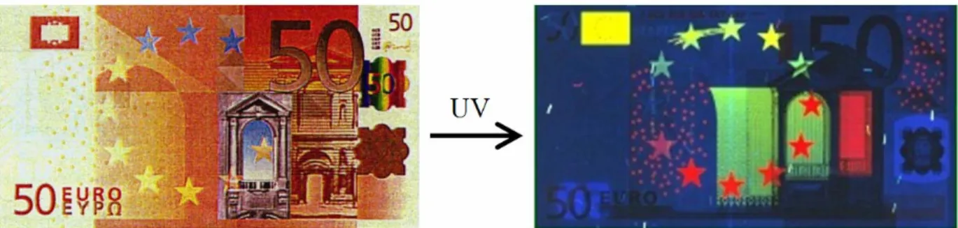
Lanthanides and lanthanide luminescence have found a variety of uses in both biological and non-biological systems.16 Perhaps one of the most interesting (and appropriate) “real-world” applications is in the security measures of bank-notes. Luminescent europium chelates are believed to be responsible for the red color that shows up on the Euro paper currency under ultraviolet light (Figure 1-4);17 it need not be pointed out that the metal and the currency share a
Figure 1-4. The security measures on euro banknotes are believed to contain europium chelates that emit red luminescence under ultraviolet light.
1-3. The Lanthanide Ions as Biological Probes1,2
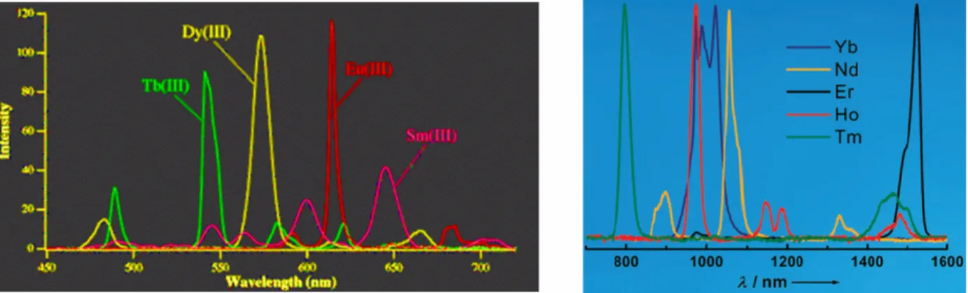
Complementing the unique emission profile, perhaps the most useful property of Tb3+ luminescence is that it is extremely long-lived, with a lifetime in the milliseconds; this, too, is a result of the parity-forbidden f–f transitions. Eu3+, while it cannot be sensitized by any canonical amino acid, also has a millisecond-length lifetime, making these lanthanides popular for use in time-resolved luminescence measurements.18-20 Other lanthanides luminesce as well, although
most of these have slightly shorter (microsecond) lifetimes.16 Some, such as Nd3+ and Yb3+, emit in the near-IR, and are growing in interest for applications such as tissue-imaging.19,21,22
The broad availability of spectroscopic equipment has made luminescence a popular choice for studying biological processes. The millisecond lifetimes of Tb3+ and Eu3+ luminescence, being significantly longer than the nanosecond lifetimes of most organic fluorophores, provide greatly increased sensitivity by elimination of background fluorescence. This, in turn, has generated considerable interest for the use of these ions in biological and biochemical assays,15,23-27 and in resonance energy transfer experiments.28-30 With regards to lanthanide luminescence it is important to note, as was alluded to previously, that excited lanthanide ions may also undergo nonradiative decay. When a molecule of water is present in the inner coordination sphere of Tb3+ or Eu3+, excited-state energy is rapidly transferred into vibrational energy of the O–H bond.31 Lanthanide complexes used for bioluminescent assays are therefore almost invariably made of ligands of sufficient number or bulk such that water molecules are prevented from directly coordinating the metal ion.
Although this thesis is concerned exclusively with the study of proteins, it should be noted that non-protein analytes are also amenable to study via lanthanide-based sensors: recent studies were published utilizing terbium luminescence in the detection of sugars32 and
phosphopeptides.33 In addition, there have been preliminary explorations into the use of lanthanides as probes of protein redox processes.34
Lanthanide ions have also greatly assisted protein structure determination both in the solid and solution state. This versatility is a result of various physical properties. For example, the abundant non-valence electrons interact strongly with X-rays. In protein X-ray crystallography, a series of crystal diffraction patterns are collected, integrated, and scaled. These contain information about the amplitude of the X-rays, but the phase information is needed to solve the structure via Fourier Transformation (Figure 1-5). The strong scattering power and anomalous scattering of lanthanides can be used to phase X-ray diffraction data in macromolecular crystallography;35-37 the anomalous signal is calculated to be roughly four times as powerful as that from a selenomethionine.38
Figure 1-5. Cartoon representation of ray crystallography. A protein crystal is bombarded with X-rays, yielding diffraction patterns that are integrated and scaled. In order to solve the phase information, the presence of an ordered, anomalously scattering heavy atom such as a lanthanide ion or selenium can be used. One lanthanide ion has the phasing power of about four selenium atoms.
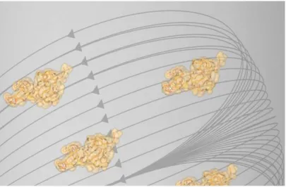
Furthermore, all lanthanide ions, with the exception of La3+ and Lu3+, have one or more unpaired electrons in f orbitals. And because these paramagnetic ions have an anisotropic susceptibility tensor, ordered Ln3+ ions can be used in NMR spectroscopy to partially orient proteins in the magnetic field, represented in Figure 1-6.39-42 This leads to magnetic dipolar
interactions between nearby spins that are otherwise averaged to zero in solution due to molecular tumbling. These so-called residual dipolar couplings (RDCs) can be observed and have proven to yield useful long-range restraints for structure determination, and in fact the measurement and use of RDCs has become an essential tool for structure determination of proteins.43-45 Unlike NOEs and scalar 3J-couplings that report on short-range distances (< 5 Å)
and dihedral angles, RDCs deliver valuable long-range orientational information. In addition, paramagnetic pseudocontact shifts can be used for the determination of the binding geometry of small ligands to protein receptors.46
Figure 1-6. When biomolecules such as proteins contain one or more lanthanide ions, the ions’ anisotropic magnetic susceptibility promote orientation in a magnetic field. This enables long-range coupling interactions to be observed. (This figure was designed and created by Nicholas Silvaggi.)
Finally, Gd3+ is unique among the trivalent lanthanide ions in that, with a [Xe]4f7 configuration, it contains seven unpaired electrons. It therefore has a strong paramagnetic relaxation enhancement effect as well as a relaxation time that is significantly longer than other lanthanides.39 This, coupled with the aforementioned rapid ligand exchange, makes it a useful metal for chelates used in Magnetic Resonance Imaging (MRI).39,47,48 It is noteworthy, though, that in the case of MRI it is necessary to have a molecule of water in the inner coordination sphere of the Gd3+ (where the relaxation enhancement takes place), and it is beneficial to have
two or more. Therefore, protein tags that work well for MRI will likely be suboptimal for luminescence-based applications and vice versa. Recently, other lanthanides have begun to attract interest for MRI applications as well.48
1-4. Introduction to Lanthanide-Binding Tags (LBTs)1,2
The wide range of physical information that can be obtained from the localization of different lanthanide ions, coupled with the absence of the rare earths from natural biological systems, has fostered significant interest in engineering tools to incorporate lanthanides into biomolecules. Various methods for the incorporation of lanthanide ions into proteins have been explored. In specialized applications, the similarity of trivalent lanthanides (Ln3+) to divalent
calcium (Ca2+) in ionic radius and oxophilicity enables direct incorporation into calcium-binding
proteins.35,40,42,49 The majority of proteins, however, lack native calcium-binding sites, and therefore this technique is clearly limited in scope. Another approach has been to incorporate lanthanide-chelating prosthetic groups as either the side-chain of a non-natural amino acid,50 or via the chemical modification of a uniquely reactive amino acid residue.36,51,52 These chelates can bind the lanthanide extremely tightly, and may incorporate a sensitizer, but this method requires considerable manipulation and significant case-by-case optimization.
A more viable tool for proteomics would be a tag that is amenable to incorporation into proteins at the DNA level via standard molecular biology techniques, thereby avoiding cumbersome, and often inefficient, post-translational chemical modifications. Indeed, despite the relatively large size (ca. 240 residues), the great success of the GFPs is in no small part due to the convenient methods for engineering GFP-fusion proteins.53 Short peptide sequences that comprise encoded amino acids and selectively bind lanthanide ions would be advantageous probes for proteomic analysis. These sequences would enable the introduction of the physical properties of lanthanide ions while retaining the ease of protein tagging via genetic manipulation. An early example of this, by Szabo and coworkers, involved a calcium-binding protein in which one of the calcium-binding loops was modified to show greater preference for terbium.54 Kaback and coworkers went further and expressed a similar loop on a membrane protein so as to be able to conduct terbium-based Resonance Energy Transfer experiments.55
Utilizing information about calcium-binding loops,56,57 previous members of the Imperiali lab conducted design and engineering studies which resulted in the development of short polypeptides that bind tightly and selectively to lanthanide ions, specifically terbium.58,59
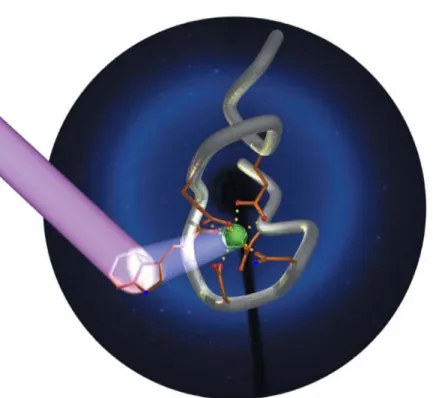
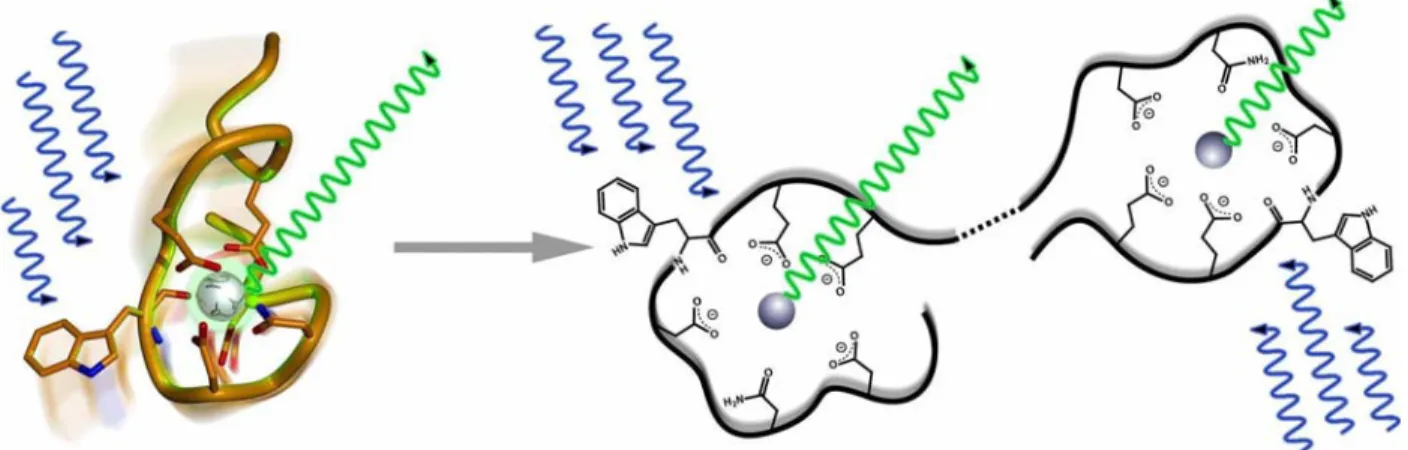
These peptides, dubbed “Lanthanide Binding Tags” (LBTs), show low-nM affinities and are selective for lanthanides over other common metal ions.1,58-60 An LBT is depicted in Figure 1-7.
Figure 1-7. Graphical Representation of an LBT, based on the crystal structure of SE2. 60 (The sequence of SE2 is YIDTNNDGWYEGDELLA; see also section 1-6, vide infra). The peptide backbone is shown in grey, and the chelating side-chains and back-bone carbonyl are shown in orange, with oxygen red and nitrogen blue. The side-chain indole of Trp7 is excited by UV light, and may transfer energy to Tb3+, shown in green, leading to luminescence. The diffraction pattern of this crystal was used as the background. (This figure was designed and created by Ezra Peisach.)
Lanthanide-Binding Tags are composed of twenty or fewer amino acids, and are thus less than 10% the size of GFP (Figure 1-8). Use of only encoded amino acids facilitates the incorporation of LBTs at the genetic level, and thus expression of the tagged protein may be accomplished by the natural cellular machinery. Once expressed, addition of the cofactor Ln3+ arms the tag for exploitation of the unique and versatile properties conferred by the metal ion (Figure 1-9).
Figure 1-8. Comparison of the size of a Lanthanide-Binding Tag to that of Green Fluorescent Protein. (This figure was created by Ryu Yoshida.)
Figure 1-9. Expression of an LBT-protein construct is straightforward.
1-5. Preliminary, Disulfide Bond-Containing Lanthanide-Binding Tags1
The similarity in atomic radii and oxophilic ligand preference of Ca2+ and Ln3+ ions (Table 1-1, vide supra) inspired researchers to base the design of lanthanide-binding sequences on ion-binding motifs selected from native calcium-binding proteins. Initially, studies by Richardson and Martin began with full length calcium-binding proteins to test for sensitization of Tb3+ lumninescence.61 Two important factors were found to govern the luminescence. First, a sensitizing group such as the aromatic side chain of tryptophan or tyrosine must be in close proximity (5-10 Å) to the Tb3+ to excite it, due to the inherently low absorbance of lanthanides.62 Second, the coordination sites of the terbium need to be fully occupied to avoid luminescence quenching due to the O-H bonds in water.31,63,64 In order to identify a binding motif for terbium luminescence, Szabo and coworkers conducted a small screen of short peptide sequences to optimize the type and position of the sensitizing agent.57 In this study, researchers found that a tryptophan residue at position 7 of the reference peptide (LBT-0, which has the sequence GDYNADGWIEFEEL; see also Table 1-2 below) was optimal for terbium luminescence. Unfortunately, once this short peptide sequence is removed from the native protein architecture,
the affinity for lanthanide ions decreases precipitously (by around two orders of magnitude) and dissociation constants on the order of 10 µM are observed.
In order for LBTs to be useful tags for proteins that do not already include native calcium-binding loops, an increase in lanthanide affinity is critical. The lanthanide-binding tag should be of sufficient affinity to preclude protein aggregation from non-specific interactions of lanthanide ion with the protein. Members of our lab therefore implemented methods to identify peptide motifs which demonstrated both increased affinity for lanthanide ions and improved luminescence. An initial attempt to rationally design short peptides with increased lanthanide affinity was spearheaded by Dr. Kathy Franz. It was hypothesized that tethering the ends of the loop might pre-organize the peptide backbone and facilitate lanthanide binding. For this reason, disulfide-bridge containing peptides based on LBT-0 were synthesized and the dissociation constants for Tb3+ determined (Table 1-2).58 From this small library, binding affinity was improved by over an order of magnitude to yield LBT-8 with a KD for Tb3+ of 220 nM (Table
1-2). While these initial results were encouraging, this method was not high-throughput, and therefore unsuitable for obtaining protein tags of even stronger lanthanide affinity without depending on disulfide bond formation. With this in mind, a more rapid and comprehensive screen was desired, both to increase the likelihood of success and to reduce the time-consuming, and often fallible, method of rational design, which allows only individual testing of a limited number of sequences.
Table 1-2. Rationally-Designed LBT Sequences Derived from Calcium-Binding Proteins.a,b (This table was designed by Bianca Sculimbrene.)
Position Peptide –4 –3 –2 –1 0 1 2 3 4 5 6 7 8 9 10 11 12 13 14 15 16 17 KD, Tb 3+ [µM] LBT-0 Ac G D Y N A D G W I E F E E L 9.0±1.0 LBT-1 A A C G D Y N A D G W I E F E E L A C A 9.3±0.4 LBT-2 Ac G G D Y N A D G W I E F E E L L 8.0±1.0 LBT-3 A C A G D Y N A D G W I E F E E L A C A 6.4±0.8 LBT-4 A C A G D Y N A D G W I E F E E L A A C A 5.9±0.5 LBT-5 A C A A G D Y N A D G W I E F E E L A C A 5.3±0.9 LBT-6 A A C G D Y N A D G W I E F E E L A A C A 5.0±0.3 LBT-7 C G D Y N A D G W I E F E E L C 0.6±0.1 LBT-8 A C A G D Y N A D G W I E F E E L C A A 0.220c
a Peptides LBT1 and LBT3 to LBT8 were oxidized to afford disulfide-containing macrocycles. Amino acids in
bold-face correspond to Tb3+–ligating residues. The tryptophan residue at position 7 coordinates Tb3+ through the backbone carbonyl and sensitizes Tb3+ luminescence. Amino acids are given as one-letter codes. Ac corresponds to an acetyl-capped N-terminus; all others are free amines. All peptides were synthesized as C-terminal amides.
b All LBTs are aligned to LBT-0. The residue numbering system is based on the literature.56,57
1-6. Development of a Powerful Combinatorial Screen1
The limiting step in the discovery of new peptides with a particular function is not the synthesis but the screening process. An enormous number of compounds would be generated from a library of completely randomized peptides, even if each position were limited to the encoded amino acids. Thousands of individual peptides can be made by solid-phase peptide synthesis (SPPS), and the ability to generate random peptide sequences in SPPS is easily achieved via split-and-pool combinatorial libraries (diagrammed in Figure 1-10).65-67 Therefore,
development of a rapid screening method was undertaken to take advantage of all the diversity available from this method.
Figure 1-10. Generation of a split-and-pool combinatorial library. Resin is loaded with peptide residues until the location in the sequence to be varied. The resin is then split into equal portions corresponding to the number of variations (two in the diagram). One of the varied amino acids is added to each portion, and then all the resin is pooled. Amino acids are added until the next variation, where the split-react-pool cycle is repeated, or until the N-terminus is reached. This methodology enables every permutation in the library to be made, while maintaining a single sequence on each individual bead.
Fluorescence spectroscopy has emerged as a highly efficient method for screening combinatorial libraries.68-71 The sensitivity of fluorescence allows small quantities of material to be analyzed using instrumentation including fluorescence microscopes and fluorescence plate readers. For this study, it was reasoned that screening combinatorial peptide libraries for Tb3+
luminescence could chemically evolve tighter and brighter LBTs. Hundreds of individual peptides could be screened on-bead simultaneously, identifying the brightest ones for selection and sequencing.
A new screen was developed in our lab to select tight and bright LBT sequences, spearheaded by Dr. Mark Nitz. Peptide sequences with high lanthanide affinity are of particular importance, to enable LBTs to be fully occupied in the presence of few equivalents of Tb3+; high [Tb3+] often induces protein aggregation. The screen utilizes Tb3+ luminescence with experimental strategies to avoid signal interference from the solid support and to reduce the cost and time associated with peptide sequencing by Edman degradation. This strategy is diagrammed in Figure 1-11.59 As solid support, TentaGel macrobeads (280-320 µm) were chosen based on compatibility with both organic solvents (during the peptide synthesis) and aqueous solution (during the Tb3+ luminescence screening). The high loading capacity of 2-3 nmol per bead ensures that sufficient peptide will be available for the orthogonal screen and sequence determination procedures. The resin is first derivatized with para-nitrophenylalanine to help quench background fluorescence. Resin is then co-functionalized with 20% of a photocleavable linker (3-amino-3-(2-nitrophenyl)propionic acid, ANP) and 80% of a base-labile linker (4-hydroxymethylbenzoic acid, HMBA). This co-functionalization allows a small amount of the peptide to be released from the bead with long wavelength UV light during screening, while the remaining portion of the peptide remains on the resin for sequencing after the selection process. A five-residue spacer is then added to facilitate ionization and to allow the shortest capped sequences to be above the background level of MALDI mass spectroscopy (MALDI-MS), which is used for sequence deconvolution.
The peptide is elongated using standard Fmoc-based SPPS protocols, including piperidine deprotection and amino-acid activation with HOBt/HBTU. Some of the varied residues are introduced simultaneously with an encoded capping reagent to generate a mass ladder for sequence determination. This method, developed by Griesinger and coworkers, utilizes an algorithm to calculate the minimum number of encoded capping steps necessary to generate a non-degenerate mass ladder.72 This approach necessitates fewer truncation sequences on each bead, compared with conventional mass spectroscopy ladder techniques that cap after every variation.
Figure 1-11. The synthesis and screening process for the combinatorial libraries to generate tighter and brighter LBTs. Reproduced and modified from the literature.60 (1) Coupling of Fmoc-4-nitrophenylalanine. (2) Coupling of orthogonal linkers (ANP/HMBA, 1:4). (3) Introduction of the spacer peptide sequence. (4) Coupling of the split-and-pool peptide library and mass-spectral ladder capping groups. (5) Amino acid side-chain deprotection, and casting of 2% agarose gel containing 50 µM Tb3+, 100 mM NaCl and 10 mM HEPES at pH 7.0 in a Petri dish. (6) Photolysis of the ANP linker using > 320 nm light. (7) Visualization of the beads at 280 nm. Beads are then selected that have bright luminescent halos, removal of agarose from selected beads followed by cleavage of the HMBA linker in 28% NH4OH, followed by MALDI-MS sequence deconvolution and single-bead Tb3+–affinity titrations. (This figure was originally designed and created by Mark Nitz, and has been previously published.1,59)
Once the split-and-pool encoded library is generated and side-chain protecting groups are cleaved by treatment with 94% trifluoroacetic acid, 2.5% water, 2.5% ethanedithiol and 1% triisopropylsilane, several hundred beads are dispersed in a buffered agarose gel containing Tb3+ ions. Nitrilotriacetic acid (NTA) or another competitive ligand may be included to ensure that only the tightest LBTs are bound to Tb3+. Release of the peptide portion attached via the photolabile linker is accomplished by illuminating the gel for six minutes on a long-wavelength UV transilluminator. The gel is visualized on a transilluminator with excitation at 280 nm; beads that contain the best LBTs exhibit a green, luminescent “halo” as shown in Figure 1-11. Beads with these halos are selectively removed from the gel. Treatment with ammonium hydroxide enables the release of these beads’ remaining peptide, the sequence of which is then determined by MALDI-MS. This selection assay avoids signal interference from the solid support while still taking advantage of the large number and diversity of peptides accessible through solid phase chemistry.
1-7. Lanthanide-Binding Tags Evolved Through the Combinatorial Screening Process1 Despite the power that comes with the ability to screen large numbers of peptides for a specific function on resin, a certain amount of rational design is still required when developing a library. For instance, complete randomization of a trideca-peptide to all twenty natural amino acids would require a library of 2013 = 8.2 × 10¹⁶ individual sequences. Approximately five
grams of resin are required to make 100,000 peptides, meaning that four billion kilograms of resin would be necessary to synthesize the aforementioned library! It is thus imperative to limit the number of variations or randomizations in order to avoid a prohibitively monumental screen or inadequate coverage of sequence space. Therefore, for the LBT libraries, no more than seven residues were ever varied in any screen, and selections were based on known or presumed function of the position whenever possible. Also, because MALDI-MS was used in sequence deconvolution, mass-degenerate amino acids were generally avoided at any one positional variation.
This prohibition on complete randomization led to the use of the original LBT-0 as the initial terbium-binding model. From this sequence, a series of four combinatorial libraries were designed, with each consensus peptide serving as the starting point for future library generations.59 The first library (Figure 1-12) contained 3,600 peptides, and addressed primarily whether the ligating glutamate residues (E9 and E12) were indeed optimized for Tb3+-binding. The sequence at positions 10 and 11 was varied to include residues biased towards turn formation by introduction of glycine and proline residues. Lastly, in order to compensate for the lack of a pre-organizational disulfide bond, explorations towards a stronger interaction between the N- and C-termini of the LBT were commenced. This results of library conclusively showed that the glutamate residues at positions 9 and 12 in LBT-0 were already optimized. Nevertheless, the best LBT, dubbed SE1, had a dissociation constant for Tb3+ that was lowered from 9 µM to 4 µM. (LBTs discovered through these combinatorial libraries were nicknamed “Sticky-Ends”, abbreviated “SE” in appreciation of the fact that the goal was to stick the termini together via non-covalent interactions.)
Position −1 0 1 2 3 4 5 6 7 8 9 10 11 12 13 Peptide KD, Tb 3+ LBT-Ref G D Y N K D G W Y E E L E L ~9 µM L A D G G D L T G E P P E T V W N N V Y Q Q S S SE1 V Y D Y N K D G W Y E G P E L ~4 µM
Figure 1-12. Results from Library 1: “Bidentate Ligating Residues and Turn Sequence”. Residues at varied positions in the starting LBT-0 are shown in red. Residues selected as optimal for each position are shown in blue. The resulting consensus sequence, the LBT “SE1,” is outlined in blue.
The second library (Figure 1-13) involved a much more thorough study—utilizing 14,700 peptides—of possible interactions between the two termini. By creating this sort of interaction, the entropic penalty upon metal-binding and organization could be minimized, lowering the dissociation constant. Calcium-binding motifs, for example, have a much stronger affinity while embedded within a protein, as a result of the more rigid structure around the loop holding the ends together. It was therefore logical to create stronger, hydrophobic interactions between the termini in an attempt to better mimic this environment. The resulting peptides from this second generation library, SE1a-α and SE1a-β, showed that increasing the length and modifying the terminal residues was indeed extremely beneficial. These sequences were significantly more selective terbium-binders than SE1, with dissociation constants now in the high nanomolar range.
Position −1 0 1 2 3 4 5 6 7 8 9 10 11 12 13 14 15 Peptide KD, Tb 3+ SE1 V Y D Y N K D G W Y E G P E ~4 µM A A W A A A L L L Y L L L V V P P T W P T T V Y W V V Y Y W W Y Y SE1a-α W V D W N K D G W Y E G P E L L A ~700 nM SE1a-β Y Y
Figure 1-13. Results from Library 2: “Interactions at the Termini”. Varied positions in the parent SE1 LBT are shown in red. Residues selected as optimal for each position are shown in blue. The resulting consensus sequences, the LBTs “SE1a-α” and “SE1a-β,” are outlined in blue.
The third library (Figure 1-14) was the largest of the libraries, involving 18,000 peptides. It continued the variation of the aromatic residues at the −1 position, introduced an additional variation at position 0 (isoleucine was not included in the previous library because it is indistinguishable from leucine by MALDI-MS), and randomized the internal, non-ligating residues at positions 4, 10 and 11. Finally, the aromatic residues at positions 7 and 8 were varied in an attempt to identify brighter LBTs. The large investment in peptide quantities paid off, for the resulting SE1b-α, SE1b-β, and SE1b-γ were an order-of-magnitude improved in KD from the
SE1a generation. This was the most significant improvement observed in any of the libraries.
Position −1 0 1 2 3 4 5 6 7 8 9 10 11 12 13 14 15 Peptide KD, Tb 3+ SE1a X V D X N K D G W Y E L A ~700 nM F I F G W W D D W V G K Y G P E L Y E G Y L N G N P P N P W R S S Y SE1b-α Y I D F N N D G W Y E G D E L L A ~70 nM SE1b-β L SE1b-γ W
Figure 1-14. Results from Library 3: “Internal, Non-ligating Residues”. Varied positions in the parent SE1a LBT are shown in red. Residues selected as optimal for each position are shown in blue. The resulting consensus sequences, the LBTs “SE1b-α,” “SE1b-β,” and “SE1b-γ,” are outlined in blue.
While this result was exciting, the apparent variability of preference at position 2 was curious, so a fourth library (Figure 1-15) was constructed to address this. Position 2 was randomized with all sulfur-free, mass-non-degenerate amino acids. The resulting peptide, which Dr. Nitz dubbed “SE2,” unambiguously showed that threonine was optimal at this position, with a 57 nM dissociation constant for Tb3+. Also, competitive titrations of SE2 between Tb3+ and most other Ln3+ metal ions clearly showed terbium to be the tightest binder.60 The trend indicated a high dependence on effective ionic radius: while Dy3+ and Eu3+ were only marginally
weaker, using lanthanides with ionic radii much different than Tb3+ (especially the larger lanthanides, e.g. La3+) led to significant drops in affinity.60 Furthermore, competitive titrations of SE2 between Tb3+ and Ca2+ or Mg2+ show only weak binding of the latter two ions. These results underscore that these libraries have successfully identified a selective terbium-binding