ORIGINAL ARTICLE
Differentiated dysplasia is a frequent precursor or associated
lesion in invasive squamous cell carcinoma of the oral cavity
and pharynx
Ruza Arsenic&Michael O. Kurrer
Received: 16 November 2012 / Revised: 29 March 2013 / Accepted: 2 April 2013 / Published online: 16 April 2013 # Springer-Verlag Berlin Heidelberg 2013
Abstract The source of precursor lesions of squamous cell carcinoma (SCC) of the oral cavity and pharynx, their classification, and grading are controversial. In contrast, vulvar and penile cancer precursor lesions are known to be related to human papillomavirus or chronic inflamma-tion and can be described using the vulvar intraepithelial neoplasia (VIN) classification system (VIN 1–3) or as differentiated vulvar intraepithelial neoplasia (dVIN), respectively. Oral and pharyngeal SCC precursor lesions are more etiologically diverse, and the spectrum of lesions may thus be wider. No international consensus exists re-garding the histological types of precursor lesions or the significance of individual types. We therefore reviewed resection specimens and preceding biopsies of 155 patients with SCC of the oral cavity and pharynx (excluding tonsils) and identified five basic patterns of SCC-associated or precursor lesions: (1) pleomorphic (22/155), (2) basaloid (5/155), (3) differentiated (63/155), (4) mixed (42/155), and (5) verrucous (12/155). Keratinization was a common but variable feature in differentiated, mixed, and verrucous
dysplasia. In 11/155 patients, no precursor lesion could be identified. Progression of isolated differentiated dysplasia (ranging from months to years) was documented in 13/155 (8 %) of patients. Our data suggest that full-thickness epithelial dysplasia of pleomorphic or basaloid type is present in <20 % of oral and pharyngeal SCC, and differ-entiated dysplasia is a frequent precursor or associated in situ lesion. Failure to recognize differentiated dysplasia results in the underdiagnosis of many patients at risk for invasive carcinoma. These results indicate a need to refine criteria to distinguish differentiated dysplasia from morpho-logically related lichenoid lesions.
Keywords Oropharyngeal cancer . Cancer precursor lesions . Dysplasia . Differentiated dysplasia of squamous epithelium Abbreviations
SCC Squamous cell carcinoma
VIN Vulvar intraepithelial neoplasia
dVIN Differentiated vulvar intraepithelial neoplasia
HPV Human papilloma virus
CIN Cervical intraepithelial neoplasia
PeIN Perineal intraepithelial neoplasia
Introduction
The grading of invasive squamous cell carcinoma (SCC)
can be traced back to Broders [1], and the grading of SCC
precursor lesions, to Papanicolaou [2]. Subsequently, the
progression from mild to moderate to severe dysplasia in the squamocolumnar epithelial junction of the uterine cervix has been considered paradigmatic. This paradigm still in-fluences concepts of cancer precursor lesions in various
epithelia [3–5].
R. Arsenic
Institute of Pathology, Cantonal Hospital Aarau, Aarau, Switzerland
M. O. Kurrer
University of Zurich, Zurich, Switzerland R. Arsenic (*)
Institute for Pathology, University Hospital Charité, Charitéplatz 1, 10117 Berlin, Germany
e-mail: [email protected] M. O. Kurrer (*)
Gemeinschaftspraxis für Pathologie, Cäcilienstrasse 3, Postfach 1520, 8032 Zurich, Switzerland
e-mail: [email protected] DOI 10.1007/s00428-013-1412-6
Historically, cervical cancer precursor lesions were graded according to the cervical intraepithelial neoplasia
(CIN) system (CIN 1–3), where dysplastic changes are
confined to the lower third of the epithelium in CIN 1, found in the lower two thirds of the epithelium in CIN 2, and involve the full thickness of the epithelium in CIN 3. Although later challenged by patient follow-up studies, epidemiological data were interpreted as evidence that progression from CIN 1 to invasive SCC was a stepwise process with single-step intervals of approximately 7 years. Eventually, it was recognized that this concept
does not reflect biology [6]. The different grades of
dysplasia are due to low- and high-risk human papillo-mavirus (HPV) types, which are responsible for more than 99 % of cervical dysplasia. Low-risk HPV types cause low-grade squamous intraepithelial lesions and can give rise to condylomata, whereas high-risk HPV types cause high-grade squamous intraepithelial lesions. Viral proteins E6 and E7 of the high-risk HPV types form complexes with cellular p53, resulting in p53 inactivation and the accumulation of mutations, leading to invasive SCC. The concept of stepwise progression from CIN 1 to 3 appears to represent an exception to the rule, typically reflecting an infection with both high- and low-risk
HPVs [7]. Therefore, the traditional idea of
morpholog-ical progression is, at least in the uterine cervix, a re-flection of independent etiologies rather than a true
biological progression [8–12].
In vulvar cancer, different etiologies are associated with distinct morphological features. HPV-associated carcino-genesis can be classified by using the traditional vulvar intraepithelial neoplasia (VIN) grading system (VIN 1–3), which is gradually being replaced by the low/high-grade squamous intraepithelial lesion system. Chronic inflammation-associated carcinogenesis (e.g., lichen planus or lichen sclerosus) is classified using the simplex or differentiated VIN system. In vulvar carcinogenesis, conventional high-grade dysplasia associated with high-risk HPV has been linked to higher-grade invasive SCC (G2/3), whereas inflammation-associated differentiated dysplasia most
of-ten gives rise to lower grades of invasive SCC (G1/2) [6,
13–18]. Analogous findings have been reported for
pe-nile SCC and its precursor lesions [19–24].
The recognition and grading of SCC precursor lesions of
the upper aerodigestive tract is controversial [3–5, 25].
Many facets appear heavily influenced by the traditional cervical cancer precursor lesion progression paradigm, and innovation, as reflected by internationally accepted
classifi-cation schemes, appears limited [26].
The field is complicated by a wider spectrum of etiolog-ical agents, namely, HPV, inflammation (e.g., oral lichen planus), smoking, alcohol consumption, and substance
chewing (e.g., areca nut and betel quid) [27].
Accordingly, a wider spectrum of histological precursor lesions could be expected that do not well fit into the one-dimensional cervical cancer precursor lesion progression paradigm. Additionally, a wide spectrum of clinical lesions has been recognized, including leukoplakia, erythroplakia, lichenoid lesions, and proliferative verrucous leukoplakia, which do not correlate well with histological precursor
lesions and cancer risk [28,29].
The current grading system considers full- or near-full-thickness dysplasia with significant nuclear atypia as high grade and high risk for invasive SCC development, and non-full-thickness dysplasia with milder nuclear atypia as low risk. Non-full-thickness dysplasia can be categorized as high
grade/high risk if basal/suprabasal atypia is significant [3].
Other lesions are predominantly defined by clinical features,
such as proliferative verrucous leukoplakia [30] and
lichenoid dysplasia [31,32]. These defy classification
with-in a conventional gradwith-ing system and carry an ill-defwith-ined but often high risk of progression to invasive SCC.
We carried out this retrospective study of 155 patients with oral and pharyngeal SCC to obtain an unbiased over-view of precursor lesions adjacent to invasive or preceding SCC. We addressed the following questions: (1) What are the relative frequencies of conventional full-thickness dys-plasia versus non-full-thickness dysdys-plasia adjacent to inva-sive SCC? (2) Are full-thickness dysplasia and non-full-thickness dysplasia associated with distinct carcinoma types? (3) What is the role of full-thickness dysplasia in the progression of precursor lesions to invasive SCC?
Patients, materials, and methods Patient selection
We searched the database of the Institute of Pathology of the Cantonal Hospital Aarau for patients with SCC of the oral cavity and pharynx (excluding tonsils). The search covered the period from 1990 to 2009. We retrieved all excision specimens and preceding biopsies on file. Mean patient age was 59.6 years (median 59 years), and the male/female
ratio was 2:1 (Table 1). We did not have anamnestic data
regarding carcinogen exposure or HPV status. Histological review
We evaluated the excision specimens of invasive cancer for cancer grade and adjacent cancer precursor lesions. The preceding biopsies without invasive carcinoma were evalu-ated for carcinoma precursor lesions. We classified the carcinoma-associated or preceding lesions as full- versus non-full-thickness dysplasia. The series was reviewed three times by two independent pathologists. Invasive SCC was
graded according to Anneroth's multifactorial grading
sys-tem [43].
Dysplasia categories
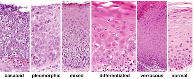
Dysplasia was divided into five categories (Table2, Figs.1
and 2). Full-thickness dysplasia was divided into basaloid
and pleomorphic types, both of which are considered severe dysplasia by conventional grading. Non-full-thickness dys-plasia was divided into differentiated, verrucous, and a mixed form of non-full-thickness dysplasia with either iso-lated basal and suprabasal dysplasia with regular superficial maturation or additional superficial features of differentiated or verrucous dysplasia. Differentiated dysplasia has previ-ously been referred to as lichenoid or simplex dysplasia, and verrucous dysplasia shares features with proliferative verrucous leukoplakia. Cases of mixed dysplasia included cases of moderate dysplasia according to the World Health Organization grading system and cases of keratinizing dysplasia.
Time course in patients with sequential biopsies
Special attention was paid to patients who did not show invasive SCC on the initial biopsy. Review of clinical infor-mation ascertained that the specimen showing invasive car-cinoma originated from the same anatomical location as the earlier biopsy.
Results
In our retrospective series of 155 patients, we determined the number and relative frequency of each SCC precursor lesion type adjacent to or preceding invasive SCC of the oral
cavity and pharynx (Table3). Full-thickness forms of SCC
precursor lesions were observed in only 17 % of cases with detectable dysplasia. Even when adding mixed dysplasia to basaloid and pleomorphic dysplasia to represent conven-tional forms of dysplasia, they comprised less than half of our cases (44 %). Non-full-thickness forms of dysplasia
(i.e., mixed, differentiated, and verrucous) represented more than 80 % of all dysplasia. Differentiated forms of dysplasia were identified as the sole associated cancer precursor lesion in nearly half of the cases with identifiable dysplasia. This suggests that development of invasive SCC of the oral cavity and pharynx via conventional full-thickness dysplasia represents the exception, whereas development via non-full-thickness dysplasia represents the rule.
Table 4 shows precursor lesions according to grade of
invasive SCC. Full-thickness forms of dysplasia were ex-clusively associated with higher grades of invasive SCC, with basaloid dysplasia associated mainly with poorly dif-ferentiated (G3) SCC, and pleomorphic dysplasia associated with both G2 and G3 SCCs.
Non-full-thickness forms of dysplasia (i.e., mixed, differ-entiated, and verrucous) showed a heterogeneous pattern. Mixed dysplasia was associated primarily with G2 SCC, whereas differentiated and verrucous dysplasia were associ-ated primarily with G1 and G2 SCC. This suggests that full-thickness dysplasia leads to higher-grade SCC, whereas non-full-thickness dysplasia, specifically differentiated and verrucous dysplasia, leads to lower-grade SCC.
We next evaluated the progression from dysplasia to invasive SCC in 24 patients. Most patients had invasive
SCC on the initial biopsy. Figure 3 shows only those
pa-tients who did not show invasive SCC on their initial biopsy. Of these 24 patients, four had clinically obvious malignant lesions and underwent a second biopsy within weeks. Twen-ty patients had initial diagnoses including hyperplasia, in-flammation, and mild/moderate dysplasia and underwent additional biopsies at variable intervals. The initial biopsy was used to categorize precursor lesions. In accordance with the study design, all patients eventually developed invasive
SCC within the time range shown in Fig.3.
In all but five cases, the category of dysplasia adjacent to the invasive carcinoma was identical to the dysplasia cate-gory on the first biopsy specimen. In one case of mixed dysplasia on initial biopsy, the next excision specimen with invasive SCC showed adjacent pleomorphic dysplasia. In another case of mixed dysplasia, the excision specimen 2 years later showed both mixed and verrucous dysplasia.
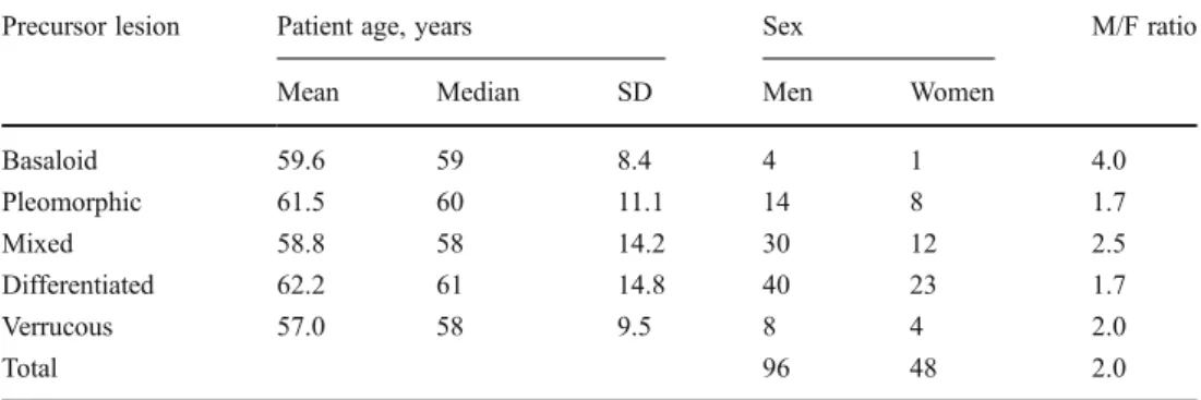
Table 1 Precursor lesions according to patient characteristics
Precursor lesion Patient age, years Sex M/F ratio Mean Median SD Men Women
Basaloid 59.6 59 8.4 4 1 4.0 Pleomorphic 61.5 60 11.1 14 8 1.7 Mixed 58.8 58 14.2 30 12 2.5 Differentiated 62.2 61 14.8 40 23 1.7 Verrucous 57.0 58 9.5 8 4 2.0 Total 96 48 2.0
T able 2 Features of squamous cell carcinoma precursor lesions Superficial maturation Compartment/epithelial layer Basal Suprabasal Superficial Keratinization Basaloid None Densely packed basaloid cells in all layers, with their long axis perpendicular to the epithelial surface, scant cytoplasm, hyperchro matic ovoid to elongated nuclei, coarse chromatin, no or indistinct nucleoli None or minimal Pleomorphic Minimal Pleomorphic cells, variation in internuclear spacing (i.e., variable cytoplasm), nuclear orientation (i.e., polarity), siz e, shape, and membrane thickness, chromatin coarseness, nucleolar size, shape, distribution, and number . Atypical mitoses could often be found but were not required T ypically none or minimal Mixed Distinct Similar to “pleomorphic, ” discernable at low power magnification V ariable, either similar to pleomorphic, “dif ferentiated, ” “verrucous, ” or regular squamous mucosa V ariable, either similar to dif ferentiated, verrucous, or regular mucosa Often present Dif ferentiated Y es Distinct clustering of often small basal cells , with small nuclei, either hyperchromatic or with open chromatin with small but distinct nucleoli, variable nuclear atypia Larg e cells, abundant eosinophilic cytoplasm, distinct desmosomes, distinctly large nuclei with open chromatin with distinct lar ge nucleoli, nuclear atypia with variable binucleation , variation in nuclear shape , membrane thickness , chr omatin structur e, nucleolar size , shape , distribution , and number . Atypical mitosis and dyskeratosis could be found but were not required Often minimal changes of verrucous V ariable V errucous Y es N o o r minimal atypia At most minimal changes of dif ferentiated Striking expansion of cells with moderate to abundant pale eosinophilic cytoplasm and nuclei with irregular thin membrane with membrane folding Often present and striking
In two cases of differentiated dysplasia, the excision speci-men obtained years later showed both differentiated and verrucous dysplasia. In one case of differentiated dysplasia, the precursor lesion shifted or progressed to mixed dyspla-sia. This suggests that dysplasia tends to remain in the same category over time rather than progress from one category to another. Specifically, there is no evidence for progression from non-full- to full-thickness dysplasia in the
develop-ment of invasive SCC. Table 4 shows that verrucous and
differentiated dysplasia tended to develop into lower-grade (G1 and G2) invasive SCC, mixed dysplasia into G2 SCC, and pleomorphic and basaloid dysplasia into both G2 and G3 SCCs, even if detection or development of invasive SCC was delayed after the initial biopsy.
Discussion
We analyzed a series of 155 patients with invasive SCC of the oral cavity and pharynx to evaluate associated adjacent or preceding cancer precursor lesions. We categorized pre-cursor lesions into five different groups rather than grades of dysplasia. Grading can be applied within individual catego-ries, each category with its own cytological and architectural grading criteria. A precedent for this can be found in data on the oral cavity and squamous epithelia of the penile and
vulvar mucosa [19,21,23,24], encompassing the
conven-tional VIN/penile intraepithelial neoplasia (PeIN) and dif-ferentiated VIN/difdif-ferentiated PeIN paradigms, and in colorectal cancer, in which sessile serrated adenomas have
Fig. 2 Microphotographs of different features of dysplasia. a– c Examples of cellular features of the superficial compartment in verrucous dysplasia; d–f examples of cellular features of the suprabasal compartment in differentiated dysplasia, showing atypical mitosis (d), nuclear and nucleolar atypia with
irregularities in number, shape, size, and distribution (e), as well as dyskeratosis; g–i examples of basal cell atypia with
hyperchromasia and smaller size or variability in size as well as nuclear and nucleolar irregularities with clustering of basal cells; j, k examples of differentiated dysplasia to show that they could both be found in thin and thick hyperplastic epithelias
recently been separated from the traditional adenomatous
pathway [33]. In both organ systems, grading criteria are
specific to the individual morphological pathways.
The Japanese Society of Oral Pathology (JSOP) advo-cates a system with three categories (i.e., basaloid,
differen-tiated, and mixed), which is similar to our system [26,29].
In contrast to the JSOP system, we divided full-thickness dysplasia into basaloid and pleomorphic types, which we hypothesize to have distinct etiologies and molecular
carci-nogenic pathways [34]. In a recent study, the spectrum of
mixed and differentiated dysplasia was addressed by
Kobayashi et al. [35] and referred to as orthokeratotic
dys-plasia. We chose to separate differentiated dysplasia from verrucous dysplasia, which is known as proliferative
verrucous leukoplakia [30,36,37] but has also been referred
to as verrucous hyperplasia [38–40]. Within penile and
vulvar squamous epithelia, the separation of differentiated dysplasia (including warty or verrucous forms) from basaloid or pleomorphic dysplasia is well established.
In contrast to Chaux et al. [24], we categorized cases with
striking basal and/or suprabasal atypia apparent at low mag-nification as mixed dysplasia, reserving the categories of differentiated and warty/verrucous dysplasia to cases with more subtle and/or suprabasal atypia, most of which are easily detected at higher magnification.
van de Nieuwenhof [15] recognized differentiated
dys-plasia in vulvar squamous epithelium by the presence of
elongated rete ridges with anastomosis, a disorderly basal cell layer with dyskeratosis, parakeratosis, prominent nucle-oli, and atypical mitoses. We recognized a disordered basal cell layer or basal layer atypia in the presence of distinct clustering of three to five often small basal cells with small nuclei, either hyperchromatic or with open chromatin and small but distinct nucleoli. Unlike van de Nieuwenhof, we required distinct changes in the suprabasal cell layer, with large cells containing abundant eosinophilic cytoplasm, dis-tinct desmosomes, large nuclei with open chromatin and large prominent nucleoli, nuclear atypia with variable binucleation, and variation in nuclear shape, membrane thickness, chromatin structure, nucleolar size, shape, distri-bution, and number. Atypical mitosis and dyskeratosis were sometimes observed but not required for diagnosis.
The criteria for the differentiated dysplasia referred to by
Kobayashi et al. [35] as orthokeratotic dysplasia included
“disturbed basal cell alignment, conspicuous pleomorphic, or atypical cellular features,” and “atypical cells with bizarre nuclei were occasionally scattered in the lower prickle cell layer.” We extended these criteria to include the distinct clustering of (often small) basal cells.
Our data show that conventional full-thickness dysplasia represents the exception in oral and pharyngeal SCC devel-opment, and non-full-thickness dysplasia (including mixed, differentiated, and verrucous forms) represent the rule. This is consistent with penile and vulvar carcinogenesis, in which differentiated dysplasia represent approximately half of
can-cer precursor lesions [19,41]. Similarly, the JSOP reported
that most premalignant lesions of the oral mucosa show
superficial maturation and differentiation [26], and
Kobayashi et al. [35] reported orthokeratotic dysplasia
(i.e., mixed and differentiated dysplasia) in approximately one third of the oral leukoplakia-type SCC cases assessed. In our study, basaloid dysplasia was uncommon. We attri-bute this result to our exclusion of tonsils, where basaloid carcinomas predominate.
In our series, conventional full-thickness dysplasia is asso-ciated with higher-grade SCC, whereas non-full-thickness dysplasia is associated with lower-grade SCC. This is similar to penile and vulvar carcinogenesis, in which differentiated dysplasia are typically associated with well-differentiated
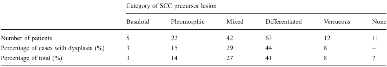
Table 3 Absolute and relative frequencies of invasive squamous cell carcinoma (SCC) associated and preceding lesions among our cohort of 155 patients
Category of SCC precursor lesion
Basaloid Pleomorphic Mixed Differentiated Verrucous None
Number of patients 5 22 42 63 12 11
Percentage of cases with dysplasia (%) 3 15 29 44 8 –
Percentage of total (%) 3 14 27 41 8 7
Table 4 Number of precursor lesions according to squamous cell carcinoma (SCC) grade
Precursor lesion Invasive SCC
G1 G2 G3 None 3 7 1 Basaloid 0 1 4 Pleomorphic 0 12 10 Mixed 6 29 7 Differentiated 37 23 3 Verrucous 6 5 1 Total 52 77 26
carcinomas [6,13–18]. Similarly, in penile and vulvar carci-nogenesis, conventional high-grade dysplasia has no role in
well-differentiated invasive SCC [19–22,24].
We acknowledge that the categories used in this study
and those described by others [19] were defined within a
morphological continuum. However, we hypothesize that different categories may be associated with distinct
etiolo-gies and molecular pathways [19,21,22,24,42], and it is
likely that carcinogenesis is caused by more than one car-cinogen and involves more than one pathway. Our results indicate that regardless of the carcinogen and molecular pathway involved, the associated morphological changes and dysplasia category tend to remain stable over time. When sequential biopsies were available over longer pe-riods of time, the initial biopsies showed precursor catego-ries that were almost always identical to the precursor lesion found adjacent to the invasive carcinoma. This supports the concept that morphological findings in dysplasia primarily relate to dysplasia categories, rather than dysplasia grades.
We recruited our patients by searching the pathology database for invasive SCC. Therefore, patients with dyspla-sia on biopsy and subsequent complete excision were not
included in this study because they would not have devel-oped invasive SCC. For this reason, our study cannot esti-mate the relative proportion of each dysplasia category in all patients but focuses on dysplasia categories in cases that progressed to invasive SCC. Our findings are consistent
with a previous report [26].
Likewise, our study does not provide definitive data on the dynamics (likelihood and time course) of progression of non-full-thickness dysplasia, specifically differentiated
dys-plasia, to invasive carcinoma. The JSOP [26,29] suggests
that differentiated dysplasia progress to invasive carcinoma within 5 years, whereas conventional dysplasia progress within 6 months. This is in line with our data showing that isolated pleomorphic and basaloid dysplasia typically progressed to invasive carcinoma within 6 months, whereas mixed and differentiated categories progressed to invasive carcinoma over longer intervals (≥8 years), with most progressing within 3 years. The more rapid progression of verrucous precursor lesions supports their relationship to proliferative verrucous leukoplakia.
We did not address the morphological criteria used to distinguish differentiated forms of dysplasia from changes
Fig. 3 Time line for the progression of the distinct forms of dysplasia to invasive squamous cell carcinoma in the subset of 24 of our 155 patients, not showing invasive squamous cell carcinoma on the first biopsy specimen (basaloid, n=1; pleomorphic, n=3; mixed, n=5; differentiated, n=13; verrucous; n=2). Each case of later invasive squamous cell carcinoma is identified in the figure by its grade (G1, G2, or G3). Cases are placed on the time line corresponding to the relevant dysplasia category to show the time interval in months between the first biopsy and eventual tissue diagnosis of invasive squamous cell carcinoma on repeat biopsy or excision. In most cases, the category of dysplasia adjacent to the invasive carcinoma was identical to the category observed in the first biopsy specimen. In cases, where the category of dysplasia adjacent to the invasive carcinoma differed from the category retained on the first biopsy specimen, this latter category of dysplasia is indicated by a foot note
observed in reactive lesions, such as lichen planus and regenerative squamous epithelium. Instead, we applied criteria used in our daily practice and influenced by the
literature on penile and vulvar neoplasia [6,8,11,13,14,
17–20]. However, under study conditions, these
morpholog-ical changes may also represent a continuum, as both lichen planus and chronic regeneration can eventually lead to dys-plasia and invasive carcinoma. Until further study, it may be better to limit definitive diagnoses of differentiated forms of dysplasia to cases that do not show significant interface inflammation or causes of regenerative response such as ulceration, chronic trauma, or fungal infection. Alternative-ly, the criteria for diagnosing differentiated forms of dyspla-sia may need to be applied stringently.
Nevertheless, we feel confident that the criteria of signif-icant basal cell atypia for mixed dysplasia, basal cell clus-tering and suprabasal atypia in differentiated dysplasia, and shriveled nuclei in a hyperplastic superficial layer for verrucous dysplasia can provide relevant clinical informa-tion. Clinically suspicious lesions that later progress to invasive carcinoma typically show changes that we and
others [15, 22] have variably referred to as differentiated
forms of dysplasia, simplex dysplasia, or lichenoid dyspla-sia. The data should thus sensitize both clinicians and pa-thologists to the relevance of differentiated forms of dysplasia in general and to subtler forms detectable only at high magnification using cytological criteria.
Finally, our data support a conceptual shift, previously
suggested by others [14,26,42], away from primary
dys-plasia grades to SCC precursor categories that may be associated with specific but overlapping carcinogenic and molecular pathways. We hope that this shift will help to
overcome the dissatisfaction [3,4] with the current
classifi-cation systems of SCC precursor lesions of the oral cavity and pharynx.
Acknowledgments We thank Prof. Dr. Manfred Dietel and Dr. Anja Schmitt for their critical reading of the manuscript.
Conflict of interest The authors declare that they have no conflict of interest.
References
1. Broders AC (1920) Squamous-cell epithelioma of the lip: a study of five hundred and thirty-seven cases. JAMA 74:656–664 2. Papanicolaou GN, Traut HF (1997) The diagnostic value of
vag-inal smears in carcinoma of the uterus. 1941. Arch Pathol Lab Med 121:211–224
3. Warnakulasuriya S, Reibel J, Bouquot J, Dabelsteen E (2008) Oral epithelial dysplasia classification systems: predictive value, utility, weaknesses and scope for improvement. J Oral Pathol Med 37:127–133
4. Fleskens S, Slootweg P (2009) Grading systems in head and neck dysplasia: their prognostic value, weaknesses and utility. Head Neck Oncol 1:11
5. Bouquot JE, Speight PM et al (2006) Epithelial dysplasia of the oral mucosa—diagnostic problems and prognostic features. Curr Diagn Pathol 12:11–21
6. Pinto AP, Crum CP (2000) Natural history of cervical neoplasia: defining progression and its consequence. Clin Obstet Gynecol 43:352–362
7. Chen EY, Tran A, Raho CJ et al (2010) Histological‘progression’ from low (LSIL) to high (HSIL) squamous intraepithelial lesion is an uncommon event and an indication for quality assurance re-view. Mod Pathol 23:1045–1051
8. Ostor AG (1993) Natural history of cervical intraepithelial neopla-sia: a critical review. Int J Gynecol Pathol 12:186–192
9. Ibeanu OA (2011) Molecular pathogenesis of cervical cancer. Cancer Biol Ther 11:295–306
10. Gravitt PE (2011) The known unknowns of HPV natural history. J Clin Invest 121:4593–4599
11. Crum CP, McLachlin CM (1995) Cervical intraepithelial neopla-sia. J Cell Biochem Suppl 23:71–79
12. Bamford PN, Beilby JO, Steele SJ, Vlies R (1983) The natural history of cervical intraepithelial neoplasia as determined by cy-tology and colposcopic biopsy. Acta Cytol 27:482–484
13. Yang B, Hart WR (2000) Vulvar intraepithelial neoplasia of the simplex (differentiated) type: a clinicopathologic study including analysis of HPV and p53 expression. Am J Surg Pathol 24:429– 441
14. van der Avoort IA, Shirango H, Hoevenaars BE et al (2006) Vulvar squamous cell carcinoma is a multifactorial disease following two separate and independent pathways. Int J Gynecol Pathol 25:22–29 15. van de Nieuwenhof HP, Bulten J et al (2011) Differentiated vulvar intraepithelial neoplasia is often found in lesions, previously diag-nosed as lichen sclerosus, which have progressed to vulvar squa-mous cell carcinoma. Mod Pathol 24:297–305
16. Nascimento AF, Granter SR, Cviko A et al (2004) Vulvar acanthosis with altered differentiation: a precursor to verrucous carcinoma? Am J Surg Pathol 28:638–643
17. Medeiros F, Nascimento AF, Crum CP et al (2005) Early vulvar squamous neoplasia: advances in classification, diagnosis, and differential diagnosis. Adv Anat Pathol 12:20–26
18. Kokka F, Singh N, Farugi A et al (2011) Is differentiated vulval intraepithelial neoplasia the precursor lesion of human papillomavirus-negative vulval squamous cell carcinoma? Int J Gynecol Cancer 21:1297–1305
19. Velazquez EF, Chaux A, Cubilla AL (2012) Histologic classifica-tion of penile intraepithelial neoplasia. Semin Diagn Pathol 29:96– 102
20. Soskin A, Vieillefond A, Carlotti A et al (2012) Warty/basaloid penile intraepithelial neoplasia is more prevalent than differentiat-ed penile intraepithelial neoplasia in nonendemic regions for penile cancer when compared with endemic areas: a comparative study between pathologic series from Paris and Paraguay. Hum Pathol 43:190–196
21. Renaud-Vilmer C, Cavelier-Balloy B et al (2009) Analysis of alterations adjacent to invasive squamous cell carcinoma of the penis and their relationship with associated carcinoma. J Am Acad Dermatol 62:284–290
22. Oertell J, Caballero C, Iglesias M et al (2011) Differentiated precursor lesions and low-grade variants of squamous cell carci-nomas are frequent findings in foreskins of patients from a region of high penile cancer incidence. Histopathology 58:925–933 23. Chaux A, Cubilla AL (2012) Diagnostic problems in precancerous
lesions and invasive carcinomas of the penis. Semin Diagn Pathol 29(2):72–82
24. Chaux A, Velazquez EF et al (2012) Distribution and characteri-zation of subtypes of penile intraepithelial neoplasia and their association with invasive carcinomas: a pathological study of 139 lesions in 121 patients. Hum Pathol 43:1020–1027
25. Fleskens SA, Bergshoeff VE, Voogd AC et al (2011) Interobserver variability of laryngeal mucosal premalignant lesions: a histopath-ological evaluation. Mod Pathol 24:892–898
26. Izumo T (2011) Oral premalignant lesions: from the pathological viewpoint. Int J Clin Oncol 16:15–26
27. van der Waal I (2009) Potentially malignant disorders of the oral and oropharyngeal mucosa; terminology, classification and present concepts of management. Oral Oncol 45:317–323
28. Ferlito A, Devaney KO, Woolgar JA et al (2012) Squamous epithelial changes of the larynx: diagnosis and therapy. Head Neck 34:1810–1816
29. Amagasa T, Yamashiro M, Uzawa N (2011) Oral premalignant lesions: from a clinical perspective. Int J Clin Oncol 16:5–14 30. Bagan J, Scully C, Jimenez Y, Martorell M (2010) Proliferative
verrucous leukoplakia: a concise update. Oral Dis 16:328–332 31. van der Meij EH, Mast H, van der Waal I (2007) The possible
premalignant character of oral lichen planus and oral lichenoid lesions: a prospective five-year follow-up study of 192 patients. Oral Oncol 43:742–748
32. Gonzalez-Moles MA, Scully C, Gil-Montoya JA (2008) Oral lichen planus: controversies surrounding malignant transforma-tion. Oral Dis 14:229–243
33. Torlakovic EE, Gomez JD et al (2008) Sessile serrated adenoma (SSA) vs. traditional serrated adenoma (TSA). Am J Surg Pathol 32:21–29 34. Chernock RD, Lewis JS Jr, Zhang Q et al (2010) Human
papillomavirus-positive basaloid squamous cell carcinomas of
the upper aerodigestive tract: a distinct clinicopathologic and molecular subtype of basaloid squamous cell carcinoma. Hum Pathol 41:1016–1023
35. Kobayashi T, Maruyama S, Abe T et al (2012) Keratin 10-positive orthokeratotic dysplasia: a new leucoplakia-type precancerous en-tity of the oral mucosa. Histopathology 61:910–920
36. van der Waal I, Reichart PA (2008) Oral proliferative verrucous leukoplakia revisited. Oral Oncol 44:719–721
37. Batsakis JG, Suarez P, el-Naggar AK (1999) Proliferative verrucous leukoplakia and its related lesions. Oral Oncol 35:354–359
38. Zhu LK, Ding YW, Liu W et al (2012) A clinicopathological study on verrucous hyperplasia and verrucous carcinoma of the oral mucosa. J Oral Pathol Med 41:131–135
39. Wang YP, Chen HM, Kuo RC et al (2009) Oral verrucous hyper-plasia: histologic classification, prognosis, and clinical implica-tions. J Oral Pathol Med 38:651–656
40. Shear M, Pindborg JJ (1980) Verrucous hyperplasia of the oral mucosa. Cancer 46:1855–1862
41. Chiesa-Vottero A, Dvoretsky PM, Hart WR (2006) Histopatholog-ic study of thin vulvar squamous cell carcinomas and associated cutaneous lesions: a correlative study of 48 tumors in 44 patients with analysis of adjacent vulvar intraepithelial neoplasia types and lichen sclerosus. Am J Surg Pathol 30:310–318
42. Pinto AP, Lin MC, Sheets EE et al (2000) Allelic imbalance in lichen sclerosus, hyperplasia, and intraepithelial neoplasia of the vulva. Gynecol Oncol 77:171–176
43. Anneroth G, Hansen LS (1984) A methodologic study of histolog-ic classifhistolog-ication and grading of malignancy in oral squamous cell carcinoma. Scand J Dent Res 92:448–468