Tobias Krauss Thomas Pfammatter Dieter Mayer Mario Lachat Lukas Hechelhammer Borut Marincek Thomas Frauenfelder Received: 3 April 2009 Revised: 26 June 2009 Accepted: 6 August 2009
Published online: 30 September 2009 # European Society of Radiology 2009
Hybrid-repair of thoraco-abdominal
or juxtarenal aortic aneurysm:
what the radiologist should know
Abstract Purpose: Endovascular aneurysm repair of the infrarenal or thoracic aorta has been shown to be a less invasive alternative to open sur-gery. A combined aneurysm of the thoracic and abdominal aorta is com-plex and challenging; the involvement of renal and/or visceral branches requires new treatment methods. Methods: A hybrid approach is cur-rently an accepted alternative to conventional surgery. Renal and/or visceral revascularisation enables subsequent stent-graft placement into the visceral portion of the aorta. Results: Knowledge of the surgical procedure and a precise assessment of the vascular morphology are crucial for pre-procedural planning and for detection of post-procedural compli-cations. Multi-detector computed tomography angiography (MDCTA) combined with two- and three-dimensional (2D and 3D) rendering is useful for pre-interventional planning
and for the detection of post-procedural complications. Three-dimensional rendering allows proper anatomical analyses, influencing interventional strategies and resulting in a better outcome. Conclusions: With the knowledge of procedure-specific MDCTA findings in various vascular conditions, the radiologist and surgeon are able to perform an efficient pre-interventional planning and follow-up examination. Based on our experience with this novel tech-nique of combined open and endo-vascular aortic aneurysm treatment, this pictorial review illustrates proce-dure-specific imaging findings, including common and rare compli-cations, with respect to 2D and 3D post-processing techniques.
Keywords Endovascular repair . Hybrid repair . Thoraco-abdominal aortic aneurysm . Debranching
Introduction
A hybrid open and endovascular approach offers a new and attractive alternative to open surgery (OS) for the treatment of complex thoraco-abdominal aortic aneur-ysms (TAAAs).
Since its introduction in the early 1990s by Volodos and Parodi [1], endovascular aneurysm repair (EVAR) has revolutionised the treatment of infrarenal and thoracic aortic aneurysms. Nowadays, EVAR represents a safe and less invasive alternative to OS for the treatment of an infrarenal aortic aneurysm. Compared with OS, EVAR
shows a 66% relative risk reduction concerning 30-day mortality [2,3].
Endovascular treatment of TAAA is a relatively more complex situation; overall experience is limited and long-term results have yet to be obtained. The involvement of renal and/or visceral branches requires new and innovative methods. In 1999, Quinones-Baldrich et al. [4] reported the first experience in using a combined surgical and endovascular approach for the treatment of TAAA. The “hybrid technique” with renovisceral debranching (i.e. extra-anatomical bypasses enabling stent-graft deployment into the visceral portion of the aorta) followed by aortic T. Krauss . T. Pfammatter .
L. Hechelhammer . B. Marincek . T. Frauenfelder (*)
Department of Medical Radiology, Institute of Diagnostic Radiology, University Hospital Zurich, Raemistrasse 100, 8091 Zurich, Switzerland e-mail: thomas.frauenfelder@usz.ch Tel.: +41-44-2559383 Fax: +41-44-2554443 D. Mayer . M. Lachat
Clinic for Cardiovascular Surgery, University Hospital of Zurich, Zurich, Switzerland
stent-graft placement is currently an accepted therapeutic alternative to endografts with customised fenestrations (F-EVAR), branched stent-graft technologies or conventional OS, especially for patients who are at high risk of undergoing open repair.
Current literature provides case reports and only a few patient series that evaluate the feasibility, as well as short-and mid-term outcomes after hybrid treatment [5–11]. But the number of patients treated using the hybrid approach increases.
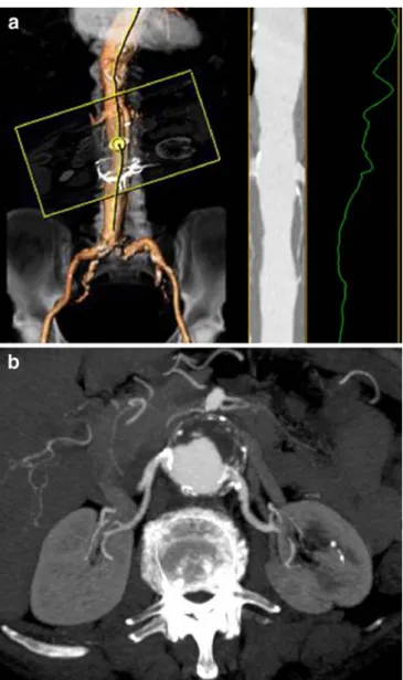
Fig. 1a, b Centreline analysis used for semi-automatic diameter calculation in preoperative MDCTA. a Perpendicular plane MPR. b VR illustrates the path of centreline through the aorta
Fig. 2 A 64-year-old woman with TAAA Crawford type I. VR technique illustrates the complex anatomy after hybrid treatment Table 1 MDCTA for post-EVAR follow-up using 64-Slice CT
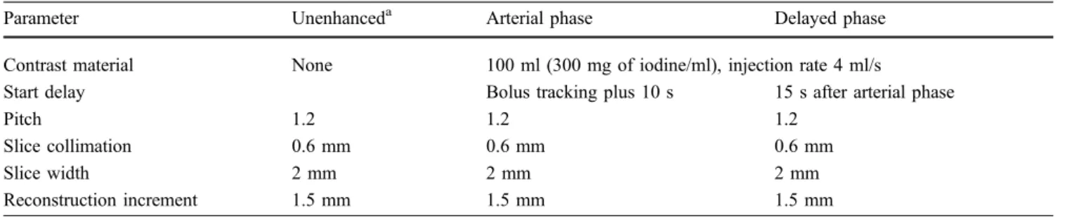
Parameter Unenhanceda Arterial phase Delayed phase
Contrast material None 100 ml (300 mg of iodine/ml), injection rate 4 ml/s
Start delay Bolus tracking plus 10 s 15 s after arterial phase
Pitch 1.2 1.2 1.2
Slice collimation 0.6 mm 0.6 mm 0.6 mm
Slice width 2 mm 2 mm 2 mm
Reconstruction increment 1.5 mm 1.5 mm 1.5 mm
a
Aside from surgical and anaesthetic risks, possible graft-and bypass-related complications include endoleak, graft occlusion, organ ischaemia, graft dislocation, pseudoaneu-rysm, anastomotic bleeding, perigraft seroma and haema-toma [6,12–14].
Because each vascular intervention is complex and challenging, a precise assessment of the vascular morphol-ogy is crucial for preoperative planning and detection of post-procedural complications. Because of the limited experience with patients undergoing the hybrid approach, experience in hybrid-specific imaging is lacking. More-over, in our institution VORTEC (Viabahn open revascu-larisation technique) is applied to most renal arteries and
frequently to the visceral arteries [15]. This technique requires a detailed analysis of the target vessels in order to implant the appropriate stent-graft device.
Based on our experience with this novel technique, this pictorial review illustrates procedure-specific imaging findings, including common and rare complications, with respect to multi-detector computed tomography angiography (MDCTA) and two-dimensional (2D) and three-dimensional (3D) post-processing techniques [i.e. multi-planar reforma-tion (MPR) and volume rendering technique (VRT)].
Imaging technique
Accurate imaging is necessary for both selecting patients suitable for hybrid treatment and the follow-up examination after hybrid repair. Multi-detector computed tomography (MDCT) combines many advantages over other imaging investigations: rapid image acquisition, few motion artefacts, reproducibility, good delineation of the aorta and its side branches, and an excellent spatial resolution. The MDCTA dataset, in combination with a variety of 2D and 3D post-processing options, allows an accurate qualitative and quantitative assessment of the vascular structures. Therefore, MDCTA is nowadays well accepted for pre- and post-procedural aortic aneurysm imaging [16,17].
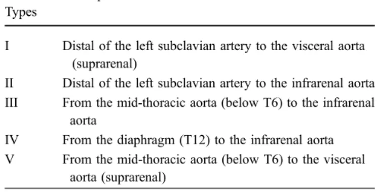
Optimised MDCTA protocols are mandatory for com-plete and accurate assessment of vascular structures before Table 2 Classification scheme for TAAA
Crawford Types
Description
I Distal of the left subclavian artery to the visceral aorta (suprarenal)
II Distal of the left subclavian artery to the infrarenal aorta III From the mid-thoracic aorta (below T6) to the infrarenal
aorta
IV From the diaphragm (T12) to the infrarenal aorta V From the mid-thoracic aorta (below T6) to the visceral
aorta (suprarenal)
Fig. 3 a Picture during bypass surgery. Feeding graft to supe-rior mesenteric artery (arrow), side grafts to right renal artery (RRA), left renal artery (LRA), and coeliac trunk (*). b Corre-lating VR from control MDCTA
and after intervention. The intravenous (IV) contrast material injection protocol has to be specified for every examined vascular region. For optimal depiction of vascular structures, the IV contrast material injection protocol has to be tailored to the area of interest in a certain anatomical region.
At our institute, the examination of thoracic and abdom-inal vascular structures is performed using a Somatom Definition 64 slice system (Siemens Medical Solution, Forchheim, Germany). Reconstruction of the axial images is performed using a slice thickness of 2 mm. A 100-ml volume of non-ionic contrast material (300 mg of iodine/ml Ultravist, BayerSchering, Berlin) is injected at a rate of 4 ml/s. Modern imaging protocols for MDCTA recommend the use of higher concentrated contrast material (>350 mg of iodine/ml) for a
better arterial enhancement [18,19]. We use contrast material with a moderate iodine concentration at our institute and get a good imaging quality with the possibility to decrease the risk of nephrotoxicity. For precise planning and follow-up a multiphasic MDCTA approach is recommended. Images are acquired throughout the whole thorax and abdomen to characterise blood supply and drainage. The imaging protocols include a non-contrast image, an arterial phase image and a venous phase image. Unenhanced images throughout the whole thorax and abdomen are useful for the identification of high-density structures such as calcifications or residual contrast material after EVAR and serve as baseline exams for follow-up.
Fig. 4a, b Preoperative 64-channel multi-detector-row MDCTA evaluation. a Centreline analysis is required for semi-automatic diameter calculation in regions of tortuosity. b Transverse recon-struction shows a TAAA Crawford type IV and depicts involvement of both renal arteries
Fig. 5 a Centreline analysis is used for diameter calculation of the renal arteries before placement of self-expanding stent grafts. b Curved multiplanar reformatted image (cMPR) of the left renal artery
Arterial phase images are acquired for pre-interventional vessel analysis. The control examination after intervention should include an unenhanced acquisition and two con-trast-enhanced CT data sets (arterial and delayed phase). Some authors recommend delayed phase imaging because endoleaks show variable flow rates and are, therefore, not necessarily detected during the early contrast-enhanced phase [16,20].
To reduce radiation dose, we consider the possibility of generating non-contrast images only in the first post-EVAR examination as previously described by Iezzi et al. [21]. An MDCTA protocol typically used at our institute is shown in Table1.
Post-processing
High spatial and temporal resolution, provided by MDCTA, are the prerequisites for high quality 3D images, which allow the radiologist to illustrate complex anatom-ical situations more clearly.
The possibility of showing pathological features inter-actively on single images from infinite projections makes 3D rendering a powerful tool for surgical planning in a close interdisciplinary approach. The visualisation of vessels, especially when dealing with complex surgery, is a major advantage of this method. Three-dimensional
imaging permits the surgeon to make crucial decisions by facilitating the depiction of vascular structures.
Besides thin transverse-section images, multi-planar reformation (MPR), maximum intensity projection (MIP) and volume rendering (VR) are currently the 2D and 3D reconstruction standards for pre- and post-procedural evaluation of aortic aneurysm.
MPR is a post-processing technique that is derived from axial slices and can be generated in any plane determined by the viewer. The spatial resolution is equal to the original source images. MPR with double oblique planes that are exactly perpendicular to the vessel lumen can be useful for a precise quantitative analysis of diameter and volume, especially in regions of tortuosity. A curved plane along a predefined data volume is used for creating a curved MPR. Curved MPR can be used for better display of regions of tortuosity, but diameter measurements with this technique are usually highly prone to errors [22]. A semi-automatic centreline analysis allows automatic diameter calculations by generating an optimal track through the centreline of a vascular segment (Fig.1).
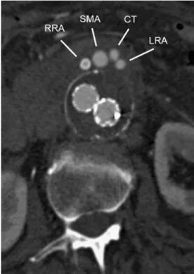
MIP rendering technique is required to visualise high-intensity structures within one volumetric dataset. MIP algorithms evaluate and display the voxel with the maximum attenuation along a ray through the volume dataset. The vascular morphology and anatomy can be evaluated using MIP images, but the generated images do not provide a good sense of the depth of the original data. VR is considered the standard method in 3D image post-processing [23]. It provides a volumetric display by rendering the entire volume of data and not only the anatomical surface contours. Each attenuation value is attributed to a relative shading of opacity and colour. The brightness of each voxel is determined by calculating a virtual light source. VR is particularly helpful for the visualisation of complex anatomical structures (Fig.2). Fig. 6 Post-procedural final control. Final MDCTA confirms a
correctly placed, patent endograft without an endoleak. Patent grafts to the right renal artery (RRA), left renal artery (LRA), superior mesenteric artery (SMA) and coeliac trunk (CT)
Table 3 Classification scheme for endoleaks (adapted from Veith et al. [38])
Endoleak Types
Description
I Attachment site leaks A Proximal end of graft B Distal end of graft C Iliac occluder II Branch leak
A Simple (from only one branch)
B Complex (two or more vessels creating flow through) III Graft failure
A Junctional leak
B Endograft fracture or holes IV Graft-wall porosity V Endotension
Classification of thoraco-abdominal aortic aneurysm
Thoraco-abdominal aortic aneurysms involve the descend-ing thoracic and the abdominal portion of the aorta. A correct classification has important therapeutic and diag-nostic impact. As proposed by Crawford et al. [24] and modified by Safi et al. [25], the classification of TAAAs is based on the proximal and distal anatomical extent of the aneurysm (Table2).
Surgical and endovascular technique
The hybrid approach for the treatment of TAAAs is a two-stage procedure:
The first step consists of a surgical retrograde visceral and/or renal revascularisation based on extra-anatomical bypasses to the renal arteries, the coeliac axis and the superior mesenteric artery (Fig. 3). Depending on local status (redo procedure) or patient pulmonary condition Fig. 7a, b MDCTA of type Ia
endoleak in an 83-year-old man. a Transverse image during the arterial phase and b oblique sagittal MIP demonstrate con-trast material outside the proxi-mal fixation of the stent-graft (arrow). Due to undersizing, the stent-graft shows a poor adap-tion on the inner aortic wall within the proximal landing zone, which is the cause of type I endoleak
Fig. 8a, b MDCTA of type Ib endoleak in an 82-year-old man. a VR image, b curved MPR demonstrate contrast material outside the distal fixation of the stent-graft (endoleak originating from insufficient distal landing zone of the right iliac limb, arrow)
(severe chronic obstructive lung disease), a retroperitoneal or a transperitoneal abdominal approach is chosen. Choice of inflow site depends on the extent of the aneurysm and on concomitant vascular pathological conditions and is in most cases provided from the aorta or iliac arteries. Alternative inflow sites based on the hepatic artery and the splenic artery (i.e. splenorenal or hepatorenal bypass) are also possible.
The second step consists of the endovascular aneurysm repair. Debranching and EVAR can be done simultaneously or sequentially. Haemodynamic instability has been identified as an independent risk factor for developing paraplegia in open thoraco-abdominal aortic repair [26]. Haemodynamic instability can also occur during or after major abdominal surgery, such as renovisceral bypass surgery. SIRS (systemic inflammatory response syndrome) and SIRS associated complications (haemodynamic in-stability due to fever, atrial fibrillation and vasoplegia and/
or anaemia) are also not uncommon after major surgery and often are unpredictable.
With EVAR, there is no direct way to increase spinal cord perfusion and haemodynamic stability must be ensured for a good spinal tissue perfusion after EVAR.
Therefore, we do prefer to perform EVAR in non-emergency cases after the patient has recovered from open surgical procedure, which is usually achieved within 1 week of time.
A bolus of 100 IE/kg of heparin is administrated during debranching and EVAR. Further intraoperative re-hepar-inisation is based on activated clotting time, which we hold on >1.5 the value before heparin administration. Between debranching and EVAR, heparin is administered at a therapeutic dose. The long-term antithrombotic regimen consists of aspirin at 100 mg/day life-long, combined with warfarin for a 12 month-period. In cases where antic-oagulation with warfarin is not possible, we combine aspirin with clopidogrel at 75 mg/day for 12 months. Fig. 9a, b MDCTA of type II
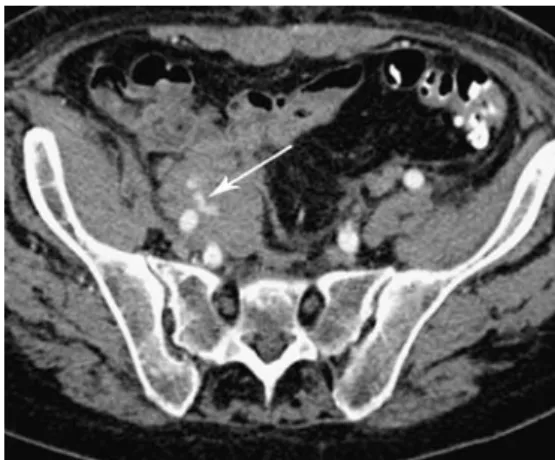
endoleak with supply from ac-cessory renal arteries in a 62-year-old man. a MDCTA during the arterial phase reveals con-trast material outside the stent-graft (arrow). b Coronal MIP depicts supply from the superior accessory renal artery (sARA) and inferior accessory renal ar-tery (iARA)
Fig. 10a, b MDCTA of a 65-year-old man who had pre-viously undergone repair of an infrarenal aortic aneurysm. Eight years later the patient presented with a juxtarenal aor-tic aneurysm and underwent hybrid treatment with de-branching followed by two stent grafts. a A 5-months post-EVAR MDCTA (VR) confirms correct positioning of the proximal and distal endovascular grafts. b Two months later MDCTA (VR) reveals proximal endograft dis-location (arrow). The proximal stent has dislodged resulting in graft discontinuity
Case
A 68-year-old woman presented with an asymptomatic but continuously increasing TAAA Crawford type IV. Because of the patient’s concomitant chronic obstructive lung disease and coronary artery disease, she was deemed a high-risk candidate for OS; a two-stage procedure was planned, which included initial visceral and renal revascu-larisation, followed by EVAR. An 8-mm polytetrafluoro-ethylene (PTFE) feeding graft was placed transperitoneally from the right iliac artery with an end-to-side anastomosis to the superior mesenteric artery (SMA). A 5-mm side graft was placed end-to-side to the coeliac trunk. Two self-expanding stent grafts (VIABAHN, W.L. Gore & Associ-ates, Flagstaff, Ariz.) based on the feeding graft enabled retrograde renal revascularisation [15]. Subsequent ultra-sound and MDCTA confirmed graft patency before EVAR. Ten days later, EVAR was attempted through interdisci-plinary cooperation between a vascular specialist and an interventional radiologist (Figs.4,5and6).
Relevant pre-interventional findings at MDCTA
Pre-interventional planning before hybrid treatment of the thoraco-abdominal aorta includes a precise assessment of
the anatomical structures and landmarks. Landmarks relevant for pre-interventional planning include the orifices of aortic branches (superior mesenteric artery, coeliac trunk, renal arteries and accessory branches), and evalua-tion of proximal and distal sealing zones. The maximum diameter of the aneurysm, the aortic bifurcation, and the diameter and tortuosity of the iliac arteries are measured using dedicated software (3Surgery, 3Mensio Medical Imaging, BS Bilthoven, Netherlands).
Neck
In the thoracic aorta, the proximal neck is defined as the distance between the left subclavian artery (LSA) and the proximal part of the aortic aneurysm. A minimum distance of 15–20 mm is recommended for successful insertion and stability of the endograft [27, 28]. Unfavourable neck morphology with circumferential thrombus or athero-sclerotic plaque is an exclusion criterion for EVAR.
Aneurysm sac
A precise localisation of the aortic aneurysm, length, diameter, its exact anatomy and morphology is essential Fig. 11 a A 68-year-old woman
with acute abdominal pain, who developed acute graft
thrombosis after multivisceral debranching. MDCTA transverse reconstruction demonstrates a total occlusion of the graft to the coeliac trunk (arrow). b A 77-year-old man who developed bilateral renal graft thrombosis. MDCTA reveals totally occluded grafts to both renal arteries (arrows)
Fig. 12a, b An 83-year-old man who underwent renal re-vascularisation in preparation for EVAR. a MDCTA oblique transverse MIP and b cMPR demonstrate a significant steno-sis at the anastomosteno-sis to the left renal artery (arrows)
and allows a graduation according to the Crawford classification system [24, 25]. In regions of tortuosity MPR-based measurements should be obtained [29]. The following measurements are needed: aneurysm length, maximum and minimum diameter of aneurysm sac and residual lumen, angulation, tortuosity, distance to aortic bifurcation, calcifications, and position and amount of mural thrombus, if present.
Iliac axis
Access for EVAR is usually gained through an ilio-femoral artery approach. Additionally, the iliac arteries can be used as a landing zone for the distal stent-graft, and the extra-anatomical bypasses for renovisceral revascularisation are often based on the iliac arteries. Therefore the iliac axis has to be assessed carefully for diameter, length, tortuosity and presence of calcifications or thrombus, using MPR techniques.
Abdominal aortic branches
An exact description of the relationships between the aortic branch vessels and the aortic aneurysm is crucial, especially for planning hybrid repair with the VORTEC. Involvement of the coeliac axis, superior mesenteric artery and renal arteries requires initial retrograde revascularisation. To avoid end-organ ischaemia and post-interventional endoleak, information about the presence of accessory branch vessels and anatomical variants must be obtained. If present, it might be necessary to exclude the origin of accessory renal arteries during EVAR. Most commonly, this does not lead to detectable renal infarction. Also, worsening of
renal function and hypertension is fairly uncommon after exclusion of accessory renal arteries [30, 31].
Follow-up and complications
The EUROSTAR collaborators recommend follow-up examinations after EVAR of abdominal aortic aneurysm at 1, 3, 6, 12, 18 and 24 months, and yearly thereafter [32, 33]. It is unclear, however, if these intervals also meet the needs of patients after hybrid treatment. However, as there is no specific recommendation for these patients, the suggested strategy may be applied to hybrid repair, but the follow-up intervals should be adapted to patients with higher co-morbidity.
For a correct evaluation of MDCTA after hybrid repair, it is necessary that the radiologist be informed about the surgical and interventional procedures formerly performed (i.e. exact number and course of bypasses and endografts). Each follow-up examination should be evaluated focusing on changes in diameter and morphology of the aneurysm sac, patency and integrity of bypasses and endograft, their position, presence of endoleak and other potential complications [34].
Diameter measurement
The primary aim of hybrid repair is to isolate the aneurysm sac from blood flow and blood pressure. The maximum aortic diameter is a major parameter of follow-up evaluation.
Shrinkage is indicative of a successful treatment, whereas diameter increase is considered a progression of the aneurysm disease due to incomplete isolation of the aneurysm sac. In consideration of aortic kinking and irregular aneurysm morphology, diameter should be mea-sured on multi-planar images. Three-dimensional reconstruc-Fig. 13 MDCTA of an 80-year-old woman who underwent hybrid
treatment with renal and multivisceral bypasses based on the right iliac artery. Transverse MPR demonstrates active arterial extravasa-tion and right iliac haematoma indicating acute anastomotic bleeding (arrow)
Fig. 14 Post-procedural ischaemia of the spleen in a 71-year-old man. MDCTA reveals a focal splenic infarction (arrow)
tions and volumetric measurements are helpful in certain patients who do not show aneurysm regression [35,36].
Stent-graft related complications
Endoleak
Development of an endoleak, a significant complication after EVAR, is defined as a persistent perfusion external to the stent-graft and inside the aneurysm sac. Its early detection is crucial, because endoleaks can lead to an expansion of the aneurysm sac. Primary endoleak occurs during the first 30 days after EVAR, whereas secondary leak occurs during follow-up [37]. An endoleak classifi-cation system has evolved in which endoleaks are organised into five categories based on the source of blood flow (Table3) [38].
The two major endoleaks are endoleak type I and endoleak type II. Type I is defined as blood flow into the aneurysm sac originating from the proximal or distal end of the prosthesis (Figs. 7, 8) Type II endoleak is defined as a retrograde blood flow through tributary arteries into the aneurysm sac. Sources are lumbar arteries, the inferior mesenteric artery and accessory renal arteries (Fig. 9).
Graft dislocation
Possible late complications include stent-graft migration and dislocation. This is a rare and challenging complication after EVAR and can lead to side-branch occlusion with subsequent ischaemia [34, 39]. None of the previous studies has reported stent-graft dislocation after hybrid treatment (Fig.10).
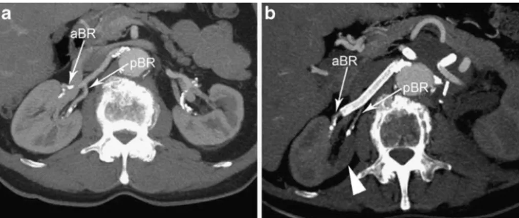
Fig. 15a, b A 64-year-old man with juxtarenal aortic aneurysm. a Initial MDCTA (MIP) demonstrates a normal branching pattern of the renal arteries with a single renal artery supplying the right kidney. Near the hilum of the kidney the renal artery divides into an anterior and a posterior branch (aBR and pBR). b Post-procedural
MDCTA (MIP) reveals a heterogeneous right nephrogram with a zone of low attenuation through the right kidney (arrowhead). The distal end of the Viabahn to the right kidney was placed into the anterior branch (aBR) of the renal artery causing occlusion of the posterior branch (pBR)
Fig. 16 A 77-year-old man who developed mesenteric ischaemia after visceral revascularisation. MDCTA through the lower abdomen demonstrates wall thickening and pneumatosis (arrows) of several small bowel loops
Fig. 17 MDCTA of a 64-year-old woman who underwent renal and visceral debranching. MDCTA demonstrates a seroma surrounding the extra-anatomical bypass grafts and a perigraft seroma-related urinary tract obstruction with mild dilatation of the left renal pelvis (arrow)
Debranching-graft related complications
Bypass occlusion
Bypass occlusion is a serious, but rarely reported compli-cation after renal or visceral debranching. Long-term experience is sparse. Recent studies describe primary patency rates that range from 98–100% after hybrid treatment (Fig.11) [6,40].
Anastomotic stenosis
Stenoses within bypasses are a commonly known problem of vascular reconstructive surgery. Stenoses often occur at the anastomotic sites of the vascular graft. There are no studies reporting this problem after complex renal and visceral debranching as part of hybrid TAAA repair. We saw one case with significant bypass stenosis at the anastomosis (sutured anastomosis) to the left renal artery (Fig. 12). Long-term studies are needed to validate the formation of intimal hyperplasia beside the anastomosis.
Anastomotic aneurysm and bleeding
Partial or total disruption of the anastomosis between an artery and a vascular graft can result in anastomotic aneurysm and/or anastomotic bleeding. Factors leading to this problem include degenerative disease, inflammatory disease and technical faults during the surgical procedure (Fig.13) [13].
Organ ischaemia
Surgical treatment of TAAA incurs a significant risk of end-organ ischaemia, leading to organ dysfunction [41].
Avoidance of high aortic cross clamping is an advantage of the hybrid approach and may lead to a significant reduction in visceral and renal ischaemia time. Ischaemic complications due to mechanical obstruc-tion, graft thrombosis, or atheroembolism involve the legs, feet, bowel, kidney, liver and other end-organs. End-organ ischaemia after a hybrid treatment may result from both renovisceral revascularisation and EVAR (Figs.14,15,16) [6,9,10].
Perigraft seroma
Perigraft seroma is an accumulating fluid collection surrounding an intact graft and presents as a soft tissue swelling around the graft. It might be caused by transu-dation of an ultrafiltrate of serum through the graft wall into the extrinsic milieu. Perigraft seroma is a rarely reported, late complication of PTFE and Dacron grafts. These seromas are mostly found in superficial grafts such as haemodialysis arteriovenous fistulas [12]. Depending on the size and location, an abdominal perigraft seroma may lead to urinary tract or bowel obstruction [42] (Fig.17).
Summary
The hybrid approach is a promising alternative to OS for the treatment of TAAA. Precise pre-interventional imaging and the knowledge of common and rare complications are crucial for a successful outcome for each patient. Procedure-specific complications vary along the spectrum, from small endoleak to stent dislocation with side branch occlusion. MDCTA with 2D and 3D post-processing methods is a reliable imaging technique for an effective and exact pre- and post-procedural evaluation.
References
1. Parodi JC, Palmaz JC, Barone HD (1991) Transfemoral intraluminal graft implantation for abdominal aortic an-eurysms. Ann Vasc Surg 5:491–499 2. Prinssen M, Verhoeven EL, Buth J et al
(2004) A randomized trial comparing conventional and endovascular repair of abdominal aortic aneurysms. New Engl J Med 351:1607–1618
3. Greenhalgh RM, Brown LC, Kwong GP, Powell JT, Thompson SG (2004) Comparison of endovascular
aneurysm repair with open repair in patients with abdominal aortic aneurysm (EVAR trial 1), 30-day operative mortality results: randomised controlled trial. Lancet 364:843–848
4. Quinones-Baldrich WJ, Panetta TF, Vescera CL, Kashyap VS (1999) Repair of type IV thoracoabdominal aneurysm with a combined endovascular and surgical approach. J Vasc Surg 30:555– 560
5. Fulton JJ, Farber MA, Marston WA, Mendes R, Mauro MA, Keagy BA (2005) Endovascular stent-graft repair of pararenal and type IV thoracoab-dominal aortic aneurysms with adjunc-tive visceral reconstruction. J Vasc Surg 41:191–198
6. Black SA, Wolfe JH, Clark M, Hamady M, Cheshire NJ, Jenkins MP (2006) Complex thoracoabdominal aortic aneurysms: endovascular exclusion with visceral revascularization. J Vasc Surg 43:1081–1089 discussion 1089
7. Chiesa R, Tshomba Y, Melissano G et al (2007) Hybrid approach to thoraco-abdominal aortic aneurysms in patients with prior aortic surgery. J Vasc Surg 45:1128–1135
8. Resch TA, Greenberg RK, Lyden SP et al (2006) Combined staged procedures for the treatment of thoracoabdominal aneurysms. J Endovasc Ther 13:481– 489
9. Donas KP, Czerny M, Guber I, Teufelsbauer H, Nanobachvili J (2007) Hybrid open-endovascular repair for thoracoabdominal aortic aneurysms: current status and level of evidence. Eur J Vasc Endovasc Surg 34:528–533 10. Donas KP, Schulte S, Krause E, Horsch S (2007) Combined endovascular stent-graft repair and adjunctive visceral vessel reconstruction for complex tho-racoabdominal aortic aneurysms. Int Angiol 26:213–218
11. Bockler D, Kotelis D, Geisbusch P et al (2008) Hybrid procedures for thoraco-abdominal aortic aneurysms and chronic aortic dissections—a single center experience in 28 patients. J Vasc Surg 47:724–732
12. Dauria DM, Dyk P, Garvin P (2006) Incidence and management of seroma after arteriovenous graft placement. J Am Coll Surg 203:506–511
13. Mii S, Mori A, Sakata H, Kawazoe N (1998) Para-anastomotic aneurysms: incidence, risk factors, treatment and prognosis. J Cardiovasc Surg 39:259– 266
14. Mita T, Arita T, Matsunaga N et al (2000) Complications of endovascular repair for thoracic and abdominal aortic aneurysm: an imaging spectrum. Radiographics 20:1263–1278 15. Lachat M, Mayer D, Criado FJ et al
(2008) New technique to facilitate renal revascularization with use of telescop-ing self-expandtelescop-ing stent grafts: VORTEC. Vascular 16:69–72 16. Rozenblit AM, Patlas M, Rosenbaum
AT et al (2003) Detection of endoleaks after endovascular repair of abdominal aortic aneurysm: value of unenhanced and delayed helical CT acquisitions. Radiology 227:426–433
17. Stavropoulos SW, Charagundla SR (2007) Imaging techniques for detec-tion and management of endoleaks after endovascular aortic aneurysm repair. Radiology 243:641–655
18. Cademartiri F, Mollet NR, van der Lugt A et al (2005) Intravenous contrast material administration at helical 16-detector row CT coronary angiography: effect of iodine concentration on vas-cular attenuation. Radiology 236:661– 665
19. Fleischmann D (2003) Use of high concentration contrast media: principles and rationale-vascular district. Eur J Radiol 45(Suppl 1):S88–S93
20. Golzarian J, Dussaussois L, Abada HT et al (1998) Helical CT of aorta after endoluminal stent-graft therapy: value of biphasic acquisition. AJR Am J Roentgenol 171:329–331
21. Iezzi R, Cotroneo AR, Filippone A et al (2006) Multidetector CT in abdominal aortic aneurysm treated with endovas-cular repair: are unenhanced and delayed phase enhanced images effec-tive for endoleak detection? Radiology 241:915–921
22. Abada HT, Sapoval MR, Paul JF, de Maertelaer V, Mousseaux E, Gaux JC (2003) Aneurysmal sizing after endo-vascular repair in patients with abdominal aortic aneurysm: interobser-ver variability of various measurement protocols and its clinical relevance. Eur Radiol 13:2699–2704
23. Dalrymple NC, Prasad SR, Freckleton MW, Chintapalli KN (2005)
Informatics in radiology (infoRAD): introduction to the language of three-dimensional imaging with multidetec-tor CT. Radiographics 25:1409–1428 24. Crawford ES, Crawford JL, Safi HJ et
al (1986) Thoracoabdominal aortic aneurysms: preoperative and intraoper-ative factors determining immediate and long-term results of operations in 605 patients. J Vasc Surg 3:389–404 25. Safi HJ, Winnerkvist A, Miller CC 3rd
et al (1998) Effect of extended cross-clamp time during thoracoabdominal aortic aneurysm repair. Ann Thorac Surg 66:1204–1209
26. Kawanishi Y, Okada K, Matsumori M et al (2007) Influence of perioperative hemodynamics on spinal cord ischemia in thoracoabdominal aortic repair. Ann Thorac Surg 84:488–492
27. Garzon G, Fernandez-Velilla M, Marti M, Acitores I, Ybanez F, Riera L (2005) Endovascular stent-graft treat-ment of thoracic aortic disease. Radio-graphics, 25(Suppl 1):S229–244 28. Makaroun MS, Dillavou ED, Kee ST et
al (2005) Endovascular treatment of thoracic aortic aneurysms: results of the phase II multicenter trial of the GORE TAG thoracic endoprosthesis. J Vasc Surg 41:1–9
29. Huber A, Matzko M, Wintersperger BJ, Reiser M (2001) [Reconstruction methods in postprocessing of CT- and MR-angiography of the aorta]. Radi-ologe 41:689–694
30. Aquino RV, Rhee RY, Muluk SC, Tzeng EY, Carrol NM, Makaroun MS (2001) Exclusion of accessory renal arteries during endovascular repair of abdominal aortic aneurysms. J Vasc Surg 34:878–884
31. Karmacharya J, Parmer SS, Antezana JN et al (2006) Outcomes of accessory renal artery occlusion during endovas-cular aneurysm repair. J Vasc Surg 43:8–13
32. Leurs LJ, Laheij RJ, Buth J (2005) What determines and are the consequences of surveillance intensity after endovascular abdominal aortic aneurysm repair? Ann Vasc Surg 19:868–875
33. Harris PL, Buth J, Mialhe C, Myhre HO, Norgren L (1997) The need for clinical trials of endovascular abdomi-nal aortic aneurysm stent-graft repair: The EUROSTAR Project. EUROpean collaborators on Stent-graft Techniques for abdominal aortic Aneurysm Repair. J Endovasc Surg 4:72–77 discussion 78–79
34. Schlensak C, Doenst T, Hauer M et al (2001) Serious complications that re-quire surgical interventions after endo-luminal stent-graft placement for the treatment of infrarenal aortic aneur-ysms. J Vasc Surg 34:198–203 35. Kritpracha B, Beebe HG, Comerota AJ
(2004) Aortic diameter is an insensitive measurement of early aneurysm expansion after endografting. J Endovasc Ther 11:184–190 36. Lee JT, Aziz IN, Lee JT et al (2003)
Volume regression of abdominal aortic aneurysms and its relation to successful endoluminal exclusion. J Vasc Surg, 38:1254–1263
37. Chaikof EL, Blankensteijn JD, Harris PL et al (2002) Reporting standards for endovascular aortic aneurysm repair. J Vasc Surg 35:1048–1060
38. Veith FJ, Baum RA, Ohki T et al (2002) Nature and significance of endoleaks and endotension: summary of opinions expressed at an international confer-ence. J Vasc Surg 35:1029–1035 39. Maleux G, Koolen M, Heye S,
Nevelsteen A (2008) Limb occlusion after endovascular repair of abdominal aortic aneurysms with supported endo-grafts. J Vasc Interv Radiol 19:1409–1412 40. Gawenda M, Aleksic M, Heckenkamp
J, Reichert V, Gossmann A, Brunkwall J (2007) Hybrid-procedures for the treatment of thoracoabdominal aortic aneurysms and dissections. Eur J Vasc Endovasc Surg 33:71–77
41. Schepens MA, Defauw JJ, Hamerlijnck RP, De Geest R, Vermeulen FE (1994) Surgical treatment of thoracoabdominal aortic aneurysms by simple cross-clamping. Risk factors and late results. J Thorac Cardiovasc Surg 107:134–142 42. Burns P, Bradbury AW (2000) Duodenal
obstruction following aortic aneurysm repair caused by an aneurysm sac sero-ma. Eur J Vasc Endovasc Surg 20:487– 488