REVIEW
Induction of intestinal lymphoid tissue formation by intrinsic
and extrinsic signals
Daniela Finke
Received: 20 April 2009 / Accepted: 20 May 2009 / Published online: 9 June 2009
# Springer-Verlag 2009
Abstract Since the discovery of inducer cells as a separate lineage for organogenesis of Peyer’s patches in the small intestine of fetal mice, a lot of progress has been made in understanding the molecular pathways involved in the generation of lymphoid tissue and the maintenance of the lymphoid architecture. The findings that inducer cells also exist in adult mice and in humans, have a lineage relationship to natural killer cells, and can be stimulated during infections highlight their possible role in establish-ing innate and adaptive immune responses. Novel concepts in the development of intestinal lymphoid tissues have been made in the past few years suggesting that lymphoid organs are more plastic as previously thought and depend on antigenic stimulation. In addition, the generation of novel lymphoid organs in the gut under inflammatory conditions indicates a function in chronic diseases. The present review summarizes current knowledge on the basic framework of signals required for developing lymphoid tissue under normal and inflammatory conditions.
Keywords Lymphoid tissue inducer cell . Peyer’s patch . Isolated lymphoid follicle . Lympho-organogenesis . Inflammation . IBD
Introduction
The gastrointestinal tract is continuously exposed to antigens (Ags) and discrimination between pathogens and
commensal bacteria is achieved by the interaction of the intestinal epithelium with lymphoid cells. Intestinal lym-phoid cells can form organized cell aggregates called Peyer’s patches (PP) where immune responses are initiated leading to the generation of IgA-secreting plasma cells [1]. These sites belong to the mucosa-associated lymphoid tissue (MALT), since they collect Ags from mucosal surfaces. The epithelium overlaying the PPs forms the interface between the MALT and the luminal microenvi-ronment. It is critically involved in host defense and maintenance of immune homeostasis through two major pathways. Firstly, it contains specialized cells named M cells able to transport luminal Ags to the PPs. Secondly, it secretes antimicrobial products as well as cytokines, which are essential for recruitment of immune cells to the Ag entry site. The commensal and pathogenic microorganisms in the luminal content engage innate receptors expressed by intestinal epithelial cells. As a consequence, epithelial cytokines and activated dendritic cells (DCs) regulate the quality of adaptive immune responses leading to either tolerance or immunity.
In addition to PPs, the lamina propria of the murine gut contains multiple small cryptopatches (CPs), which can emerge into isolated lymphoid follicles (ILFs) in response to the gut flora [2,3]. Whereas the number and localization of PPs is developmentally fixed before birth, CPs and ILFs represent the most dynamic compartments of the small intestine, since they develop after birth and number and size are highly variable, depending on the bacterial load [3]. ILFs were shown to be alternative sites of generating IgA responses [4].
The process of lymphoid tissue induction in the gut depends on two mutually nonexclusive pathways, including (1) tumor necrosis factor super family member signals provided by hematopoietic cells and (2) innate signals D. Finke (*)
Department of Biomedicine, Developmental Immunology, University of Basel,
Mattenstrasse, 28, 4058 Basel, Switzerland e-mail: [email protected]
through toll-like receptors (TLRs) and proinflammatory cytokines leading to the expression of lymphoid chemo-kines and adhesion molecules. In fetal mice, a particular subset of hematopoietic cells was identified, which is indispensable for the development of secondary lymphoid organs (SLOs) [5, 6]. These cells engage lymphotoxin β receptor (LTβR) on stromal cells followed by the produc-tion of chemokines that recruit T and B cells. In adult mice, similar cell subsets might exist, which, together with mature lymphocytes and proinflammatory cytokines, contribute to shaping of preexisting lymphoid organs. In addition, novel lymphoid tissues can form within organs during chronic inflammation and autoimmune diseases [7,8]. The cellular architecture of these tissues is reminiscent to the T and B cell compartments seen in SLOs.
In this chapter, we attempt to compare the pathways, which lead to the formation of intestinal lymphoid organs (a) before birth, (b) upon antigenic challenge, and (c) during chronic inflammation. We discuss the contribution of lymphoid tissue inducer cells, lymphocytes, and intesti-nal bacteria to the development and maturation of intestiintesti-nal lymphoid organs in mouse and man and emphasize on the role of intestinal lymphoid organs for the development of inflammatory bowel disease (IBD).
Intestinal lymphoid organs in mouse and man Organized lymphoid microenvironments in the gut Organized lymphoid aggregates in the gut represent the primary sites for eliciting adaptive immune responses toward mucosal Ags. Joseph Hans Conrad Peyer, a Swiss anatomist and physician (1653–1712), described for the first time PPs as the most prominent organized SLOs of the small intestine, with greatest density in the ileum. In humans, they emerge as follicles localized in the antime-senteric border of the gut at week 19 of gestation [9]. In contrast to lymph nodes (LNs), PPs are not connected to afferent lymphatic vessels draining Ag entry sites. Instead,
they are overlaid by a follicle-associated epithelium (FAE), which is specialized to uptake luminal microorganisms and Ags. The development of PPs is related to age, and number and size is at maximum at puberty and involutes upon aging [10]. Studies in other species reveal that jejunal and ileal PPs differ in their capacity to mount an immune response, to export lymphocytes via efferent lymphatic vessels and to produce cytokines [11–13]. Like other SLO, PPs are composed of segregated B and T cell zones, which form in an Ag-independent manner [14] (Fig.1a). Unlike LNs, where T cells are mainly found in the paracortex, PP T cells localize in T cell zones between B cell follicles (Fig.1b). The subepithelial dome (SD) of PPs contains two distinct subsets of DCs [15] that are distinguished by the expression of either CX3CR1 or CCR6 [16] (Fig. 2). The CX3CR1 DCs appear to be noninflammatory residual DCs collecting Ag from the luminal site under homeostatic conditions. These cells are likely identical to intraepithelial CD11b−CD8α−DCs that were found within the FAE and in the interfollicular region (IFR) [17]. A third subset of DCs localizes to the IFR that is CD11b−and CD8α+. DCs are the key players in eliciting primary immune responses, as in the absence of DCs specific T cell responses in PPs are impaired [16]. Moreover, under pathogen-free condi-tion, DCs are the dominant subset of cells underneath the FAE, whereas B and T cells only enter the subepithelial dome after colonization with bacteria [18]. Most of the mucosal IgA Abs are produced by germinal center (GC) B cells found in PPs of the intestine. The formation of GCs requires the activation of T helper cells by Ag-loaded DCs, the cognate interaction between activated T cells and Ag-specific B cells, and the presence of follicular CD4+ T helper (Th) cells (Fig.1b). These follicular Th cells can be generated from Foxp3+ CD4+ T cells demonstrating an unexpected link between Tregs and GC formation [19]. TGF-β, Il4, Il6, and Il10 are locally synthesized in PPs and help to trigger B cell responses and expand IgA-secreting plasma cells [20]. Although PPs represent an important site for the induction of mucosal immune responses, in PP-deficient mice, a surprising capacity of generating oral
Fig. 1 a Peyer’s patch in mouse small intestine. b Immunohisto-chemistry of Peyer’s patch using antimouse CD4 (green) and antimouse CD11c (red) Ab
tolerance and producing intestinal IgA responses was uncovered suggesting alternative sites for inducing adaptive immune responses. These sites were identified as multiple lymphoid aggregates called ILF scattered throughout the entire small intestine and colon, with the highest abundance in the distal ileum [21, 22] (Fig. 3a). Unlike PPs, they develop only after colonization of the gut with bacteria and represent highly dynamic lymphoid structures, which can vary in number and size [21]. ILF are composed of a single B cell follicle, surrounded by DCs, T cells, and Il7r+c-kit+ cells, the latter one closely localized to the subepithelial dome [21,23] (Fig.3b). They are overlain by an FAE, can uptake Ag, and generate GCs, a fact that drew them into attention as inductive sites for immune responses [24,25]. Beside ILFs, smaller lymphoid structures named crypto-patches (CPs) are scattered throughout the intestine [26]. They appear 1 to 2 weeks after birth and, in contrast to ILFs, localize to the bottom of crypt areas and barely contain B and T cells [26]. In contrast to ILFs, the presence of CPs has so far only been described in mice, but CPs are absent from the small intestine in humans, rats, and pigs [27]. Morphological intermediates between CPs and ILFs suggest that CPs can mature into ILFs [27].
Follicle-associated epithelium and M cells
The SD region of PPs is covered by the FAE, which is defined by its high density of specialized Ag-sampling cells called membrane or microfold (M) cells [28]. In addition, the FAE is, unlike the villus epithelium, not underlain by subepithelial myofibroblasts and goblet cells are almost absent [29]. M cells lack an absorptive capability, have only poorly organized short brush-border microfilli, and do not produce enzymes with digestive activity. Hence, alkaline phosphatase and sucrase-isomaltase, which are typical for enterocytes, were both used as negative markers of M cells [30]. M cells have an enormous propensity of transcytosis of Ags and particles from the luminal side of the gut. One characteristic of M cells is that they form large invaginated pockets harboring lymphocytes and DCs. This facilitates the contact between the incoming Ag and the adaptive immune system. There is strong evidence that the viral and bacterial uptake of M cells is receptor-mediated [31, 32]. Pattern recognition receptors on the surface of M cells have been identified as important receptors including TLR4, platelet-activating factor receptor, and α5β1 integrin [32]. The engagement of TLRs can stimulate the microparticle uptake and recruitment of DCs [33, 34]. In addition, the number of M cells increases in germ-free mice upon bacterial stimulation reflecting the dynamic of shaping intes-tinal microcompartments through environmental changes [35]. It is therefore likely that upon intestinal infection, inflammatory signals promote the generation and activation of M cells thereby increasing the efficiency of transcytosis and recruitment of DCs to the subepithelial dome. These DCs induce Ab isotype switching in B cells from IgM to IgA through their secretion of Il6 and expression of retinal dehydrogenases, the enzymes required for the generation of retinoid acid from vitamin A [36,37].
Studies in rabbits and humans have shown that the FAE does not express the polymeric Ig receptor or secretory component, the cleaved extracellular domain of the poly-meric Ig receptor [38,39]. Secretory IgA (SIgA), however, exhibits a striking affinity to M cells in mouse and man Fig. 2 Dendritic cell subpopulations in Peyer’s patch. SD
subepithe-lial dome, FAE follicle-associated epithelium, IFR interfollicular region, DC dendritic cell
Fig. 3 a Immunohistochemistry with anti-CD45 (green) Ab of small intestine and b organiza-tion of isolated lymphoid follicles
FAE [40], and immune complexes of SIgA and bacteria can translocate to SD DCs via M cells [41] thereby eliciting an immune response in PPs. SIgA might not only absorb microorganism and Ags in the intestinal lumen but also direct them specifically to M cells thereby allowing uptake, processing, and presentation in lymphoid organs. Under noninflammatory conditions, steady-state Ag acquisition of luminal content including commensal bacteria and cell debris may favor tolerance induction.
Using microarray analysis and in situ hybridization, various genes have been identified that were restricted to mouse, macaque, or human FAE [42–46]. These studies clarified that some but not all genes expressed by the FAE were unique for M cells. The FAE harbors to date poorly investigated subsets of epithelial cells that differ in their genetic profile from the normal villus epithelium and from M cells. Whether these cells represent predetermined M cell precursors that can become fully-functional Ag-sampling cells upon microbial stimulation remains to be investigated. In mice, the FAE of the small intestine constitutively expresses the chemokines CCL9 [47] and CCL20 (Mip3α) [15, 48], whereas in humans, CCL20 is also expressed in colonic epithelial cells [49]. CCL20 is chemotactic for cells expressing the corresponding chemokine receptor CCR6 such as γδ T cells, memory T cell subsets, B cells, and myeloid CD11b+ DCs subpopulations [15, 50–53]. Under inflammatory conditions, normal enterocytes can produce CCL20 as well [54] reconciling the concept that intestinal epithelial cells are highly plastic and can connect the innate and adaptive immune system through recruiting CCR6-expressing DCs and lymphocytes to sites of inflammation. Although it was reported that CCR6−/− mice had a perturbed intestinal immune system [16,55] and a reduced number of M cells [56], investigation of various CCR6−/− mice revealed a relatively normal ratio of subepithelial DCs and lymphocytes [47, 56]. This would suggest that other FAE-specific chemokines such as CCL9 could compensate for the lack of CCR6 or, alternatively, that some SD DCs are independent of CCR6, whereas others are recruited through CCR6 and inflammatory signals as previously proposed [16].
There are two models explaining the differentiation of the FAE and the origin of M cells (Fig. 4). One model proposes that the FAE develops from the crypts surround-ing the PP anlage. Stem cells reside within the crypts and give rise to progressively differentiating absorptive enter-ocytes and mucous cells, which migrate up toward the tip of the villi, where they are shed a few days later [57]. The follicle-associated crypts might contain stem cells, which have to give rise to cells migrating in two different axis. On one side, crypt stem cells differentiate into absorptive enterocytes, goblet cells, and enteroendocrine cells. On the other side of the crypt wall, cells give rise to the FAE
containing M cells. The stem cells that can give rise to the FAE or the specific differentiation program, which is required to coordinate the development of the two axes, has not been identified so far. Alternatively, it was proposed that M cells can be generated through conversion of enterocytes upon contact with underlying lymphocytes. This hypothesis was supported by an in vitro assay demonstrating for the first time that human enterocyte cell lines cultured in the presence of B cells could be converted into M cells [58]. This model was further developed by adding PP cells to human or mouse epithelial cell lines [59,
60]. In these studies, however, human adenocarcinoma cells (Caco-2) were used that might not behave like conventional enterocytes. In addition, the appearance of M-like cells was reported to occur in Caco-2 cells even in the absence of lymphocytes [61]. Although in vitro and in vivo studies using B cell-deficient mice have let to the conclusion that B lymphocytes play a role for M cell conversion [58, 62], RAG-1−/−mice develop small but detectable PPs with M-cell harboring FAE indicating that M M-cells can develop in the absence of lymphocytes [63]. This is also confirmed in SCID mice, which lack B and T lymphocytes but develop detectable PP anlagen [64]. It is likely that in the absence of lymphocytes, M cells can develop but that a mature FAE depends on the presence of lymphocytes. The reconstitution of immunodeficient mice with WT bone marrow (BM) enlarges preexisting PP anlagen and may promote the maturation of the FAE [65]. Since an early appearance of M cell markers was already detected in the vicinity of the follicle-facing crypts [66], factors produced by underlying lymphocytes must act very early in the differentiation of crypt cells. Altogether, we think that the development of M cells is a result of commitment from particular progenitor cells, the interaction of progenitor cells with lymphocytes and contact with microbial Ags.
The paradigm of a specialized FAE required for Ag uptake has become challenged by more recent findings that single M cells are also found along the villus axis in normal and PP-deficient mice [67]. Moreover, CCL20 expression can be extended to normal enterocytes upon stimulation with flagellin [54] suggesting that inflamed enterocytes can Fig. 4 Models of M cell development
adopt“FAE-like” features. The discovery of M cells in the villus epithelium [67] together with the fact that a specialized subset of CX3CR1+ lamina propria DCs can form transepithelial dendrites to uptake luminal Ag [68,69] suggest that efficient Ag uptake does not only occur in organized lymphoid aggregates.
Stromal cell networks in the intestine
It is well established that stromal cells not only have an essential role in generating a lymphoid tissue architecture but are also required for adaptive immune responses. For example, PP stromal cells secrete factors such as TGFβ1, Il6, and Il10, which promotes the differentiation of Ag-stimulated B cells into IgA-producing plasma cells. In addition, lymphoid stroma cells secrete BAFF, SCF, and Il7, which may enhance the survival of B and T cells and class switching of Igs. Various subsets of stromal cells in the gut-associated lymphoid tissue (GALT) were identified which exert different functions in the intestinal immune system. In PPs, CPs and ILFs of adult mice, vascular cell adhesion molecule-1 (VCAM-1)+ stromal cells are detect-able around the periphery and within B cell areas, a pattern reminiscent of follicular dendritic cells (FDCs) [70]. FDCs are of mesenchymal origin and, in addition to VCAM-1 and LTβR, express complement receptors 1 (CD35) and 2 (CD21) and the FcγRIIb, which allow the trapping of immune complexes and retention of Ag. The display of Ag, the production of B cell growth factors, and the secretion of chemokines support the activation and differentiation of follicular B cells and the generation of GCs. It is generally accepted that B cells, lymphotoxin, and tumor necrosis factor (TNF) are required for FDC development and organization of T/B zones [71]. The loss of LTβ expression
in gene-targeted mice reveals a direct correlation between the level of organization and immunosurveillance.
In LNs, the T cell zone consists of gp38+ VCAM-1+ stromal cells, which form a three-dimensional network of cells and fibers [72, 73]. These fibroblast reticular cells (FRCs) produce Il7 which has a key function in T cell homeostasis and function [74]. Moreover, LN FRCs create the conduit system consisting of a sheet of FRCs around reticular fibers and extracellular matrix proteins [75–77]. The conduit and fiber network can be identified by ER-TR7 immunohistochemistry. The conduits are coated with various chemokines, which allow the distribution and locomotion of DCs and lymphocytes attaching to the FDC fibers [78]. It is currently unknown if similar network of ER-TR7+conduits exist in PPs and ILFs that may regulate lymphocyte trafficking in intestinal lymphoid tissues.
The recruitment of lymphocytes is coordinated by chemokines produced by stromal cells such as FDCs, FRCs, endothelial cells, and perivascular myofibroblasts.
Naïve lymphocytes leave the circulation and enter SLOs via high endothelial venules (HEVs). CXCL13 produced by FDCs localizes incoming B cells to the follicular zone, and in the T zone, FRCs express CCL19 and CCL21 that recruits T cells [79]. In contrast to mouse PP, in which HEV produce CCL21, HEVs in human PPs lack detectable CCL21 transcripts, although CCL21 protein is readily detectable [80]. One possible explanation was provided by another study demonstrating that HEVs possess basolateral binding sites for CCL19 [81]. This indicates that trans-cytosis of chemokines through endothelial cells may be responsible for attracting naïve CCR7+ T cells to human PPs. It is conceivable that HEVs have a capacity of transcytosis similar to the aforementioned M cells in the FAE.
In addition to regulating migration and survival of lymphocytes, under homeostatic conditions, stromal cells can potentially contribute to the induction of peripheral tolerance. LN stromal cells express autoimmune regulator (AIRE) [82], the regulator of promiscuous gene expression. They help generating regulatory DCs [83] and produce the vitamin A metabolite retinoid acid (RA), a key molecule in generating gut-homing T cells and intestinal Foxp3+Tregs [84]. Whether stromal cells support Ag-specific immune responses or rather tolerance induction might depend on additional inflammatory signals provided by the innate immune system.
Stromal cells are also active players in the initiation of lymphoid tissue development (see also “Regulation of Peyer’s patch development during fetal life” section). In PP and LN anlagen of fetal mice, mesenchymal stromal cells named “organizer cells” express LTβR, VCAM-1, intercellular adhesion molecule 1 (ICAM-1), and mucosal vascular addressin cell adhesion molecule 1 (MAdCAM-1). The gene expression profile of organizer cells isolated from PP anlagen differs from mesenteric LN organizer cells [85]. This is consistent with the notion that PP and LN development requires the activity of different genes. Recently, LTβR+
VCAM-1+ ICAM-1+ MAdCAM-1+cells designated as marginal reticular cells (MRC) were identi-fied in adult SLO [86]. Interestingly, MRCs were localized in the subepithelial dome of PPs, the marginal zone of the spleen, and underneath the subcortical sinus of LNs. These are the sites where Ag enters lymphoid tissues. It will be important to study if MRCs have a role for recruiting DCs or coordinating immune responses upon Ag entry.
Regulation of Peyer’s patch development during fetal life
PP development has been extensively studied in various animal models. One of the first fundamental discoveries
toward understanding the molecular events driving PP organogenesis was the finding that mice with a mutation of alymphoplasia (aly) were completely devoid of PPs and LNs [87]. The aly allele encoded a single mutation in the C-terminal interaction domain of NF-κB-inducing kinase (NIK) [88, 89], a signaling molecule of the LTβR. A
phenotype similar to the aly mutation was found in mice with a deletion of LTα, a TNF superfamily chain, which can associate to LTβ thereby forming a heterodimeric ligand for LTβR [90]. Moreover, a deletion of NIK resulted in loss of PPs and LNs [91]. When LTβR−/− mice were analyzed, PPs were found to be absent [92,93]. Altogether, these data highlight the importance of LTβR and its downstream signaling molecules for the development of PPs during fetal life.
Studies in the last 10 years have led to the paradigm that in mice, at least two cell types act as key players in PP development: hematopoietic LTαβ+Il7r+ c-kit+ CD4+ lin− cells [64, 94, 95], named lymphoid tissue inducer (LTi) cells and mesenchymal LTβR+
VCAM-1+ ICAM-1+ orga-nizer cells [96,97]. Both cell subsets express corresponding adhesion molecules and cytokine receptor/ligand pairs, which enable them to attach to each other and form cellular aggregates forming the primordium of PPs. LTi cells colonize the developing gut at E12.5 of gestation [98]. Since they mainly localize to blood vessels, it is likely that the entry site for LTi cells is the vascular endothelium. In addition to the developing gut, LTi cells migrate to putative sites of LN formation, to the fetal spleen, and, as recently reported, to the fetal thymus [99]. In developing primary and secondary lymphoid organs, they have specific but probably diverse functions dependent on which cellular partner they engage.
LTi cells express the nuclear hormone receptor RORγt [98] as well as a number of TNF superfamily member molecules (LTα1β2, LIGHT, TNF-α, RANK, DR3, 4-1BB) [95, 100, 101], among them LTα1β2 can engage LTβR
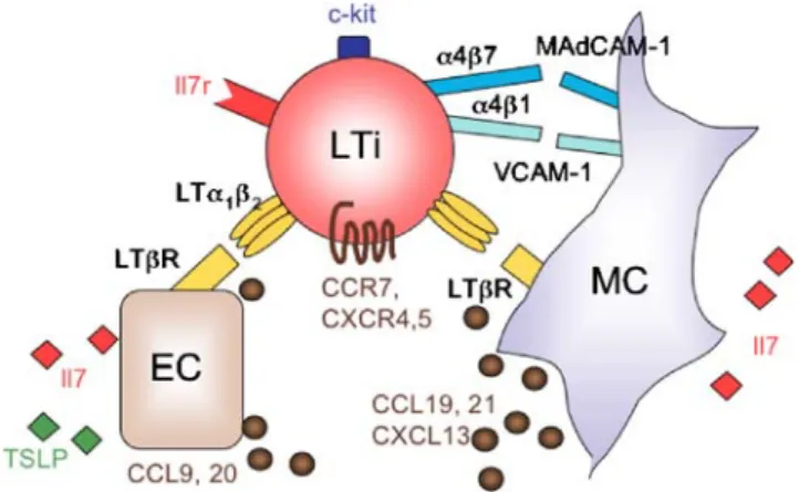
expressed on the surface of organizer cells [97] (Fig. 5). The adhesion of LTi cells to organizer cells is coordinated by activation of α4β1-integrin on LTi cells, the high affinity ligand of VCAM-1 [102]. Moreover, LTi cells express α4β7, which can bind to MAdCAM-1 expressed by the organizer cells. After attachment, signaling via LTβR leads to the expression of chemokines and adhesion molecules that are involved in lympho-organogenesis [103– 105]. The production of CCL19, CCL21, and CXCL13 probably helps to recruit more LTi cells, since they express the corresponding chemokine receptors CCR7 and CXCR5 [97, 101]. In addition, these chemokines promote the colonization of PPs with lymphocytes. It is possible that LTi cells not only engage LTβR on the surface of organizer cells but also on endothelial cells before entering the parenchyma. The activation of endothelial LTβR is
re-quired for the formation of HEV and for the development of lymphatic vessels [106–108]. Finally, LTi cells can trigger LTβR signaling in epithelial cells, which is an important step for CCL20 expression and maturation of the FAE [109] (Fig. 5). The local release of Il7 by enterocytes and organizer cells might help to maintain the pool of LTi cells through anti-apoptotic signals.
It is surprising that in LTα−/− mice treated with an agonist anti-LTβR Ab from E13 onward, unlike some LNs, the formation of PPs could not be restored [110]. These data suggest that either the threshold of LTβR signals required for PP development exceed LN development or, alternatively, that additional signals are required for PP development. The latter explanation is supported by the observation that in some mouse models with a null mutation for TNFRI (TNFp55), PP formation is abrogated [111,112]. Although treatment of pregnant mice with the TNFRp55-Ig fusion protein failed to block PP development in the offspring, it substantially suppressed the proportion of organizer cells [97]. TNFα can activate both TNFRI and
TNFRII. Since TNFRII−/− mice have normal PPs, TNFRI together with LTβR seem to be the major receptors triggering the expression of genes involved in PP develop-ment. There is indeed evidence for a synergistic activity of TNFRI and LTβR signaling in mesenteric LN genesis [113]. TNF can induce LTαβ expression by LTi cells [114], and therefore, the local release of TNF may indirectly promote the engagement of LTβR. A relative increase in the amount of TNFα together with LTαβ was found in fetal as compared to adult LNs [115] and this may contribute to lympho-organogenesis during fetal life. Altogether, despite the essential role of LTβR for the development of PPs and LNs, this pathway alone is not sufficient to induce organogenesis. The identification of additional signals triggered by early fetal hematopoietic cells will be a matter of future investigations in order to better understand the requirements for lymphoid organ development.
Fig. 5 Development of Peyer’s patches and FAE through engagement of LTβR. LTi lymphoid tissue inducer cell, MC mesenchymal cell, EC epithelial cell
Signaling via the heterodimeric Il7r complex composed of the IL-7Rα and the common γ chain (γc) is the canonical cytokine pathway for the development of PPs and LNs [116]. Despite that, there is evidence that additional cytokines may contribute to PP and LN development. For example, mice double deficient in the fms-like tyrosine kinase-3 (flt3) ligand and IL-7Rα show further impairment in the development of LNs and PPs [117]. Il7−/− mice have normal numbers of PP anlagen, whereas mice having a null mutation of IL-7Rα, γc, or JAK3, a kinase involved in the Il7r-signaling pathway, are completely devoid of PP anlagen [6, 94, 118–121]. This could potentially reflect the action of one or more alternative ligands promoting organogenesis in the fetal intestine. The Il7-like cytokine thymic stromal lympho-poietin (TSLP) binds to a heterochimeric receptor com-posed of IL-7Rα and a receptor subunit called TSLPR [122,123]. Both Il7 and TSLP are constitutively expressed by intestinal epithelial cells (Fig.5). TSLPR−/−mice have no defect in PP development [124] emphasizing the commanding role of Il7r in organogenesis. Therefore, alternative ligands regulating PP anlagen formation in fetal Il7−/− mice remain to be identified. A collaboration of signals regulating lympho-organogenesis was also proposed for IL-7Rα, CXCL13, and CCR7 [120] as well as for IL-7Rα and TRAF6 [114]. Mice deficient for CCR7, CCL19, or CCL21 have a normal phenotype with respect to SLO development [125, 126]. By combining the ablation of CXCR5 and CCR7, peripheral LN numbers were severely reduced, although PP numbers were almost normal [127]. These observations are in agreement with findings that the combinatorial interactions between two or more cytokines are frequently required for development and differentiation [128]. The collaboration of chemokines, cytokines, and adhesion molecules might promote the entry and accumu-lation of LTi cells, as well as their survival, expansion, and expression of LTαβ in the fetal gut.
Despite substantial knowledge on the molecular path-ways that are involved in lympho-organogenesis, the sequence of inductive events in PP ontogeny and the cellular requirements to generate PPs are still not fully understood. At the early stages, PP anlagen appear as cluster of VCAM-1+ organizer cells in the proximal jejunum at E15.5 [64] before they extent to the distal part of the gut. One to 2 days after VCAM-1+ spots have emerged, LTi cell cluster are detectable at the same location. The formation of organizer cluster does not occur cell autonomously, as the lack of LTi cells in RORγ−/− mice, the Ab-mediated neutralization of IL-7Rα, or the deletion of LTα prevents PP anlagen formation. These data suggest that few LTαβ-expressing LTi cells are sufficient to initiate PP anlagen formation before LTi cell cluster are detectable. Il7 has a major role in inducing LTαβ
expression on LTi cells [95, 114, 129]. Hence, the neutralizing activity of α-IL-7Rα Ab treatment may rely on insufficient expression of LTαβ. This is further supported by the fact that the migration of LTi cells to the intestine is maintained in the absence of IL-7Rα or LTαβ, although the cells are only scattered [94]. It is likely that after entering the gut via capillaries discharging into the antimesenteric wall of the gut, single LTi cells require stimulation by Il7 in the intestine in order to express high levels of LTαβ and activate mesenchymal organizer cells. In a second step, VCAM-1 is upregulated by organizer cells that were previously exposed to incoming LTi cells. The formation of organizer cluster is then a consequence of LTβR signals activating neighboring VCAM-1− organizer cells according to the concentration of LTi cells. Alterna-tively, VCAM-1+ organizer cell cluster are a result of proliferation and/or migration of VCAM-1+ cells. This would, however, not explain why LTα−/− mice are completely devoid of single VCAM-1+ cells. In a final step, the chemokine-driven reinforced influx of LTi cells might lead to the formation of detectable LTi cell cluster.
We demonstrated the dependence of organizer cluster formation on LTi cells in two mouse models with increased number of LTi cells. In one model, the adoptive transfer of LTi cells was able to restore organizer cell cluster in CXCR5−/− mice [102]. In the other model, a striking increase in the absolute cell number of organizer cells was observed in mice expressing Il7 transgene (tg) under the control of an ubiquitous promoter [129]. Il7 tg expression increased both the absolute cell number and LTαβ expression of LTi cells in fetal mice. Accordingly, a continuous band of VCAM-1+organizer cells accumulated in the antimesenteric wall at E16.5 demonstrating for the first time that there is no positional signal for PP anlagen formation. At birth, however, this band of organizer cells gave way to multiple clusters along the entire intestine. The mechanism for this pattern formation is still unsolved. Nishikawa et al. proposed that analogously to the reaction– diffusion paradigm in other biological systems, inhibitory signals interfere with activation signals coming from LTi and organizer cells and result in a periodic pattern [6,130]. For example, cytokine signals may be modulated by inhibitory soluble cytokine receptors or suppressors of cytokine signaling [131, 132]. Altogether, a model has evolved where PP anlagen are formed as soon as LTi cells and organizer cells assemble [6].
Together with the abundance of LTi cells in the fetal gut, numerous CD11c+ cells are found scattering over the gut [64,95]. At E17.5, CD11c+cells are mainly localized in the periphery of PPs but eventually fuse to LTi cell cluster after birth [96]. This cell subset was recently characterized in more detail [133]. The cells are of hematopoietic origin, express CD11b, CCR7, c-kit, and high levels of LTαβ, but
are negative for CD4, CD3, CXCR5, and IL-7Rα. [96]. In contrast to conventional DCs, they lack DEC205, but express Gr-1 and NK1.1. Forty percent of these cells express RET, a receptor tyrosine kinase involved in the formation of the enteric nervous system. The corresponding ligand artemin (artn) is expressed by intestinal VCAM-1+ cells, which include mesenchymal organizer cells and endothelial cells. Artn was found to promote the recruit-ment of LTi cells and CD11c+ cells in vitro. Mice with a deletion or hypomorphic allele of RET had a reduced number of PP anlagen suggesting that the RET/Artn axis has a role in gut lymphoid tissue development. It remains to be studied if the number and activity of RET CD11c+cells is normal in mouse models such as IL-7Rα−/−, RORγ−/−, and Id2−/−mice. Altogether, TNFRI, LTβR, Il7r, and RET are instrumental in PP development. The availability of Il7 determines the number and activity of LTi cells thereby influencing the size of the pool of VCAM-1+ organizer cells and PP anlagen.
Origin, life cycle, and fate of LTi cells in mouse and man LTi cells are present in RAG−/−, scid, and nude mice indicating that they do not need RAG recombination or a thymus for their development. However, the generation of conventional LTi cells is strictly dependent on the expres-sion of the retinoid acid-related orphane receptor RORγt
[134] and the inhibitory HLH transcription factor Id2 [135]. In RORγ−/−and Id2−/−mice, LTi cells were absent resulting in the complete lack of PPs and LNs [98,134–136]. The effect of Id2 on LTi cell development appears to be indirect via suppressing E2A, an E protein transcription factor promoting B cell development but limiting LTi cell and NK cell development. In double mutant Id2−/−E2A−/−mice, LTi cells, LNs, and PPs develop normally [137].
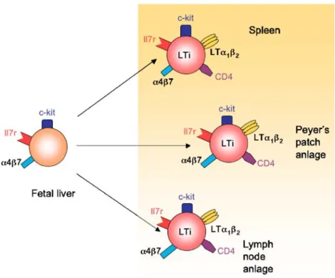
LTi cells originate fromα4β7+Il7r+c-kit+lin cells in the fetal liver (FL) [138]. Il7r+c-kit+lin−FL cells were found to give rise to all lymphoid lineages and LTi cells suggesting that they corresponded to a common lymphoid progenitor (CLP)-like cell [139]. In our hands, purified α4β7+
Il7r+ c-kit+ lin− cells had lost the potential to differentiate into B cells. It is possible that Il7r+c-kit+lin− cells represent a heterogenous population comprising already committed α4β7+ Il7r+ c-kit+ lin− LTi cells and precursor cells harboring a CLP activity. Il7r+ c-kit+ FL cells do not express LTαβ indicating that they lack properties of mature LTi cells [95]. After exit from the FL and entry into the fetal intestine, spleen, and putative sites of LN development, they get exposed to local cytokines such as Il7, which might induce further maturation and expression of LTαβ (Fig. 6). In line with this, Il7 transcripts were found in the fetal gut already at E13 and remained high until birth, whereas in the FL, only weak levels of Il7 transcripts were detectable between E16 and 18 [140]. Il7 not only promotes the expression of LTαβ but
Fig. 6 Colonization of the periphery with LTi precursor cells from fetal liver
also the differentiation of CD4− FL precursors and the survival of both FL precursor and mature LTi cells [114,
129, 138, 139]. There is evidence that the expression of CD4 starts in the periphery and that CD4− Il7r+ c-kit+ lin cells exist, which have some properties comparable to LTi cells, and might represent an intermediate between FL precursors and fully differentiated LTi cells [95].
A LTi-like cell subset was recently identified in the mesenteries and LNs of human fetuses [141]. These cells express IL-7Rα and RORγt, LTαβ, and TRANCE but are negative for CD4. Coculture with fetal mesenchymal cells resulted in the upregulation of adhesion molecules on the mesenchymal cells. This effect could be blocked by inhibiting the LTβR and TNFR pathway suggesting that, like LTi cells in fetal mice, human LTi cells were able to induce the maturation of mesenchymal cells by signaling through both LTβR and TNFR.
There is a debate if LTi cells are terminally differentiated or still harbor the potential to differentiate into other lineages. Initially, the commitment of murine LTi cells into Ag-presenting cells (APCs) and natural killer (NK) cells was described [101]. Using RORγt knockin mice
express-ing the enhanced green fluorescence protein under the control of the RORγt promoter, it was reported that in peripheral lymphoid tissue of fetal mice, RORγt was almost exclusively expressed in LTi cells and not in any other lineages [98]. This does not fully exclude the possibility that LTi cells harbor the capacity to differentiate into other lineages and that under noncompetitive con-ditions, e.g., in immunodeficient mice, LTi cells can give rise to NK cells and APCs. In mice and man, a subpopulation of CD4−cells in the intestinal crypts express RORγt and other markers characteristic for LTi cells [142]. Unlike LTi cells, however, these cells are NKp46+ [142– 145], a type 1 transmembrane protein found on NK cells. NKp46+ cells have no conventional NK cell function such as IFNγ or perforin production. Instead, they produce high levels of Il22, a cytokine that has been shown to be essential for host defense and epithelial cell homeostasis in humans [143]. There is evidence for a developmental relationship between LTi cells and RORγt+
NKp46+ NK-like cells in mice and humans. Human LTi cells display surface markers characteristic for NK cells [141]. In addition, LTi cells in mice and man express Il22 and Il17 [141, 146]. As for mouse LTi cells, the commitment of human LTi cells into NK cells was reported in vitro [141] and a LTi-like subset isolated from the lamina proporia of adult intestine gave rise to NK cells both in vitro and in vivo [147]. The number of RORγt+ NK-like cells is severely reduced in germfree mice, suggesting that the intestinal microflora could influence their generation from a precursor cell present in the intestine. The idea that either LTi cells and NK cells originate from a bipolar precursor or
that LTi cells can give rise to NK cells is supported by the findings that the HLH protein Id2 is required for the development of both LTi cells and NK cells [137, 148]. Altogether, a developmental relationship of LTi and NK cells is likely, and the generation of the cells might be regulated by cytokines provided in the FL and intestine, as well as by exposure to commensal bacteria.
There is evidence that LTi cells are not only generated during fetal but also adult life and mainly persist in SLOs. Adult CD4+CD3−cells were found in the spleen, LNs, PPs, lamina propria, and in ILFs and CPs [149,150]. The cells share a transcript expression profile with fetal LTi cells and were termed LTi-like cells [151]. In contrast to LTi cells, however, adult LTi-like cells express OX40L and CD30L after in vitro culture [152]. Coculture experiments with T helper 2 cells and the in vivo colocalization with T cells let to the hypothesis that they have a role for TH cells promoting long-lived B cell responses [153]. In fact, the Ag-driven expression of OX40 and CD30 on T cells allows the receiving of survival signals from OX40L and CD30L-expressing cells.
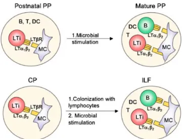
Mice deficient in RORγt lack adult LTi-like cells and CPs suggesting that after birth, these cells contribute to the generation and/or maintenance of CPs. Adoptive transfer experiments have shown that the pool of adult LTi-like cells in CPs and ILFs could be reconstituted by the transfer of BM, indicating that adult BM can be the source of adult LTi-like cells [70]. Similarly to the link between LTi-like cells and CPs, the same cells can induce the formation of ILFs in adult mice [4]. The effect of adult LTi-like cells was further increased in the presence of LPS (Fig.7). In a recent report, LPS was shown to induce Il7 expression thereby regulating T cell responses [154]. It is possible that the
Fig. 7 Mature lymphocytes, LTi cells, and microbial stimulation shape the size and organization of intestinal lymphoid tissues after birth. MC mesenchymal cell, DC dendritic cell, PP Peyer’s patch, CP cryptopatch, ILF isolated lymphoid follicle
number and activity of adult LTi-like cells is regulated by a similar mechanism, since they are Il7-responsive cells. Altogether, these data suggest that there is a link between innate signals and generation of local sites for adaptive immune responses in the gut. The effect of adult LTi-like cells on stromal cells has also been shown in lymphocyte choriomeningitis virus (LCMV)-infected mice. Here, the adoptive transfer of BM into LCMV-infected mice led to the accumulation of LTi-like cells in the spleen of recipient mice, where they could help reorganizing the splenic architecture by promoting the establishment of a normal FRC network [155]. There are a number of open questions that remain to be investigated. For example, the lineage relationship between fetal LTi cells and adult LTi-like cells and the identity of a BM precursor cell that gives rise to LTi cells remain unknown. In addition, the signals that regulate LTi-like cell generation and recruitment to the gut are still unclear. Further studies in mouse models lacking adult LTi-like cells will help to clarify their role for the generation of inducible lymphoid tissue and adaptive immune responses in the gut.
PP maturation in the postnatal life
The formation of PPs cannot be disrupted by injecting a LTβR inhibitor (LTβR:Ig) after birth or by transgene expression of LTβR:Ig postnatally, indicating that there is a critical time window in which LTβR signaling is required for PP development [156,157]. In contrast, PPs are absent in adult mice treated only once during fetal life with a blocking LTβR-Ig fusion protein [93]. These data indicate that the interruption of LT signaling during fetal develop-ment has irreversible consequences for PP developdevelop-ment and cannot be compensated by the activity of lymphocytes in adults. B, T, and NK cells arrive in the gut around E18.5. A mature PP architecture with segregated B and T cell zones is not achieved before 1 week after birth and is independent of the presence of lymphocytes [96]. Similarly, in the spleen, the cellular organization that directs T and B cells into different compartments is already in place before the entry of lymphocytes [158]. The maintenance of lymphoid follicle architecture and the development of FDC networks in adult PPs, however, require the presence of B cells expressing TNF and LTαβ [159]. Indeed, activated B, T, and NK cells express LTαβ [160], and it is therefore likely that they can play a significant role in maintaining lymphoid tissue architecture and organization in adult SLOs. Interestingly, B cell follicles are maintained in LNs of LTβR-Ig-treated adult mice despite the loss of FDC networks [161]. Therefore, in contrast to PPs, LN B cell follicle organization remains functionally independent of LTβR signaling and FDC networks.
In the postnatal life, there is a relationship between the maturation of PPs and antigenic stimulation (Fig. 7). If animals are kept in a germ free environment, PPs remain small and disorganized, but as soon as mice are exposed to conventional or specific pathogen-free conditions, PPs return to normal size and organization [162]. There is, however, no evidence that the number of PPs increases upon antigenic stimulation indicating that PP organogenesis is developmentally fixed.
Importantly, intestinal infections with mouse mammary tumor virus (MMTV), a retrovirus that is transmitted to the pubs via milk, can accelerate PP maturation [163]. MMTV enters the mucosa through infecting enterocytes and probably also via M cells [164, 165]. After invasion of intestinal lymphoid tissue, MMTV infects DC and B cells. Infected DCs present a MMTV superantigen (Sag) to T cells in PPs. The SAg-driven activation and amplification of specific T cells is followed by T cell/B cell interaction and GC formation [166]. In 10 days old pubs, Ig-producing plasma cells formed large aggregates after MMTV infec-tion, whereas in uninfected controls, follicles appear to be still disorganized and small. It is not known if this MMTV-driven maturation of lymphoid architecture is due to programmed increase of B cells and/or an effect on lymphoid stroma cells. The number of VCAM-1+ mesen-chymal cells in MMTV-infected PPs is increased suggest-ing that probably both the lymphocyte compartments and the stroma cell niche are affected. Altogether, mature lymphocytes and microbial stimulation can shape the size and organization of PPs, but have no effect on the number of PPs in mice after birth.
Induction of ILF formation
In germ-free mice, the spectrum of organized intestinal lymphoid tissue comprises small PPs and hundreds of CPs, which are detectable 1–2 weeks after birth [3]. Bacterial colonization does not affect the overall number of intestinal lymphoid tissues, but increases the number of ILFs to the expense of CPs. These data strongly suggest that the intestinal flora can stimulate the progression of CPs to ILFs rather than the de novo generation of ILFs (Fig. 7). The invariant localization of both structures in the gut together with the observation that intermediates of CPs and ILFs can be easily identified, let to the conclusion that ILFs can emerge from CPs and that both represent a dynamic pool of lymphoid aggregates termed “solitary intestinal lymphoid structures” [27].
Bacteria are recognized by receptors for pathogen-associated molecular patterns. Hence, the generation of ILFs might be regulated via the activation of such innate receptors. In line with this, the number of ILFs is decreased
in TLR2/4-, Myd88-, TRIF-, and NOD2-deficient mice [2]. It was further demonstrated that the bacteria-driven transi-tion of CPs into ILFs was mediated through the recognitransi-tion of Gram-negative probiotics by nucleotide-binding oligo-merization domain containing 1 (NOD1) [2]. NOD1 is a member of pattern recognition molecules that sense bacterial products within the cytoplasm and is expressed by intestinal epithelial cells [167]. After the immediate production of proinflammatory cytokines and antimicrobial factors by epithelial cells, a second line of defense might involve the generation of ILFs serving as inductive sites for pathogen-specific immune responses. Perhaps, the most important role of commensal bacteria is to continuously trigger the formation of ILFs in adults. In mice deficient for activation-induced cytidine deaminase, an enzyme that regulates class switch recombination, the absence of IgA leads to an abnormal expansion of anaerobic bacteria [168]. These mice have a dramatic increase in the number of ILFs. The ILF hyperplasia was reversible when the bacterial load was reduced by antibiotics further illustrating the link between microflora and ILF formation.
The epithelial and stromal-derived signals, which induce the transition from CPs to ILFs after exposure to bacteria, are unknown, but it is likely that factors regulating the recruitment of lymphocytes play an essential role. The integrin α4β7 expressed by lymphocytes and the ligand MAdCAM-1 expressed by vascular endothelial cells are required for the adherence of lymphocytes to HEVs of PPs [169]. In the absence of β7 integrin, lymphocyte migration into PP is abolished and the transition of CPs into ILFs is impaired [170, 171]. Moreover, the colonization of CPs with B cells requires CXCR5 [172] and CCR6 [173] expressed by B cells, whereas CCR7 acts as a negative regulator of ILF formation [3]. CCR6 has two known ligands, CCL20 expressed by the FAE [109] and β-defensin 3 (mBD3) found in inflamed epithelia and in crypts [174,175]. The reduction of ILFs in mBD3-deficient mice and in animals treated with blocking anti-CCL20 Abs suggest that both CCR6 ligands have a nonredundant function in ILF formation. CCR6 is expressed by mature B cells, subsets of CD4 and CD8 T cells as well as CD11b+ myeloid DCs [51]. It is possible that both the activation and recruitment of lymphocytes and DCs is affected in mice lacking CCR6 signals.
In addition to bacterial stimuli, LTβR signaling is essential for the generation and maintenance of ILFs, and ILFs and CPs can be induced in LTα−/−mice lacking such structures by transplantation of WT BM [22, 24, 70]. In utero treatment of mice with LTβR-Ig, however, did not prevent the development of ILFs illustrating that the LT βR-dependent formation of ILFs occurred after birth [21,22]. Unlike PP, ILFs require continuous LTβR signaling for maintenance through adulthood, since treatment of adult mice with LTβR-Ig abolishes ILFs but not PPs [176]. The
development and maintenance of ILFs in adults was also shown to depend on TNFRI [22, 177]. Finally, another mutation that can lead to the absence of CPs and ILFs is the deficiency of RORγt [149]. As previously reported, these mice lack fetal LTi cells and have no apparent LTi-like cells in the adult intestine. Since CPs mainly consist of RORγ+ LTi-like cells, it was possible that adult LTi-like cells were the source of LTβ thereby initiating the development of CPs after birth, before or during bacterial stimuli promoted the maturation of these structures into ILFs. This question has been directly addressed by reconstituting RORγ-deficient mice with adult LTi-like cells [4]. In these mice, the formation of CPs and ILFs as well as the number of IgA plasma cells was considerably restored. In addition, it was shown for the first time that the activity of adult LTi-like cells was augmented by bacterial stimuli. The interaction of LTi-like cells with stromal cells augmented the production of CCL20 and BAFF, a TNF family member molecule involved in the proliferation of B cells and the survival of plasma cells. This study explains the dynamic crosstalk between bacteria, nonhematopoietic and hematopoietic cells in preexisting CPs leading to the generation of mature ILFs and to local IgA synthesis. Altogether, the generation of ILFs requires LTi-like cells, LTβR and TNF-R-expressing stromal cells, bacteria, and the integrin and chemokine-dependent influx of mature lymphocytes. The maintenance of ILFs in the gut by commensal bacteria and LTβR signaling might be protective in generating immunogenic or tolerogenic responses. In line with this, with the aim of commensal bacteria, the progression of intestinal inflamma-tion could be inhibited [178].
Role of gut-associated lymphoid tissue for the development of IBD
Human inflammatory bowel disease (IBD) including Crohn’s disease and colitis ulcerosa is a multifactorial disease that is characterized by aberrant cytokine expres-sion, leukocyte infiltration, inflammation, and tissue dam-age. Since the GALT is required for priming or tolarizing Ag-specific intestinal T cells, a link between GALT dysfunction and IBD has been proposed. Various studies in mice have addressed this question by studying the development of colitis in GALT-deficient mice. Dependent on the model used, GALT were either protective or nonprotective for the development of chronic intestinal inflammation. Mice with a targeted disruption of Gαi2, a candidate gene for IBD, or RA-inducible gene I, a regulator of Gαi2, had a significantly reduced number and size of PPs and were susceptible to DSS-induced colitis [179–181]. The disease was more active in mice deficient for both mesenteric LNs and PPs than in mice lacking only PP
[182]. Elimination of GALT with LTβR-Ig prevented mice
from a Th1-type CD45RBhi-driven [183] and a Th2-mediated form of experimental colitis but had no effect on Th1-type TNBS-induced colitis [184]. Collectively, it can be assumed that mesenteric LNs can have a role in controlling intestinal inflammation, probably through in-duction of peripheral tolerance mechanisms.
Both LTαβ and LIGHT are ligands for LTβR and the overexpression of these molecules is associated with the development of various autoimmune diseases including colitis and the de novo formation of ectopic lymphoid tissues [185]. These so-called tertiary lymphoid organs (TLO) show remarkable similarities with normal SLOs to form an organized microarchitecture and to mount T and B cell responses [7, 8]. Indeed, increased numbers of lymphoid follicles are detectable in the human intestine under inflammatory conditions, including IBD [186]. The pathogenetic role of lymphoid neogenesis is not fully understood but it is possible that TLOs provide the infrastructure for Ag-driven clonal expansion, accumulation of activated T and B cells, and chronic autoimmune responses. The formation of HEVs and the expression of adressins and chemokines in TLOs might lead to massive influx with leukocytes resulting in escalation of the inflammatory disease. In line with this, MAdCAM-1, an addressin found on endothelial cells of GALT HEVs, is aberrantly upregulated in chronically inflamed intestines of patients with IBD [187], and blocking of the α4β7/ MAdCAM-1 pathway has been developed as a therapeutic strategy to treat IBD [188, 189]. In mucosal IBD lesions, CXCL13, a chemokine playing an important role in normal GALT development, was found [190]. The ectopic expres-sion of CXCL13 can induce TLO development [191] suggesting that the aberrant CXCL13 expression in the inflamed gut of IBD patients might contribute to the formation of irregular lymphoid aggregates. Altogether, chronic inflammatory conditions can lead to the aberrant expression of lymphoid adhesion molecules and chemo-kines followed by the abnormal recruitment and persistence of leukocytes.
Numerous cytokines contribute to the homeostatic regulation of the mucosal immune system [192]. The cytokine-driven pathways which orchestrate T cell priming, differentiation, and survival in the GALT might influence the balance between tolerance and immunity in the intestine. Here, DCs have a key role in eliciting immune responses and preventing inflammation. For example, the production of RA by DCs in PPs mediates the generation of gut-homing IgA-secreting B cells and gut-homing T cells and, on the other hand, the differentiation of naïve T cells into regulatory T cells [36,193,194]. The Il7-like cytokine TSLP produced by intestinal epithelial cells can stimulate subepithelial DCs to produce Il10 [195]. Hence, these
“conditioned” DCs may stabilize a noninflammatory protective Th2-type response in PP, unless an infection occurs, which generates a rather proinflammatory environ-ment. In a mouse model of IBD, TSLPR−/−mice exhibited an increased severity of inflammation, which was correlated with increased levels of proinflammatory cytokines [196]. TSLP might therefore have an immunoregulatory role for preventing the development of a Th1-mediated colitis. Interestingly, the inflammatory cytokines Il1β and TNF-α, as well as TLR stimulation, can induce TSLP expression in human airway epithelial cells via activation of NF-κB [197,
198]. This cytokine-driven induction of TSLP secretion might represent a mechanism to self-limit inflammation at epithelial surfaces. Il7 and SCF, two pleiotropic cytokines with activity of B cell growth and T cell homeostasis, are produced by various nonhematopoietic cells including intestinal epithelial cells [199, 200]. Inflammatory signals and bacterial pathogens can induce Il7 and SCF production by enterocytes, which might indirectly stimulate B and T cell responses. Indeed, studies in Il7 tg mice revealed that there is a link between expression of Il7 in the colonic epithelium and development of chronic colitis [201]. Furthermore, Il7 was shown to be essential for the development and persistence of colitis in an inflammatory bowel disease model where colitogenic T cells were adoptively transferred into syngenic RAG−/− mice [202]. Notably, in this model, systemic rather than intestinal Il7 was required for the development of the disease [203]. In mice expressing Il7tg ubiquitously (H-Il7 mice) already before birth, we observe a large increase in PPs and additional colonic patches [129]. These mice did not develop IBD spontaneously, although we cannot fully exclude that the reconstitution of H-Il7 mice with colito-genic T cells could cause an exacerbated intestinal inflammation. Since in H-Il7 animals, the number of Il7r-expressing B, T, and LTi cells is strikingly increased, the activity of Il7 in the gut might be rather low through high consumption of Il7. Collectively, the proportion of protec-tive and proinflammatory cytokines are key factors in polarizing intestinal DCs, which determines T cell priming in the GALT and disposition of developing chronic colitis.
Concluding remarks
In the last decade, studies in mice on the molecular program that controls PP development have led to a paradigm that two cellular subsets, lymphoid tissue inducer and organizer cells, are key players in the framework of signals for organogenesis. The identification of LTi-like cells in adult mice and in humans had led to a new concept where these cells have a broader role in establishing and maintaining lymphoid compartments and immune responses. In addition
to organizer cells, LTi cells have the potential to interact with other hematopoietic and nonhematopoietic subsets such as endothelial and epithelial cells. They can be stimulated upon inflammatory signals and, due to their operative role as lymphoid tissue inducers, may regulate immunosurveillance against pathogens. Finally, the obser-vations that IgA+ lymphoid follicles can form upon bacterial colonization and inflammation in the gut add new information on the dynamic process of lymphoid tissue formation and the role of the innate immune system in generating adaptive immune responses.
Acknowledgments The author sincerely apologizes to all colleagues whose work has been omitted due to space limitations. D.F. is supported by the Swiss National Science Foundation (SNF) grant PP00A-116894/1, the Mobiliar, and the Julia Bangerter Rhyner foundation. The author has no conflicting financial interest.
References
1. Brandtzaeg P, Pabst R (2004) Let's go mucosal: communication on slippery ground. Trends Immunol 25:570–577. doi:10.1016/j. it.2004.09.005
2. Bouskra D, Brezillon C, Berard M, Werts C, Varona R, Boneca IG, Eberl G (2008) Lymphoid tissue genesis induced by commensals through NOD1 regulates intestinal homeostasis. Nature 456:507–510. doi:10.1038/nature07450
3. Pabst O, Herbrand H, Friedrichsen M, Velaga S, Dorsch M, Berhardt G, Worbs T, Macpherson AJ, Forster R (2006) Adaptation of solitary intestinal lymphoid tissue in response to microbiota and chemokine receptor CCR7 signaling. J Immunol 177:6824–6832
4. Tsuji M, Suzuki K, Kitamura H, Maruya M, Kinoshita K, Ivanov II, Itoh K, Littman DR, Fagarasan S (2008) Requirement for lymphoid tissue-inducer cells in isolated follicle formation and T cell-independent immunoglobulin a generation in the gut. Immunity 29:261–271. doi:10.1016/j.immuni.2008.05.014 5. Mebius RE (2003) Organogenesis of lymphoid tissues. Nat Rev
Immunol 3:292–303. doi:10.1038/nri1054
6. Nishikawa S, Honda K, Vieira P, Yoshida H (2003) Organogen-esis of peripheral lymphoid organs. Immunol Rev 195:72–80. doi:10.1034/j.1600-065X.2003.00063.x
7. Aloisi F, Pujol-Borrell R (2006) Lymphoid neogenesis in chronic inflammatory diseases. Nat Rev Immunol 6:205–217. doi:10. 1038/nri1786
8. Drayton DL, Liao S, Mounzer RH, Ruddle NH (2006) Lymphoid organ development: from ontogeny to neogenesis. Nat Immunol 7:344–353. doi:10.1038/ni1330
9. Spencer J, MacDonald TT, Finn T, Isaacson PG (1986) The development of gut associated lymphoid tissue in the terminal ileum of fetal human intestine. Clin Exp Immunol 64:536–543 10. Cornes JS (1965) Peyer's patches in the human gut. Proc R Soc
Med 58:716
11. Mutwiri G, Watts T, Lew L, Beskorwayne T, Papp Z, Baca-Estrada ME, Griebel P (1999) Ileal and jejunal Peyer's patches play distinct roles in mucosal immunity of sheep. Immunology 97:455–461. doi:10.1046/j.1365-2567.1999.00791.x
12. Pabst R, Reynolds JD (1987) Peyer's patches export lymphocytes throughout the lymphoid system in sheep. J Immunol 139:3981– 3985
13. Yasuda M, Nasu T, Murakami T (2009) Differential cytokine mRNA expression in single lymphatic follicles of the calf ileal and jejunal Peyer's patches. Dev Comp Immunol 33:430–433. doi:10.1016/j.dci.2008.09.007
14. Crabbe PA, Nash DR, Bazin H, Eyssen H, Heremans JF (1970) Observations on lymphoid tissues from conventional and germ free mice. Lab Invest 22:448
15. Iwasaki A, Kelsall BL (2000) Localization of distinct Peyer's patch dendritic cell subsets and their recruitment by chemokines macrophage inflammatory protein (MIP)-3alpha, MIP-3beta, and secondary lymphoid organ chemokine. J Exp Med 191:1381– 1394. doi:10.1084/jem.191.8.1381
16. Salazar-Gonzalez RM, Niess JH, Zammit DJ, Ravindran R, Srinivasan A, Maxwell JR, Stoklasek T, Yadav R, Williams IR, Gu X, McCormick BA, Pazos MA, Vella AT, Lefrancois L, Reinecker HC, McSorley SJ (2006) CCR6-mediated dendritic cell activation of pathogen-specific T cells in Peyer's patches. Immunity 24:623–632. doi:10.1016/j.immuni.2006.02.015 17. Iwasaki A, Kelsall BL (2001) Unique functions of CD11b+,
CD8 alpha+, and double-negative Peyer's patch dendritic cells. J Immunol 166:4884–4890
18. Yamanaka T, Helgeland L, Farstad IN, Fukushima H, Midtvedt T, Brandtzaeg P (2003) Microbial colonization drives lympho-cyte accumulation and differentiation in the follicle-associated epithelium of Peyer's patches. J Immunol 170:816–822 19. Tsuji M, Komatsu N, Kawamoto S, Suzuki K, Kanagawa O,
Honjo T, Hori S, Fagarasan S (2009) Preferential generation of follicular B helper T cells from Foxp3+ T cells in gut Peyer's patches. Science 323:1488–1492. doi:10.1126/science.1169152 20. Cerutti A, Rescigno M (2008) The biology of intestinal
immunoglobulin A responses. Immunity 28:740–750. doi:10. 1016/j.immuni.2008.05.001
21. Hamada H, Hiroi T, Nishiyama Y, Takahashi H, Masunaga Y, Hachimura S, Kaminogawa S, Takahashi-Iwanaga H, Iwanaga T, Kiyono H, Yamamoto H, Ishikawa H (2002) Identification of multiple isolated lymphoid follicles on the antimesenteric wall of the mouse small intestine. J Immunol 168:57–64
22. Lorenz RG, Chaplin DD, McDonald KG, McDonough JS, Newberry RD (2003) Isolated lymphoid follicle formation is inducible and dependent upon lymphotoxin-sufficient B lym-phocytes, lymphotoxin beta receptor, and TNF receptor I function. J Immunol 170:5475–5482
23. Ivanov II, Diehl GE, Littman DR (2006) Lymphoid tissue inducer cells in intestinal immunity. Curr Top Microbiol Immunol 308:59–82. doi:10.1007/3-540-30657-9_3
24. Glaysher BR, Mabbott NA (2007) Isolated lymphoid follicle maturation induces the development of follicular dendritic cells. Immunology 120:336–344. doi:10.1111/j.1365-2567.2006. 02508. x
25. Lorenz RG, Newberry RD (2004) Isolated lymphoid follicles can function as sites for induction of mucosal immune responses. Ann N Y Acad Sci 1029:44–57. doi:10.1196/annals.1309.006 26. Kanamori Y, Ishimaru K, Nanno M, Maki K, Ikuta K, Nariuchi
H, Ishikawa H (1996) Identification of novel lymphoid tissues in murine intestinal mucosa where clusters of c-kit + IL-7R + Thy1+ lympho-hemopoietic progenitors develop. J Exp Med 184:1449– 1459. doi:10.1084/jem.184.4.1449
27. Pabst O, Herbrand H, Worbs T, Friedrichsen M, Yan S, Hoffmann MW, Korner H, Bernhardt G, Pabst R, Forster R (2005) Cryptopatches and isolated lymphoid follicles: dynamic lymphoid tissues dispensable for the generation of intraepithelial lymphocytes. Eur J Immunol 35:98–107. doi:10.1002/eji.200 425432
28. Kraehenbuhl JP, Neutra MR (2000) Epithelial M cells: differen-tiation and function. Annu Rev Cell Dev Biol 16:301–332. doi:10.1146/annurev.cellbio.16.1.301
29. Sierro F, Pringault E, Assman PS, Kraehenbuhl JP, Debard N (2000) Transient expression of M-cell phenotype by enterocyte-like cells of the follicle-associated epithelium of mouse Peyer's patches. Gastroenterology 119:734–743. doi:10.1053/gast.2000. 16481
30. Gebert A, Rothkotter HJ, Pabst R (1996) M cells in Peyer's patches of the intestine. Int Rev Cytol 167:91–159. doi:10.1016/ S0074-7696(08)61346-7
31. Fotopoulos G, Harari A, Michetti P, Trono D, Pantaleo G, Kraehenbuhl JP (2002) Transepithelial transport of HIV-1 by M cells is receptor-mediated. Proc Natl Acad Sci U S A 99:9410– 9414. doi:10.1073/pnas.142586899
32. Tyrer PC, Ruth Foxwell A, Kyd JM, Otczyk DC, Cripps AW (2007) Receptor mediated targeting of M-cells. Vaccine 25:3204–3209. doi:10.1016/j.vaccine.2007.01.028
33. Chabot S, Wagner JS, Farrant S, Neutra MR (2006) TLRs regulate the gatekeeping functions of the intestinal follicle-associated epithelium. J Immunol 176:4275–4283
34. Gebert A, Steinmetz I, Fassbender S, Wendlandt KH (2004) Antigen transport into Peyer's patches: increased uptake by constant numbers of M cells. Am J Pathol 164:65–72
35. Man AL, Prieto-Garcia ME, Nicoletti C (2004) Improving M cell mediated transport across mucosal barriers: do certain bacteria hold the keys? Immunology 113:15–22. doi:10.1111/j.1365-2567. 2004.01964.x
36. Mora JR, Iwata M, Eksteen B, Song SY, Junt T, Senman B, Otipoby KL, Yokota A, Takeuchi H, Ricciardi-Castagnoli P, Rajewsky K, Adams DH, von Andrian UH (2006) Generation of gut-homing IgA-secreting B cells by intestinal dendritic cells. Science 314:1157–1160. doi:10.1126/science.1132742
37. Sato A, Hashiguchi M, Toda E, Iwasaki A, Hachimura S, Kaminogawa S (2003) CD11b+ Peyer's patch dendritic cells secrete IL-6 and induce IgA secretion from naive B cells. J Immunol 171:3684–3690
38. Bjerke K, Brandtzaeg P (1988) Lack of relation between expression of HLA-DR and secretory component (SC) in follicle-associated epithelium of human Peyer's patches. Clin Exp Immunol 71:502–507
39. Pappo J, Owen RL (1988) Absence of secretory component expression by epithelial cells overlying rabbit gut-associated lymphoid tissue. Gastroenterology 95:1173–1177
40. Mantis NJ, Cheung MC, Chintalacharuvu KR, Rey J, Corthesy B, Neutra MR (2002) Selective adherence of IgA to murine Peyer's patch M cells: evidence for a novel IgA receptor. J Immunol 169:1844–1851
41. Kadaoui KA, Corthesy B (2007) Secretory IgA mediates bacterial translocation to dendritic cells in mouse Peyer's patches with restriction to mucosal compartment. J Immunol 179:7751– 7757
42. Anderle P, Rumbo M, Sierro F, Mansourian R, Michetti P, Roberts MA, Kraehenbuhl JP (2005) Novel markers of the human follicle-associated epithelium identified by genomic profiling and microdissection. Gastroenterology 129:321–327. doi:10.1053/j.gastro.2005.03.044
43. Hase K, Ohshima S, Kawano K, Hashimoto N, Matsumoto K, Saito H, Ohno H (2005) Distinct gene expression profiles characterize cellular phenotypes of follicle-associated epithelium and M cells. DNA Res 12:127–137. doi:10.1093/dnares/12.2.127 44. Lo D, Tynan W, Dickerson J, Scharf M, Cooper J, Byrne D, Brayden D, Higgins L, Evans C, O'Mahony DJ (2004) Cell culture modeling of specialized tissue: identification of genes expressed specifically by follicle-associated epithelium of Peyer's patch by expression profiling of Ca2/Raji co-cultures. Int Immunol 16:91–99. doi:10.1093/intimm/dxh011 45. Pielage JF, Cichon C, Greune L, Hirashima M, Kucharzik T,
Schmidt MA (2007) Reversible differentiation of Caco-2 cells
reveals galectin-9 as a surface marker molecule for human follicle-associated epithelia and M cell-like cells. Int J Biochem Cell Biol 39:1886–1901. doi:10.1016/j.biocel.2007.05.009 46. Verbrugghe P, Waelput W, Dieriks B, Waeytens A, Vandesompele
J, Cuvelier CA (2006) Murine M cells express annexin V specifically. J Pathol 209:240–249. doi:10.1002/path.1970 47. Zhao X, Sato A, Dela Cruz CS, Linehan M, Luegering A,
Kucharzik T, Shirakawa AK, Marquez G, Farber JM, Williams I, Iwasaki A (2003) CCL9 is secreted by the follicle-associated epithelium and recruits dome region Peyer's patch CD11b+ dendritic cells. J Immunol 171:2797–2803
48. Finke D, Kraehenbuhl JP (2001) Formation of Peyer's patches. Curr Opin Genet Dev 11:561–567. doi:10.1016/S0959-437X(00) 00233-1
49. Izadpanah A, Dwinell MB, Eckmann L, Varki NM, Kagnoff MF (2001) Regulated MIP-3alpha/CCL20 production by human intestinal epithelium: mechanism for modulating mucosal im-munity. Am J Physiol Gastrointest Liver Physiol 280:G710– G719
50. Kondo T, Takata H, Takiguchi M (2007) Functional expression of chemokine receptor CCR6 on human effector memory CD8+ T cells. Eur J Immunol 37:54–65. doi:10.1002/eji.200636251 51. Kucharzik T, Hudson JT 3rd, Waikel RL, Martin WD,
Williams IR (2002) CCR6 expression distinguishes mouse myeloid and lymphoid dendritic cell subsets: demonstration using a CCR6 EGFP knock-in mouse. Eur J Immunol 32:104–112. doi: 10.1002/1521-4141(200201)32:1<104::AID-IMMU104>3.0.CO;2-C
52. Liao F, Rabin RL, Smith CS, Sharma G, Nutman TB, Farber JM (1999) CC-chemokine receptor 6 is expressed on diverse memory subsets of T cells and determines responsiveness to macrophage inflammatory protein 3 alpha. J Immunol 162:186– 194
53. Tanaka Y, Imai T, Baba M, Ishikawa I, Uehira M, Nomiyama H, Yoshie O (1999) Selective expression of liver and activation-regulated chemokine (LARC) in intestinal epithelium in mice and humans. Eur J Immunol 29:633–642. doi:10.1002/(SICI) 1521-4141(199902)29:02<633::AID-IMMU633>3.0.CO;2-I 54. Sierro F, Dubois B, Coste A, Kaiserlian D, Kraehenbuhl JP,
Sirard JC (2001) Flagellin stimulation of intestinal epithelial cells triggers CCL20-mediated migration of dendritic cells. Proc Natl Acad Sci U S A 98:13722–13727. doi:10.1073/pnas.24130 8598
55. Cook DN, Prosser DM, Forster R, Zhang J, Kuklin NA, Abbondanzo SJ, Niu XD, Chen SC, Manfra DJ, Wiekowski MT, Sullivan LM, Smith SR, Greenberg HB, Narula SK, Lipp M, Lira SA (2000) CCR6 mediates dendritic cell localization, lymphocyte homeostasis, and immune responses in mucosal tissue. Immunity 12:495–503. doi:10.1016/S1074-7613(00) 80201-0
56. Lugering A, Floer M, Westphal S, Maaser C, Spahn TW, Schmidt MA, Domschke W, Williams IR, Kucharzik T (2005) Absence of CCR6 inhibits CD4+ regulatory T-cell development and M-cell formation inside Peyer's patches. Am J Pathol 166:1647–1654
57. Cheng H, Leblond CP (1974) Origin, differentiation and renewal of the four main epithelial cell types in the mouse small intestine. V. Unitarian Theory of the origin of the four epithelial cell types. Am J Anat 141:537–561. doi:10.1002/aja.1001410407 58. Kerneis S, Bogdanova A, Kraehenbuhl JP, Pringault E (1997)
Conversion by Peyer's patch lymphocytes of human enterocytes into M cells that transport bacteria. Science 277:949–952. doi:10.1126/science.277.5328.949
59. El Bahi S, Caliot E, Bens M, Bogdanova A, Kerneis S, Kahn A, Vandewalle A, Pringault E (2002) Lymphoepithelial interactions trigger specific regulation of gene expression in the M