HAL Id: hal-02944172
https://hal.inrae.fr/hal-02944172
Submitted on 22 Sep 2020
HAL is a multi-disciplinary open access
archive for the deposit and dissemination of sci-entific research documents, whether they are pub-lished or not. The documents may come from teaching and research institutions in France or abroad, or from public or private research centers.
L’archive ouverte pluridisciplinaire HAL, est destinée au dépôt et à la diffusion de documents scientifiques de niveau recherche, publiés ou non, émanant des établissements d’enseignement et de recherche français ou étrangers, des laboratoires publics ou privés.
Distributed under a Creative Commons Attribution| 4.0 International License
Unraveling the infection process of garlic by Fusarium
proliferatum, the causal agent of root rot
Paul L. Chretien, Sandrine Laurent, Isabelle Bornard, Claire Troulet,
Mohamed El Maâtaoui, Christel Leyronas
To cite this version:
Paul L. Chretien, Sandrine Laurent, Isabelle Bornard, Claire Troulet, Mohamed El Maâtaoui, et al.. Unraveling the infection process of garlic by Fusarium proliferatum, the causal agent of root rot. Phytopathologia Mediterranea, Firenze University Press, 2020, 59, pp.285-293. �10.14601/Phyto-11103�. �hal-02944172�
Phytopathologia Mediterranea 59(2): 285-293, 2020 Mediterranean Phytopathological Union
ISSN 0031-9465 (print) | ISSN 1593-2095 (online) | DOI: 10.14601/Phyto-11103
Citation: P.L. Chrétien, S. Laurent, I.
Bornard, C. Troulet, M. El Maâtaoui, C. Leyronas (2020) Unraveling the infec-tion process of garlic by Fusarium
pro-liferatum, the causal agent of root rot. Phytopathologia Mediterranea 59(2):
285-293. DOI: 10.14601/Phyto-11103
Accepted: June 1, 2020 Published: August 31, 2020
Copyright: © 2020 P.L. Chrétien, S.
Laurent, I. Bornard, C. Troulet, M. El Maâtaoui, C. Leyronas. This is an open access, peer-reviewed article published by Firenze University Press (http:// www.fupress.com/pm) and distributed under the terms of the Creative Com-mons Attribution License, which per-mits unrestricted use, distribution, and reproduction in any medium, provided the original author and source are credited.
Data Availability Statement: All
rel-evant data are within the paper and its Supporting Information files.
Competing Interests: The Author(s)
declare(s) no conflict of interest.
Editor: Antonio Moretti, National
Research Council, (CNR), Bari, Italy.
Author’s contributions: PLC, IB, SL,
CT & MEM performed experiments and analysed the results.
IB & PLC performed the electron microscopy experiments and realized the figures.
PLC, SL, MEM and CL participated to the redaction of the manuscript. CL led the research project and super-vised the redaction of the manuscript.
Research Papers
Unraveling the infection process of garlic by
Fusarium proliferatum, the causal agent of root rot
Paul L. CHRÉTIEN1, Sandrine LAURENT2, Isabelle BORNARD1, Claire
TROULET1, Mohamed EL MAÂTAOUI2, Christel LEYRONAS1,*
1 INRAE, Pathologie Végétale, 84140 Montfavet, France 2 Avignon Université, Qualisud UMR95, 84000 Avignon, France
*Corresponding author: christel.leyronas@inrae.fr
Summary. Since the mid-2000s, and despite demanding production rules, Fusarium
proliferatum (Matsushima) Niremberg has been found on garlic heads during storage inducing root and bulbs rots. Brown spots on the surface of garlic cloves and water-soaking of heads were noted. Histological observations of the fungus during early stages of infection were made from clove to the cellular levels. Fusarium proliferatum germinates, colonizes roots and degrades the outer root and parechchyma cell layers 72 h post inoculation. Conidium germination and host colonization are facilitated by the emergence of garlic roots, creating cellular debris and natural wounds. Hyphae of the pathogen did not penetrate healthy host cells and appeared to degrade them before penetration. These results provide understanding of when and how quickly F. prolife-ratum penetrates garlic bulbs. This is a primary step towards elucidating the life cycle of this pathogen during the garlic drying process, and development of an efficient and sustainable bulb rot management strategy.
Keywords. Histology, electron microscopy, garlic rot, host-parasite interaction.
INTRODUCTION
From Hippocratic medicine (Totelin 2015) to modern and sophisticated molecular cuisine (This 2006), garlic has been used throughout history and has a special place in human civilization. World garlic production reached a peak of more than 26 million tons produced in 2016, rising from 11 million in 2000 (FAOSTAT, 2018). Global average price is $US 2.35 kg-1 and
contin-ues to increase (Tridge, 2019). China produces and exports the most of the world’s garlic (80% of world production and 25 millions of tons per year).
France ranks 6th among European producers of garlic, with 21,000 tons produced each year in two main areas of cultivation: the South-East and the South-West of the country. Quality is an important objective for French pro-ducers in that French garlic benefits from high quality standards and certifi-cation, which ensure control of production such as the restricted geographic areas of production (one AOP – Appellation d’Origine Protégée – and four IGP – Indication Géographique Protégée – labels) (INAO, 2019). For the seed
286 Paul L. Chrétien et alii certification, garlic production in each field must be
separated by 5 years, in order to reduce propagation of the nematode Ditylenchus dipsaci (Robert and Matthews 1995) and white rot caused by Sclerotium cepivorum (Basallote-Ureba and Melero-Vara 1993). All certified varieties are free from Onion yellow dwarf virus (OYDV) and Leek yellow stripe virus (LYSV), and have been obtained by meristem culture. In the field, all plants that show differences compared to the source mate-rial are destroyed. During all production steps before and after storage, damaged garlic heads are destroyed. Despite these control and certification standards for seed and plants, Fusarium rot of garlic emerged in France in approx. 2006 (Ricard 2017). During storage, browning of cloves commences from the basal plates and extends to the tops of the heads. Tissues become soft and water-soaked before complete rotting. Losses are variable: less than 1% in 2006 year, increasing to 25% on average in 2015. Some plots have been more affected than the oth-ers (from 1 to 80%), and high losses drastically reduce the volumes sold and therefore the income for produc-ers. In extreme cases, farmers have ceased producing several varieties that were highly susceptible to root rot.
Eight Fusarium species have been found on symp-tomatic garlic, with the majority being F. proliferatum followed by F. oxysporum and then F. solani (Koleva 2004; Stankovic et al., 2007; Ochoa-Fuentes et al., 2012; Moharam et al., 2013; Delgado-Ortiz et al., 2016; Ign-jatov et al., 2017). The disease caused by F.
prolifera-tum on garlic is common wherever the crop is grown,
and was first reported in 2002 in Germany (Seefelder
et al., 2002) and in 2003 in North America (Dugan et al., 2003). The number of countries affected by F. pro-liferatum on garlic has increased to include Serbia in
2007 (Stankovic et al., 2007), India in 2012 (Sankar and Babu 2012) and Egypt in 2013 (Moharam et al., 2013). In France, the pathogen was recently identified as F.
proli-feratum (Matsushima) Niremberg in 2018 (Leyronas et al., 2018). Fusarium proliferatum is within the F. fuji-kuroi species complex native to Asia (O’Donnell et al.,
2013), and is responsible for bulb, root or fruit diseases on many crop plants, including onion (Toit et al., 2003), soybean (Diaz Arias et al., 2011), chive (Yamazaki et al., 2013), lily (Lebiush-Mordechai et al., 2014), welsh onion (Alberti et al., 2017), peach (Xie et al., 2018) and straw-berry (Borrero et al., 2019).
There is no officially accepted prophylactic or chemi-cal control method for Fusarium spp. on garlic in France. One of the hurdles to developing control meth-ods is the lack of knowledge of the pathogen infection processes into garlic bulbs. Mycelium starts to develop around the basal plates of bulbs where the roots emerge
(Stankovic et al., 2007; Tonti et al., 2012). Fungal growth also occurs on the bulb apices, where the skin cracks as leaves emerge. Then tissues become brown, and wilt from the bottom to the top at a regular rate. Wilting also starts from wounds on the bulb surfaces. Microscope observations have been made of infection of sorghum plants, showing that hyphae of F. proliferatum quickly penetrate (about 2 weeks after sowing) the endodermal and xylem parenchyma layers of roots, colonize com-plete root cortices (Ndambi et al., 2012).
The objectives of the present study were to investigate the infection processes of garlic by F. proliferatum, and in particular to determine when and where the pathogen conidia enter host tissues. Observations were focused on the basal parts of the garlic heads, including the roots and tissues around the heads and the basal plates. Using light and electron microscopy, interactions were observed at different scales, from overall aspects to his-tological levels.
MATERIALS AND METHODS Preparation of biological material
Biological material
A F. proliferatum strain (FA3-E01) isolated from pink garlic cloves cultivated in France during the summer 2017 was used in this study. This strain was previously purified and added to a laboratory fungal collection, and this strain has been shown to be aggressive on garlic bulbs (Leyronas et al., 2018).
All garlic cloves used in this study were from a single lot of pink garlic from the south of France harvested in late June 2018. The cloves were all asymptomatic at the time of inoculation.
Preparation of conidium suspensions
Inoculum was produced on potato dextrose agar medium (Difco Laboratories) at 21°C under cool white light (12 h photoperiod, 23.8 μmol m-2 s-1). Mycelium
plugs were taken from 1-week old cultures and were added on 250 mL capacity Erlenmeyer flasks contain-ing 150 mL of potato dextrose broth (Difco Laboratories, Detroit). The flasks were placed on a rotary shaker at 100 rpm at 21°C under cool white light (12 h photoperiod, 23.8 μmol m-2 s-1). After 7 d, the medium was filtered
through etamine filters (25–35 μm pores) to remove mycelium fragments and retain microconidia. The con-centration was evaluated with a haemocytometer, and
adjusted to 1.0 × 10⁶ conidia mL-1 using sterile deionized
water. This concentration was known to recreate symp-toms on cloves.
Inoculation of garlic cloves
Healthy peeled cloves of pink garlic (n = 6) were sur-face-disinfected with 1% NaOCl for 1 min and rinsed in 3 successive baths of sterile water. Disinfected cloves were soaked in 200 mL of conidial suspension (or ster-ile water for negative control) inside a beaker placed on a rotary shaker at 100 rpm for 24 h at 21°C. The cloves were then placed inside a sterile plastic box at 23°C with saturated humidity and constant obscurity. Samples were collected after 6, 12, 24, 48 and 72 h for microscop-ic analyses.
Observations of samples at different scales
Sample preparation for light microscopy
All samples were first observed with an illuminated binocular magnifier. General structures of cloves, basal plates and tissues were described.
Control (uninoculated) and inoculated garlic cloves were longitudinally sliced and fixed in FAA (1/1/8, V/V/V, 37% formalin/glacial acetic acid/90% ethanol). To promote penetration of the fixative products, sam-ples were subjected to vacuum for 20 min. After 48 h of fixation, the specimens were rinsed in distilled water and stored in 70% ethanol at 4°C until required. They were then dissected to collect 5 x 5 mm basal fragments that were processed for cytohistology. Briefly, the sam-ples were dehydrated in a graded ethanol series (80-100 %) and infiltrated in methacrylate resin (Kit Technovit 7100, Heraeus-Kulzer GmbH), according to the manu-facturer instructions). Sections (3 μm thick) were seri-ally cut using a retraction microtome (Supercut 2065, Reichert-Young), and collected on microscope slides. For routine observations, sections were stained by toluidine blue, a metachromatic dye (Clark, 1981). For each treat-ment, sections were stained to visualize major cell com-ponents using periodic acid-Schiff’s reagent (PAS) for polysaccharides (starch and cellulose) (pink) and naph-thol blue-black (NBB) for protein (blue) (El Maâtaoui and Pichot 1999). All microscope analyses were per-formed using a Leica DMR photomicroscope equipped for bright field, dark field, phase contrast and UV illu-minations. Images were captured using a Leica DFC 300 FX digital camera and processed using Leica LAS soft-ware. For each treatment, approx. 60 serial sections from
five different garlic cloves were analyzed, and representa-tive images were selected to illustrate the major histolog-ical traits. Attention was paid to initiation of infection, mycelium progression in clove tissues, and cyto-patho-logical effects.
Sample preparation for scanning electron microscopy Garlic tissue samples were fixed in 2.5% (v/w) glu-taraldehyde for 1 h and then washed three times with 0.2M sodium cacodylate buffer (pH 7.2). The samples were then dehydrated with a series of ethanol baths of increasing concentration (30% to 100%). The ethanol was replaced by HMDS until it completely invaded the samples and evaporated. After drying, the samples were mounted on aluminium stubs, sputter-coated with a lay-er of gold, and then obslay-erved with an FEI/Philips XL30 scanning electron microscope at 10 kV.
RESULTS Structure of healthy garlic tissues
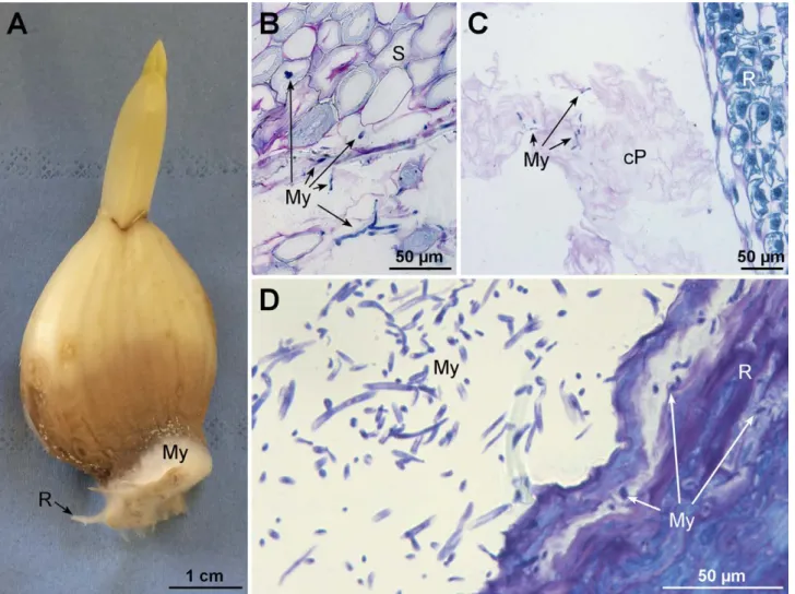
Each peeled garlic clove usually had bulbil-like mor-phostructure, with a basal plate supporting a fleshy external leaf or scale (Figure 1A) attached to a short stem (not shown) that bore numerous root primordia (Figure 1B). After clove germination, root emergence took place between the external scale and the abscission zone of the basal plate, thus enlarging the space at the junction between the external scale and the abscission zone. Microscope analyses of sections from the abscis-sion zone showed that this area was composed of thick-walled, dead cells (Figure 1C). Condensation and retrac-tion of cytoplasm and degradaretrac-tion of chromatin inside nuclei indicated that the cells were dying. Inside cloves, roots embedded in leaf parenchyma (Figure 1D) created each an aperture and many dead collapsed cells while emerging (Figure 1E).
Progression of Fusarium proliferatum in cloves
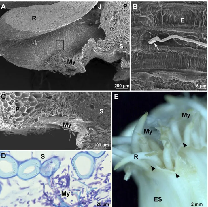
After 72 hpi, hyphae adhered and colonized the junc-tions between roots and parenchyma of external scale edges (Figure 2A). Inoculated conidia germinated on the surface of epidermis cells (Figure 2B). Mycelium then invaded the entire area composed of the parenchyma of the external scale and suberized tissue (Figure 2C). Abscission zones (AZ) were covered with white myce-lium (not shown). Microscope analyses of these areas
288 Paul L. Chrétien et alii
showed dense mycelium growth in the cellular debris close to suberized tissue (Figure 2D). On each clove, a coat of white hyphae appeared all around the roots from the base to the top and at the edge of the external scale (Figure 2E). Specific infection structure were not observed. Fusarium proliferatum proliferated as myce-lium in a saprophyte-like manner.
Browning of cloves progressed from the base to the top at 72 hpi. The boundary between healthy and dis-eased tissues was easily discernible, and mycelium entirely covered roots (Figure 3A). At 48 hpi, germi-nated conidia and mycelium were observed against de-structured suberised regions (Fig. 3B). The pathogen was also observed to be growing inside collapsed dead cells from the external clove scales resulting from the root emergence (Figure 3C). At 72 hpi and beneath the mycelium coat, root tissues were degraded and hyphae penetrated only inside the first layers of dead cells (Fig-ure 3D).
De-structuring of clove cell layers
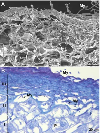
In tissues infected by F. proliferatum, the cells died and shrank particularly within the parenchyma of exter-nal scales and in the superficial layers of the roots (Figure 4A and B). Three levels of degraded tissue could be dis-cerned; entirely degraded tissue, collapsed cells filled with secondary metabolites (deep blue) and dead matrices of cells filled with fungal mycelium (III), a border between dead and healthy cells (II) and healthy parenchyma cells (I) (Figure 4B). The dead cells accumulated proteins, as indicated by the azure colour of their contents.
DISCUSSION
This study has clarified the pathway by which F.
proliferatum enters garlic gloves. Fusarium species (e.g. F. oxysporum, F. solani and F. verticillioides) are well
Figure. 1. Morphology and structure of healthy garlic cloves. A, Peeled clove after 3 d in sterile distilled water. B, Abscission zone. C,
Sec-tion of the abscission zone. D, Longitudinal secSec-tion of the basal part of an ungerminated clove. E, SecSec-tion from the area indicated by a square in D. ES: external scale, AZ: abscission zone, R: roots, P: parenchyma, S: thick-walled cells, cP: collapsed parenchymatous cells. Black arrowheads indicate junctions between roots and between AZ boundaries and root primordia. A, B and D, Stereomicrographs. C and E, Light micrographs of PAS/NBB stained samples.
known for their capacity to infect vascular and root tis-sues causing wilt on plants (Agrios, 2005). In the present study, rapid degradation of garlic root cells was observed after inoculation with F. proliferatum, but the pathogen did not invade host conductive tissues. Fusarium
pro-liferatum progressed from the points of infection in all
directions. Proliferation of the fungus in garlic tissue was reduced in the presence of thick-walled cells, and was directed toward the parenchyma. Although this pathogen possesses the enzymes required to degrade
Figure 2. Mycelium development of Fusarium proliferatum. A, SEM micrograph of a germinating garlic root. B, High magnification of the
area indicated by the square in A. C, Section of the basal part of an infected clove. D, Detail of suberized tissue. E, Basal part of a clove 72 h post inoculation. R: roots, J: junction, P: parenchyma, My: mycelium, E: epidermis, S: thick-walled cells, ES: external scale. White arrow in B indicates a germinated conidium. Black arrowheads in E indicate junctions between roots and between the abscission zone boundary and root primordia. A and B, Scanning electron micrographs. D, Light micrograph after TB staining. E, Stereomicrograph.
290 Paul L. Chrétien et alii
suberized cells, such as laccases (Regalado et al., 1999; Hernández Fernaud et al., 2006), chitinases, glucosidases and galactosidases (Keshri and Magan 2000), F.
prolife-ratum probably takes the path of least host resistance. Fusarium proliferatum rapidly infected garlic root
tissues before infecting clove tissues. Germination of roots may have triple positive indirect effects on the development of the root rot. First, while emerging, roots degrade parenchyma cells and release cellular debris that may be utilized for growth of the fungus. Second, the physical barrier of the host epidermis breaks and allows penetration of the pathogen into host tissues. In some preliminary experiments, we have observed that F.
pro-liferatum was unable to enter through intact garlic
epi-dermis surfaces. Third, the presence of host and non-host roots in the soil are known to induce germination
of fungal conidia and attract hyphae of F. oxysporum (Nelson 1981). Further studies could assess if this is also the case for F. proliferatum and garlic roots.
In the present study, F. proliferatum was observed to colonize decayed host tissues, but not to penetrate healthy cells. Tissues destroyed beyond the margins where fungal growth occurred were seen, indicating that the fungus deployed strategies, such as enzymes and toxin production, to destroy host tissues prior to colonization. The limits between dead and living cells were clearly visible. This could indicate activity of the many organosulfur compounds produced and stored inside garlic cells. These molecules are responsible for the characteristic fragrance of garlic. These com-pounds, such as allicin, have been widely studied as potential biocontrol agents against fungi, bacteria and
Figure 3. Morphology and cytohistological aspects of Fusarium proliferatum infected garlic cloves 48–72 h post inoculation. A, Image of an
infected clove. B and C, Micrographs illustrating the progression of clove infection 48 h post inoculation. D, Detail of the decayed root tis-sue. R: roots, My: mycelium, S: thick-walled cells, cP: collapsed parenchyma cells. A, Stereomicrograph. B and C, Light micrographs after PAS/NBB staining. D, Light micrograph with TB staining.
other pests (Curtis et al., 2004). Allicin is synthetized from a precursor molecule (alliin) through the action of alliinase activated when cells are damaged. The fact that F. proliferatum is able to grow, colonize and devel-op on a matrix containing organosulfur compounds was interesting. Previous studies have found that gar-lic extracts inhibit F. proliferatum on culture media at pH 3 to 7 (Chen et al., 2018). One hypothesis to explain pathogenicity is the production of desulfurization enzymes (sulfatases) by some F. proliferatum strains (Shvetsova et al., 2015). This overcoming of host resist-ance could also be linked to glutathione metabolism (Leontiev et al., 2018). Another example of fungi being able to grow in the presence of organosulfur is Coriolus
versicolor known to degrade this type of compound in
wood (Linder 2018). One hypothesis about the emer-gence of root rot of garlic is that some pathogen strains may have acquired resistance to garlic compounds. In further studies, we will assess the impacts of garlic
extracts on in vitro development of several F.
prolifera-tum strains, and evaluate the ability of F. proliferaprolifera-tum
strains, collected from other crops, to develop disease symptoms on garlic.
Like most Fusarium spp., F. proliferatum has a soil-borne phase. The present study showed that this fungus can penetrate garlic cloves at the outline/contour of their basal plates when the roots emerge. This indicates that F.
proliferatum remains dormant in the soil, is attracted to
germinating garlic cloves (at early stages of their devel-opment), and then penetrates through wounds without generating visible symptoms. Airborne phases cannot be excluded. Fusarium proliferatum is a prolifically sporu-lating species, and in Spain, conidia collected in rainwa-ter were shown to be aggressive on garlic (Gil-Serna et
al., 2016). In the field, sprouting garlic leaves take
fun-nel shapes, and rainwater is directly led to germinated cloves where the epidermis is weakened. In further experiments, it could be interesting to test independent inoculation of shoots and of the basal plates could be investigated. Knowledge of the origin of inoculum that induces rot on garlic is crucial for development of effi-cient, sustainable and environmentally-friendly root rot management strategies.
Through of French and European actions, French garlic producers are encouraged to reduce their use of phytosanitary products (European Directive 2009/128/ EC). There is a need for efficient alternative disease con-trol methods, among which are aimed at management of garlic root rot. Soil solarization could be explored to control the soil-borne population of Fusarium spp. In Japan, this method was efficient for control Fusarium wilt of strawberry caused by F. oxysporum, in fields and greenhouses (Koike and Gordon 2015). Results could be linked to heat alone or to selection of antagonistic soil microorganisms. In another research, solarization with
Medicago sativa amendment was shown to be an
effi-cient, non-chemical, method for control of Fusarium wilt of cucumber caused by F. oxysporum (Yao et al., 2016). Although soil solarization has proved to be effi-cient against species other than F. proliferatum, it would be worth testing this method for garlic crops. The use of biocontrol agents such as bacteria applied on garlic cloves may also be promising. Four strains of B. subtilis have been shown to reduce severity of disease caused by
F. proliferatum strains on garlic (Bjelic et al., 2018).
Biocontrol and solarization need to be applied at the right time, in optimum conditions, to have appropri-ate activity during or before crop planting. To this end, some of our current studies are focusing on the impacts of abiotic factors on F. proliferatum mycelium growth and sporulation.
Figure 4. Histopathological effects of Fusarium proliferatum on
infected clove tissues. A and B, Detail of the first root cells layers 72 h post inoculation. My: mycelium, dC: dead cells, P: parenchy-ma. A, Scanning electron micrograph. B, Light micrograph after TB staining.
292 Paul L. Chrétien et alii
ACKNOWLEDGEMENTS
This research was supported by the microscopy facilities of the Platform 3A, funded by the Europe-an Regional Development Fund, the French Ministry of Research, Higher Education and Innovation, the Provence-Alpes-Côte d’Azur region, the Departmental Council of Vaucluse and the Urban Community of Avi-gnon. The authors thank M. Duffaud for her work in the purification of strains included in the laboratory fungal collection. We thank Top’Alliance Alinéa for providing the garlic cloves.
LITERATURE CITED
Agrios GN, 2005. Plant Pathology. 5th edition. Elsevier. Alberti I., Prodi A., Montanari M., Paglia G., Asioli C.,
Nipoti P., 2017. First report of Fusarium proliferatum associated with Allium fistulosum L. in Italy. Journal
of Plant Diseases and Protection 125: 231–233.
Basallote-Ureba M.J., Melero-Vara J.M., 1993. Control of garlic white rot by soil solarization. Crop Protection 12: 219–223.
Bjelić D., Ignjatov M., Marinković J., Milošević D., Nikolić Z., Gvozdanović-Varga J., Karaman M., 2018.
Bacillus isolates as potential biocontrol agents of Fusarium clove rot of garlic. Zemdirbyste-Agriculture
105: 369–376.
Borrero C., Capote N., Gallardo M.A., Avilés M., 2019. First report of vascular wilt caused by Fusarium
pro-liferatum on strawberry in Spain. Plant Disease 103:
581.
Clark G., 1981. Staining Procedures. 4th Edition. Wil-liams & Wilkins. Baltimore.
Chen C., Liu C.-H., Cai J., Zhang W., Qi W.-L., … Yang Y., 2018. Broad-spectrum antimicrobial activity, chemical composition and mechanism of action of garlic (Allium sativum) extracts. Food Control 86: 117–125.
Curtis H., Noll U., Störmann J., Slusarenko A.J., 2004. Broad-spectrum activity of the volatile phytoanti-cipin allicin in extracts of garlic (Allium sativum L.) against plant pathogenic bacteria, fungi and oomy-cetes. Physiological and Molecular Plant Pathology 65: 79–89.
Delgado-Ortiz J.C., Ochoa-Fuentes Y.M., Cerna-Chávez E., Beltrán-Beache M., Rodríguez-Guerra R., Agu-irre-Uribe L.A., Vázquez-Martínez O., 2016. Pato-genicidad de especies de Fusarium asociadas a la pudrición basal del ajo en el centro norte de México.
Revista Argentina de Microbiologia 48: 222–228.
Díaz Arias M.M., Munkvold G.P., Leandro L. F., 2011. First Report of Fusarium proliferatum causing root rot on soybean (Glycine max) in the United States.
Plant Disease 95: 1316.
Dugan F.M., Hellier B.C., Lupien S. L., 2003. First report of Fusarium proliferatum causing rot of garlic bulbs in North America. Plant Pathology 52: 426.
El Maâtaoui M., Pichot C., 1999. Nuclear and cell fusion cause polyploidy in the megagametophyte of com-mon cypress, Cupressus sempervirens L. Planta 208: 345–351.
European Parliament. Establishing a framework for Com-munity action to achieve the sustainable use of pesti-cides. DIRECTIVE 2009/128/EC.
Hernández Fernaud J.R., Marina A., González K., Vázquez J., Falcón M.A., 2006. Production, partial characterization and mass spectrometric studies of the extracellular laccase activity from Fusarium
pro-liferatum. Applied Microbiology and Biotechnology 70:
212–221.
FAOSTAT, 2018. Production/Yield quantities of Garlic in World + (Total). Available at: http://www.fao.org/ faostat/en/#data/QC/visualize. Accessed September 6, 2018.
Gil-Serna J., Gálvez L., París M., Palmero D., 2016.
Fusar-ium proliferatum from rainwater and rooted garlic
show genetic and pathogenicity differences. European
Journal of Plant Pathology 146: 199–206.
Ignjatov M., Bjelić D., Nikolić Z., Milošević D., Gvozdanović-Varga J., Marinković J., Ivanović Ž., 2017. First report of Fusarium acuminatum causing garlic bulb rot in serbia. Plant Disease 101: 1047. INAO, 2019. Les signes officiels de la qualité et de
l’origine, SIQO. Available at: https://www.inao.gouv. fr/Les-signes-officiels-de-la-qualite-et-de-l-origine-SIQO. Accessed June 6, 2019.
Keshri G., Magan N., 2000. Detection and differentia-tion between mycotoxigenic and non-mycotoxigenic strains of two Fusarium spp. using volatile produc-tion profiles and hydrolytic enzymes. Journal of
Applied Microbiology 89: 825–833.
Koike S.T., Gordon T.R., 2015. Management of Fusarium wilt of strawberry. Crop Protection 73: 67–72.
Koleva K., 2004. Variety of species and spread of fungi of genus Fusarium related to rotting of garlic. Bulgarian
Journal of Agricultural Science 10: 177–180.
Lebiush-Mordechai S., Erlich O., Maymon M., Freeman S., Ben-David T., … Tsror Lahkin L., 2014. Bulb and root rot in lily (Lilium longiflorum) and onion (Allium cepa) in Israel. Journal of Phytopathology 162: 466–471. Leontiev R., Hohaus N., Jacob C., Gruhlke M.C.H.,
antibac-terial and antifungal activities of thiosulfinate ana-logues of allicin. Scientific Reports 8: 1–19.
Leyronas C., Chrétien P.L., Troulet C., Duffaud M., Ville-neuve F., Morris C.E., Hunyadi H., 2018. First Report of Fusarium proliferatum causing garlic clove rot in France. Plant Disease 102: 2658.
Linder T., 2018. Assimilation of alternative sulfur sources in fungi. World Journal of Microbiology and
Biotech-nology 34: 51.
Moharam M.H.A., Farrag E.S.H., Mohamed M.D.A., 2013. Pathogenic fungi in garlic seed cloves and first report of Fusarium proliferatum causing cloves rot of stored bulbs in upper Egypt. Archives of
Phytopathol-ogy and Plant Protection 46: 2096–2103.
Ndambi B., Cadisch G., Elzein A., Heller A., 2012. Tissue specific reactions of sorghum roots to the mycoher-bicide Fusarium oxysporum f. sp. strigae versus the pathogenic F. proliferatum. Biocontrol Science and
Technology 22: 135–150.
Nelson P.E., 1981. Chapter 3: Life cycle and epidemiology of Fusarium oxysporum. In: Fungal Wilt Diseases of Plants. M.E. Mace, A.A. Bell & C.H. Beckman, eds. Academic press, New York, U.S., p. 51–80.
Ochoa Fuentes Y.M., Cerna Chávez E., Gallegos Morales G., Landeros Flores J., Delgado Ortiz J.C., … Olalde Portugal V., 2012. Identificación de especies de
Fusar-ium en semilla de ajo en Aguascalientes, México. Revista Mexicana de Micologia 36: 27–32.
O’Donnell K., Rooney A.P., Proctor R.H., Brown D.W., McCormick S.P., … Aoki T., 2013. Phylogenetic analyses of RPB1 and RPB2 support a middle Creta-ceous origin for a clade comprising all agriculturally and medically important fusaria. Fungal Genetics and
Biology 52: 20–31.
Regalado V., Perestelo F., Rodríguez A., Carnicero A., Sosa F.J., De la Fuente G., Falcón M.A., 1999. Activated oxygen species and two extracellular enzymes: laccase and aryl-alcohol oxidase, novel for the lignin-degrading fungus Fusarium
prolif-eratum. Applied Microbiology and Biotechnology 51:
388–390.
Ricard P., 2017. Les premiers pas vers l’étiologie et l’épidémiologie d’une maladie multifactorielle et en expansion : la fusariose de l’ail (Allium sativum L.). https://dumas.ccsd.cnrs.fr/dumas-01618306. Accessed 14 Oct 2019.
Roberts P.A., Matthews W.C., 1995. Disinfection alterna-tives for control of Ditylenchus dipsaci in garlic seed cloves. Journal of Nematology 27: 448–456.
Sankar N.R., Babu G.P., 2012. First Report of Fusarium
proliferatum causing rot of garlic bulbs (Allium sati-vum) in India. Plant Disease 96: 290.
Seefelder W., Gossmann M., Humpf H.-U., 2002. Analysis of fumonisin B(1) in Fusarium proliferatum-infected asparagus spears and garlic bulbs from Germany by liquid chromatography-electrospray ionization mass spectrometry. Journal of Agricultural and Food
Chem-istry 50: 2778–2781.
Shvetsova S.V., Zhurishkina E.V., Bobrov K.S., Ron-zhina N.L., Lapina I.M., … Kulminskaya A.A., 2015. The novel strain Fusarium proliferatum LE1 (RCAM02409) produces α- L -fucosidase and aryl-sulfatase during the growth on fucoidan: Alfa-L-fucosidase and arylsulfatase from the novel strain
Fusarium proliferatum. Journal of Basic Microbiology
55: 471–479.
Stankovic S., Levic J., Petrovic T., Logrieco A., Moretti A., 2007. Pathogenicity and mycotoxin production by
Fusarium proliferatum isolated from onion and garlic
in Serbia. European Journal of Plant Pathology 118: 165–172.
Toit L.J., Inglis D.A., Pelter G.Q., 2003. Fusarium
prolif-eratum pathogenic on onion bulbs in Washington. Plant Disease 87: 750.
Tonti S., Prà M.D., Nipoti P., Prodi A., Alberti I., 2012. First Report of Fusarium proliferatum causing rot of stored garlic bulbs (Allium sativum L.) in Italy.
Jour-nal of Phytopathology 160: 761–763.
This H., 2006. Molecular gastronomy: exploring the sci-ence of flavor. Columbia University Press.
Totelin L., 2015. When foods become remedies in ancient Greece: The curious case of garlic and other sub-stances. Journal of Ethnopharmacology 167:3 0–37. Tridge, 2019. Garlic (Allium sativum) – Garlic Price
Trend. Available at: https://www.tridge.com/prod-ucts/garlic. Accessed August 10, 2019.
Xie S.H., Zhang C., Chen M.S., Wei Z.P., Jiang Y.B.., Lin X., Zheng Y.Q., 2018. Fusarium proliferatum : a new pathogen causing fruit rot of peach in Ningde, China.
Plant Disease 102: 1858.
Yamazaki M., Morita Y., Kashiwa T., Teraoka T., Arie T., 2013. Fusarium proliferatum, an additional bulb rot pathogen of Chinese chive. Journal of General Plant
Pathology 79: 431–434.
Yao Y., Xue Z., Hong C., Zhu F., Chen X., … Yang X., 2016. Efficiency of different solarization-based eco-logical soil treatments on the control of Fusarium wilt and their impacts on the soil microbial commu-nity. Applied Soil Ecology 108: 341–351.