HAL Id: hal-02546568
https://hal.umontpellier.fr/hal-02546568
Submitted on 18 Apr 2020
HAL is a multi-disciplinary open access
archive for the deposit and dissemination of
sci-entific research documents, whether they are
pub-lished or not. The documents may come from
teaching and research institutions in France or
abroad, or from public or private research centers.
L’archive ouverte pluridisciplinaire HAL, est
destinée au dépôt et à la diffusion de documents
scientifiques de niveau recherche, publiés ou non,
émanant des établissements d’enseignement et de
recherche français ou étrangers, des laboratoires
publics ou privés.
Ameliorates Muscular Dystrophy in the mdx Mouse
Diaphragm
Marion Pauly, Frederic Daussin, Yan Burelle, Tong Li, Richard Godin, J.
Fauconnier, Christelle Koechlin-Ramonatxo, Gerald Hugon, Alain
Lacampagne, Marjorie Coisy-Quivy, et al.
To cite this version:
Marion Pauly, Frederic Daussin, Yan Burelle, Tong Li, Richard Godin, et al.. AMPK Activation
Stimulates Autophagy and Ameliorates Muscular Dystrophy in the mdx Mouse Diaphragm.
Amer-ican Journal of Pathology, AmerAmer-ican Society for Investigative Pathology, 2012, 181 (2), pp.583-592.
�10.1016/j.ajpath.2012.04.004�. �hal-02546568�
Molecular Pathogenesis of Genetic and Inherited Diseases
AMPK Activation Stimulates Autophagy and
Ameliorates Muscular Dystrophy in the mdx
Mouse Diaphragm
Marion Pauly,* Frederic Daussin,
†Yan Burelle,
†Tong Li,
‡Richard Godin,
†Jeremy Fauconnier,*
Christelle Koechlin-Ramonatxo,
§Gerald Hugon,*
Alain Lacampagne,* Marjorie Coisy-Quivy,*
Feng Liang,
‡Sabah Hussain,
‡Stefan Matecki,*
and Basil J. Petrof
‡From the Physiology and Experimental Medicine Heart-Muscle Unit,* INSERM U1046, Montpellier 1 University, Montpellier,
France; the Faculty of Pharmacy,†University of Montreal,
Montreal, Quebec, Canada; the Meakins-Christie Laboratories and Respiratory Division,‡McGill University Health Centre and
Research Institute, Montreal, Quebec, Canada; and the Cellular Differentiation and Growth Unit,§National Agronomic Research
Institute-INRA-UMR A866, Montpellier, France
Duchenne muscular dystrophy (DMD) is character-ized by myofiber death from apoptosis or necrosis, leading in many patients to fatal respiratory muscle weakness. Among other pathological features, DMD muscles show severely deranged metabolic gene reg-ulation and mitochondrial dysfunction. Defective mi-tochondria not only cause energetic deficiency, but also play roles in promoting myofiber atrophy and injury via opening of the mitochondrial permeability transition pore. Autophagy is a bulk degradative mechanism that serves to augment energy production and eliminate defective mitochondria (mitophagy). We hypothesized that pharmacological activation of AMP-activated protein kinase (AMPK), a master meta-bolic sensor in cells and on-switch for the autophagy-mitophagy pathway, would be beneficial in themdx mouse model of DMD. Treatment ofmdx mice for 4 weeks with an established AMPK agonist, AICAR (5-aminoimidazole-4-carboxamide-1-!-D-ribofuranoside), potently triggered autophagy in themdx diaphragm without inducing muscle fiber atrophy. In AICAR-treatedmdx mice, the exaggerated sensitivity of mdx diaphragm mitochondria to calcium-induced perme-ability transition pore opening was restored to nor-mal levels. There were associated improvements in mdx diaphragm histopathology and in maximal
force-generating capacity, which were not linked to increased mitochondrial biogenesis or up-regulated utrophin expression. These findings suggest that ago-nists of AMPK and other inducers of the autophagy-mitophagy pathway can help to promote the elimina-tion of defective mitochondria and may thus serve as useful therapeutic agents in DMD. (Am J Pathol 2012,
181:583–592;http://dx.doi.org/10.1016/j.ajpath.2012.04.004)
Duchenne muscular dystrophy (DMD), the most common X-linked lethal disorder in humans, is caused by defects in the dystrophin gene.1Absence of dystrophin is associated
with muscle fiber death involving both apoptosis and ne-crosis.2 Because DMD affects the diaphragm and other
respiratory muscles, many patients die of respiratory failure. Although the ideal treatment for DMD would be restoration of dystrophin expression to all muscles of the body, this is not currently feasible, and there is an urgent need for new therapies. The mdx mouse also lacks dystrophin, and is a commonly used animal model for studying the disease and its potential responsiveness to new treatments.3
Muscles lacking dystrophin exhibit multiple cellular de-fects, including abnormal fragility of the sarcolemma, in-creased oxidative stress, and elevated cytosolic calcium levels.4DMD muscles also show mitochondrial
dysfunc-tion and diminished expression of energy-producing met-abolic genes.5,6 Conversely, forced expression of the
mitochondrial biogenesis factor peroxisome proliferator-activated receptor-! coactivator 1-" (PGC-1"; official
Supported by grants from the Canadian Institutes of Health Research, the Health Research Fund of Quebec, the French National Health and Medical Research Institute, Researcher of the Future award from the Regional Council of Langue-doc Roussillon, and the French Association Against Myopathies.
Accepted for publication April 5, 2012.
S.M. and B.J.P. contributed equally to this work.
Supplemental material for this article can be found at http://ajp. amjpathol.orgor athttp://dx.doi.org/10.1016/j.ajpath.2012.04.004.
Address reprint requests to Basil J. Petrof, M.D., McGill University Health Centre, Royal Victoria Hospital Site, Respiratory Division, Room L4.11, 687 Pine Ave. West, Montreal, QC, H3A 1A1, Canada. E-mail:
basil.petrof@mcgill.ca.
The American Journal of Pathology, Vol. 181, No. 2, August 2012 Copyright © 2012 American Society for Investigative Pathology. Published by Elsevier Inc. All rights reserved.
http://dx.doi.org/10.1016/j.ajpath.2012.04.004
symbol, PPARGC-1-") ameliorates dystrophic pathology in mdx mice.7 AMP-activated protein kinase (AMPK), a
major sensor of cellular energy status, switches on mecha-nisms favoring ATP generation under conditions of ener-getic deficiency.8Thus, AMPK can stimulate mitochondrial
biogenesis through the PGC-1-" pathway, as well as through catabolic pathways that fuel energy production. Chief among these catabolic mechanisms is the lyso-somally mediated process of macroautophagy (hence-forth referred to simply as autophagy), which targets cel-lular constituents that are too large to be removed by other degradative pathways.9,10
In addition to its energy-producing function, au-tophagy plays a critical role in cellular quality control by preferentially eliminating proteins and organelles that are nonessential or dysfunctional, including defective mito-chondria.9,10 Recent work has linked defects in
au-tophagic removal of mitochondria (mitophagy) to Parkin-son’s disease and other neurodegenerative diseases, as well as to muscular dystrophy associated with collagen VI deficiency.11,12Impaired mitophagy permits the
accumu-lation of damaged mitochondria, which not only perform poorly in their energy-generating role but also have a greater propensity to undergo opening of the mitochondrial membrane permeability transition pore (PTP) complex.12,13
This leads to mitochondrial swelling, collapse of the mito-chondrial membrane potential, and release of proapoptotic factors, which have been implicated not only in cell death but also in muscle fiber atrophy and injury.14,15
In the present study, we postulated that therapeutic activation of AMPK might stimulate autophagic removal of defective mitochondria in mdx mice, thereby leading to beneficial effects on mitochondrial PTP opening, as well as on the overall muscular dystrophy phenotype. To test this hypothesis, we treated mdx mice with AICAR (5-aminoimidazole-4-carboxamide-1-#-D-ribofuranoside),
an established pharmacological activator of AMPK16that
has been shown to promote mitophagy.17 We focused
the present study on the mdx mouse diaphragm, be-cause this model closely mimics human DMD with re-spect to both fiber loss and weakness.3Here, we show
that chronic AICAR treatment effectively stimulates au-tophagy, increases the ability of muscle fiber mitochon-dria to resist PTP opening, and ameliorates histological features of muscular dystrophy, as well as muscle strength in the mdx diaphragm. These results support the concept that promoting the autophagic removal of defec-tive mitochondria via AMPK stimulation could be a useful therapeutic strategy in DMD patients.
Materials and Methods
Animals and Cell Culture
Six-week-old male mdx mice (Jackson Laboratories, Bar Harbour, ME) received intraperitoneal injections of AICAR (Toronto Research Chemicals, Toronto, ON, Canada) at a daily dose of 500 mg/kg body weight (5 consecutive days per week) for 4 weeks.16 Untreated littermate mdx mice
were injected in the same manner with vehicle (0.9% NaCl);
wild-type (WT) (C57BL6) mice (Janvier SAS, Le Genest Saint Isle, France) did not receive any treatment. Primary myotube cultures from mdx mice were derived from single living myofibers, as described previously.18The
investiga-tion complied with the Guide for the Care and Use of Lab-oratory Animals (2011 edition).
Immunoblotting and Gene Expression Analysis
Immunoblotting was performed by standard methods us-ing antibodies against phospho-AMPK" (Thr172), totalAMPK", phospho-ACC (Ser79), total ACC, LC3, Beclin-1,
phospho-mTOR (Ser2448), total mTOR, phospho-raptor
(Ser792), total raptor, phospho-p70S6K (Thr389), and total
p70S6K from Cell Signaling Technology (Danvers, MA); Ulk1, Bnip3, #-actin, and tubulin from Sigma-Aldrich (St. Louis, MO); mitochondrial respiratory complex subunits (I-20 kDa; II-30 kDa; III-47 kDa; V-53 kDa) from MitoSci-ences (Eugene, OR); GAPDH from Abcam (Cambridge, UK); and utrophin, with a polyclonal antibody previously characterized by our laboratory.19Analysis and
quantifica-tion (normalized in all cases to indicated loading control proteins) were performed with ImageJ software version 1.43u (NIH, Bethesda, MD). Real-time PCR to quantify mRNA levels was performed using Fast SYBR Green (Ap-plied Biosystems) and the cycle threshold method.
Immunohistochemistry and Morphometry
Transverse cryosections stained by H&E were used for determination of fiber size and the percentage of cen-trally nucleated fibers, with a minimum of five cross sec-tions per muscle, each containing an average of 100 fibers.20For determination of fiber types, adjacentsec-tions were immunostained with anti-slow type 1 (M-8421; Sigma-Aldrich) and anti-fast type 2a (mAb SC-71; Santa Cruz Biotechnology, Santa Cruz, CA) antibodies. Elec-tron microscopy was performed on muscle tissues as described previously.20
Mitochondrial Enzyme and Respiration Assays
Mitochondrial enzyme and respiration assays were per-formed as described previously.21 Briefly, activities ofcitrate synthase and cytochrome c oxidase (COX) were determined spectrophotometrically using standard cou-pled enzyme assays. Mitochondrial respiration was ana-lyzed in an oximeter equipped with a Clarke type of electrode. The chamber was filled with the following so-lution: 2.77 mmol/L CaK2-EGTA, 7.23 mmol/L K2-EGTA
(100 nmol/L free Ca2!), 6.56 mmol/L MgCl
2(1 mmol/L
free Mg2!), 20 mmol/L taurine, 0.5 mmol/L dithiothreitol,
50 mmol/L potassium-methane sulfonate (160 mmol/L ionic strength), and 20 mmol/L imidazole (pH 7.1). After the baseline oxygen content in the chamber had been recorded, one bundle of 1 to 2 mg dry weight of saponin-permeabilized myofibers was placed into the chamber. Readings were taken of the rate of O2consumption per
unit time, first basally in the presence of glutamate plus malate (10:5, mmol/L), and then after the addition of ADP (2 mmol/L). Respiration rates were measured at 23°C under
continuous stirring. At the end of each test, fibers were carefully removed from the oxygraphic cell, blotted, and dried for determination of fiber weight. Rates of O2
con-sumption were expressed in nmol O2/minute per per
milli-gram dry weight.
Mitochondrial H
2O
2Release and Scavenging
Mitochondrial H2O2dynamics were measured in dissected
fiber bundles with the fluorescent probe Amplex Red (20 $mol/L; Invitrogen), as described previously.21After
sapo-nin permeabilization, the fibers were rinsed three times in the following buffer maintained at 4°C and at pH 7.3: 110 mmol/L K-2-(N-morpholino)ethanesulfonic acid, 35 mmol/L KCl, 1 mmol/L EGTA, 5 mmol/L K2HPO4, 3 mmol/L MgCl2·
6H2O, and 0.5 mg/mL BSA. Fiber bundles (0.3 to 1.0 mg
dry weight) were then incubated in a quartz microcuvette with continuous magnetic stirring in the same buffer sup-plemented with 1.2 U/mL horseradish peroxidase at 37°C. Baseline fluorescence readings were taken in the absence of any exogenous respiratory substrates. The following additions were then made sequentially: 5 mmol/L succinate, 10 mmol/L ADP, and 8 $mol/L antimy-cin A. To determine H2O2scavenging capacity,
perme-abilized fiber bundles were placed in the above buffer containing additionally 50 $mol/L pyruvate and 20 $mol/L malate in a thermally controlled chamber set at 37°C with continuous stirring. An aliquot of the buffer was removed immediately after adding 40 $mol/L of H2O2
and subsequently at 20, 40, and 60 seconds. H2O2
con-tent in aliquots was determined immediately on a fluores-cence plate reader in a buffer containing 10 $mol/L of Amplex Red and 0.5 U/mL horseradish peroxidase. The rate of H2O2scavenging by mitochondria was determined
as the difference between fluorescence levels obtained at time t " 0. Rates of H2O2production and scavenging were
calculated from standard curves established under corre-sponding experimental conditions. All measurements were performed at least in duplicate, and results were expressed in nmol H2O2 scavenged per minute per milligram dry
weight.
Mitochondrial PTP Function
The PTP opening time and calcium retention capacity were determined by fluorimetrically monitoring changes in extramitochondrial calcium concentration, using the probe Calcium Green-5N (Invitrogen-Life Technologies, Carlsbad, CA), after exposing fibers to a single pulse of external calcium.21,22Ghost fibers were first prepared by
incubating saponin-permeabilized bundles in a high-KCl medium, to extract myosin. The ghost fibers were then incubated at 23°C in a quartz microcuvette under contin-uous stirring in the following buffer: 250 mmol/L sucrose, 10 mmol/L 3-(N-morpholino)propanesulfonic acid, 0.005 mmol/L EGTA, and 10 mmol/L propidium iodide-Tris (pH 7.3), supplemented with glutamate plus malate (5:2.5, mmol/L) and 0.5 nmol/L oligomycin. After adding 20 nmol Ca2!to the buffer, PTP opening time was taken as the time
lapse between addition of the Ca2!pulse and the time at
which Ca2!release was first noted to occur. Calcium
re-tention capacity was defined as the total amount of Ca2!
accumulated by mitochondria before Ca2!release caused
by PTP opening, expressed per milligram wet fiber weight. Ca2! concentration in the cuvette was calculated from a
standard curve relating [Ca2!] to the fluorescence of
Cal-cium Green-5N.
Measurement of Skeletal Muscle Contractile
Properties
Diaphragm strips were electrically stimulated to deter-mine intrinsic contractile properties, as described pre-viously.23 After euthanasia, the diaphragm was
surgi-cally excised and immediately transferred to chilled Krebs solution perfused with 95% O2:5% CO2(pH 7.4).
From the central tendon to the rib, a 2-mm-wide muscle strip was dissected free and mounted between two electrodes within a jacketed tissue bath chamber filled with continuously perfused Krebs solution warmed to 25°C. A 4-0 silk thread was used to secure the central tendon to an isometric force transducer. After a 15-minute thermoequilibration period, muscle length was gradually adjusted to optimal length (Lo, the length at
which maximal twitch force is obtained). The force-frequency relationship was determined by sequential supramaximal stimulation for 1 second over a range of stimulation frequencies (from 10 to 120 Hz), with 2 minutes between each stimulation train. Fatigue resis-tance was assessed by measuring the rate of loss of muscle force during repetitive stimulation at 30 Hz over a 10-minute period. At the end of the experiment, Lo
was directly measured with a microcaliper and the muscle was blotted dry and weighed. Specific force (force/cross-sectional area) was calculated and ex-pressed in newtons per square centimeter, assuming a muscle density of 1.056 g/cm3.
Statistical Analysis
Data are expressed as means # SEM. Statistical signifi-cance was defined as P $ 0.05, using Student’s unpaired
t-test or analysis of variance (one- or two-way), followed by a Bonferroni selected-comparison test.
Results
Premature Mitochondrial PTP Opening and
Energetic Deficiency in Dystrophic Muscle
Dystrophic diaphragms contained numerous fibers with morphologically abnormal mitochondria, charac-terized by a rounded swollen appearance and grossly disrupted internal structure (Figure 1A). To examine the ability to resist PTP opening, which is a critically important aspect of mitochondrial function, permeabil-ized fibers were exposed to an external calcium load. The mitochondria of mdx diaphragms underwent sig-nificantly earlier PTP opening than observed in WT controls (Figure 1B). In keeping with these morpholog-ical and functional signs of defective mitochondria,AMPK Activation in mdx Mouse Diaphragm 585
baseline levels of AMPK activation (phosphorylation) were also higher in mdx than in WT diaphragms (Figure 1, C and D), which is consistent with cellular sensing of
relative energetic deficiency within dystrophic myo-fibers.
AICAR Treatment Activates AMPK and ACC in
Dystrophic Muscle
To determine whether short-term AICAR treatment could further boost AMPK activation in dystrophic muscle, we first examined AICAR effects on phosphorylation of AMPK and its downstream target, ACC (acetyl CoA car-boxylase) in primary mdx myotubes. AICAR induced phosphorylation of the "-subunit of AMPK and to an even greater extent ACC in vitro (Figure 2A). As expected, AICAR also induced increased activation of AMPK in vivo, such that increased phosphorylation levels of both AMPK and ACC were observed in the diaphragms of mdx mice within 48 hours after AICAR treatment was initiated (Figure 2B). Food intake did not differ significantly between the untreated and AICAR-treated mdx groups for the study pe-riod as a whole, from 6 to 10 weeks of age, although it was briefly increased in the AICAR-treated group from days 10 to 12. Similarly, there were no significant differences in body weight between the untreated and AICAR-treated mdx mice over the total treatment period (Figure 2C).
AICAR Treatment Induces Autophagy and
Improves Mitochondrial PTP Function
After 4 weeks of AICAR treatment in vivo, there was a major up-regulation of multiple components of the autophagy pro-gram in mdx diaphragms. This was reflected by increases in the mammalian Atg1 homolog, Ulk1 (an autophagy-initi-ating kinase that is directly activated by AMPK), as well as in Beclin-1, a component of the class III phosphoinositide 3-kinase complex involved in the initiation of autophago-0 1 2 3 p-AMPKα/AMPKα CTL mdx
*
mdx diaphragm CTL diaphragm p-AMPK AMPKA
Pr ot ei n op " ca l d en si tyC
0 100 200 300 400 500 600 700 CTL mdx*
B
Ti m e to P TP o pe ni ng (s ec )D
Ac"nFigure 1. Defective mitochondria and premature PTP opening in mdx diaphragm fibers. A: Electron micrographs demonstrating differences in mitochondrial morphol-ogy between normal WT (CTL) and mdx diaphragms. Note numerous swollen mitochondria with abnormal cristae (arrow) in the mdx group. Scale bar " 1 $. B: Time to mitochondrial permeability transition pore (PTP) opening after calcium challenge in mdx versus WT diaphragm fibers. C: Representative immunoblots showing baseline levels of phosphorylated and total forms of AMPK in WT and mdx diaphragms. D: Optical density quantification of p-AMPK"/total AMPK" protein levels (arbitrary units normalized to actin). Data are expressed as means # SEM. *P $ 0.05. n " 7 to 9 per group (B and D).
AICAR AMPK p-AMPK p-ACC ACC mdx myotubes in vitro Untreated Untreated mdx diaphragm in vivo AICAR AMPK p-ACC p-AMPK ACC 0 0.5 1 1.5 2 2.5 3 3.5
p-AMPKα/ AMPKα p-ACC/ ACC untreated AICAR * * 0 0.5 1 1.5 2 p-AMPKα/AMPKα p-ACC/ACC untreated AICAR * * 0 1 2 3 4 5 0 2 4 6 8 10 12 14 16 18 20 24 28 Fo od i nt ake (g /d ay) Time (Days) mdx mdx+AICAR 0 5 10 15 20 25 30 35 0 2 4 6 8 10 12 14 16 18 20 24 28 Body w ei ght (g ) Time (Days) Pr ot ein o p" ca l d en sit y Pr ot ein o p" ca l d en sit y
A
B
C
Tubulin Ac"nFigure 2. AICAR promotes AMPK activation in dystrophic muscles in vitro and in vivo. A and B: Phosphorylated and total forms of AMPK and acetyl CoA carboxylase (ACC) in primary myotube cultures derived from mdx mice, either without or with AICAR treatment (1 mmol/L for 24 hours) (A) and in dystrophic mdxdiaphragms in vivo, either without or with AICAR treatment (500 mg/kg/ day intraperitoneally for 48 hours) (B). Optical density quantification is in arbi-trary units, normalized to actin or tubulin. C: Food intake and body weight of mdxmice during 4 weeks of receiving AICAR treatment or vehicle alone. Data are expressed as means # SEM. n " 8 per group. *P $ 0.05.
Figure 3. AICAR activates the autophagy pathway in mdx diaphragms. Representative immunoblots showing protein expression levels of Ulk1 and Beclin-1 (A), LC3-I and LC3-II (B), and Bnip3 (C) within the diaphragms of WT mice (CTL), untreated mdx mice, and AICAR-treated mdx mice. All quantifications of autophagy pathway proteins are normalized to GAPDH in terms of fold-change relative to WT mice. Data are expressed as means # SEM. n " 4 to 6. *P $ 0.05 versus wild type;†P $0.05 versus mdx.
somes (Figure 3A). In addition, the AICAR-treated group demonstrated higher levels of LC3-II, the lipidated form of LC3 that is generated during the process of autophago-some formation, and in the ratio of LC3-II to LC3-I (Figure 3B). Bnip3, a mitochondria-associated protein that plays a crucial role in the autophagic removal mitochondria in skel-etal muscle was also found to be significantly up-regulated in the diaphragms of AICAR-treated mdx mice (Figure 3C). The levels of Ulk1, LC3, and Bnip3 were all mildly increased above WT levels in untreated mdx mice, suggesting that a degree of autophagic induction occurs basally in mdx dia-phragms, most likely as an adaptive response to the dys-trophic disease process itself.
The mammalian target of rapamycin (mTOR) exists within two functional complexes, mTORC1 and mTORC2, with the former being directly regulated by cellular en-ergy status.24 To determine whether the stimulation of
autophagy by AICAR was associated with mTORC1 mod-ulation in mdx diaphragms, we measured total and phos-phorylated protein levels of key mTORC1 members, mTOR and raptor, as well as p70S6K, the downstream target of activated mTOR (Figure 4A). Levels of total mTOR, raptor, and p70S6K were all increased above WT levels in the two groups of mdx mice (Figure 4, B–D). However, the phosphorylated forms of these proteins in-creased in a proportionate fashion, and the ratios of phosphorylated to total forms of these proteins did not differ significantly between the untreated and AICAR-treated mdx mice (Figure 4, E–G).
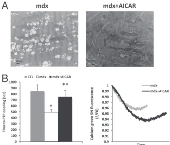
Given that autophagy is an important mechanism for eliminating damaged mitochondria, we next sought to determine whether AICAR treatment led to improved mitochondrial PTP function. In AICAR-treated mdx mice, electron microscopy suggested qualitative im-provements in mitochondrial morphology (Figure 5A). Importantly, the abnormal susceptibility to PTP opening observed in mdx diaphragm fibers was reversed in AICAR-treated mdx mice (Figure 5B). There were no
sta-tistically significant differences in global calcium reten-tion capacity of mitochondria between WT, mdx, and
mdx!AICAR groups (1.17 # 0.18, 1.37 # 0.27, and 1.69 # 0.28 nmol Ca2!per milligram, respectively). Thus,
the major findings are that the autophagic process in dystrophic muscles is greatly boosted by chronic admin-istration of AICAR, and that this is associated with a significantly greater ability of mitochondria to resist cal-cium-induced membrane permeabilization.
0.0 0.1 0.2 0.3 0.4 0.5 0.6 0.7 0.8 0.9 p-raptor/raptor Pr ot ein E xp re ss io n (A U ) CTL mdx mdx+AICAR 0.0 0.1 0.2 0.3 0.4 0.5 0.6 0.7 0.8 0.9 1.0 raptor/GAPDH Protein Ex pressi on (A U) CTL mdx mdx+AICAR 0.0 0.1 0.2 0.3 0.4 0.5 0.6 0.7 0.8 0.9 1.0 p70S6K/GAPDH Protein Ex pressi on (A U) CTL mdx mdx+AICAR 0.0 0.1 0.1 0.2 0.2 0.3 0.3 0.4 0.4 0.5 0.5 p-p70S6K/p70S6K Pr ot ein Ex pr es si on (A U ) CTL mdx mdx+AICAR * * * * 0.0 0.1 0.2 0.3 0.4 0.5 0.6 0.7 0.8 0.9 1.0 mTOR/GAPDH Pr ot ein Ex pr es si on (A U ) CTL mdx mdx+AICAR * * 0.0 0.5 1.0 1.5 2.0 2.5 3.0 p-mTOR/ mTOR Pr ot ein E xp re ss io n (A U ) CTL mdx mdx+AICAR CTL mdx mdx+AICAR p70S6K raptor mTOR CTL mdx mdx+AICAR ph-raptor ph-mTOR ph-p70S6K
B
G
D
F
C
E
A
Figure 4. AICAR treatment does not alter mTORC1 activation in mdx diaphragms. Repre-sentative immunoblots (A) and quantification of protein levels for the total forms of mTOR (B), raptor (C), and p70S6K (D), as well as their relative phosphorylation levels (E–G), within the diaphragms of WT mice (CTL), untreated mdx mice, and AICAR-treated mdx mice. Optical den-sity quantification is in arbitrary units, normal-ized to GAPDH in all cases. Data are expressed as means # SEM. n " 4 or 5. *P $ 0.05 versus wild type. 0.9 0.91 0.92 0.93 0.94 0.95 0.96 0.97 0.98 0.99 1 Ca lciu m gr een 5N flu orescen ce (F /F 0) Time mdx mdx+AICAR
B
500nm 500nm mdx+AICAR mdxA
0 100 200 300 400 500 600 700 800 900 1000 Ti m e to P TP o pe ni ng (s ec ) CTL mdx mdx+AICAR * **Figure 5. AICAR improves mitochondrial PTP function in mdx diaphragm fibers. A: Electron microscopic images of mitochondria in mdx and mdx!AICAR diaphragms. B: Effects of AICAR treatment on PTP opening time in mdx dia-phragms (left) in WT mice (CTL), untreated mdx mice, and AICAR-treated mdx mice, as well as a representative tracing of mitochondrial calcium uptake and release within permeabilized fibers after calcium challenge (right). Data are expressed as means # SEM. n " 7 to 10 per group. *P $ 0.05 versus WT; **P $ 0.05 versus mdx. Scale bar " 500 nm.
AMPK Activation in mdx Mouse Diaphragm 587
Lack of AICAR Effects on Mitochondrial
Biogenesis and Oxidative Functions
In addition to autophagy, improved mitochondrial PTP function after AICAR treatment could be the result of enhanced mitochondrial biogenesis and its accompany-ing antioxidant effects. Therefore, we evaluated whether AICAR reprogrammed the mdx diaphragm to a more oxidative phenotype. At 4 weeks after initiation of AICAR treatment, the mRNA levels for PGC-1-" and PGC-1-#, two transcription factors typically associated with activa-tion of the mitochondrial biogenesis program, were un-changed compared with untreated mice (Figure 6A). In AICAR-treated mdx mice, there was a small increase in type 1 (slow oxidative) fibers, but without any alteration in type 2a (fast oxidative) fibers in the diaphragm (Figure 6B). Immunoblotting revealed increased mitochondrial complex I protein expression without changes in com-plexes II, III, or V (Figure 6C). Levels of the dystrophin homolog protein utrophin, which is up-regulated in mdx muscles and typically is expressed at higher levels in oxidative fibers,25 were similarly unaltered by AICAR
treatment in mdx diaphragms (see Supplemental Figure S1 athttp://ajp.amjpathol.org).
The above findings suggesting minimal effects of chronic AICAR administration on the overall oxidative
capacity of mdx diaphragms were further examined at a functional level. Enzymatic activity levels of both mitochon-drial citrate synthase and cytochrome c oxidase (COX) in the diaphragm were unaffected by AICAR administration (Figure 7A). In addition, basal (V0) and maximal (Vmax) rates
of oxygen consumption by mdx diaphragm fibers were un-changed (Figure 7B). Antioxidant function was determined by measuring H2O2fluxes from permeabilized fibers using
Amplex Red. Neither the H2O2scavenging capacity (Figure 7C) nor H2O2release under conditions of altered substrate
supply or of inhibition of the mitochondrial electron transport chain (Figure 7D) was affected by AICAR treatment. Over-all, these findings indicate that improvements in mitochon-drial PTP function in the mdx diaphragm after AICAR treat-ment were not linked to the developtreat-ment of a substantially more oxidative fiber type.
AICAR Treatment Improves Dystrophic Muscle
Structure and Contractile Function
To establish whether autophagic induction and im-proved mitochondrial PTP function were associated with any amelioration of the muscular dystrophy
phe-A
0 10 20 30 40 50 MHC1 MHC2a Rel ati ve pr opor tion (% ) mdx mdx+AICAR*
MHC1 MHC2aC
B
0 10 20 30 40 50Complex I Complex II Complex III Complex V
Pr ot ei n op tic al de ns ity (A U ) mdxmdx+AICAR
*
CV CIII CII CI GAPDH 0 0.5 1 1.5 2 PGC1 α PGC1 β mRN A ex pressi on (F ol d ch an ge) mdx mdx+AICAR mdx mdx+AICARFigure 6. AICAR effects on oxidative fiber types and mitochondrial complexes. A: mRNA levels of transcription factor genes associated with mitochondrial biogenesis, reported in terms of fold change relative to untreated mdx dia-phragms. Scale bars: 20 $. B: Comparison of relative proportion of fiber types in the diaphragms of untreated mdx and AICAR-treated mdx mice; micrographs show serial transverse muscle sections (with same fibers indicated by small circle and square) immunostained for type 1 or type 2a myosin heavy chain (MHC) expression. C: Quantification of respiratory chain complexes in untreated mdx and AICAR-treated mdx mice, with immunoblotting for the same complexes (CI, CII, CIII, and CV). Protein and mRNA levels are normalized to housekeeping GAPDH. Data are expressed as means # SEM. n " 6 to 10 per group. *P $ 0.05.
A
0.0 0.5 1.0 1.5 2.0 2.5 3.0 3.5 4.0 4.5 V0 Vmax Oxy gen co ns um p" on (n mo l/ min /mg d ry wt) mdx mdx+AICAR 0 10 20 30 40 50 60 70 80 90 CS ac " vi ty (mU /min /mg ) mdx mdx + AICAR 0 5 10 15 20 25 30 35 40 45 CO X ac " vi ty (mU /mi n/mg)C
B
0.0 0.1 0.2 0.3 0.4 0.5 0.6 0.7 0.8 0.9 60sec H2 O2 sc av en gi ng (p mo l/ mg dr y w ei ght ) mdx mdx+AICARD
0 1 2 3 4 5 6 7Fiber Succinate ADP An"mycine A
H2 O2 relea se (p mo l/ min /mg d ry weig ht ) mdx mdx+AICAR
Figure 7. Lack of AICAR treatment effects on oxidative function. A: Citrate synthase (CS) and cytochrome c oxidase (COX) activity levels. B: Rate of O2
consumption per unit of time, basally in the presence of glutamate plus malate (V0) and after the addition of ADP (Vmax). C: H2O2scavenging as indicated by
levels at 60 seconds after addition of H2O2(40 $mol/L) to permeabilized fibers.
D: Net rate of mitochondrial H2O2release from permeabilized diaphragm fibers
under basal conditions (Fiber) and after the addition of succinate, ADP, or antimycin A. Data are expressed as means # SEM. n " 8 to 10 per group.
notype in the diaphragm, we first performed histologi-cal analysis of untreated versus AICAR-treated mdx mice. Muscle fibers with centrally located nuclei are a hallmark of previously regenerated muscle, and their relative proportion is therefore considered reflective of prior episodes of muscle fiber necrosis in mdx mice.26,27In AICAR-treated mdx mice, the diaphragm
showed a reduction in the percentage of centrally nu-cleated fibers (Figure 8A), consistent with a mitigation of prior necrosis. Moreover, despite the increased level of autophagy in the AICAR group, the mean cross-sectional area of individual diaphragmatic muscle fi-bers was unaffected (Figure 8B), indicating that chronic AICAR administration did not induce fiber at-rophy. In keeping with the lack of atrophy, AICAR treat-ment also partially eliminated the pathological pres-ence in mdx diaphragms of a 14-kDa actin cleavage product that has previously been shown to be a reliable biomarker of skeletal muscle wasting (Figure 8C).28,29
Finally, to determine effects on contractile function, we compared the force-generating capacities of un-treated and AICAR-un-treated mdx diaphragm muscle strips electrically stimulated ex vivo. AICAR-treated
mdxmice demonstrated significantly greater diaphrag-matic force production over a broad range of stimula-tion frequencies (30 to 120 Hz) (Figure 8D). In this regard, the maximal tetanic force production by the diaphragm increased by a mean of 21% (P $ 0.005) in AICAR-treated mdx mice. On the other hand, endur-ance properties (fatigue resistendur-ance) of the diaphragm were unaffected by AICAR treatment (Figure 8E), which is consistent with its lack of physiologically im-portant effects on oxidative capacity.
Discussion
Skeletal muscles lacking dystrophin have a reduced ca-pacity for oxidative phosphorylation, and the diminished energy-producing potential of dystrophic muscle has been characterized as a metabolic crisis.5,6 The
meta-bolic sensor AMPK plays a key role in orchestrating the adaptive changes, including autophagy, that permit the survival of cells faced with energetic stress.8Autophagy
not only supplies substrates for cellular energy produc-tion, but also provides a mechanism for more efficient use of these substrates via the removal of dysfunctional mi-tochondria.11,17In the present study, we postulated that
pharmacological activation of AMPK would promote this normal adaptive response and thus have beneficial ef-fects on the physiological function of the mdx diaphragm. The diaphragm is the primary muscle of respiration, and its involvement by the disease in DMD is responsible for the majority of patient deaths. In addition, in the mdx mouse model the diaphragm is the most severely af-fected muscle and bears the greatest resemblance to the human DMD phenotype.3
Our investigation revealed several new findings. First,
mdx mice show evidence for increased autophagy and augmented AMPK activation at baseline, which suggests an adaptive response to the presence of mitochondrial damage and energetic stress. Second, AICAR treatment led to a further major increase in activation of the au-tophagy pathway, as indicated by the characteristic bio-chemical changes of increased LC3-II content and up-regulation of other prototypical autophagy-associated proteins.9Third, in comparison with untreated mdx mice,
the AICAR-treated group showed an improved ability to maintain mitochondrial integrity, as indicated by a greater
Figure 8. Beneficial effects of AICAR-treated relative to untreated mdx diaphragms. A: Repre-sentative H&E staining and percentages of dia-phragm myofibers with centrally located nuclei. B: Mean cross-sectional area of diaphragm myo-fibers. C: Immunoblotting for actin, as well as the ratio between cleaved (14 kDa) and full-length forms of the protein, relative to untreated mdxdiaphragms. D: Maximal force-generating capacity of the diaphragm ex vivo at different frequencies of electrical stimulation. E: Fatigue resistance of the diaphragm during repetitive elec-trical stimulation. Data are expressed as means # SEM. n " 8 per group (B, D, and E); n " 5 per group (C). *P $ 0.05. CTL, WT control. Scale bar " 50 $m.
AMPK Activation in mdx Mouse Diaphragm 589
resistance to PTP opening in the face of calcium over-load. Fourth, and most importantly, AICAR treatment of
mdxmice for 4 weeks led to significant improvements in both muscle structure and maximum force-generating capacity of the mdx diaphragm.
Autophagy has been linked to situations associated with an inhibition of mTORC1 activation, such as nutrient deprivation, hypoxia, endoplasmic reticulum stress, and infections.9,10However, autophagy can also be triggered
without the direct participation of mTORC1. For example, Beclin-1 can be activated by the stress-responsive c-Jun amino-terminal kinase 1 (JNK1) and death-associated protein kinase (DAPK) in an mTOR-independent fash-ion.30,31 Acute AMPK stimulation by AICAR has been
shown to initiate autophagy through complex interrelated mechanisms involving activation of the tuberous sclerosis complex (TSC), inhibition of mTORC1, phosphorylation of raptor, and activation of Ulk1.17,32,33In the present study,
chronic AICAR administration induced autophagy in mdx diaphragms, but this was not associated with evidence of mTORC1 inhibition. Thus, the phosphorylation status (ie, the phosphorylated fraction) was unaltered not only for mTOR, but also for its partner raptor and the downstream target p70S6K. We therefore speculate that AICAR-in-duced autophagy in our model may have occurred, at least in part, through an mTOR-independent mechanism. This could potentially involve the direct phosphorylation of Ulk1 by AMPK, as recently described by different groups of investigators.17,32,33Rapamycin therapy from 6
to 12 weeks of age in mdx mice was recently reported to improve dystrophic histopathology in the diaphragm and tibialis anterior muscles, but once again with no consis-tent relationship to mTOR phosphorylation status.34
Whether autophagy is beneficial or harmful for skeletal muscle is dependent on its magnitude and on the spe-cific context in which it occurs. Both excessive and inad-equate autophagy can lead to muscle fiber atrophy in various disease states.35 AICAR administration did not
induce muscle fiber atrophy in the present study, sug-gesting that the autophagic process induced by AICAR was tightly regulated in its magnitude and/or was selec-tive for dysfunctional cellular components. Although it may seem somewhat counterintuitive, previous work has shown that autophagy is actually required to maintain normal muscle mass.36Thus, autophagy-deficient
knock-out mice exhibit fiber atrophy, as well as increases in abnormal mitochondria and apoptosis.36,37Along these
same lines, muscular dystrophies linked to collagen VI deficiency have an autophagy defect leading to the ac-cumulation of dysfunctional mitochondria and exagger-ated apoptosis, which can be significantly ameliorexagger-ated by the forced activation of autophagy.12At an appropriate
level, therefore, autophagy and, more particularly, mi-tophagy appear to be important homeostatic mecha-nisms in skeletal muscle that are necessary for ensuring mitochondrial quality control through the removal of dam-aged or dysfunctional mitochondria, as well as for the maintenance of normal muscle mass.
In addition to their primordial role in cellular energy production, mitochondria act as a calcium sink that can buffer and locally modulate cytosolic calcium levels.38
When this mechanism is overwhelmed, however, mito-chondrial calcium overload induces opening of the PTP.38,39In the present study, we evaluated sensitivity to
calcium-induced PTP opening in a skinned myofiber preparation, thereby eliminating the potential for selec-tion bias or experimentally induced damage associated with mitochondrial isolation procedures.40We found that
mitochondria from mdx diaphragms exhibit premature PTP opening, compared with WT mice, when challenged with a calcium load. This is in keeping with the fact that damaged mitochondria, such as observed in mdx fibers, have a lower threshold for PTP opening. In addition, the pathological elevations of intracellular calcium and in-creased oxidative stress found in dystrophin-deficient muscles4are potent sensitizers of the PTP.39AICAR
ther-apy improved the ability of mdx mitochondria to withstand an increased calcium load, as indicated by a normaliza-tion of the calcium exposure time needed to induce PTP opening under these conditions.
In AICAR-treated mdx mice, there was significant up-regulation of Ulk1, which, as noted above, has recently been identified as a direct target of AMPK that links cellular energy sensing to the process of mitophagy.17In
addition, we observed increased expression of Bnip3, a mitochondrial BH3-only protein of the Bcl-2 family, which recruits the autophagy proteins LC3-II and Gabarap to mitochondria.41Bnip3 has also been strongly implicated
in the process of mitophagy, and inhibition of Bnip3 im-pedes autophagosome formation in skeletal mus-cles.42,43 Furthermore, Bnip3 is capable of stimulating
mitophagy even without the requirement for mitochon-drial membrane permeabilization.44Thus, the combined
up-regulation of the mitophagy-associated proteins Ulk1 and Bnip3 in the AICAR-treated group, together with an improvement in mitochondrial PTP function, suggests that increased autophagy in treated mdx mice may have preferentially eliminated the mitochondrial population with a lower threshold for PTP opening.
In addition to stimulating mitophagy, AMPK activation by AICAR has the potential to induce mitochondrial bio-genesis, which could also have beneficial effects on mus-cle function.16In this regard, forced expression of
PGC-1-" has been shown to mitigate aging-associated muscle atrophy,45as well as muscle pathology in mdx mice.7The
latter effect could be, at least in part, related to the fact that utrophin, a protein capable of functionally compen-sating for the absence of dystrophin, is expressed at higher levels in fibers with a greater oxidative capacity.7
In the present study, however, there was no increase in utrophin protein within the diaphragms of AICAR-treated
mdx mice. This is consistent with the absence of any detectable change in mitochondrial content (as reflected by citrate synthase) or functional indices of oxidative capacity (as determined by several complementary mo-lecular and physiological assays, including direct mea-surements of fatigue resistance during repetitive muscle contractions induced by electrical stimulation). Our data are thus in keeping with prior observations that AICAR effects on mitochondrial biogenesis are minimal in mus-cles that are already highly oxidative,16,46 such as the
effects of AICAR treatment on the mdx diaphragm were not the result of increased mitochondrial biogenesis or other factors specifically linked to oxidative metabolism (although it has recently been reported that this could be an additional advantage of activating AMPK in more gly-colytic mdx muscles25). Furthermore, we do not exclude
the possibility that AICAR exerts other biological effects beyond autophagy that could be beneficial for dystrophic muscles (eg, altered glucose uptake), nor that the relative importance of these effects may also vary among differ-ent muscles.25
Given that many DMD patients ultimately die of respi-ratory muscle failure, the most clinically relevant finding of the present study is that AICAR administration im-proved mdx diaphragm force-generating capacity. There are several ways in which the autophagic removal of damaged mitochondria could account for this finding. Although we initially postulated that eliminating damaged mitochondria would reduce oxidative stress arising from dystrophic fibers, diminished reactive oxygen species production from mitochondria (as determined by direct H2O2release measurements) could not be demonstrated
in the AICAR-treated group. However, damaged mito-chondria are also impaired in their ability to effectively buffer elevated cytosolic calcium levels, which has been implicated in numerous aspects of dystrophic patho-physiology.4This can include activation of calpains and
other proteolytic enzymes,23as well as the triggering of
proinflammatory pathways regulated by NF-%B.47In
ad-dition, mitochondrial membrane permeabilization leads to the release of several apoptosis and muscle injury-promoting factors.14,15 By initiating disassembly of the
sarcomeric apparatus, these factors play an important role in the early phases of muscle atrophy and in de-pressing specific force production even when atrophy is not yet present.48In this regard, we observed that levels
of a 14-kDa actin cleavage product previously associ-ated with caspase-3 activation in muscle-wasting condi-tions28,29 was elevated in mdx but not in WT mice in
vivo, and that the presence of this cleavage product was also significantly attenuated by AICAR treatment. Our results are thus compatible with previous studies showing muscle fiber necrosis and wasting to be at-tenuated by interventions that inhibit or desensitize the PTP in dystrophic mice.13,49
In summary, treatment of mdx mice with the AMPK ago-nist AICAR significantly mitigated histological signs of pa-thology and improved contractile function of the diaphragm in the mdx mouse model of DMD. These beneficial effects were associated with induction of the autophagy program and evidence for improved mitochondrial integrity in dys-trophic muscle fibers. Given the current availability of com-monly used drugs (eg, metformin) that stimulate AMPK, our findings suggest that AMPK could represent a useful therapeutic target in DMD. Indeed, at a dose of metformin often prescribed for diabetes therapy (2 g/day), provided over 10 weeks, both AMPK activity and phospho-AMPK levels increased by approximately 80% over baseline levels in skeletal muscle of human subjects.50 This is
similar to the magnitude of increase in phospho-AMPK levels observed with AICAR treatment in the present
study. We therefore propose that pharmacological stim-ulation of AMPK to enhance the autophagic removal of damaged cellular constituents, including mitochondria, is worthy of further clinical exploration as a therapy in DMD, and may also have broader applicability to other forms of skeletal muscle pathology.
Acknowledgments
We thank Johanne Bourdon and Christian Lemaire for expert technical assistance.
References
1. Koenig M, Hoffman EP, Bertelson CJ, Monaco AP, Feener C, Kunkel LM: Complete cloning of the Duchenne muscular dystrophy (DMD) cDNA and preliminary genomic organization of the DMD gene in normal and affected individuals. Cell 1987, 50:509–517
2. Tidball JG, Albrecht DE, Lokensgard BE, Spencer MJ: Apoptosis precedes necrosis of dystrophin-deficient muscle. J Cell Sci 1995, 108:2197–2204
3. Stedman HH, Sweeney HL, Shrager JB, Maguire HC, Panettieri RA, Petrof B, Narusawa M, Leferovich JM, Sladky JT, Kelly AM: The mdx mouse diaphragm reproduces the degenerative changes of Duch-enne muscular dystrophy. Nature 1991, 352:536–539
4. Petrof BJ: Molecular pathophysiology of myofiber injury in deficien-cies of the dystrophin-glycoprotein complex. Am J Phys Med Rehabil 2002, 81(11 Suppl):S162–S174
5. Kuznetsov AV, Winkler K, Wiedemann FR, von BP, Dietzmann K, Kunz WS: Impaired mitochondrial oxidative phosphorylation in skeletal muscle of the dystrophin-deficient mdx mouse. Mol Cell Biochem 1998, 183:87–96
6. Chen YW, Zhao P, Borup R, Hoffman EP: Expression profiling in the muscular dystrophies: identification of novel aspects of molecular pathophysiology. J Cell Biol 2000, 151:1321–1336
7. Handschin C, Kobayashi YM, Chin S, Seale P, Campbell KP, Spiegel-man BM: PGC-1alpha regulates the neuromuscular junction program and ameliorates Duchenne muscular dystrophy. Genes Dev 2007, 21:770–783
8. Hardie DG: AMP-activated/SNF1 protein kinases: conserved guard-ians of cellular energy. Nat Rev Mol Cell Biol 2007, 8:774–785 9. Levine B, Kroemer G: Autophagy in the pathogenesis of disease. Cell
2008, 132:27–42
10. Mizushima N, Levine B, Cuervo AM, Klionsky DJ: Autophagy fights disease through cellular self-digestion. Nature 2008, 451:1069–1075 11. Youle RJ, Narendra DP: Mechanisms of mitophagy. Nat Rev Mol Cell
Biol 2011, 12:9–14
12. Grumati P, Coletto L, Sabatelli P, Cescon M, Angelin A, Bertaggia E, Blaauw B, Urciuolo A, Tiepolo T, Merlini L, Maraldi NM, Bernardi P, Sandri M, Bonaldo P: Autophagy is defective in collagen VI muscular dystrophies, and its reactivation rescues myofiber degeneration. Nat Med 2010, 16:1313–1320
13. Palma E, Tiepolo T, Angelin A, Sabatelli P, Maraldi NM, Basso E, Forte MA, Bernardi P, Bonaldo P: Genetic ablation of cyclophilin D rescues mitochondrial defects and prevents muscle apoptosis in collagen VI myopathic mice. Hum Mol Genet 2009, 18:2024–2031
14. Marzetti E, Hwang JC, Lees HA, Wohlgemuth SE, Dupont-Ver-steegden EE, Carter CS, Bernabei R, Leeuwenburgh C: Mitochondrial death effectors: relevance to sarcopenia and disuse muscle atrophy. Biochim Biophys Acta 2010, 1800:235–244
15. Powers SK, Kavazis AN, McClung JM: Oxidative stress and disuse muscle atrophy. J Appl Physiol 2007, 102:2389–2397
16. Narkar VA, Downes M, Yu RT, Embler E, Wang YX, Banayo E, Mihay-lova MM, Nelson MC, Zou Y, Juguilon H, Kang H, Shaw RJ, Evans RM: AMPK and PPARdelta agonists are exercise mimetics. Cell 2008, 134:405–415
17. Egan DF, Shackelford DB, Mihaylova MM, Gelino S, Kohnz RA, Mair W, Vasquez DS, Joshi A, Gwinn DM, Taylor R, Asara JM, Fitzpatrick J, Dillin A, Viollet B, Kundu M, Hansen M, Shaw RJ: Phosphorylation
AMPK Activation in mdx Mouse Diaphragm 591
of ULK1 (hATG1) by AMP-activated protein kinase connects energy sensing to mitophagy. Science 2011, 331:456–461
18. Demoule A, Divangahi M, Danialou G, Gvozdic D, Larkin G, Bao W, Petrof BJ: Expression and regulation of CC class chemokines in the dystrophic (mdx) diaphragm. Am J Respir Cell Mol Biol 2005, 33: 178–185
19. Chazalette D, Hnia K, Rivier F, Hugon G, Mornet D: alpha7B integrin changes in mdx mouse muscles after L-arginine administration. FEBS Lett 2005, 579:1079–1084
20. Jaber S, Petrof BJ, Jung B, Chanques G, Berthet JP, Rabuel C, Bouyabrine H, Courouble P, Koechlin-Ramonatxo C, Sebbane M, Similowski T, Scheuermann V, Mebazaa A, Capdevila X, Mornet D, Mercier J, Lacampagne A, Philips A, Matecki S: Rapidly progressive diaphragmatic weakness and injury during mechanical ventilation in humans. Am J Respir Crit Care Med 2011, 183:364–371
21. Daussin FN, Godin R, Ascah A, Deschênes S, Burelle Y: Cyclophi-lin-D is dispensable for atrophy and mitochondrial apoptotic signal-ling in denervated muscle. J Physiol 2011, 589:855–861
22. Ascah A, Khairallah M, Daussin F, Bourcier-Lucas C, Godin R, Allen BG, Petrof BJ, Des Rosiers C, Burelle Y: Stress-induced opening of the permeability transition pore in the dystrophin-deficient heart is attenuated by acute treatment with sildenafil. Am J Physiol Heart Circ Physiol 2011, 300:H144–H153
23. Bellinger AM, Reiken S, Carlson C, Mongillo M, Liu X, Rothman L, Matecki S, Lacampagne A, Marks AR: Hypernitrosylated ryanodine receptor calcium release channels are leaky in dystrophic muscle. Nat Med 2009, 15:325–330
24. Inoki K, Kim J, Guan KL: AMPK and mTOR in cellular energy homeo-stasis and drug targets. Annu Rev Pharmacol Toxicol 2012, 52:381–400 25. Ljubicic V, Miura P, Burt M, Boudreault L, Khogali S, Lunde JA, Renaud JM, Jasmin BJ: Chronic AMPK activation evokes the slow, oxidative myogenic program and triggers beneficial adaptations in mdx mouse skeletal muscle. Hum Mol Genet 2011, 20:3478–3493 26. Karpati G, Carpenter S, Prescott S: Small-caliber skeletal muscle
fibers do not suffer necrosis in mdx mouse dystrophy. Muscle Nerve 1988, 11:795–803
27. Yang L, Lochmüller H, Luo J, Massie B, Nalbantoglu J, Karpati G, Petrof BJ: Adenovirus-mediated dystrophin minigene transfer im-proves muscle strength in adult dystrophic (mdx) mice. Gene Ther 1998, 5:369–379
28. Du J, Wang X, Miereles C, Bailey JL, Debigare R, Zheng B, Price SR, Mitch WE: Activation of caspase-3 is an initial step triggering accel-erated muscle proteolysis in catabolic conditions. J Clin Invest 2004, 113:115–123
29. Workeneh BT, Rondon-Berrios H, Zhang L, Hu Z, Ayehu G, Ferrando A, Kopple JD, Wang H, Storer T, Fournier M, Lee SW, Du J, Mitch WE: Development of a diagnostic method for detecting increased muscle protein degradation in patients with catabolic conditions. J Am Soc Nephrol 2006, 17:3233–3239
30. Wei Y, Pattingre S, Sinha S, Bassik M, Levine B: JNK1-mediated phosphorylation of Bcl-2 regulates starvation-induced autophagy. Mol Cell 2008, 30:678–688
31. Zalckvar E, Berissi H, Mizrachy L, Idelchuk Y, Koren I, Eisenstein M, Sabanay H, Pinkas-Kramarski R, Kimchi A: DAP-kinase-mediated phosphorylation on the BH3 domain of beclin 1 promotes dissociation of beclin 1 from Bcl-XL and induction of autophagy. EMBO Rep 2009, 10:285–292
32. Lee JW, Park S, Takahashi Y, Wang HG: The association of AMPK with ULK1 regulates autophagy. PLoS One 2010, 5:e15394 33. Kim J, Kundu M, Viollet B, Guan KL: AMPK and mTOR regulate
autophagy through direct phosphorylation of Ulk1. Nat Cell Biol 2011, 13:132–141
34. Eghtesad S, Jhunjhunwala S, Little SR, Clemens PR: Rapamycin ameliorates dystrophic phenotype in mdx mouse skeletal muscle. Mol Med 2011, 17:917–924
35. Sandri M: Autophagy in skeletal muscle. FEBS Lett 2010, 584:1411– 1416
36. Masiero E, Agatea L, Mammucari C, Blaauw B, Loro E, Komatsu M, Metzger D, Reggiani C, Schiaffino S, Sandri M: Autophagy is required to maintain muscle mass. Cell Metab 2009, 10:507–515
37. Raben N, Hill V, Shea L, Takikita S, Baum R, Mizushima N, Ralston E, Plotz P: Suppression of autophagy in skeletal muscle uncovers the accumulation of ubiquitinated proteins and their potential role in muscle damage in Pompe disease. Hum Mol Genet 2008, 17:3897– 3908
38. Gunter TE, Sheu SS: Characteristics and possible functions of mito-chondrial Ca(2!) transport mechanisms. Biochim Biophys Acta 2009, 1787:1291–1308
39. Rasola A, Bernardi P: Mitochondrial permeability transition in Ca(2!)-dependent apoptosis and necrosis. Cell Calcium 2011, 50:222–233 40. Picard M, Ritchie D, Wright KJ, Romestaing C, Thomas MM, Rowan SL, Taivassalo T, Hepple RT: Mitochondrial functional impairment with aging is exaggerated in isolated mitochondria compared to permeabilized myofibers. Aging Cell 2010, 9:1032–1046
41. Novak I, Kirkin V, McEwan DG, Zhang J, Wild P, Rozenknop A, Rogov V, Löhr F, Popovic D, Occhipinti A, Reichert AS, Terzic J, Dötsch V, Ney PA, Dikic I: Nix is a selective autophagy receptor for mitochon-drial clearance. EMBO Rep 2010, 11:45–51
42. Hamacher-Brady A, Brady NR, Logue SE, Sayen MR, Jinno M, Kir-shenbaum LA, Gottlieb RA, Gustafsson AB: Response to myocardial ischemia/reperfusion injury involves Bnip3 and autophagy. Cell Death Differ 2007, 14:146–157
43. Mammucari C, Milan G, Romanello V, Masiero E, Rudolf R, Del PP, Burden SJ, Di LR, Sandri C, Zhao J, Goldberg AL, Schiaffino S, Sandri M: FoxO3 controls autophagy in skeletal muscle in vivo. Cell Metab 2007, 6:458–471
44. Quinsay MN, Thomas RL, Lee Y, Gustafsson AB: Bnip3-mediated mitochondrial autophagy is independent of the mitochondrial perme-ability transition pore. Autophagy 2010, 6:855–862
45. Wenz T, Rossi SG, Rotundo RL, Spiegelman BM, Moraes CT: In-creased muscle PGC-1alpha expression protects from sarcopenia and metabolic disease during aging. Proc Natl Acad Sci USA 2009, 106:20405–20410
46. Leick L, Fentz J, Bienso RS, Knudsen JG, Jeppesen J, Kiens B, Wojtaszewski JF, Pilegaard H: PGC-1{alpha} is required for AICAR-induced expression of GLUT4 and mitochondrial proteins in mouse skeletal muscle. Am J Physiol Endocrinol Metab 2010, 299:E456– E465
47. Acharyya S, Villalta SA, Bakkar N, Bupha-Intr T, Janssen PM, Cara-thers M, Li ZW, Beg AA, Ghosh S, Sahenk Z, Weinstein M, Gardner KL, Rafael-Fortney JA, Karin M, Tidball JG, Baldwin AS, Guttridge DC: Interplay of IKK/NF-kappaB signaling in macrophages and myo-fibers promotes muscle degeneration in Duchenne muscular dystro-phy. J Clin Invest 2007, 117:889–901
48. Supinski GS, Callahan LA: Caspase activation contributes to endo-toxin-induced diaphragm weakness. J Appl Physiol 2006, 100:1770– 1777
49. Reutenauer J, Dorchies OM, Patthey-Vuadens O, Vuagniaux G, Ruegg UT: Investigation of Debio 025, a cyclophilin inhibitor, in the dystrophic mdx mouse, a model for Duchenne muscular dystrophy. Br J Pharmacol 2008, 155:574–584
50. Musi N, Hirshman MF, Nygren J, Svanfeldt M, Bavenholm P, Rooy-ackers O, Zhou G, Williamson JM, Ljunqvist O, Efendic S, Moller DE, Thorell A, Goodyear LJ: Metformin increases AMP-activated protein kinase activity in skeletal muscle of subjects with type 2 diabetes. Diabetes 2002, 51:2074–2081