HAL Id: hal-01455668
https://hal-amu.archives-ouvertes.fr/hal-01455668
Submitted on 3 Feb 2017
HAL is a multi-disciplinary open access
archive for the deposit and dissemination of
sci-entific research documents, whether they are
pub-lished or not. The documents may come from
teaching and research institutions in France or
abroad, or from public or private research centers.
L’archive ouverte pluridisciplinaire HAL, est
destinée au dépôt et à la diffusion de documents
scientifiques de niveau recherche, publiés ou non,
émanant des établissements d’enseignement et de
recherche français ou étrangers, des laboratoires
publics ou privés.
Natural killer cells modulation in hematological
malignancies
Céline Baier, Aurore Fino, Carole Sanchez, Laure Farnault, Pascal Rihet,
Brigitte Kahn-Perlès, Régis Costello
To cite this version:
Céline Baier, Aurore Fino, Carole Sanchez, Laure Farnault, Pascal Rihet, et al.. Natural killer cells
modulation in hematological malignancies. Frontiers in Immunology, Frontiers, 2013, NK and
lym-phoid cell biology (4), pp.459. �10.3389/fimmu.2013.00459�. �hal-01455668�
Natural killer cells modulation in hematological
malignancies
Céline Baier
1,2, Aurore Fino
1,2, Carole Sanchez
1, Laure Farnault
1,3, Pascal Rihet
1,2, Brigitte Kahn-Perlès
1,2and
Régis T. Costello
1,2,3*
1UMR1090 TAGC, INSERM, Marseille, France
2UMR1090 TAGC, Aix-Marseille Université, Marseille, France 3
Service d’hématologie, APHM, Hôpital de la Conception, Marseille, France
Edited by:
Massimo Vitale, IRCCS A.O.U. S.Martino-IST, Italy
Reviewed by:
Laurel L. Lenz, National Jewish Health, USA
Jacques Zimmer, Centre de Recherche Public de la Santé, Luxembourg
Nicolas Dulphy, Universite Paris Diderot, France
Renato Zambello, Padova University School of Medicine, Italy
*Correspondence:
Régis T. Costello, Service d’hématologie, APHM, Hôpital de la Conception, 147 Bd Baille, 13005 Marseille 13005, France e-mail: [email protected]
Hematological malignancies (HM) treatment improved over the last years resulting in
increased achievement of complete or partial remission, but unfortunately high relapse
rates are still observed, due to remaining minimal residual disease. Therefore, sustainment
of long-term remission is crucial, using either drug maintenance treatment or by boosting
or prolonging an immune response. Immune system has a key role in tumor surveillance.
Nonetheless, tumor-cells evade the specific T-lymphocyte mediated immune surveillance
using many mechanisms but especially by the down-regulation of the expression of HLA
class I antigens. In theory, these tumor-cells lacking normal expression of HLA class I
molecules should be destroyed by natural killer (NK) cells, according to the missing-self
hypothesis. NK cells, at the frontier of innate and adaptive immune system, have a central
role in tumor-cells surveillance as demonstrated in the setting of allogenic stem cell
trans-plantation. Nevertheless, tumors develop various mechanisms to escape from NK innate
immune pressure. Abnormal NK cytolytic functions have been described in many HM. We
present here various mechanisms involved in the escape of HM from NK-cell surveillance,
i.e., NK-cells quantitative and qualitative abnormalities.
Keywords: hematological malignancies, natural killer cells, natural cytotoxicity receptors, immune escape, immunotherapy
INTRODUCTION
The natural killer (NK) cells are central players in innate immunity
particularly regarding the surveillance against malignant tumors
(
1
,
2
). NK role in tumor-cells clearance is proved by allogenic
stem cells transplantation, since a better engraftment and a low
relapse rate are observed when the graft NK inhibitory
recep-tors mismatch with recipient HLA molecules (
3
) (Figure 1). The
triggering event of NK-cells activation and killing of target cells
results from a balance between activating and inhibitory
sig-nals sent by membrane receptors that either enhance or block
the NK-mediated cytotoxicity (
4
). Inhibitory signals arise from
interaction between HLA-specific inhibitory receptors, as the
killer immunoglobulin-like receptors (KIR), NK group protein
2A (NKG2A), or Immunoglobulin-like transcript 2 (ILT-2) with
HLA class I molecules, whereas the absence or abnormal
expres-sion of the later molecules induces NK-cells cytotoxicity (
5
). The
down-regulation of HLA class I molecules is an immune escape
mechanism frequently used by tumor cells (
6
) that, accordingly,
should not be recognized by the T-lymphocyte receptor (TCR).
The absence of normal HLA class I molecule on tumor cells should
lead to NK-cells activation, more efficiently when co-stimulatory
molecules and ligands for NK activating receptor are present
at tumor cell surface. The natural cytotoxicity receptors (NCR)
NKp46/NCR1, NKp30/NCR3, and NKp44/NCR2 (
7
), NKG2D,
DNAM-1 and also co-receptors such as 2B4/CD244 and NTBA,
play a central role in NK activation. Once activated, NK
lym-phocytes kill tumor cells via FcgRIIIA (CD16) which can trigger
antibody-dependent cellular cytotoxicity (ADCC) on
encounter-ing target cells opsonized with IgG, via the Fas/Fas-L pathway and
via cytotoxic granules (perforin/granzyme) secretion (
1
,
8
).
Defects in NK-cell cytotoxicity have been described in most
hematological malignancies (HM) (
9
–
12
) (Table 1).
Interest-ingly, tumor-cells develop various escape mechanisms to
NK-cell surveillance and contribute to the dysfunction of NK-NK-cell
cytotoxicity (
4
).
NK-CELLS DYSFUNCTIONS IN HEMATOLOGICAL
MALIGNANCIES
QUANTITATIVE ABNORMALITIES
The first mechanism explaining tumor escape is the quantitative
abnormalities of NK cells. In myelodysplastic syndrome (MDS)
patients, decreased NK-cell cytolytic functions correlate with a
low number of circulating NK cells and a high level of sIL-2R (
13
,
14
). In chronic myelogenous leukemia (CML), functional NK-cell
deficiency can be reversed in vitro by IL-2 (
15
), but this effect is
progressively lost while a progressive decrease in NK-cell number
is observed (
16
). In Philadelphia (Ph1)-negative
myeloprolifera-tive syndrome (MPS), NK cytotoxic activity is decreased, mostly
in idiopathic myelofibrosis (IMF) patients. The percentage of NK
cells is decreased in IMF and increased in polycythemia vera (PV)
(
17
). We have confirmed that the percentage and absolute
num-ber of NK cells are significantly increased in PV, but we failed to
detect any abnormalities in the expression of activating NK-cell
receptors or cytotoxic functions (personal data, C. Sanchez). An
Baier et al. NK-cells modulation in hematological malignancies
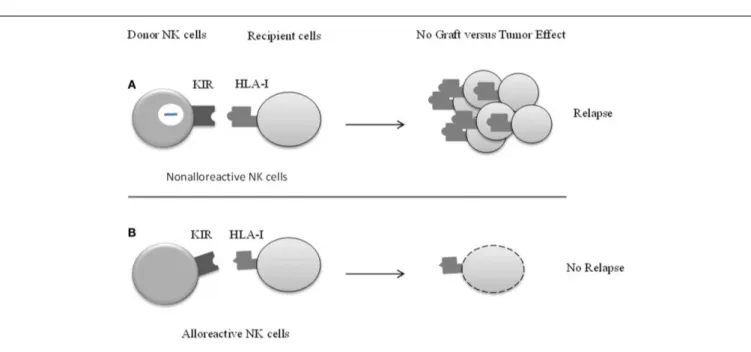
FIGURE 1 | The effect of KIR/HLA class I mismatch in patients undergoing allogenic stem cells transplantation (HSTC).The main
challenge in HSTC is to keep the balance between conserving the graft versus leukemia (GvL) effect on the one hand and preventing risk of relapse on the other.(A) Killer cell immunoglobulin-like receptor (KIR) of donors NK
cells is engaged by corresponding HLA ligand, which inhibits NK-cell function.
Donor NK cells are non-alloreactive and do not kill recipient’s blasts, which leads to the relapse of patient.(B) The concept and recognition of KIR-ligand
incompatibility (also known as KIR-ligand mismatch) has important
implications. KIR-mismatch is an independent predictor of survival in patients. Graft versus tumor NK-cell alloreactivity reduces the risk of leukemia relapse, and markedly improves event-free survival.
increase in the total number of NK cells in the peripheral blood
has also been described in chronic lymphocytic leukemia (CLL)
but still associated with defective cytolytic functions (
18
).
ALTERED ACTIVATING RECEPTORS PROFILES
In acute myeloid leukemia (AML) the down-regulation of
acti-vating receptors NKp30/NCR3 and NKp46/NCR1 correlates with
defective NK-cell cytotoxicity and poor leukemia prognosis (
9
,
19
).
In patients attaining complete remission (CR) after
chemother-apy, NKp46/NCR1 expression returns to normal levels while
patients who do not achieve CR or who relapse maintain
abnormal NCR expression (
9
,
19
). The defect in NCRs
expres-sion could be potentiated by the low expresexpres-sion of NCR and
NKG2D ligands by leukemic cells (
20
–
22
). Down-regulation of
the NK activating receptors/co-receptor DNAM-1, 2B4/CD244,
and CD94/NKG2C have also been reported in AML (
23
,
24
).
Leukemic blasts that express 1 ligands induce
DNAM-1 down-regulation at the NK-cell surface (
25
), thus impending
NK-cell functions.
In acute lymphoblastic leukemia (ALL), expression of the
NKG2D activating receptor ligands MICA/B was only observed in
NK sensitive T-ALL cell line, while NK-resistant B-ALLs did not
express detectable amounts of MICA/B (
26
). Deficient
engage-ment of other activating receptors may also contribute to ALL
resistance to NK lysis, since B-ALL cells lose or express low
lev-els of several other NK activating ligands such as UL-16 binding
proteins (ULBPs), PVR (polio virus receptor, CD155), Nectin-2
(CD112), or CD48 (
27
).
In MDS, a pre-leukemic stage, Epling-Brunette et al. (
13
)
have shown that expression of NKp30/NCR3 and NKG2D was
decreased, in contrast with the data of Kiladjian et al. (
28
); this
discrepancy could be related to the heterogeneity of MDS patients.
In CML patients, Boissel et al. (
29
) reported high serum
sMICA levels and weak NKG2D expression on NK cells, that
correlate with low NK-cell cytotoxicity capacities. Imatinib
mesy-late, the first inhibitor of tyrosine kinase used in CML, increases
NKG2D expression and decreases MICA protein production and
release, thus contributing to normal NK cytotoxicity through the
restoration of a functional NKG2D signaling (
29
).
Monoclonal gammopathy of undetermined significance
(MGUS) is a common disorder of aging and a precursor lesion
to multiple myeloma (MM). In MGUS, tumor-cells express high
levels of MICA, whereas low levels of sMICA are detected in
peripheral blood (
30
). This explains the capacities of NK cells to
kill MGUS tumor cells by interaction between MICA and NKG2D.
Conversely, MM patients present high plasma level of sMICA
while tumor-cells express low level of MICA, thus impending NK
stimulation via NKG2D (
31
). This reveals that the alterations in
the NKG2D pathway signaling are associated with the
progres-sion from MGUS to MM (
30
,
32
). In peripheral blood from
patients with MM a normal expression of the NCRs and NKG2D
is observed, while 2B4/CD244 and the low-affinity Ig- Fc
recep-tor CD16 display significantly weaker expression in comparison
with healthy donors (
31
). Nonetheless, when NK are studied at
the site of tumor location, i.e., bone marrow, (
33
) a drastic
down-regulation of three major activating NK receptors (NKp30/NCR3,
NKG2D, and 2B4/CD244) is observed in comparison with bone
marrow from healthy donors (
34
). This suggests that some NK
abnormalities may be underestimated if only peripheral blood is
studied.
Table 1 | Mechanisms of immune escape and drugs used with their effects on NK cells in hematological malignancies. Hematological
malignancies
Principal mechanisms of escape References Principal class of drugs used
Effects of drugs on NK activation
References
MDS NK-cell quantitative deficiency Elevated TNF
(13,14,49) Demethylating agents IMIDs
Up-regulation of KIR and NKG2D ligands
(76,81)
NKp46 upregulation
AML Upregulation of HLA-I
Decrease expression of NKp30, 2B4/CD244 Production of ROS (9,19,39, 40,54) HDACIs Histamine dihydrochloride All-trans retinoic acid Monoclonal antibody (IPH2101) Upregulation of MICA/B expression (65,67,72, 73,84,85) Suppress ROS production Promote NK-cells cytotoxicity
CLL Upregulation of HLA-I low level of MICA or ULBP
(18,37) Monoclonal antibody Mediate NK-cells cytotoxicity (88) MM Production of sMICA/sMICB Weak expression of CD16 (30,31) IMIDs Proteasome inhibitor IPH2101 NKp46 upregulation NKG2D ligand upregulation (79,82–84, 86)
CML Production of sMICA and weak expression of NKG2D
(29) Tyrosine kinase inhibitor sMICA down-regulation and NKG2D upregulation
(29)
ALL Low production of MICA/B and ULBP (26,27,41) Down regulation of HLA-A and HLA-Bw6
MDS, myelodysplastic syndrome; AML, acute myeloid leukemia; CLL, chronic lymphocytic leukemia; MM, multiple myeloma; CML, chronic myelogenous leukemia; ALL, acute lymphoblastic leukemia; s/MICA/B, soluble/stress-induced molecules human class I-like molecules A and B; ULBPs, UL-16 binding proteins; ROS, reactive oxygen species; KIR, killer immunoglobulin-like receptors; PDGF, platelet-derived growth factor; HDC, histamine dihydrochloride; IMIDs, immune-modulatory drugs; HDACIs, Histone deacetylase inhibitors.
In CLL, NK cells have weak cytotoxic functions which can be
restored by stimulation with recombinant IFN-γ or IL-2 (
35
). We
failed to detect a difference in NCR expression between patients
and age-matched healthy donors (
36
), but we found a correlation
with abnormal activating molecule expression and poor
progno-sis factors. In CLL decreased NK functions could be explained in
part by absent or low level of MICA or ULBP (
18
,
37
) at CLL
lymphocyte cell surface, thus impeding NKG2D engagement.
ABNORMAL KIRs PHENOTYPE AND INHIBITORY MOLECULES
Killer immunoglobulin-like receptor-mismatch in allogenic stem
cells transplantation improves the disease-free survival in AML
(
38
). In fact, NK-cell cytotoxicity is down-regulated by the
engage-ment of HLA-specific inhibitory receptors, including KIRs and
CD94 and NKG2A/B heterodimers. The analysis of KIR phenotype
in AML patients shows that the frequency of particular inhibitory
KIRs in association with their putative HLA class I ligands is
sig-nificantly increased compared to healthy donors (
10
,
39
,
40
). This
supports the hypothesis that AML blasts escape from immune
surveillance according to the dominance of inhibitory over
acti-vating KIR signals. In ALL, the resistance of B-cell precursors to
cytotoxicity is explained by the interaction between HLA-G and
KIR2DL4 (
26
). Demanet et al. (
41
) have observed in ALL and CLL
a selective down-regulation of HLA-A and HLA-Bw6 associated
with HLA-Bw4 preservation, which provided an escape
mecha-nism from NK-cell immune surveillance. Maki et al. (
18
) reported
in CLL cells an increased expression of HLA-G1, a class I molecule
that engages NK-cell inhibitory molecules and which has for
lig-and p49/KIR2DL4/CD158d (expressed on NK cells lig-and a fraction
of T cells), ILT-2 [expressed on NK, T, B cells, dendritic cells (DC),
and monocytes], and ILT-4 [expressed on antigen-presenting cells
(APC)] (
42
).
TUMOR ENVIRONMENT AND ROLE OF CYTOKINES
One of the strategies used by tumor cells having an effect on
NK-cell function is the production of inhibitory molecules, which
decrease NK-cells number and inhibit NK-cell activation.
Increas-ing evidence supports the role of the tumor microenvironment
in conferring drug resistance, a major cause of relapse and
incur-ability of cancers. Tumor microenvironment includes tumor-cells
contact and interaction, but also production of soluble factors,
which provide signals for tumor growth and survival or inhibition
of NK.
ROLE OF CYTOKINES AND CELLULAR LIGANDS
Several cytokines decrease NK-cell activation and cytotoxicity,
such as the Transforming Growth Factor beta (TGF-β). High
circu-lating TGF-β level correlates with poor prognosis in acute leukemia
(
43
) and is linked to reduced NK-cell activity with reduced
expres-sion of NKp30/NCR3 and NKG2D (
44
). TGF-β antagonizes
IL-15, a cytokine that induces NK-cell proliferation and activation.
Thus, TGF-β inhibits the expression of both NK-cell activation
Baier et al. NK-cells modulation in hematological malignancies
receptor molecules and components of the cytotoxic apparatus
(
45
,
46
). Several studies have also underlined that low INF-γ
producing capacity of NK cells was correlated with loss of NK-cell
cytotoxicity (
47
,
48
).
A physiologic concentration of platelet-derived growth factor
(PDGF) significantly inhibits human NK-cell cytotoxicity. Patients
suffering from IMF and essential thrombocythemia have
signifi-cantly elevated circulating levels of plasma PDGF. Pretreatment
of normal NK cells with concentrated PDGF-containing
platelet-poor plasma from patients with these diseases significantly inhibits
NK cytotoxicity. This inhibitory effect is reversed by neutralization
of plasma PDGF with anti-PDGF (
17
,
49
). All these data suggest
that PDGF is probably a key factor of NK functional deficiency
in MPS.
Interaction between tumor cells and NK impairs
NK-cell-mediated cytotoxicity and thus induces tolerance to tumor
inva-sion. Up-regulation of the immunosuppressive cell surface
gly-coprotein CD200 and of soluble GITRL (glucocorticoid-induced
TNFR related protein ligand) on AML cells specifically
com-promises NK-cell anti-tumor responses (
50
,
51
) and is a poor
prognosis factor. AML cells exert direct immunosuppressive effects
on NK cells mediated by immunosuppressive ligands or soluble
factors and induce regulatory T lymphocytes (Treg) that weaken
NK-cell responses (
52
). NK cells can also interact with DC
lead-ing to activation of Treg and inhibition of NK cells (
53
). Recently,
ligand of NKp44/NCR2 (NKp44L) was identified as an isoform
of mixed-lineage leukemia-5 (MLL5) (
54
). This ligand is not
detectable in the normal tissues but is present in hematopoietic,
non-hematopoietic tumor and transformed cells. The expression
of MLL5 is a good prognosis factor in AML (
55
). Thus we can
speculate that the prognostic value of MLL5 expression is linked
to its capacity to activate anti-leukemia NK cells.
REACTIVE OXYGEN SPECIES
Non-malignant phagocytic cells down-modulate lymphocyte
functions by producing and releasing NADPH oxidase-derived
reactive oxygen species (ROS) (
56
). Monocytic and
myelo-monocytic (French-American-British classification M4/M5
sub-types) AML cells, but not cells from myeloblastic (FAB class M2) or
immature (FAB class M1) AML, produce ROS via the NADPH
oxi-dase component gp91phox, and trigger extensive apoptosis of NK
cells via a poly-[ADPribose] polymerase-1 dependent pathway,
together with a down-regulation of NKp46/NCR1. This suggests
a novel mechanism of immune evasion in myelo-monocytic and
monocytic AML (
57
).
MATERIAL TRANSFER DURING CELL–CELL CONTACT
Tumor membrane-derived microvesicles (tMV) are important
mediators of cell-to-cell communication. These circular
mem-brane fragments are enriched in various bioactive molecules and
directly stimulate cells as a kind of “signaling complex.” An
important mode of communication between carcinoma cells and
immune cells involves tMV, also known as exosomes, ectosomes, or
microparticles. These microvesicles carry lipids, proteins, mRNAs,
and microRNAs and travel short or long distances to deliver
unde-graded and undiluted material to other cells (
58
). Microvesicles
present in AML patients’ sera contain TGF-β that down-regulates
the expression of NKG2D and thus interfere with NK-cell
acti-vation. Nonetheless, IL-15 protects NK cells from adverse effects
of tMV and could thus contribute to maintain their anti-tumor
response (
59
). Baj-Krzyworzeka et al. (
60
) have observed that
tMV carry mRNA of tumor cells and transfer some of them to
monocytes and modify their activity. This type of mRNA transfer
could participate to NK inactivation and tumor escape to innate
immunity.
IMMUNOTHERAPY APPROACHES
Modulation of NK-cell function by down-regulation of
recep-tors and/or ligand corresponds to an immune escape mechanism
for tumors. Restoring the expression of activating receptors on
NK cells, or corresponding ligands on cancer cells, is an effective
approach to cancer immunotherapy in order to improve
disease-free survival after therapy. Currently, in most AML patients, the
induction treatment leads to CR, defined as microscopic
disap-pearance of leukemic disease along with the return of normal
hematopoiesis. However, many patients in CR relapse with poor
prospects of long-term survival. One of developing
immunother-apy that enhances NK-cell ability to kill tumor cells is the allogenic
transplantation after chemotherapy. In AML, a recipient of
hap-loidentical allogenic transplant with a NK HLA-specific
receptor-mismatch is associated with a favorable prognosis because this
increases the anti-leukemic graft reactivity (
61
,
62
). Rapid
recov-ery of NK cells after hematopoietic stem cell transplantation
has been associated with a reduction in the rates of relapse and
acute graft-versus-host disease (GvHD) (
63
). The transfer of NK
cell from haploidentical origins into AML recipients is a potent
immunotherapy intervention that is, unfortunately, associated
with a significant transplant-related morbidity and mortality that
limit its use (
64
).
The use of cytokines is another therapeutic approach to
enhance NK-cell cytotoxicity. IL-2, IL-12, Il-15 (
65
), and IL-18
have been used in culture to increase cell cytotoxic prior to the
injection of NK cells in cancer patients (
66
). Another strategy
consists in restoring normal NCR expression since these
mole-cules are pivotal for the anti-leukemia response. A phase III study
in 320 AML patients has demonstrated that immunotherapy with
histamine dihydrochloride (HDC) and IL-2 decreases and delays
relapses in AML (
67
). HDC suppresses or inhibits ROS formation
in mononuclear and polymorphonuclear myeloid cells. This
pre-vents from oxygen radical-induced NK apoptosis, restores NK-cell
capacity to respond to IL-2, and improves NK proliferation and
production of immuno-stimulatory cytokines (
56
,
68
–
71
).
More-over, in presence of HDC, cytotoxic functions of NK cells remain
intact due to the preserved expression of the activating receptors
NKG2D and NKp46/NCR1.
DNA methylation also has a key role in the control of gene
activ-ity in cancer cells. Two agents are currently used in MDS treatment:
5-azacytidine (Vidaza) and 5-aza-20-deoxycytidine (Decitabine)
(
72
). These two hypomethylating agents up-regulate NKG2D
lig-ands MICA/B leading to enhanced NK-cell cytotoxicity (
73
,
74
).
Conversely we have observed the down-regulation of 2B4/CD244
in NK from AML patients treated with 5-azacytidine, that could
have an opposite effect, i.e., cytotoxicity down-regulation (Leclerq
et al. personal data).
The immune-modulatory drugs (IMIDs) such as thalidomide
and lenalidomide are used in MM and MDS treatment and
have anti-angiogenic and anti-inflammatory properties. They also
act as IMIDs by cytokine release and activating effector cells
by enhancing ADCC and NK-cell cytotoxicity thanks to the
up-regulation of NKp46/NCR1 (
75
–
77
).
Bortezomib is a proteasome inhibitor used in MM treatment.
Physiologically, proteasome is involved in protein degradation. Its
inhibition by the drug interferes with tumor growth and with
innate immunity. Bortezomib is involved in the down-regulation
of HLA I molecules and in the up-regulation of NKG2D, TRAIL,
and DNAM ligands, thus leading to increased NK-cell cytotoxicity
against plasma cell (
78
,
79
).
Another attractive therapeutic approach consists in blocking
the NK inhibitory receptors. A phase 1 trial has tested the IPH2101,
a fully humanized IgG4 anti-KIR monoclonal antibody, in patients
with relapsed/refractory AML (clinical trial registration
num-ber NCT01256073) and MM (clinical trial registration numnum-ber
NCT00999830). IPH2101 promotes immune complex formation
and NK-cell cytotoxicity specifically against MM cell targets but
not normal cells. No evidence of autoimmunity was observed.
These findings suggest that IPH2101 is safe and tolerable and that
this approach warrants further development in MM and AML
(
80
–
82
) as we are waiting for clinical results.
Several studies revealed ADCC as one major mode of action
of antibody-based therapeutics and stimulated more interest in
how to mobilize, expand, and activate NK cells in humans (
83
).
In CLL, the ADCC pathway via the Fc receptor (FcgRIIIa) CD16
at surface of NK cells is pivotal in the clinical effect of mAbs such
as rituximab or ofatumumab which mediate ADCC by NK cells
(
84
). Second generation mAb are designed in order to maximize
both direct apoptosis and ADCC. The role of ADCC is underlined
by better clinical responses to rituximab when NK cells expressed
the high-affinity form of the FcgRIII (
85
,
86
).
Bi-specific NK-cell engagers (BiKE) simultaneously bind
CD16α and c-Met (a receptor overexpressed in many tumors) and
thus may increase NK-cell ADCC (
87
).
Another type of therapeutic strategy consists in taking
advan-tage from anti-cancer drugs properties (Table 1). Chemotherapy
drugs can be separated by their ability to inhibit (such as
vinblas-tine, chlorambucil, docetaxel) or enhance (such as asparaginase,
bleomycin, doxorubicin) NK-cell-mediated killing of target cells
(
88
,
89
). In cancer, epigenetic changes are also involved in
dysreg-ulating NK-cell ligand expression. Histone deacetylase inhibitors
(HDACIs) such as trichostatin, are epigenetic anti-cancer agents
that modulate innate immunity by the regulation of expression of
NKG2D or DNAM-1 ligands. Indeed, HDACIs increase
expres-sion of ligands of these two activating receptors, MICA/B or
PVR and Nectin-2 respectively, on acute leukemia cells (
90
,
91
).
This suggests that epigenetic drugs make tumor cells more
sen-sitive to NK-cell-mediated lysis (
92
). However, it has also been
demonstrated that HDACIs suppress NK-cell cytotoxic activity by
down-regulation of NKp30/NCR3, NKp46/NCR1 (
93
). Thus, the
same drug can have contradictory effects on NK cells.
CONCLUDING REMARKS
The more precise and exhaustive analysis of NK dysfunction in
HM has opened the way to novel therapeutic strategies
involv-ing either specifically developed drugs/antibodies or innovative
use of “old” drugs such as IMIDs. Due to the complexity of the
immune response and the putative opposed effects of a drug on
the various partners of the immune network, data obtained by
in vitro experiments or in vivo in animal models have to be
eval-uated in clinically and biologically carefully monitored clinical
trials.
ACKNOWLEDGMENTS
This work was supported by Grants from INSERM and Region
Provence-Alpes-Côtes d’Azur “Bourse Doctorale Regionale 2011–
2014.”
REFERENCES
1. Vivier E, Tomasello E, Baratin M, Walzer T, Ugolini S. Functions of natural killer cells. Nat Immunol (2008) 9:503–10. doi:10.1038/ni1582
2. Vivier E, Raulet DH, Moretta A, Caligiuri MA, Zitvogel L, Lanier LL, et al. Innate or adaptive immunity? The example of natural killer cells. Science (2011) 331:44–9. doi:10.1126/science.1198687
3. Ruggeri L, Mancusi A, Burchielli E, Capanni M, Carotti A, Aloisi T, et al. NK cell alloreactivity and allogeneic hematopoietic stem cell transplantation. Blood Cells Mol Dis (2008) 40:84–90. doi:10.1016/j.bcmd.2007.06.029
4. Costello RT, Gastaut JA, Olive D. Tumor escape from immune surveillance. Arch Immunol Ther Exp (Warsz) (1999) 47:83–8.
5. Ljunggren HG, Karre K. In search of the ‘missing self ’: MHC molecules and NK cell recognition. Immunol Today (1990) 11:237–44. doi:10.1016/0167-5699(90) 90097-S
6. Garrido F, Cabrera T, Lopez-Nevot MA, Ruiz-Cabello F. HLA class I antigens in human tumors. Adv Cancer Res (1995) 67:155–95.
7. Moretta A, Biassoni R, Bottino C, Mingari MC, Moretta L. Natural cytotoxi-city receptors that trigger human NK-cell-mediated cytolysis. Immunol Today (2000) 21:228–34. doi:10.1016/S0167-5699(00)01596-6
8. Campbell KS, Hasegawa J. Natural killer cell biology: an update and future direc-tions. J Allergy Clin Immunol (2013) 132:536–44. doi:10.1016/j.jaci.2013.07.006 9. Costello RT, Sivori S, Marcenaro E, Lafage-Pochitaloff M, Mozziconacci MJ, Rev-iron D, et al. Defective expression and function of natural killer cell-triggering receptors in patients with acute myeloid leukemia. Blood (2002) 99:3661–7. doi:10.1182/blood.V99.10.3661
10. Sanchez CJ, Le Treut T, Boehrer A, Knoblauch B, Imbert J, Olive D, et al. Nat-ural killer cells and malignant haemopathies: a model for the interaction of cancer with innate immunity. Cancer Immunol Immunother (2011) 60:1–13. doi:10.1007/s00262-010-0898-x
11. Costello RT, Fauriat C, Sivori S, Marcenaro E, Olive D. NK cells: innate immu-nity against hematological malignancies? Trends Immunol (2004) 25:328–33. doi:10.1016/j.it.2004.04.005
12. Rey J, Veuillen C, Vey N, Bouabdallah R, Olive D. Natural killer and gammadelta T cells in haematological malignancies: enhancing the immune effectors. Trends Mol Med (2009) 15:275–84. doi:10.1016/j.molmed.2009.04.005
13. Epling-Burnette PK, Bai F, Painter JS, Rollison DE, Salih HR, Krusch M, et al. Reduced natural killer (NK) function associated with high-risk myelodysplas-tic syndrome (MDS) and reduced expression of activating NK receptors. Blood (2007) 109:4816–24. doi:10.1182/blood-2006-07-035519
14. Yokose N, Ogata K, Ito T, An E, Tamura H, Dan K, et al. Elevated plasma soluble interleukin 2 receptor level correlates with defective natural killer and CD8+ T-cells in myelodysplastic syndromes. Leuk Res (1994) 18:777–82. doi:10.1016/0145-2126(94)90060-4
15. Chang WC, Fujimiya Y, Casteel N, Pattengale P. Natural killer cell immunodefi-ciency in patients with chronic myelogenous leukemia. III. Defective interleukin-2 production by T-helper and natural killer cells. Int J Cancer (1989) 43:591–7. doi:10.1002/ijc.2910430410
16. Pierson BA, Miller JS. CD56+bright and CD56+dim natural killer cells in patients with chronic myelogenous leukemia progressively decrease in number,
Baier et al. NK-cells modulation in hematological malignancies
respond less to stimuli that recruit clonogenic natural killer cells, and exhibit decreased proliferation on a per cell basis. Blood (1996) 88:2279–87. 17. Gersuk GM, Carmel R, Pattamakom S, Challita PM, Rabinowitz AP, Pattengale
PK. Quantitative and functional studies of impaired natural killer (NK) cells in patients with myelofibrosis, essential thrombocythemia, and polycythemia vera. I. A potential role for platelet-derived growth factor in defective NK cytotoxicity. Nat Immun (1993) 12:136–51.
18. Maki G, Hayes GM, Naji A, Tyler T, Carosella ED, Rouas-Freiss N, et al. NK resistance of tumor cells from multiple myeloma and chronic lympho-cytic leukemia patients: implication of HLA-G. Leukemia (2008) 22:998–1006. doi:10.1038/leu.2008.15
19. Fauriat C, Just-Landi S, Mallet F, Arnoulet C, Sainty D, Olive D, et al. Deficient expression of NCR in NK cells from acute myeloid leukemia: evolution dur-ing leukemia treatment and impact of leukemia cells in NCRdull phenotype induction. Blood (2007) 109:323–30. doi:10.1182/blood-2005-08-027979 20. Pende D, Cantoni C, Rivera P, Vitale M, Castriconi R, Marcenaro S, et al. Role of
NKG2D in tumor cell lysis mediated by human NK cells: cooperation with nat-ural cytotoxicity receptors and capability of recognizing tumors of nonepithelial origin. Eur J Immunol (2001) 31:1076–86. doi:10.1002/1521-4141(200104)31: 4<1076::AID-IMMU1076>3.0.CO;2-Y
21. Biassoni R, Cantoni C, Marras D, Giron-Michel J, Falco M, Moretta L, et al. Human natural killer cell receptors: insights into their molecular function and structure. J Cell Mol Med (2003) 7:376–87. doi:10.1111/j.1582-4934.2003. tb00240.x
22. Hilpert J, Grosse-Hovest L, Grunebach F, Buechele C, Nuebling T, Raum T, et al. Comprehensive analysis of NKG2D ligand expression and release in leukemia: implications for NKG2D-mediated NK cell responses. J Immunol (2012) 189:1360–71. doi:10.4049/jimmunol.1200796
23. Sanchez-Correa B, Morgado S, Gayoso I, Bergua JM, Casado JG, Arcos MJ, et al. Human NK cells in acute myeloid leukaemia patients: analysis of NK cell-activating receptors and their ligands. Cancer Immunol Immunother (2011) 60:1195–205. doi:10.1007/s00262-011-1050-2
24. Lion E, Willemen Y, Berneman ZN, Van Tendeloo VF, Smits EL. Natural killer cell immune escape in acute myeloid leukemia. Leukemia (2012) 26:2019–26. doi:10.1038/leu.2012.87
25. Sanchez-Correa B, Gayoso I, Bergua JM, Casado JG, Morgado S, Solana R, et al. Decreased expression of DNAM-1 on NK cells from acute myeloid leukemia patients. Immunol Cell Biol (2012) 90:109–15. doi:10.1038/icb.2011.15 26. Romanski A, Bug G, Becker S, Kampfmann M, Seifried E, Hoelzer D, et al.
Mechanisms of resistance to natural killer cell-mediated cytotoxicity in acute lymphoblastic leukemia. Exp Hematol (2005) 33:344–52. doi:10.1016/j.exphem. 2004.11.006
27. Pende D, Castriconi R, Romagnani P, Spaggiari GM, Marcenaro S, Dondero A, et al. Expression of the DNAM-1 ligands, Nectin-2 (CD112) and poliovirus receptor (CD155), on dendritic cells: relevance for natural killer-dendritic cell interaction. Blood (2006) 107:2030–6. doi:10.1182/blood-2005-07-2696 28. Kiladjian JJ, Bourgeois E, Lobe I, Braun T, Visentin G, Bourhis JH, et al. Cytolytic
function and survival of natural killer cells are severely altered in myelodysplastic syndromes. Leukemia (2006) 20:463–70. doi:10.1038/sj.leu.2404080
29. Boissel N, Rea D, Tieng V, Dulphy N, Brun M, Cayuela JM, et al. BCR/ABL oncogene directly controls MHC class I chain-related molecule A expression in chronic myelogenous leukemia. J Immunol (2006) 176:5108–16.
30. Jinushi M, Vanneman M, Munshi NC, Tai YT, Prabhala RH, Ritz J, et al. MHC class I chain-related protein A antibodies and shedding are associated with the progression of multiple myeloma. Proc Natl Acad Sci U S A (2008) 105:1285–90. doi:10.1073/pnas.0711293105
31. Carbone E, Neri P, Mesuraca M, Fulciniti MT, Otsuki T, Pende D, et al. HLA class I, NKG2D, and natural cytotoxicity receptors regulate multiple myeloma cell recognition by natural killer cells. Blood (2005) 105:251–8. doi:10.1182/blood-2004-04-1422
32. Fauriat C, Mallet F, Olive D, Costello RT. Impaired activating receptor expression pattern in natural killer cells from patients with multiple myeloma. Leukemia (2006) 20:732–3. doi:10.1038/sj.leu.2404096
33. Costello RT, Boehrer A, Sanchez C, Mercier D, Baier C, Le Treut T, et al. Dif-ferential expression of natural killer cell activating receptors in blood versus bone marrow in patients with monoclonal gammopathy. Immunology (2013) 139:338–41. doi:10.1111/imm.12082
34. Eissens DN, Spanholtz J, van der Meer A, van Cranenbroek B, Dolstra H, Kwekkeboom J, et al. Defining early human NK cell developmental stages in primary and secondary lymphoid tissues. PLoS One (2012) 7:e30930. doi:10.1371/journal.pone.0030930
35. Katrinakis G, Kyriakou D, Papadaki H, Kalokyri I, Markidou F, Eliopou-los GD. Defective natural killer cell activity in B-cell chronic lymphocytic leukaemia is associated with impaired release of natural killer cytotoxic fac-tor(s) but not of tumour necrosis factor-alpha. Acta Haematol (1996) 96:16–23. doi:10.1159/000203709
36. Costello RT, Knoblauch B, Sanchez C, Mercier D, Le Treut T, Sebahoun G. Expres-sion of natural killer cell activating receptors in patients with chronic lympho-cytic leukaemia. Immunology (2012) 135:151–7. doi:10.1111/j.1365-2567.2011. 03521.x
37. Pende D, Spaggiari GM, Marcenaro S, Martini S, Rivera P, Capobianco A, et al. Analysis of the receptor-ligand interactions in the natural killer-mediated lysis of freshly isolated myeloid or lymphoblastic leukemias: evidence for the involve-ment of the Poliovirus receptor (CD155) and Nectin-2 (CD112). Blood (2005) 105:2066–73. doi:10.1182/blood-2004-09-3548
38. Ruggeri L, Capanni M, Urbani E, Perruccio K, Shlomchik WD, Tosti A, et al. Effectiveness of donor natural killer cell alloreactivity in mismatched hematopoietic transplants. Science (2002) 295:2097–100. doi:10.1126/science. 1068440
39. Wetzler M, Baer MR, Stewart SJ, Donohue K, Ford L, Stewart CC, et al. HLA class I antigen cell surface expression is preserved on acute myeloid leukemia blasts at diagnosis and at relapse. Leukemia (2001) 15:128–33. doi:10.1038/sj.leu.2401982 40. Verheyden S, Demanet C. Susceptibility to myeloid and lymphoid leukemia is mediated by distinct inhibitory KIR-HLA ligand interactions. Leukemia (2006) 20:1437–8. doi:10.1038/sj.leu.2404279
41. Demanet C, Mulder A, Deneys V, Worsham MJ, Maes P, Claas FH, et al. Down-regulation of HLA-A and HLA-Bw6, but not HLA-Bw4, allospecificities in leukemic cells: an escape mechanism from CTL and NK attack? Blood (2004) 103:3122–30. doi:10.1182/blood-2003-07-2500
42. Creput C, Durrbach A, Charpentier B, Carosella ED, Rouas-Freiss N. [HLA-G: immunoregulatory molecule involved in allograft acceptance]. Nephrologie (2003) 24:451–6.
43. Bergmann L, Schui DK, Brieger J, Weidmann E, Mitrou PS, Hoelzer D. The inhibition of lymphokine-activated killer cells in acute myeloblastic leukemia is mediated by transforming growth factor-beta 1. Exp Hematol (1995) 23:1574–80.
44. Lee JC, Lee KM, Kim DW, Heo DS. Elevated TGF-beta1 secretion and down-modulation of NKG2D underlies impaired NK cytotoxicity in cancer patients. J Immunol (2004) 172:7335–40.
45. Flavell RA, Sanjabi S, Wrzesinski SH, Licona-Limon P. The polarization of immune cells in the tumour environment by TGFbeta. Nat Rev Immunol (2010) 10:554–67. doi:10.1038/nri2808
46. Wilson EB, El-Jawhari JJ, Neilson AL, Hall GD, Melcher AA, Meade JL, et al. Human tumour immune evasion via TGF-beta blocks NK cell activation but not survival allowing therapeutic restoration of anti-tumour activity. PLoS One (2011) 6:e22842. doi:10.1371/journal.pone.0022842
47. Tajima F, Kawatani T, Endo A, Kawasaki H. Natural killer cell activity and cytokine production as prognostic factors in adult acute leukemia. Leukemia (1996) 10:478–82.
48. Castriconi R, Cantoni C, Della Chiesa M, Vitale M, Marcenaro E, Conte R, et al. Transforming growth factor beta 1 inhibits expression of NKp30 and NKG2D receptors: consequences for the NK-mediated killing of dendritic cells. Proc Natl Acad Sci U S A (2003) 100:4120–5. doi:10.1073/pnas.0730640100
49. Verhoef GE, De Schouwer P, Ceuppens JL, Van Damme J, Goossens W, Boogaerts MA. Measurement of serum cytokine levels in patients with myelodysplastic syndromes. Leukemia (1992) 6:1268–72.
50. Coles SJ, Wang EC, Man S, Hills RK, Burnett AK, Tonks A, et al. CD200 expression suppresses natural killer cell function and directly inhibits patient anti-tumor response in acute myeloid leukemia. Leukemia (2011) 25:792–9. doi:10.1038/leu.2011.1
51. Baessler T, Krusch M, Schmiedel BJ, Kloss M, Baltz KM, Wacker A, et al. Glucocorticoid-induced tumor necrosis factor receptor-related protein ligand subverts immunosurveillance of acute myeloid leukemia in humans. Cancer Res (2009) 69:1037–45. doi:10.1158/0008-5472.CAN-08-2650
52. Ustun C, Miller JS, Munn DH, Weisdorf DJ, Blazar BR. Regulatory T cells in acute myelogenous leukemia: is it time for immunomodulation? Blood (2011) 118:5084–95. doi:10.1182/blood-2011-07-365817
53. Fauriat C, Moretta A, Olive D, Costello RT. Defective killing of dendritic cells by autologous natural killer cells from acute myeloid leukemia patients. Blood (2005) 106:2186–8. doi:10.1182/blood-2005-03-1270
54. Baychelier F, Sennepin A, Ermonval M, Dorgham K, Debre P, Vieillard V. Iden-tification of a cellular ligand for the natural cytotoxicity receptor NKp44. Blood (2013) 122:2935–42. doi:10.1182/blood-2013-03-489054
55. Damm F, Oberacker T, Thol F, Surdziel E, Wagner K, Chaturvedi A, et al. Prognostic importance of histone methyltransferase MLL5 expression in acute myeloid leukemia. J Clin Oncol (2011) 29:682–9. doi:10.1200/JCO.2010.31.1118 56. Thoren FB, Romero AI, Hellstrand K. Oxygen radicals induce poly(ADP-ribose) polymerase-dependent cell death in cytotoxic lymphocytes. J Immunol (2006) 176:7301–7.
57. Aurelius J, Thoren FB, Akhiani AA, Brune M, Palmqvist L, Hansson M, et al. Monocytic AML cells inactivate antileukemic lymphocytes: role of NADPH oxi-dase/gp91(phox) expression and the PARP-1/PAR pathway of apoptosis. Blood (2012) 119:5832–7. doi:10.1182/blood-2011-11-391722
58. Pucci F, Pittet MJ. Molecular pathways: tumor-derived microvesicles and their interactions with immune cells in vivo. Clin Cancer Res (2013) 19:2598–604. doi:10.1158/1078-0432.CCR-12-0962
59. Szczepanski MJ, Szajnik M, Welsh A, Whiteside TL, Boyiadzis M. Blast-derived microvesicles in sera from patients with acute myeloid leukemia suppress natural killer cell function via membrane-associated transforming growth factor-beta1. Haematologica (2011) 96:1302–9. doi:10.3324/haematol.2010.039743 60. Baj-Krzyworzeka M, Szatanek R, Weglarczyk K, Baran J, Zembala M.
Tumour-derived microvesicles modulate biological activity of human monocytes. Immunol Lett (2007) 113:76–82. doi:10.1016/j.imlet.2007.07.014
61. Ruggeri L, Mancusi A, Capanni M, Urbani E, Carotti A, Aloisi T, et al. Donor natural killer cell allorecognition of missing self in haploidentical hematopoi-etic transplantation for acute myeloid leukemia: challenging its predictive value. Blood (2007) 110:433–40. doi:10.1182/blood-2006-07-038687
62. Ruggeri L, Aversa F, Martelli MF, Velardi A. Allogeneic hematopoietic transplan-tation and natural killer cell recognition of missing self. Immunol Rev (2006) 214:202–18. doi:10.1111/j.1600-065X.2006.00455.x
63. Savani BN, Mielke S, Adams S, Uribe M, Rezvani K, Yong AS, et al. Rapid natural killer cell recovery determines outcome after T-cell-depleted HLA-identical stem cell transplantation in patients with myeloid leukemias but not with acute lym-phoblastic leukemia. Leukemia (2007) 21:2145–52. doi:10.1038/sj.leu.2404892 64. Miller JS, Soignier Y, Panoskaltsis-Mortari A, McNearney SA, Yun GH, Fautsch
SK, et al. Successful adoptive transfer and in vivo expansion of human hap-loidentical NK cells in patients with cancer. Blood (2005) 105:3051–7. doi:10. 1182/blood-2004-07-2974
65. Suck G, Oei VY, Linn YC, Ho SH, Chu S, Choong A, et al. Interleukin-15 supports generation of highly potent clinical-grade natural killer cells in long-term cul-tures for targeting hematological malignancies. Exp Hematol (2011) 39:904–14. doi:10.1016/j.exphem.2011.06.003
66. Chan CJ, Andrews DM, Smyth MJ. Can NK cells be a therapeutic target in human cancer? Eur J Immunol (2008) 38:2964–8. doi:10.1002/eji.200838764 67. Brune M, Castaigne S, Catalano J, Gehlsen K, Ho AD, Hofmann WK, et al.
Improved leukemia-free survival after postconsolidation immunotherapy with histamine dihydrochloride and interleukin-2 in acute myeloid leukemia: results of a randomized phase 3 trial. Blood (2006) 108:88–96. doi:10.1182/blood-2005-10-4073
68. Hansson M, Hermodsson S, Brune M, Mellqvist UH, Naredi P, Betten A, et al. Histamine protects T cells and natural killer cells against oxidative stress. J Inter-feron Cytokine Res (1999) 19:1135–44. doi:10.1089/107999099313073 69. Hellstrand K, Asea A, Hermodsson S. Role of histamine in natural killer
cell-mediated resistance against tumor cells. J Immunol (1990) 145:4365–70. 70. Hellstrand K, Hermodsson S. Synergistic activation of human natural killer
cell cytotoxicity by histamine and interleukin-2. Int Arch Allergy Appl Immunol (1990) 92:379–89. doi:10.1159/000235169
71. Romero AI, Thoren FB, Brune M, Hellstrand K. NKp46 and NKG2D recep-tor expression in NK cells with CD56dim and CD56bright phenotype: regula-tion by histamine and reactive oxygen species. Br J Haematol (2006) 132:91–8. doi:10.1111/j.1365-2141.2005.05842.x
72. Kopp LM, Ray A, Denman CJ, Senyukov VS, Somanchi SS, Zhu S, et al. Decitabine has a biphasic effect on natural killer cell viability, phenotype, and function under proliferative conditions. Mol Immunol (2013) 54:296–301. doi:10.1016/j.molimm.2012.12.012
73. Tang KF, He CX, Zeng GL, Wu J, Song GB, Shi YS, et al. Induction of MHC class I-related chain B (MICB) by 5-aza-20-deoxycytidine. Biochem Biophys Res
Commun (2008) 370:578–83. doi:10.1016/j.bbrc.2008.03.131
74. Santourlidis S, Trompeter HI, Weinhold S, Eisermann B, Meyer KL, Wernet P, et al. Crucial role of DNA methylation in determination of clonally distributed killer cell Ig-like receptor expression patterns in NK cells. J Immunol (2002) 169:4253–61.
75. Davies FE, Raje N, Hideshima T, Lentzsch S, Young G, Tai YT, et al. Thalidomide and immunomodulatory derivatives augment natural killer cell cytotoxicity in multiple myeloma. Blood (2001) 98:210–6. doi:10.1182/blood.V98.1.210 76. Reddy N, Hernandez-Ilizaliturri FJ, Deeb G, Roth M, Vaughn M, Knight J,
et al. Immunomodulatory drugs stimulate natural killer-cell function, alter cytokine production by dendritic cells, and inhibit angiogenesis enhancing the anti-tumour activity of rituximab in vivo. Br J Haematol (2008) 140: 36–45.
77. Berg SL, Cairo MS, Russell H, Ayello J, Ingle AM, Lau H, et al. Safety, phar-macokinetics, and immunomodulatory effects of lenalidomide in children and adolescents with relapsed/refractory solid tumors or myelodysplastic syndrome: a Children’s Oncology Group Phase I Consortium report. J Clin Oncol (2011) 29:316–23. doi:10.1200/JCO.2010.30.8387
78. Shi J, Tricot GJ, Garg TK, Malaviarachchi PA, Szmania SM, Kellum RE, et al. Bortezomib down-regulates the cell-surface expression of HLA class I and enhances natural killer cell-mediated lysis of myeloma. Blood (2008) 111:1309–17. doi:10.1182/blood-2007-03-078535
79. Soriani A, Zingoni A, Cerboni C, Iannitto ML, Ricciardi MR, Di Gialleonardo V, et al. ATM-ATR-dependent up-regulation of DNAM-1 and NKG2D ligands on multiple myeloma cells by therapeutic agents results in enhanced NK-cell susceptibility and is associated with a senescent phenotype. Blood (2009) 113:3503–11. doi:10.1182/blood-2008-08-173914
80. Alici E. IPH-2101, a fully human anti-NK-cell inhibitory receptor mAb for the potential treatment of hematological cancers. Curr Opin Mol Ther (2010) 12:724–33.
81. Vey N, Bourhis JH, Boissel N, Bordessoule D, Prebet T, Charbonnier A, et al. A phase 1 trial of the anti-inhibitory KIR mAb IPH2101 for AML in complete remission. Blood (2012) 120:4317–23. doi:10.1182/blood-2012-06-437558 82. Benson DM Jr, Hofmeister CC, Padmanabhan S, Suvannasankha A,
Jagan-nath S, Abonour R, et al. A phase 1 trial of the anti-KIR antibody IPH2101 in patients with relapsed/refractory multiple myeloma. Blood (2012) 120:4324–33. doi:10.1182/blood-2012-06-438028
83. Wallace ME, Smyth MJ. The role of natural killer cells in tumor control – effec-tors and regulaeffec-tors of adaptive immunity. Springer Semin Immunopathol (2005) 27:49–64. doi:10.1007/s00281-004-0195-x
84. Weitzman J, Betancur M, Boissel L, Rabinowitz AP, Klein A, Klingemann H. Variable contribution of monoclonal antibodies to ADCC in patients with chronic lymphocytic leukemia. Leuk Lymphoma (2009) 50:1361–8. doi:10.1080/ 10428190903026500
85. Cartron G, Dacheux L, Salles G, Solal-Celigny P, Bardos P, Colombat P, et al. Therapeutic activity of humanized anti-CD20 monoclonal antibody and poly-morphism in IgG Fc receptor FcgammaRIIIa gene. Blood (2002) 99:754–8. doi:10.1182/blood.V99.3.754
86. Treon SP, Hansen M, Branagan AR, Verselis S, Emmanouilides C, Kimby E, et al. Polymorphisms in FcgammaRIIIA (CD16) receptor expression are associated with clinical response to rituximab in Waldenstrom’s macroglobulinemia. J Clin Oncol (2005) 23:474–81. doi:10.1200/JCO.2005.06.059
87. Boltz A, Piater B, Toleikis L, Guenther R, Kolmar H, Hock B. Bi-specific aptamers mediating tumor cell lysis. J Biol Chem (2011) 286:21896–905. doi:10.1074/jbc.M111.238261
88. Markasz L, Skribek H, Uhlin M, Otvos R, Flaberg E, Eksborg S, et al. Effect of frequently used chemotherapeutic drugs on cytotoxic activity of human cytotoxic T-lymphocytes. J Immunother (2008) 31:283–93. doi:10.1097/CJI. 0b013e3181628b76
89. Kittang AO, Hatfield K, Sand K, Reikvam H, Bruserud O. The chemokine network in acute myelogenous leukemia: molecular mechanisms involved in
Baier et al. NK-cells modulation in hematological malignancies
leukemogenesis and therapeutic implications. Curr Top Microbiol Immunol (2010) 341:149–72. doi:10.1007/82_2010_25
90. Kato N, Tanaka J, Sugita J, Toubai T, Miura Y, Ibata M, et al. Regulation of the expression of MHC class I-related chain A, B (MICA, MICB) via chromatin remodeling and its impact on the susceptibility of leukemic cells to the cytotoxicity of NKG2D-expressing cells. Leukemia (2007) 21:2103–8. doi:10.1038/sj.leu.2404862
91. Schmudde M, Braun A, Pende D, Sonnemann J, Klier U, Beck JF, et al. Histone deacetylase inhibitors sensitize tumour cells for cytotoxic effects of natural killer cells. Cancer Lett (2008) 272:110–21. doi:10.1016/j.canlet.2008.06.027 92. Diermayr S, Himmelreich H, Durovic B, Mathys-Schneeberger A, Siegler U,
Langenkamp U, et al. NKG2D ligand expression in AML increases in response to HDAC inhibitor valproic acid and contributes to allorecognition by NK-cell lines with single KIR-HLA class I specificities. Blood (2008) 111:1428–36. doi:10.1182/blood-2007-07-101311
93. Ogbomo H, Michaelis M, Kreuter J, Doerr HW, Cinatl J Jr. Histone deacety-lase inhibitors suppress natural killer cell cytolytic activity. FEBS Lett (2007) 581:1317–22. doi:10.1016/j.febslet.2007.02.045
Conflict of Interest Statement: The authors declare that the research was conducted in the absence of any commercial or financial relationships that could be construed as a potential conflict of interest.
Received: 20 September 2013; accepted: 02 December 2013; published online: 19 December 2013.
Citation: Baier C, Fino A, Sanchez C, Farnault L, Rihet P, Kahn-Perlès B and Costello RT (2013) Natural killer cells modulation in hematological malignancies. Front. Immunol. 4:459. doi: 10.3389/fimmu.2013.00459
This article was submitted to NK Cell Biology, a section of the journal Frontiers in Immunology.
Copyright © 2013 Baier, Fino, Sanchez, Farnault , Rihet , Kahn-Perlès and Costello. This is an open-access article distributed under the terms of the Creative Commons Attribution License (CC BY). The use, distribution or reproduction in other forums is permitted, provided the original author(s) or licensor are credited and that the original publication in this journal is cited, in accordance with accepted academic practice. No use, distribution or reproduction is permitted which does not comply with these terms.