HAL Id: hal-02947674
https://hal.archives-ouvertes.fr/hal-02947674
Submitted on 24 Sep 2020
HAL is a multi-disciplinary open access
archive for the deposit and dissemination of
sci-entific research documents, whether they are
pub-lished or not. The documents may come from
teaching and research institutions in France or
abroad, or from public or private research centers.
L’archive ouverte pluridisciplinaire HAL, est
destinée au dépôt et à la diffusion de documents
scientifiques de niveau recherche, publiés ou non,
émanant des établissements d’enseignement et de
recherche français ou étrangers, des laboratoires
publics ou privés.
pseudotuberculosis and Its Deletion during the
Emergence of Yersinia pestis
Kévin Quintard, Amélie Dewitte, Angéline Reboul, Edwige Madec, Sébastien
Bontemps-Gallo, Jacqueline Dondeyne, Michaël Marceau, Michel Simonet,
Jean-Marie Lacroix, Florent Sebbane
To cite this version:
Kévin Quintard, Amélie Dewitte, Angéline Reboul, Edwige Madec, Sébastien Bontemps-Gallo, et al..
Evaluation of the Role of the opgGH Operon in Yersinia pseudotuberculosis and Its Deletion during
the Emergence of Yersinia pestis. Infection and Immunity, American Society for Microbiology, 2015,
83 (9), pp.3638-3647. �10.1128/IAI.00482-15�. �hal-02947674�
Evaluation of the Role of the opgGH Operon in Yersinia
pseudotuberculosis and Its Deletion during the Emergence of Yersinia
pestis
Kévin Quintard,a,b,c,d,e,fAmélie Dewitte,a,b,c,d,eAngéline Reboul,a,b,c,d,eEdwige Madec,fSébastien Bontemps-Gallo,f
Jacqueline Dondeyne,fMichaël Marceau,a,b,c,d,eMichel Simonet,a,b,c,d,eJean-Marie Lacroix,fFlorent Sebbanea,b,c,d,e
Plague and Yersinia pestis Group, INSERM U1019, Lille, Francea; Centre National de la Recherche Scientifique UMR8204, Lille, Franceb; Institut Pasteur de Lille, Centre
d’Infection et d’Immunité de Lille, Lille, Francec; Université Lille Nord de France, Lille, Franced; Université du Droit et de la Santé de Lille, Centre d’Infection et d’Immunité
de Lille, Lille, Francee; Unité de Glycobiologie Structurale et Fonctionnelle, UMR CNRS 8576, Université des Sciences et Technologies de Lille, Université de Lille Nord de
France, Villeneuve d’Ascq, Francef
The opgGH operon encodes glucosyltransferases that synthesize osmoregulated periplasmic glucans (OPGs) from UDP-glucose,
using acyl carrier protein (ACP) as a cofactor. OPGs are required for motility, biofilm formation, and virulence in various
bacte-ria. OpgH also sequesters FtsZ in order to regulate cell size according to nutrient availability. Yersinia pestis (the agent of
flea-borne plague) lost the opgGH operon during its emergence from the enteropathogen Yersinia pseudotuberculosis. When
ex-pressed in OPG-negative strains of Escherichia coli and Dickeya dadantii, opgGH from Y. pseudotuberculosis restored OPGs
synthesis, motility, and virulence. However, Y. pseudotuberculosis did not produce OPGs (i) under various growth conditions or
(ii) when overexpressing its opgGH operon, its galUF operon (governing UDP-glucose), or the opgGH operon or Acp from E.
coli. A
⌬opgGH Y. pseudotuberculosis strain showed normal motility, biofilm formation, resistance to polymyxin and
macro-phages, and virulence but was smaller. Consistently, Y. pestis was smaller than Y. pseudotuberculosis when cultured at >37°C,
except when the plague bacillus expressed opgGH. Y. pestis expressing opgGH grew normally in serum and within macrophages
and was fully virulent in mice, suggesting that small cell size was not advantageous in the mammalian host. Lastly, Y. pestis
ex-pressing opgGH was able to infect Xenopsylla cheopis fleas normally. Our results suggest an evolutionary scenario whereby an
ancestral Yersinia strain lost a factor required for OPG biosynthesis but kept opgGH (to regulate cell size). The opgGH operon
was presumably then lost because OpgH-dependent cell size control became unnecessary.
Y
ersinia pestis is the bacterium that causes plague, a fatal disease
that cycles between mammalian and flea hosts (
1
). After Y.
pestis is taken up into a flea’s gut during a blood meal, the
bacte-rium forms a biofilm that ultimately obstructs the digestive tract.
The “blocked” (and thus starving) flea will bite a new host many
times in an effort to feed. During these unproductive attempts to
feed, some bacteria are dislodged from the biofilm and
regurgi-tated into the dermal biting site (
2–4
). Although it is not clear
whether these Y. pestis organisms are truly phagocytized, it
gener-ally assumed that they replicate initigener-ally within phagocytes and
produce antiphagocytic factors; this leads to extracellular
replica-tion throughout the draining lymph node and ultimately in the
blood and other deep tissues (
5
).
Y. pestis emerged from Yersinia pseudotuberculosis, a widely
spread environmental bacterium that causes a mild bowel disease
in humans following ingestion of contaminated foods (
6
). During
its emergence, Y. pestis accreted a small amount of genetic material
via horizontal transfer but also lost a large number of functional
genes (
7
). Early investigations showed that the emergence of
plague can be explained (at least in part) by the acquisition of
genetic material (
8–11
). Hence, a stepwise scenario in which
se-quential gene losses led to a flea-borne transmission route was
subsequently proposed (
12
,
13
). However, the complete set of
genetic events that led to the ability to cause plague remains
un-known.
Genomic analysis has indicated that in comparison with all
strains of Y. pseudotuberculosis for which the genome has been
sequenced, all Y. pestis strains lack a 12-kb block encompassing 8
genes (including the opgGH locus) (see Fig. S1 in the supplemental
material). The latter operon is found in many
gammaproteobac-teria and is functionally homologous to the nvbAB, chvAB, and cgs
loci found in alphaproteobacteria (
14–27
). The opgGH operon
encodes glucosyltransferases that synthesize branched glucans
from UDP glucose (UDP-Glc), using an acyl carrier protein
(ACP) as a cofactor (
28
,
29
). In most bacterial species, the rate of
glucan synthesis decreases in proportion to the osmolarity of the
external microenvironment (
20
,
21
,
25
,
26
,
28
,
30–33
).
Accord-ingly, these macromolecules are generally referred to as
osmo-regulated periplasmic glucans (OPGs). It is noteworthy that
Bru-Received 13 April 2015 Returned for modification 20 May 2015 Accepted 26 June 2015
Accepted manuscript posted online 6 July 2015
Citation Quintard K, Dewitte A, Reboul A, Madec E, Bontemps-Gallo S, Dondeyne J, Marceau M, Simonet M, Lacroix J-M, Sebbane F. 2015. Evaluation of the role of the opgGH operon in Yersinia pseudotuberculosis and its deletion during the emergence of
Yersinia pestis. Infect Immun 83:3638 –3647.doi:10.1128/IAI.00482-15. Editor: B. A. McCormick
Address correspondence to Jean-Marie Lacroix, jean-marie.lacroix@univ-lille1.fr, or Florent Sebbane, florent.sebbane@inserm.fr.
J.-M.L. and F.S. are co-senior authors.
Supplemental material for this article may be found athttp://dx.doi.org/10.1128 /IAI.00482-15.
Copyright © 2015, American Society for Microbiology. All Rights Reserved.
cella and Rhizobiaceae may secrete OPGs into the external
environment (
15
,
34
).
Hence, in some (but not all) bacteria, OPGs may act as
os-molytes that maintain turgor pressure during growth under
low-osmolarity conditions (
27
). In gammaproteobacteria, OPGs
re-portedly control not only cell motility, biofilm formation, and
virulence (via the regulation of the Rcs regulatory system) but also
protein folding and degradation and carbohydrate catabolism (via
as-yet-unknown mechanisms) (
16
,
24
,
35–40
). Furthermore,
OPGs can sequester antibiotics and detergents, thus protecting the
bacterium against these harmful molecules (
40–42
). When
se-creted in the environment, OPGs were found to prevent
phagoly-sosome biogenesis and thus enable intracellular replication of
Brucella (
34
,
43
). OPGs are also involved in nodule production in
plants that host symbiotic bacteria (
22
,
23
,
32
,
44
,
45
). Hence, in
all bacterial species studied to date, OPGs appear to be key players
in virulence and in bacterial adaptation to environmental change.
Lastly, OpgH controls bacterial cell size and shape in an
UDP-Glc concentration-dependent manner (independently of its
glu-cosidase activity and thus its effect on the Rcs system) (
46
). In
response to an increase in the intracellular concentration of
UDP-Glc (i.e., passage into a nutrient-rich environment), OpgH
re-duces the rate of the cytokinetic ring formation by directly
seques-tering the cell division protein FtsZ at midcell. This results in a
greater cell size but does not affect the mass doubling time. Hence,
OpgH’s sequestering activity might provide a selective advantage
in response to changes in nutrient availability, because bacterial
cell shape is considered to have selective value (
47
). However, this
idea remains to be proven.
Given this context, the absence of opgGH in Y. pestis is
intrigu-ing because the bacterium is much more virulent than Y.
pseudo-tuberculosis and forms biofilms for efficient flea-borne
transmis-sion (
8
). Hence, we sought to establish why Y. pestis has lost the
opgGH operon and whether this loss contributed to the emergence
of modern plague. However, we first had to study opgGH’s
previ-ously unknown role in Y. pseudotuberculosis.
MATERIALS AND METHODS
Bacterial strains and growth conditions. The strains and plasmids used
in the present study are listed in Table S1 in the supplemental material. Lysogeny broth (LB) with or without sodium chloride (NaCl), low-osmolarity medium (LOS) (21), and LB agar with or without kanamy-cin (50g/ml) or carbenicillin (100 g/ml) supplementation were used to culture bacteria at the desired temperature. opgGH-negative Y.
pseudotuberculosis mutants (in which bases 1070 to 1829 are deleted) were
generated using the pCVD442-based method and checked in a PCR anal-ysis, as previously described (48). The opgGH and galUF operon from Y.
pseudotuberculosis and the acpP gene from Escherichia coli (under the
con-trol of their respective promoters) were amplified by PCR using the primer sets Opg1 (5=-CGCGCTGCAGCTGACTTGCTAGGCTTATGC-3=) plus Opg2 (5=-GTGTGGATCCCTCTTATGGTCTGCCGCTAC-(5=-CGCGCTGCAGCTGACTTGCTAGGCTTATGC-3=); Gal1 (5=-AAAAGAGCTCAGATGTTGGATATTATTTTCA-3=) plus Gal2 (5=-ACATGCATGCACCGCCATACCGACAACGCCGA-3=); and Acp1 CCGCGGATCCTGATTTGCGTTATTGGGGGG-3=) plus Acp2; (5=-CCCGGAATTCTAAAACTCAGGCGGTCGAAC-3=). The opgGH and
galUF amplicons were digested with the restriction enzymes BamHI/PstI
and EcoRI/SphI, respectively, and were thus inserted into the correspond-ing restriction sites of the pUC18Not and pBR322 vectors (yieldcorrespond-ing the plasmids pNF400 and pRB100). The acpP amplicon was cloned into the pCRII cloning vector (Invitrogen) to yield pAcpEc. Lastly, pNF400 was used to subclone the Y. pseudotuberculosis opgGH operon at the BamHI
and PstI sites in the mini-Tn7 pUC842 vector and thus integrate the operon into the Y. pestis chromosomal at att Tn7 (49).
Extraction of OPGs. OPGs were extracted as previously described
(28). Briefly, bacteria cultured under the chosen growth conditions were pelleted by centrifugation, suspended in sterile, distilled water, and then lysed with trichloroacetic acid. After centrifugation of the lysate, the su-pernatant was collected and mixed with charcoal. After incubation at room temperature with vigorous shaking, the charcoal (which bound the OPGs) was collected by centrifugation and mixed with pyridine to release the glucans. After centrifugation, the OPG-containing supernatant was concentrated by rotary evaporation and then fractionated by gel filtration. The presence of OPGs in the eluate fractions was monitored using the anthrone-sulfuric acid method (50). The putative secretion of OPGs into the growth media was determined by applying the above-described pro-cedure to the culture supernatant.
Motility assay. Bacteria grown overnight in LB at 21°C (for Y. pseudo-tuberculosis) or 30°C (for E. coli and Dickeya dadantii [previously Erwinia chrysanthemi]) were diluted in LB to an optical density at 540 nm (OD540)
of 0.2. Five microliters of the freshly diluted suspension was spotted at the center of an LB soft agar plate containing 0.25% agar (for Y.
pseudotuber-culosis) or 0.4% agar (for E. coli and D. dadantii). Bacterial swimming
diameters were measured after a 24-hour incubation at 21°C (for Y.
pseu-dotuberculosis) or at 30°C (for E. coli and D. dadantii).
Biofilm assay. Biofilms were assayed as previously described, with
slight modifications (12). Briefly, bacteria suspended in LB supplemented with 4 mM MgCl2and 4 mM CaCl2was added to the wells of a 24-well
plate. After a 24-hour incubation at 21°C with shaking, the planktonic bacteria were rinsed away with water and the attached bacterial biofilm was stained with a crystal violet dye solution. After staining, the wells were washed with water. The dye attached to the biofilm was then released by the addition of an ethanol-acetone solution. The absorbance of each well was measured at 540 nm. Measurements were corrected by subtracting the crystal violet binding observed for control (noninoculated) wells. The absorbance ratio for each strain of interest (relative to the parental strain) was calculated.
Analysis of opgGH expression. In transcription assays, cultures were
mixed with RNAprotect reagent (Qiagen) according to the manufactur-er’s protocol and then pelleted for RNA extraction with the Nucleospin RNA kit (Macherey-Nagel). RNA samples were (i) reverse transcribed using the Superscript III kit (Invitrogen) after having been treated twice with DNase (using a DNA-free kit from Ambion), (ii) checked for integ-rity (using an Agilent Bioanalyzer), and (iii) checked for the absence of DNA contamination (using PCR). Quantitative PCR was performed us-ing SYBR green technology with the primer sets opg3 (5=-GCTGACGGT AATAGCCATTCCAT-3=) plus opg4 (5=-CCCGTTTGCATCAGCAGAT T-3=) and crr1 (5=-GCCCTCTGGCAATAAA ATGG-3=) plus crr2 (5=-A GCATGGTTGGTCTCGAAAATT-3=). Data were normalized against levels of the crr transcript, and the relative fold change was calculated using the⌬⌬CTmethod.
In translation assays, whole-cell lysates were separated by sodium do-decyl sulfate-polyacrylamide (10%) gel electrophoresis and then electro-phoretically transferred to a nitrocellulose membrane using equipment and protocols from Bio-Rad Laboratories. The membrane was blocked overnight at 4°C with 5% (wt/vol) nonfat dry milk in phosphate-buffered saline containing 0.2% Tween 20 (PBS-T), washed three times for 10 min with PBS-T, probed at room temperature for 1.5 h with a rabbit polyclonal against His-tagged OpgG from E. coli (Eurogentec) diluted in PBS-T, and then washed three times with PBS. Immunoreactive proteins were de-tected using a horseradish peroxidase-conjugated goat anti-rabbit IgG and the ECL plus Western blotting kit (GE Healthcare), according to the manufacturer’s protocols.
Measurement of bacterial cell size. Smears of mid-log-phase bacterial
cultures in LB supplemented with glucose (2 g/liter) were air dried and ethanol fixed on glass slides. Fixed bacteria were successively washed with
70% ethanol, stained with fuchsin, and then washed with water. The cell length (250 cells per slide) was measured using ImageJ software.
Interactions with serum, polymyxin, and macrophages. Survival and
growth upon contact with serum, the MIC of polymyxin, and survival within macrophages were assayed as previously described (51). Briefly, a bacterial suspension of 5⫻ 108bacteria/ml in PBS was prepared from an
overnight culture in LB at 21°C. The suspension was diluted 10-fold into fresh human serum and then incubated at 37°C. Bacterial growth was measured by counting the CFU. To determine the MIC of polymyxin, a range of polymyxin concentrations were added to inocula of 106
bacte-ria/ml (prepared from an overnight culture at 21°C or 37°C in LB). The MIC was measured after a 24-hour incubation. For the assay of survival within phagocytes, bacteria from an overnight culture at 21°C were sus-pended in Dulbecco’s modified Eagle’s medium (DMEM) medium and added to RAW macrophages suspended in DMEM at multiplicity of in-fection of 10. Cells were centrifuged for 5 min at 18⫻ g and then incu-bated at 37°C in a 5% CO2atmosphere. Thirty minutes after contact,
nonphagocytized bacteria were rinsed away with DMEM, and fresh DMEM supplemented with gentamicin was then added. At different time points, the supernatant was removed, cells were lysed with cold water on ice, and serial dilutions of the lysate were spread on blood agar to count CFU.
Determination of bacterial virulence. Groups of six to eight
9-week-old OF-1 female mice (Charles River) were inoculated (i) via intragastric and intravenous routes with Y. pseudotuberculosis strain 2777 or its iso-genic opgGH mutant or (ii) via the intradermal route with Y. pestis CO92 strain or its derivative, as previously described (5,52). Mice were moni-tored daily after inoculation. To measure the time course of organ colo-nization, organs (Peyer’s patches, mesenteric lymph nodes, spleens, and livers) were collected in PBS immediately after euthanasia and put on ice. Dilutions of the triturated organs were plated on LB agar for CFU count-ing after 48 h of incubation at 28°C. The virulence of the phytopathogen
D. dadantii on chicory leaves was determined as previously described (24). Briefly, a bacterial suspension in saline solution was prepared from an overnight culture in LB medium and placed on a wounded leaf. Macera-tion was determined after 48 h of incubaMacera-tion in a dew chamber at 30°C. At the time when the present experiments were initiated, they were not sub-ject to ethical approval. However, the legislation changed during the study. Accordingly, some (but not all) of the experiments were approved by the regional animal care and use committee (Lille, France). However, all animal experiments were performed in compliance with French and European regulations on the care and protection of laboratory animals (EC Directive 86/609 and the French Act 2001-486, issued on 6 June 2001).
Flea infection. Y. pestis strain KIM6⫹ and its derivative were cultured
in brain heart infusion (BHI) for 18 h at 37°C (without shaking), pelleted by centrifugation, and then suspended to 3⫻ 108bacteria/ml in PBS. Next, 1 ml of the suspension was homogenized with 5 ml of heparinized mouse blood. The contaminated blood was added to an artificial feeding system in which⬃400 Xenopsylla cheopis fleas were allowed to feed for 1 h. After feeding, two cohorts of infected fleas were collected, maintained at 21°C and 75% humidity, and allowed to feed twice a week for a 4-week period. Notably, one cohort (composed of 50 males and 50 females) was used to monitor blockage of the flea gut. The second cohort (composed of females only) was used to measure infection rates and bacterial loads immediately after and 27 days after the infectious blood meal. In partic-ular, 20 insects were individually triturated and plated on BHI containing 1g/ml Irgasan and 10 g/ml hemin. CFU were counted after a 48-hour incubation at 28°C.
Statistical analysis. A one-way analysis of variance, the
Gehan-Breslow-Wilcoxon test, and the Mann-Whitney U test were used to ana-lyze the results of in vitro experiments, survival curves, and data from flea infection experiments.
RESULTS
In contrast to other proteobacteria, Y. pseudotuberculosis does
not produce detectable OPGs. The role of opgGH in Y.
pseudotu-berculosis was not known prior to the present study. Hence, we
first had to determine its role in order to establish whether loss of
opgGH was likely to have been selected during the emergence of
plague or, in contrast, had no impact. Surprisingly, no OPGs
could be detected in Y. pseudotuberculosis strain 2777 grown under
low-osmolarity conditions (NaCl-free LB and LOS) when the
as-say method conventionally employed for all bacterial species
stud-ied to date (including E. coli and D. dadantii, which were used as
positive controls here) was used (
Fig. 1A
and data not shown). No
OPGs were detected in cultures of Y. pseudotuberculosis strain
2777 or in the extracellular medium when the extraction volume
was increased 10-fold (i.e., up to 1 liter of culture medium) (data
not shown). These results were not due to an artifact with strain
2777, because we did not detect any OPGs for the unrelated Y.
pseudotuberculosis strain 2515. However, Western blot
experi-ments indicated that Y. pseudotuberculosis does translate the
opgGH operon (
Fig. 2B
).
The opgGH operon from Y. pseudotuberculosis encodes
functional OPGs in E. coli and D. dadantii. The
above-men-tioned, unexpected results prompted us to determine whether the
opgGH operon from Y. pseudotuberculosis encodes functional
en-zymes. To this end, we looked at whether opgGH from Y.
pseudo-tuberculosis restores the phenotypes associated with the loss of
OPG synthesis in E. coli and D. dadantii. In the absence of OPGs,
both E. coli and D. dadantii show motility defects, and D. dadantii
is also avirulent on chicory leaves (
24
,
38
). OPGs were purified
from OPG-deficient strains (grown in NaCl-free LB) of E. coli
(opgG-negative, opgH-negative, and opgGH-negative strains) and
D. dadantii (an opgG-negative strain), in which the opgGH operon
from Y. pseudotuberculosis was expressed under the control of its
own promoter (
Fig. 1A
and data not shown). We found that
mo-tility was restored in both E. coli and D. dadantii opgG-mutant
strains and that D. dadantii was newly virulent on chicory leaves
(
Fig. 1B
and
C
).
Y. pseudotuberculosis constitutively transcribes opgGH but
does not produce detectable OPGs under various growth
condi-tions. Given that opgGH from Y. pseudotuberculosis does indeed
encode functional enzymes, we next sought to identify growth
conditions that could stimulate OPG production, i.e., whether
synthesis of OPGs is regulated at transcriptional and/or
posttran-scriptional level in Y. pseudotuberculosis. To this end, we
moni-tored (i) the transcription of opgGH using quantitative reverse
transcription-PCR (qRT-PCR) and (ii) levels of the OpgGH
glu-cosyltransferase product under various growth conditions
(high-, low-, and medium-osmolarity media; 21°C and 37°C;
aerobic and anaerobic conditions; and heat-inactivated serum
[which was used here as a surrogate model of in vivo
condi-tions]). The assay results indicated that opgGH was
constitu-tively transcribed under all the conditions tested (
Fig. 2A
) but
that no OPGs were detected.
Overexpression of opgGH by Y. pseudotuberculosis does not
induce OPG biosynthesis. The above data suggested that Y.
pseu-dotuberculosis lacks essential OPG synthesis factors and/or carries
genes encoding inhibitors of OPG biosynthesis. To distinguish
between these two possibilities, we decided to screen for OPGs in
Y. pseudotuberculosis overexpressing opgGH (since genetic
pression can override inhibition mechanisms). We therefore
transformed Y. pseudotuberculosis with a high-copy-number
plas-mid (pUC18) containing opgGH (under the control of its own
promoter), confirmed (using qRT-PCR) that the operon is highly
transcribed, and then assayed the transformed strain for OPG
syn-thesis. Even when the recombinant strain overexpressed opgGH
100-fold more intensely than the parental strain, OPGs were still
not detected (
Fig. 2A
and data not shown). Lastly, OPG synthesis
was not detected when Y. pseudotuberculosis overexpressed opgGH
from E. coli (under the control of its own promoter); the data were
identical to those shown in
Fig. 1A
. This finding suggests that Y.
pseudotuberculosis lacks factors that are important for OPG
bio-synthesis.
The absence of detectable OPGs in Y. pseudotuberculosis is
not due to a lack of functional ACP. The ACP is essential for OPG
synthesis in E. coli (
29
), and certain amino acid substitutions can
FIG 1 Y. pseudotuberculosis does not produce detectable OPGs, despite the fact that its opgGH operon encodes functional active glucosyltransferases in E. coli and D. dadantii. (A) Eluate fractions of bacterial samples (prepared for OPG extraction, as described in Materials and Methods) stained with anthrone-sulfuric acid
reagent. Blue color indicates the presence of OPGs. In these experiments, WT Y. pseudotuberculosis, E. coli, and D. dadantii strains and their derivative opgGH-or opgG-negative strains harbopgGH-oring (opgGH-or not) the Y. pseudotuberculosis opgGH operon and its own promoter on a plasmid (pNF400;⫹opgGHYpst) were grown in
NaCl-free LB. (B) Swimming motility of WT E. coli and D. dadantii strains (black bars) and their OPG-negative derivative strains harboring pNF400 (
⫹opg-GHYpst) (gray bars) or not (white bars), after a 24-hour incubation on a LB soft agar plate at 30°C. The photos are representative of bacteria after 24 h of
swimming, and the swim diameter (mean⫾ SEM) was determined from three independent experiments. The swim diameters of the ⌬opgGH E. coli strain and the⌬opgG D. dadantii strain were significantly lower than those of the corresponding WT strain and the respective OPG-deficient mutant strain expressing
opgGH from Y. pseudotuberculosis (P⬍ 0.05 in a one-way analysis of variance). However, partial complementation was observed in D. dadantii, presumably due
to plasmid instability in this species. (C) Virulence of D. dadantii on chicory leaves after 48 h of contact. Leaves were infected with 107CFU of WT D. dadantii,
affect the protein’s ability to participate in OPG biosynthesis (
14
).
Hence, our inability to detect any OPGs in Y. pseudotuberculosis
might have been caused by a nonfunctional ACP. Consistently, an
amino acid sequence alignment revealed that ACP from Y.
pseu-dotuberculosis and E. coli diverge (see Fig. S2 in the supplemental
material). However, we did not detect any OPGs in Y.
pseudotu-berculosis expressing acpP (under the control of its own promoter)
from E. coli (data not shown). Hence, ACP does not appear to
account for the lack of OPGs.
The absence of detectable OPGs in Y. pseudotuberculosis is
not due to a defect in UDP-Glc synthesis. UDP-glucose is an
essential precursor for the biosynthesis of several
polysaccha-rides, including OPGs (
27
). Hence, one can hypothesize that Y.
pseudotuberculosis does not produce enough UDP-glucose to
drive the production of OPG. To test this hypothesis, the galUF
operon (governing UDP-Glc synthesis [
53
]) was overexpressed
(using the average-copy-number plasmid pBR322) in Y.
pseudo-tuberculosis. This overexpression strongly induced the
tran-scription of the opgGH operon but did not lead to the
produc-tion of detectable OPGs (
Fig. 2A
and data not shown), further
supporting the hypothetical lack of essential factors for OPG
biosynthesis.
The loss of opgGH does not impact Rcs-dependent
pheno-types in Y. pseudotuberculosis. The above data suggested that Y.
pseudotuberculosis does not synthesize OPGs. However, we could
not rule out the presence of very small amounts of extremely
ac-tive OPGs (or other glucans) that might nevertheless control the
Rcs regulatory system. Hence, we next looked at whether a
⌬opgGH Y. pseudotuberculosis strain displayed one or more
phe-notypes (defects in motility, biofilm formation, and virulence)
attributed to aberrant stimulation of the Rcs system by OPGs (
24
,
36
,
38
). As shown in
Fig. 3A
and
B
,
⌬opgGH Y. pseudotuberculosis
remained fully motile and formed the same amount of biofilm as
the wild-type (WT) strain. Furthermore, the mutant grew
nor-mally within macrophages (
Fig. 3C
). The MICs of polymyxin for
the mutant grown at 21 and 37°C were identical to those observed
for the parental strain (48
g/ml and 192 g/ml, respectively),
suggesting that the absence of opgGH did not impact virulence.
Consistently, the time course of organ colonization in animals
inoculated by intragastric and intravenous routes revealed that the
mutant colonized the Peyer’s patches, the mesenteric lymph
nodes, the spleen, and the liver at much the same rate as the WT
strain (
Fig. 3D
and
E
).
Loss of opgGH during emergence probably explains why Y.
pestis is smaller than Y. pseudotuberculosis. Independently of its
ability to synthesize OPGs, OpgH is involved in elaboration of the
bacterial cell’s architecture by sequestering FtsZ and thus affecting
the cell length (
46
). Hence, we looked at whether the loss of opgH
in Y. pseudotuberculosis affects bacterial shape during growth in LB
supplemented with glucose and at the temperatures encountered
by the bacterium in nature (21°C and 37°C). Regardless of the
growth temperature, the opgGH mutant was significantly (11% to
14%) smaller than the WT strain (
Fig. 4
). However, the cell size
was normal when the mutant was complemented with the WT
opgGH operon. Consistently, Y. pestis (which lost opgGH during
emergence) was significantly (⬃11%) smaller than Y.
pseudotu-berculosis when cultured at 37°C. Furthermore, expression of the
Y. pseudotuberculosis opgGH operon from the chromosomally
in-tegrated mini-Tn7 increased the length of Y. pestis to the same size
as its recent ancestor (1.64
m and 1.66 m, respectively).
Sur-prisingly, Y. pestis grown at 21°C was the same size as Y.
pseudo-tuberculosis and was greater still (by 8%) when the bacillus
ex-pressed opgGH from Y. pseudotuberculosis. However, in a one-way
analysis of variance, the latter difference was not statistically
sig-nificant. Taken as a whole, and considering that OpgH (but not
OpgG) was found to control cell size in E. coli (
46
), these data
suggest that (i) opgH controls the length of Y. pseudotuberculosis
and (ii) deletion of opgH during the emergence of Y. pestis
de-creased the bacterial cell size at the mammalian host temperature
but not at a temperature required for efficient flea-borne
trans-mission.
Effect of the expression of opgGH from Y. pseudotuberculosis
on the Y. pestis life cycle. A particular bacterial shape is thought to
have selective value by facilitating specific cell functions (
47
).
Hence, we wondered whether the loss of opgGH provided Y. pestis
with a selective advantage for the emergence of flea-borne plague.
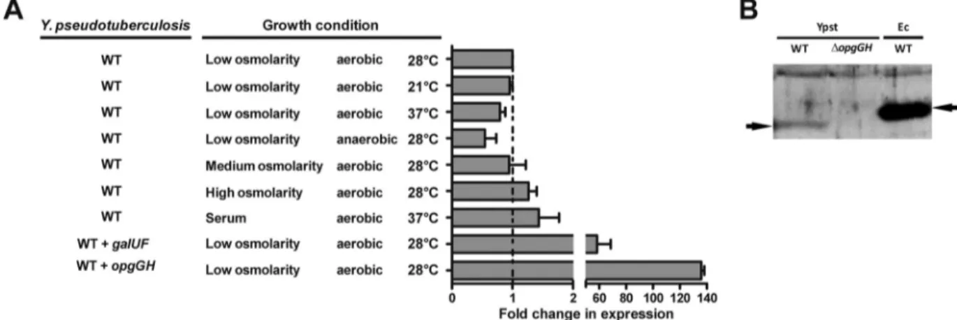
FIG 2 Y. pseudotuberculosis constitutively expresses the opgGH operon under various growth conditions. (A) Fold change expression of opgGH in Y. pseudotu-berculosis (strain 2777) grown under various conditions and in Y. pseudotupseudotu-berculosis harboring a recombinant pBR322 plasmid containing its galUF operon or
a recombinant pUC18 plasmid containing its opgGH operon, compared with the opgGH expression level in Y. pseudotuberculosis grown under low-osmolarity and aerobic conditions and at 28°C. galUF and opgGH were under the control of their own respective promoters. Under each condition, the level of opgGH transcripts was normalized to that of crr transcripts. The data are means and SEM from two independent experiments. (B) Immunoblot of a whole-cell lysate of
Y. pseudotuberculosis (strain 2777), its isogenic⌬opgGH mutant and WT E. coli grown in LB at 30°C. E. coli was used as a positive control. The blot was
immunostained with a polyclonal antibody against E. coli OpgG. Arrowheads indicate the positions of the Y. pseudotuberculosis OpgG protein.
Virulence of Y. pestis containing a copy of opgGH at the att Tn7
chromosomal site was assessed in a mouse model of bubonic
plague and in the Xenopsylla cheopis rat flea model. Regardless of
the presence or absence of opgGH in the Y. pestis genome, all mice
inoculated with
⬃10 CFU succumbed to the disease. A slight
in-crease in the median survival time was observed for animals
inoc-ulated with Y. pestis expressing opgGH (4 days, versus 3 in Y. pestis
not expressing opgGH), although the survival curves were not
sig-nificantly different (P
⬎ 0.3). Consistently, we found that
expres-sion of opgGH did not affect the bacterium’s capability to survive
within macrophages or to grow in normal human serum (see Fig.
S3 in the supplemental material). Furthermore, expression of
opgGH did not affect the bacillus’ capability to block the flea
di-gestive tract (
Fig. 5A
). Accordingly, 27 days after the infected
blood meal, the percentage of infected fleas and the bacterial loads
recovered from the infected fleas did not differ according to the
expression or nonexpression of opgGH (
Fig. 5B
and
C
). These data
are line with the fact that biofilm formation in vitro was not
af-fected in Y. pestis expressing opgGH (biofilm formation, reported
as the mean
⫾ standard error of the mean [SEM], was 89 ⫾ 9.6%
relative to the parental strain, as determined using data from two
independent experiments).
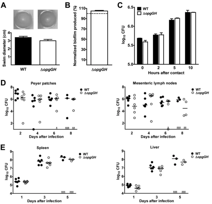
FIG 3 In contrast to other proteobacteria, Y. pseudotuberculosis lacking a functional opgGH operon does not show the pleiotropic phenotype resulting from the
activity of the OPGs through the Rcs signaling pathway. Data on swimming motility on an LB soft agar plate after a 24-hour incubation at 21°C (A), biofilm formation (relative to the WT strain) after a 24-hour incubation at 21°C with shaking in LB supplemented with Ca2⫹and Mg2⫹(B), rates of survival within RAW
264.7 macrophages up to 10 h after internalization (C), the time course of colonization of the Peyer’s patches and mesenteric lymph nodes after intragastric inoculation (109bacteria) (D), and the time course of colonization of the spleen and liver after intravenous inoculation (103bacteria) by the WT strain 2777 and
its derivative⌬opgGH strain (E) are shown. The photos are representative of bacteria after 24 h of swimming. The means and SEM from three independent experiments (A and B) and two independent experiments (C) are shown. The time course of organ colonization was determined from one experiment using groups of six animals (D and E); horizontal lines indicate the medians of the individual data points. Bacterial loads in organs did not vary significantly according to the presence or absence of opgGH (P⬎ 0.05 in a one-way analysis of variance).
DISCUSSION
All of the 20 or so bacterial species studied to date that carry the
opgGH operon or one of its functional homologs (ndvAB, chvAB,
and cgs) have been found to govern the biosynthesis of the glucose
homopolymer OPG. The latter accounts for 0.5% to 10% of the
bacterial dry weight (
22–24
,
32
,
34
,
36
,
40
,
44
,
45
). However, using
the conventional method used to extract OPGs from all the
bac-terial species studied to date, we did not detect any OPGs (i) in Y.
pseudotuberculosis or its culture supernatant after bacterial growth
under various growth conditions, (ii) when the bacterium
artifi-cially overexpressed its own opgGH operon or that of E. coli, (iii)
when Y. pseudotuberculosis overproduced the Opg enzymes’
sub-strate (UDP-Glc) via the overexpression of galUF, or (iv) when the
bacterium expressed the acpP gene from E. coli, which encodes a
cofactor required for OPG biosynthesis. Lastly, several cellular
processes (motility, biofilm formation, and virulence) known to
be controlled by OPGs via the Rcs signaling pathway in E. coli
and/or D. dadantii (
24
,
38
) were unaffected in a
⌬opgGH Y.
pseu-dotuberculosis strain. However, the Rcs system is known to control
biofilm formation and motility in Y. pseudotuberculosis (
54
).
Taken as a whole, these data suggest that Y. pseudotuberculosis has
lost the capability to produce OPGs.
One can legitimately wonder why Y. pseudotuberculosis
(prob-ably) does not produce OPGs, considering that (i) our present
data and data from the KEGG database suggest that the bacterium
produces the Opg enzymes, the ACP cofactor, and the UDP-Glc
substrate (following glucose incorporation or glucogenesis) and
(ii) the opgGH operon from Y. pseudotuberculosis encodes
func-FIG 4 Y. pestis is smaller than Y. pseudotuberculosis when grown at 37°C (but
not at 21°C), presumably because the bacterium lost opgGH during its emer-gence. The lengths of Y. pseudotuberculosis WT strain 2777, the⌬opgGH strain, the complemented mutant, and Y. pestis KIM6⫹ expressing opgGH from Y.
pseudotuberculosis (⫹opgGH) or not (WT) were measured during
exponen-tial-phase growth in LB supplemented with glucose at 21°C and at 37°C. The data are means and SEM from five independent experiments. Regardless of the growth temperature, the⌬opgGH Y. pseudotuberculosis strain (but not the complemented mutant strain) was significantly smaller than the WT strain (P⬍ 0.05 in one way analysis of variance). When grown at 37°C, Y. pestis was significantly smaller than Y. pseudotuberculosis except when it expressed
opgGH from Y. pseudotuberculosis (P⬍ 0.05 in one way analysis of variance). *, P⬍ 0.05.
FIG 5 Y. pestis expressing opgGH from Y. pseudotuberculosis is fully competent in fleas. (A) Proportion of fleas became blocked in the 4 weeks following an
infected blood meal; (B) proportion of infected fleas; (C) bacterial loads immediately after and 27 days after the infectious meal. Experiments were performed with Y. pestis WT KIM6⫹ (WT) and Y. pestis KIM6⫹ harboring a chromosomally integrated mini-Tn7 encompassing the opgGH operon from Y.
pseudotuber-culosis (⫹opgGH). Data in panels A and B are means and standard deviations (SD) from two independent experiments. In panel C, the data correspond to samples
from two independent experiments. Each circle indicates the bacterial load of an individual flea, and the horizontal lines indicate the median CFU per flea.
tional glucans in E. coli and D. dadantii. On one hand, Y.
pseudo-tuberculosis might produce OPGs under particular conditions or
might lack an essential factor for OPG biosynthesis. The latter
might be a polyprenyl-phosphate carrier because this conserved
molecule has a key role in glycan biosynthesis in several kingdoms,
and it has been suggested that UPD-Glc has to be transferred to a
polyprenyl-phosphate carrier prior to the assembly of OPGs (
55
,
56
). On the other hand, Y. pseudotuberculosis may constitutively
accumulate certain ionic and/or nonionic solutes potentially
in-volved in controlling glycosyltransferase activity in response to
osmotic stress (
57
,
58
). However, we found that 100-fold
overex-pression of the opgGH operon did not bypass this putative
repres-sion. Furthermore, the screening of
⬃1,000 strains from a Tn5
mutant library of Y. pseudotuberculosis for OPG synthesis did not
lead to the identification of this type of inhibitor (unpublished
data). Hence, the data support the hypothesis whereby Y.
pseudo-tuberculosis lacks a functional intermediate that is essential for
OPG biosynthesis.
The apparent absence of OPGs in both Y. pseudotuberculosis
and Y. pestis (the latter being naturally devoid of opgGH) raises the
question of how these species compensate for the lack of OPGs.
Suppressor mutations that rescue phenotypes associated with loss
of OPGs have been identified in E. coli, D. dadantii, and members
of the Rhizobiaceae (
35–38
,
59–61
). All of these mutations affect
regulatory systems, including OmpR-EnvZ, RcsCDB, and a
phos-phodiesterase regulating cyclic di-GMP signaling. However, none
of the suppressor mutations completely alleviates the loss of
OPGs. Hence, Y. pseudotuberculosis and Y. pestis may have several
suppressor mutations, i.e., a very different regulatory network
compared with the above-mentioned bacteria. Consistently, Y.
pestis has altered Rcs and cyclic di-GMP signaling pathways and
does not control ompF and ompC (involved in bacterial
adapta-tion to osmotic condiadapta-tions) in the same way as E. coli and D.
dadantii (
13
,
62
,
63
). However, whatever applies to Y. pestis might
not apply to Y. pseudotuberculosis, since the latter has a functional
Rcs system and does not control its c-di-GMP signaling pathways
in the same way as the former. Differences in the membrane
com-position in Y. pseudotuberculosis and Y. pestis (relative to E. coli
and D. dadantii) may constitute an alternative and/or additional
explanation (
64
). Indeed, membrane composition has a role in
bacterial adaptation to the environment (
65
,
66
). Lastly, Y.
pseu-dotuberculosis and Y. pestis may produce glucans that are
equiva-lent to OPGs, such as the “free oligosaccharides” described in
Campylobacter jejuni (
67
).
In agreement with a previous report on E. coli (
46
), we found
that OpgH controls the size of Y. pseudotuberculosis. Consistently,
Y. pestis (which naturally lacks opgGH) is smaller than Y.
pseudo-tuberculosis but was the same size as its recent ancestor when it
expressed opgGH. However, Y. pestis and Y. pseudotuberculosis
dif-fered in size when cultured at 37°C but not when cultured at 21°C,
presumably because Y. pestis has a compensatory mechanism.
These results indicate that the shape control mechanism has
evolved in Y. pestis, potentially providing a selective advantage.
Indeed, it has been found that a particular cell shape can maximize
the use of available nutrients and promote biofilm formation in
vitro and virulence (e.g., the ability to escape detection and resist
phagocytes and complement) (
68–73
). However, opgH expression
did not influence the virulence of Y. pestis in mice or its ability to
colonize and block fleas. In agreement with the virulence data for
Y. pestis, Y. pseudotuberculosis lacking OpgH was able to colonize
mice normally.
Lastly, our results suggest an evolutionary scenario in which an
ancestral strain of Yersinia lost (or did not acquire) a cofactor that
was important for OPG biosynthesis. However, opgGH was
ini-tially retained, presumably because the ability to control bacterial
size provides an advantage in the external environment. We
sug-gest that the opgGH gene was subsequently lost because the
OpgH-dependent size control mechanism was no longer necessary or was
replaced by another control mechanism. During this evolutionary
process, several events (including regulatory network
reprogram-ming) presumably occurred so as to rescue the essential cellular
functions associated with OPGs synthesis and the
OpgH-depen-dent size-control mechanism iOpgH-depen-dentified in other bacteria.
How-ever, there are other possible explanations for the loss of opgGH.
Indeed, we cannot rule out the possibility that Y.
pseudotubercu-losis synthesizes OPGs in order to survive under specific
condi-tions that have not been identified in other bacteria. Consistently,
genomic analysis indicates that Y. enterocolitica (the third
human-pathogenic Yersinia sp.) and all environmental Yersinia species
(for which the genomes have been sequenced) have an opgGH
operon. Hence, Y. pestis may also have lost the opgGH operon
because it no longer encounters conditions under which OPG
synthesis is required for survival.
ACKNOWLEDGMENTS
We thank Rodolph Beaubois for his help in screening the mutant library. This work was funded by the Institut National de la Santé et de la Recherche Médicale, the Institut Pasteur de Lille, the Université Lille Nord de France, the Centre National de la Recherche Scientifique, the Région Nord-Pas de Calais, and the European Regional Development Fund as part of the “Action de recherche concertée d’initiative régionale” program (grant 10090077-Présage 35712 to F.S.).
REFERENCES
1. Butler T. 1994. Yersinia infections: centennial of the discovery of the plague bacillus. Clin Infect Dis 19:655– 661.http://dx.doi.org/10.1093 /clinids/19.4.655.
2. Bacot AW, Martin CJ. 1914. Observations on the mechanism of the transmission of plague by fleas. J Hyg Plague 13(Plague Suppl 3):423– 439. 3. Sebbane F, Jarrett CO, Gardner D, Long D, Hinnebusch BJ. 2006. Role of the Yersinia pestis plasminogen activator in the incidence of distinct sep-ticemic and bubonic forms of flea-borne plague. Proc Natl Acad Sci U S A
103:5526 –5530.http://dx.doi.org/10.1073/pnas.0509544103.
4. Pujol C, Bliska JB. 2005. Turning Yersinia pathogenesis outside in: sub-version of macrophage function by intracellular yersiniae. Clin Immunol
114:216 –226.http://dx.doi.org/10.1016/j.clim.2004.07.013.
5. Sebbane F, Gardner D, Long D, Gowen BB, Hinnebusch BJ. 2005. Kinetics of disease progression and host response in a rat model of bu-bonic plague. Am J Pathol 166:1427–1439. http://dx.doi.org/10.1016 /S0002-9440(10)62360-7.
6. Achtman M, Morelli G, Zhu P, Wirth T, Diehl I, Kusecek B, Vogler AJ,
Wagner DM, Allender CJ, Easterday WR, Chenal-Francisque V, Wor-sham P, Thomson NR, Parkhill J, Lindler LE, Carniel E, Keim P. 2004.
Microevolution and history of the plague bacillus, Yersinia pestis. Proc Natl Acad Sci U S A 101:17837–17842.http://dx.doi.org/10.1073/pnas .0408026101.
7. Chain PS, Carniel E, Larimer FW, Lamerdin J, Stoutland PO, Regala
WM, Georgescu AM, Vergez LM, Land ML, Motin VL, Brubaker RR, Fowler J, Hinnebusch J, Marceau M, Medigue C, Simonet M, Che-nal-Francisque V, Souza B, Dacheux D, Elliott JM, Derbise A, Hauser LJ, Garcia E. 2004. Insights into the evolution of Yersinia pestis
through whole-genome comparison with Yersinia pseudotuberculosis. Proc Natl Acad Sci U S A 101:13826 –13831.http://dx.doi.org/10.1073/pnas .0404012101.
hemin storage (hms) locus in the transmission of plague by fleas. Science
273:367–370.http://dx.doi.org/10.1126/science.273.5273.367.
9. Hinnebusch BJ, Rudolph AE, Cherepanov P, Dixon JE, Schwan TG,
Forsberg A. 2002. Role of Yersinia murine toxin in survival of Yersinia pestis in the midgut of the flea vector. Science 296:733–735.http://dx.doi .org/10.1126/science.1069972.
10. Kutyrev V, Mehigh RJ, Motin VL, Pokrovskaya MS, Smirnov GB,
Brubaker RR. 1999. Expression of the plague plasminogen activator in Yersinia pseudotuberculosis and Escherichia coli. Infect Immun 67:1359 –
1367.
11. Sebbane F, Jarrett C, Gardner D, Long D, Hinnebusch BJ. 2009. The
Yersinia pestis caf1M1A1 fimbrial capsule operon promotes transmission
by flea bite in a mouse model of bubonic plague. Infect Immun 77:1222– 1229.http://dx.doi.org/10.1128/IAI.00950-08.
12. Erickson DL, Jarrett CO, Callison JA, Fischer ER, Hinnebusch BJ. 2008. Loss of a biofilm-inhibiting glycosyl hydrolase during the emergence of
Yersinia pestis. J Bacteriol 190:8163– 8170.http://dx.doi.org/10.1128/JB .01181-08.
13. Sun YC, Jarrett CO, Bosio CF, Hinnebusch BJ. 2014. Retracing the evolutionary path that led to flea-borne transmission of Yersinia pestis. Cell Host Microbe 15:578 –586.http://dx.doi.org/10.1016/j.chom.2014 .04.003.
14. Tang L, Weissborn AC, Kennedy EP. 1997. Domains of Escherichia coli acyl carrier protein important for membrane-derived-oligosaccharide biosynthesis. J Bacteriol 179:3697–3705.
15. Miller KJ, Kennedy EP, Reinhold VN. 1986. Osmotic adaptation by gram-negative bacteria: possible role for periplasmic oligosaccharides. Science 231:48 –51.http://dx.doi.org/10.1126/science.3941890. 16. Beaudoin T, Zhang L, Hinz AJ, Parr CJ, Mah TF. 2012. The
biofilm-specific antibiotic resistance gene ndvB is important for expression of eth-anol oxidation genes in Pseudomonas aeruginosa biofilms. J Bacteriol 194: 3128 –3136.http://dx.doi.org/10.1128/JB.06178-11.
17. Bhagwat AA, Jun W, Liu L, Kannan P, Dharne M, Pheh B, Tall BD,
Kothary MH, Gross KC, Angle S, Meng J, Smith A. 2009.
Osmoregu-lated periplasmic glucans of Salmonella enterica serovar Typhimurium are required for optimal virulence in mice. Microbiology 155:229 –237.http: //dx.doi.org/10.1099/mic.0.023747-0.
18. Roset MS, Ciocchini AE, Ugalde RA, Inon de Iannino N. 2006. The
Brucella abortus cyclic beta-1,2-glucan virulence factor is substituted with
O-ester-linked succinyl residues. J Bacteriol 188:5003–5013.http://dx.doi .org/10.1128/JB.00086-06.
19. Salanoubat M, Genin S, Artiguenave F, Gouzy J, Mangenot S, Arlat M,
Billault A, Brottier P, Camus JC, Cattolico L, Chandler M, Choisne N, Claudel-Renard C, Cunnac S, Demange N, Gaspin C, Lavie M, Moisan A, Robert C, Saurin W, Schiex T, Siguier P, Thebault P, Whalen M, Wincker P, Levy M, Weissenbach J, Boucher CA. 2002. Genome
se-quence of the plant pathogen Ralstonia solanacearum. Nature 415:497– 502.http://dx.doi.org/10.1038/415497a.
20. Zorreguieta A, Ugalde RA. 1986. Formation in Rhizobium and
Agrobac-terium spp. of a 235-kilodalton protein intermediate in beta-D(1-2)
glu-can synthesis. J Bacteriol 167:947–951.
21. Cogez V, Gak E, Puskas A, Kaplan S, Bohin JP. 2002. The opgGIH and
opgC genes of Rhodobacter sphaeroides form an operon that controls
back-bone synthesis and succinylation of osmoregulated periplasmic glucans. Eur J Biochem 269:2473–2484. http://dx.doi.org/10.1046/j.1432-1033 .2002.02907.x.
22. Minsavage GV, Mudgett MB, Stall RE, Jones JB. 2004. Importance of
opgHXcv of Xanthomonas campestris pv. vesicatoria in host-parasite
inter-actions. Mol Plant Microbe Interact 17:152–161. http://dx.doi.org/10 .1094/MPMI.2004.17.2.152.
23. Bhagwat AA, Mithofer A, Pfeffer PE, Kraus C, Spickers N, Hotchkiss A,
Ebel J, Keister DL. 1999. Further studies of the role of cyclic beta-glucans
in symbiosis. An NdvC mutant of Bradyrhizobium japonicum synthesizes cyclodecakis-(1¡3)-beta-glucosyl. Plant Physiol 119:1057–1064. 24. Bontemps-Gallo S, Madec E, Dondeyne J, Delrue B, Robbe-Masselot C,
Vidal O, Prouvost AF, Boussemart G, Bohin JP, Lacroix JM. 2013.
Concentration of osmoregulated periplasmic glucans (OPGs) modulates the activation level of the RcsCD RcsB phosphorelay in the phytopathogen bacteria Dickeya dadantii. Environ Microbiol 15:881– 894.http://dx.doi .org/10.1111/1462-2920.12054.
25. Komaniecka I, Choma A. 2003. Isolation and characterization of periplasmic cyclic beta-glucans of Azorhizobium caulinodans. FEMS Microbiol Lett 227: 263–269.http://dx.doi.org/10.1016/S0378-1097(03)00690-6.
26. Altabe SG, Inon de Iannino N, de Mendoza D, Ugalde RA. 1994. New osmoregulated beta(1-3),beta(1-6) glucosyltransferase(s) in Azospirillum
brasilense. J Bacteriol 176:4890 – 4898.
27. Bohin JP. 2000. Osmoregulated periplasmic glucans in Proteobacteria. FEMS Microbiol Lett 186:11–19.http://dx.doi.org/10.1111/j.1574-6968 .2000.tb09075.x.
28. Lacroix JM, Bohin JP. 2010. Osmoregulated periplasmic glucan polym-erization requires constant protein synthesis in Escherichia coli. Curr Mi-crobiol 61:396 – 400.http://dx.doi.org/10.1007/s00284-010-9625-2. 29. Therisod H, Weissborn AC, Kennedy EP. 1986. An essential function for
acyl carrier protein in the biosynthesis of membrane-derived oligosaccha-rides of Escherichia coli. Proc Natl Acad Sci U S A 83:7236 –7240.http://dx .doi.org/10.1073/pnas.83.19.7236.
30. Briones G, Inon de Iannino N, Steinberg M, Ugalde RA. 1997. Periplas-mic cyclic 1,2-beta-glucan in Brucella spp. is not osmoregulated. Micro-biology 143:1115–1124.http://dx.doi.org/10.1099/00221287-143-4-1115. 31. Talaga P, Fournet B, Bohin JP. 1994. Periplasmic glucans of
Pseudomo-nas syringae pv. syringae. J Bacteriol 176:6538 – 6544.
32. Breedveld MW, Miller KJ. 1994. Cyclic beta-glucans of members of the family Rhizobiaceae. Microbiol Rev 58:145–161.
33. Kennedy EP. 1982. Osmotic regulation and the biosynthesis of membrane-derived oligosaccharides in Escherichia coli. Proc Natl Acad Sci U S A 79: 1092–1095.http://dx.doi.org/10.1073/pnas.79.4.1092.
34. Arellano-Reynoso B, Lapaque N, Salcedo S, Briones G, Ciocchini AE,
Ugalde R, Moreno E, Moriyon I, Gorvel JP. 2005. Cyclic beta-1,2-glucan
is a Brucella virulence factor required for intracellular survival. Nat Im-munol 6:618 – 625.http://dx.doi.org/10.1038/ni1202.
35. Bouchart F, Delangle A, Lemoine J, Bohin JP, Lacroix JM. 2007. Proteomic analysis of a non-virulent mutant of the phytopathogenic bac-terium Erwinia chrysanthemi deficient in osmoregulated periplasmic glu-cans: change in protein expression is not restricted to the envelope, but affects general metabolism. Microbiology 153:760 –767.http://dx.doi.org /10.1099/mic.0.2006/000372-0.
36. Kannan P, Dharne M, Smith A, Karns J, Bhagwat AA. 2009. Motility revertants of opgGH mutants of Salmonella enterica serovar Typhimurium remain defective in mice virulence. Curr Microbiol 59:641– 645.http://dx .doi.org/10.1007/s00284-009-9486-8.
37. Fineran PC, Williamson NR, Lilley KS, Salmond GP. 2007. Virulence and prodigiosin antibiotic biosynthesis in Serratia are regulated pleiotro-pically by the GGDEF/EAL domain protein, PigX. J Bacteriol 189:7653– 7662.http://dx.doi.org/10.1128/JB.00671-07.
38. Girgis HS, Liu Y, Ryu WS, Tavazoie S. 2007. A comprehensive genetic characterization of bacterial motility. PLoS Genet 3:1644 –1660. 39. Mukhopadhyay P, Williams J, Mills D. 1988. Molecular analysis of a
pathogenicity locus in Pseudomonas syringae pv. syringae. J Bacteriol 170: 5479 –5488.
40. Mahajan-Miklos S, Tan MW, Rahme LG, Ausubel FM. 1999. Molecular mechanisms of bacterial virulence elucidated using a Pseudomonas
aerugi-nosa-Caenorhabditis elegans pathogenesis model. Cell 96:47–56.http://dx .doi.org/10.1016/S0092-8674(00)80958-7.
41. Banta LM, Bohne J, Lovejoy SD, Dostal K. 1998. Stability of the
Agro-bacterium tumefaciens VirB10 protein is modulated by growth
tempera-ture and periplasmic osmoadaption. J Bacteriol 180:6597– 6606. 42. Mah TF, Pitts B, Pellock B, Walker GC, Stewart PS, O’Toole GA. 2003.
A genetic basis for Pseudomonas aeruginosa biofilm antibiotic resistance. Nature 426:306 –310.http://dx.doi.org/10.1038/nature02122.
43. Lapaque N, Forquet F, de Chastellier C, Mishal Z, Jolly G, Moreno E,
Moriyon I, Heuser JE, He HT, Gorvel JP. 2006. Characterization of Brucella abortus lipopolysaccharide macrodomains as mega rafts. Cell
Mi-crobiol 8:197–206.http://dx.doi.org/10.1111/j.1462-5822.2005.00609.x. 44. Dylan T, Ielpi L, Stanfield S, Kashyap L, Douglas C, Yanofsky M, Nester
E, Helinski DR, Ditta G. 1986. Rhizobium meliloti genes required for
nodule development are related to chromosomal virulence genes in
Agro-bacterium tumefaciens. Proc Natl Acad Sci U S A 83:4403– 4407.http://dx .doi.org/10.1073/pnas.83.12.4403.
45. de Iannino NI, Ugalde RA. 1989. Biochemical characterization of aviru-lent Agrobacterium tumefaciens chvA mutants: synthesis and excretion of beta-(1-2)glucan. J Bacteriol 171:2842–2849.
46. Hill NS, Buske PJ, Shi Y, Levin PA. 2013. A moonlighting enzyme links
Escherichia coli cell size with central metabolism. PLoS Genet 9:e1003663. http://dx.doi.org/10.1371/journal.pgen.1003663.
47. Young KD. 2007. Bacterial morphology: why have different shapes? Curr Opin Microbiol 10:596 – 600.http://dx.doi.org/10.1016/j.mib.2007.09.009.
48. Sebbane F, Jarrett CO, Linkenhoker JR, Hinnebusch BJ. 2004. Evalua-tion of the role of constitutive isocitrate lyase activity in Yersinia pestis infection of the flea vector and mammalian host. Infect Immun 72:7334 – 7337.http://dx.doi.org/10.1128/IAI.72.12.7334-7337.2004.
49. Choi KH, Gaynor JB, White KG, Lopez C, Bosio CM,
Karkhoff-Schweizer RR, Karkhoff-Schweizer HP. 2005. A Tn7-based broad-range bacterial
cloning and expression system. Nat Methods 2:443– 448.http://dx.doi.org /10.1038/nmeth765.
50. Dische Z. 1962. General color reactions. Methods Carbohydr Chem
1:478 – 492.
51. Reboul A, Lemaitre N, Titecat M, Merchez M, Deloison G, Ricard I,
Pradel E, Marceau M, Sebbane F. 2014. Yersinia pestis requires the
2-component regulatory system OmpR-EnvZ to resist innate immunity during the early and late stages of plague. J Infect Dis 210:1367–1375.
http://dx.doi.org/10.1093/infdis/jiu274.
52. Sebbane F, Bury-Mone S, Cailliau K, Browaeys-Poly E, De Reuse H,
Simonet M. 2002. The Yersinia pseudotuberculosis Yut protein, a new type
of urea transporter homologous to eukaryotic channels and functionally interchangeable in vitro with the Helicobacter pylori UreI protein. Mol Microbiol 45:1165–1174.http://dx.doi.org/10.1046/j.1365-2958.2002 .03096.x.
53. Marolda CL, Valvano MA. 1996. The GalF protein of Escherichia coli is not a UDP-glucose pyrophosphorylase but interacts with the GalU pro-tein possibly to regulate cellular levels of UDP-glucose. Mol Microbiol
22:827– 840.http://dx.doi.org/10.1046/j.1365-2958.1996.01531.x. 54. Hinchliffe SJ, Howard SL, Huang YH, Clarke DJ, Wren BW. 2008. The
importance of the Rcs phosphorelay in the survival and pathogenesis of the enteropathogenic yersiniae. Microbiology 154:1117–1131.http://dx .doi.org/10.1099/mic.0.2007/012534-0.
55. Weissborn AC, Rumley MK, Kennedy EP. 1991. Biosynthesis of mem-brane-derived oligosaccharides. Membrane-bound glucosyltransferase system from Escherichia coli requires polyprenyl phosphate. J Biol Chem
266:8062– 8067.
56. Hartley MD, Imperiali B. 2012. At the membrane frontier: a prospectus on the remarkable evolutionary conservation of polyprenols and polypre-nyl-phosphates. Arch Biochem Biophys 517:83–97.http://dx.doi.org/10 .1016/j.abb.2011.10.018.
57. Rumley MK, Therisod H, Weissborn AC, Kennedy EP. 1992. Mecha-nisms of regulation of the biosynthesis of membrane-derived oligosaccha-rides in Escherichia coli. J Biol Chem 267:11806 –11810.
58. Ingram-Smith C, Miller KJ. 1998. Effects of ionic and osmotic strength on the glucosyltransferase of Rhizobium meliloti responsible for cyclic be-ta-(1,2)-glucan biosynthesis. Appl Environ Microbiol 64:1290 –1297. 59. Bontemps-Gallo S, Madec E, Lacroix JM. 2014. Inactivation of pecS
restores the virulence of mutants devoid of osmoregulated periplasmic glucans in the phytopathogenic bacterium Dickeya dadantii. Microbiology
160:766 –777.http://dx.doi.org/10.1099/mic.0.074484-0.
60. Dylan T, Nagpal P, Helinski DR, Ditta GS. 1990. Symbiotic pseudor-evertants of Rhizobium meliloti ndv mutants. J Bacteriol 172:1409 –1417.
61. Nagpal P, Khanuja SP, Stanfield SW. 1992. Suppression of the ndv mutant phenotype of Rhizobium meliloti by cloned exo genes. Mol Micro-biol 6:479 – 488.http://dx.doi.org/10.1111/j.1365-2958.1992.tb01492.x. 62. Gao H, Zhang Y, Han Y, Yang L, Liu X, Guo Z, Tan Y, Huang X, Zhou
D, Yang R. 2011. Phenotypic and transcriptional analysis of the osmotic
regulator OmpR in Yersinia pestis. BMC Microbiol 11:39.http://dx.doi .org/10.1186/1471-2180-11-39.
63. Sun YC, Hinnebusch BJ, Darby C. 2008. Experimental evidence for negative selection in the evolution of a Yersinia pestis pseudogene. Proc Natl Acad Sci U S A 105:8097– 8101.http://dx.doi.org/10.1073/pnas .0803525105.
64. Rebeil R, Ernst RK, Gowen BB, Miller SI, Hinnebusch BJ. 2004. Vari-ation in lipid A structure in the pathogenic yersiniae. Mol Microbiol 52: 1363–1373.http://dx.doi.org/10.1111/j.1365-2958.2004.04059.x. 65. Roy H, Dare K, Ibba M. 2009. Adaptation of the bacterial membrane to
changing environments using aminoacylated phospholipids. Mol Micro-biol 71:547–550.http://dx.doi.org/10.1111/j.1365-2958.2008.06563.x. 66. Klein G, Lindner B, Brabetz W, Brade H, Raina S. 2009. Escherichia coli
K-12 suppressor-free mutants lacking early glycosyltransferases and late acyltransferases: minimal lipopolysaccharide structure and induction of envelope stress response. J Biol Chem 284:15369 –15389.http://dx.doi.org /10.1074/jbc.M900490200.
67. Nothaft H, Liu X, McNally DJ, Li J, Szymanski CM. 2009. Study of free oligosaccharides derived from the bacterial N-glycosylation pathway. Proc Natl Acad Sci U S A 106:15019 –15024.http://dx.doi.org/10.1073/pnas .0903078106.
68. Singh SP, Montgomery BL. 2011. Determining cell shape: adaptive reg-ulation of cyanobacterial cellular differentiation and morphology. Trends Microbiol 19:278 –285.http://dx.doi.org/10.1016/j.tim.2011.03.001. 69. Cho H, Jonsson H, Campbell K, Melke P, Williams JW, Jedynak B,
Stevens AM, Groisman A, Levchenko A. 2007. Self-organization in
high-density bacterial colonies: efficient crowd control. PLoS Biol 5:e302.
http://dx.doi.org/10.1371/journal.pbio.0050302.
70. Chaput C, Ecobichon C, Cayet N, Girardin SE, Werts C, Guadagnini S,
Prevost MC, Mengin-Lecreulx D, Labigne A, Boneca IG. 2006. Role of
AmiA in the morphological transition of Helicobacter pylori and in im-mune escape. PLoS Pathog 2:e97.http://dx.doi.org/10.1371/journal.ppat .0020097.
71. Weiser JN. 2013. The battle with the host over microbial size. Curr Opin Microbiol 16:59 – 62.http://dx.doi.org/10.1016/j.mib.2013.01.001. 72. Justice SS, Hung C, Theriot JA, Fletcher DA, Anderson GG, Footer MJ,
Hultgren SJ. 2004. Differentiation and developmental pathways of
uro-pathogenic Escherichia coli in urinary tract pathogenesis. Proc Natl Acad Sci U S A 101:1333–1338.http://dx.doi.org/10.1073/pnas.0308125100. 73. Dalia AB, Weiser JN. 2011. Minimization of bacterial size allows for
complement evasion and is overcome by the agglutinating effect of anti-body. Cell Host Microbe 10:486 – 496.http://dx.doi.org/10.1016/j.chom .2011.09.009.