HAL Id: inserm-02304793
https://www.hal.inserm.fr/inserm-02304793
Submitted on 3 Oct 2019
HAL is a multi-disciplinary open access
archive for the deposit and dissemination of
sci-entific research documents, whether they are
pub-lished or not. The documents may come from
teaching and research institutions in France or
abroad, or from public or private research centers.
L’archive ouverte pluridisciplinaire HAL, est
destinée au dépôt et à la diffusion de documents
scientifiques de niveau recherche, publiés ou non,
émanant des établissements d’enseignement et de
recherche français ou étrangers, des laboratoires
publics ou privés.
First case report of Cohen syndrome in the Tunisian
population caused by VPS13B mutations
Imen Rejeb, Houweyda Jilani, Yasmina Elaribi, Syrine Hizem, Lamia Hila,
Julia Zillahrdt, Jamel Chelly, Lamia Benjemaa
To cite this version:
Imen Rejeb, Houweyda Jilani, Yasmina Elaribi, Syrine Hizem, Lamia Hila, et al.. First case report of
Cohen syndrome in the Tunisian population caused by VPS13B mutations. BMC Medical Genetics,
BioMed Central, 2017, 18 (1), pp.134. �10.1186/s12881-017-0493-5�. �inserm-02304793�
C A S E R E P O R T
Open Access
First case report of Cohen syndrome in the
Tunisian population caused by VPS13B
mutations
Imen Rejeb
1*, Houweyda Jilani
1, Yasmina Elaribi
1, Syrine Hizem
1, Lamia Hila
2, Julia Lauer Zillahrdt
3,4,5,
Jamel Chelly
3,4,5and Lamia Benjemaa
1Abstract
Background: Cohen syndrome is a rare autosomal recessive developmental disorder that comprises variable clinical features counting developmental delay, pigmentary retinopathy, myopia, acquired microcephaly, truncal obesity, joint hypermobility, friendly disposition and intermittent neutropenia.VPS13B (vacuolar protein sorting 13, yeast, homologue of B) gene is the only gene responsible for Cohen Syndrome, causative mutations include nonsense, missense, indel and splice-site variants. The integrity of the Golgi apparatus requires the presence of the peripheral membrane protein VPS13B that have an essential function in intracellular protein transport and vesicle-mediated sorting.
Case presentation: In this study, we performed whole exome sequencing (WES) in a Tunisian family with two young cases having developmental delay, hypotonia, autism spectrum disorder, ptosis and thick hair and eyebrows. The proposita presented also pigmentory retinopathy. Compound heterozygous mutation inVPS13B gene was detected by WES. This mutation inherited from healthy heterozygous parents, supports an unpredictable clinical diagnosis of Cohen Syndrome. The proband’s phenotype is explained by the presence of compound heterozygous mutations in the VPS13B gene. This finding refined the understanding of genotype-phenotype correlation.
Conclusions: This is the first report of a Tunisian family with Cohen syndrome mutated in theVPS13B gene. Keywords: Cohen syndrome,VPS13B gene, Compound heterozygous mutation
Background
Cohen syndrome (CS) (MIM# 216550) is a rare autosomal recessive developmental disorder characterized by Cohen and colleagues in 1973 [1]. Truncal obesity, intellectual disability, developmental delay, joint laxity, craniofacial dysmorphism, high myopia and/or retinal dystrophy and neutropenia are typical clinical manifestations of the syn-drome [1, 2].At present, CS has been essentially assigned to mutations in theVPS13B gene (MIM# 607817) among patients from diverse ethnicity. VPS13B, the single CS linked gene so far described, is localized on q22.2 locus of chromosome 8. Its length is about 864 kb and comprises 62 exons. The longest transcript [NM_017890.4] is 14,100 bp long encoding for a 4022 amino acid protein.
VPS13B is a peripheral membrane protein with putative transmembrane domains and functional motifs that have an essential function in the transport of intracellular pro-teins and in vesicle-mediated sorting [3]. The expression of the VPS13B is mainly noticed in the whole body and in the central nervous system, blood, muscles, and heart [4]. Approximately, 200 cases of the CS and about more than 150 deleterious mutations have been identified to date (http://www.hgmd.org); in most cases mutations are stop codon mutations that result in a functionally null protein. The diagnosis is always difficult in childhood, this is due to the fact that many of the typical traits may be nonexis-tent till scholarisation or upcoming years and intermitnonexis-tent neutropenia is not consistently observable.
Here we report the characterization of a new compound heterozygous mutation in VPS13B gene in 2 Tunisian related cases with CS.
* Correspondence:imen_rejeb@yahoo.fr;imen.rejeb@rns.tn
1Service des Maladies Congénitales et Héréditaires, CHU Mongi Slim La Marsa, Sidi Daoud La Marsa, 2046 Tunis, Tunisia
Full list of author information is available at the end of the article
© The Author(s). 2017 Open Access This article is distributed under the terms of the Creative Commons Attribution 4.0 International License (http://creativecommons.org/licenses/by/4.0/), which permits unrestricted use, distribution, and reproduction in any medium, provided you give appropriate credit to the original author(s) and the source, provide a link to the Creative Commons license, and indicate if changes were made. The Creative Commons Public Domain Dedication waiver (http://creativecommons.org/publicdomain/zero/1.0/) applies to the data made available in this article, unless otherwise stated.
Case presentation
The present study describes clinical and molecular findings in two patients with CS from a non-consanguineous Tunis-ian family. One of the authors examined the patients. DNA was extracted from peripheral blood using standard methods. The patients’ parents gave their consent.
The proposita was the first child of non-consanguineous and healthy condition parents. Pregnancy was normal and carried to term with an APGAR score of 9 and 10 at one and five minutes after birth, respectively. The parameters at birth were: weight 3300 g (60th percentile), height 50 cm (70 th percentile), occipitofrontal circumference (OFC) 35 cm (85th percentile). Hypotonia and poor sucking were noticed. She presented delayed psychomotor development: she wasn’t capable to sit without help till 12 months and walked at the age of 2 years. At 3 years, the patient was not capable to speak. Ophthalmological evaluation showed left strabismus and pigmentary retinopathy.
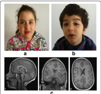
When reevaluated at 12 years, growth retardation and progressive microcephaly were noted. We noticed a weight of 31 kg (−1 SD), a height of 126 cm (−3.2 SD) and an OFC of 48 cm (−3.8SD). At neurological assessment, we noticed widespread hypotonia and joint hypermobility. Clinical diagnosis revealed dysmorphic facial features as thick hair eye brows and lashes, prominent upper central incisors, prominent lips and short philtrum; the last two features lead to a half open-mouth. The hands were small with ta-pering fingers. She also presented truncal obesity (Fig. 1). Communication and social skills were impaired. She presented intellectual disability with autistic-like traits.
Cerebral MRI showed thick and dysmorphic corpus cal-losum and lateral ventricular asymmetry. The brainstem and the cerebellum were normal (Fig. 1c). Karyotype and CGH array were normal.
Her brother presented at the age of 6 years a weight of 22 kg (normal), a height of 113 cm (normal) and an OFC of 48 cm (−3SD). Birth parameters were: weight 3350 g (50th percentile), height 50 cm (50th percentile), occipitofrontal circumference (OFC) 36 cm (85th percentile). Pregnancy was normal and carried to term with an APGAR score of 10 and 10 at one and five minutes after birth, respectively. He was born with a ptosis in the left eye operated at the age of 5 years (Fig. 1). He presented high myopia. He had, like his sister, thick hair eyebrows and lashes, he presented downslanted palpebral fissures, micrognathia, arched palate, clinodactyly of the toes, slender hands and feet and tapering fingers. Intellectual disability and stereotyped motor behav-ior were also noticed.
After the identification of the causal mutation, the reverse phenotyping showed moderate neutropenia and mild neutropenia in the proposita and her brother respectively. In fact, neutropenia is one of the important clinical signs of CS, but because of the nonexistence of clinical signs and the occasional occurrence, it is rarely identified.
The CARE guidelines were followed.
Genetic testing
Because of limitations of renseignement on neutropenia and pigmentary retinopathy we didn’t notice that the clinical features matched with a CS, so we first use WES approach. We performed WES in trio made of the proband and his two unrelated parents.
Library generation, exome enrichment and whole-exome sequencing methodology used are detailed in the article written by Poirier et al. [5].
We analyzed variants affecting coding regions and essen-tial splicing sites and excluded all variants with a frequency greater than 1% according to genomic databases (dbSNP, 1000 Genomes, Exome variant server and local platform database). All relevant variant were visually explored with Integrative Genomics Viewer (IGV: http://software.broadin-stitute.org/software/igv/) to detect false positive results.
With this method and these filters, 9 variants were de-tected in index case (7 with de novo model of inheritance and 2 in the same gene with recessive model). 5 out of the 7 “de novo” variants appeared inherited from one parent (IGV), the 2 others were absent in the affected brother. The 2 variations in VPS13B gene (identified by recessive model of analysis) were also present in the brother; the c.3582delT, p.A1194fs were inherited from the mother and the c.6295_6296delAT, p.M2124 fs one from father.
Finally, we confirmed by PCR and Sanger sequencing of all coding regions and exon-intron boundaries of the VPS13B gene the relevant variants identified by WES. Using genomic
a
b
c
Fig. 1 a The proband. b the brother of the proband. c Proband’s MRI (1) sections showing dysmorphic and thick corpus callosum and (2, 3) the lateral ventricular asymmetry
DNA from the proband, her parents and her brother, the mutations identified were tested for familial segregation.
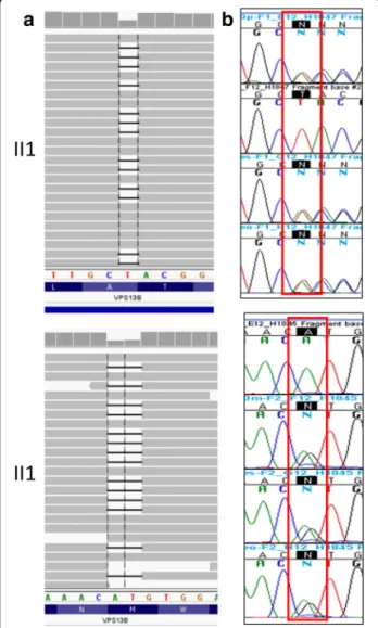
WES of individual II1 noticed the presence of a compound heterozygous variant (100,479,778 T/−, 100712001AT/−) in the VPS13B gene (Fig. 2a). This compound heterozygous mutation in theVPS13B gene (NM_017890.4) was validated by Sanger sequencing (c.3582delT, p.A1149fs + c.6295_6 296delAT, p.M2124 fs) (Fig. 2b) inherited from healthy heterozygous parents confirms the diagnosis of CS.
Sanger sequencing showed that the variations c.3582delT (p.A1149fs) and c.6295_6296delAT (p.M2124 fs) in the VPS13B gene present in both the proband and her brother were of biparental origin (Fig. 3).
Discussion and conclusions
We report a Tunisian family including two siblings with developmental delay and intellectual disability harbouring a novel compound heterozygous mutation in theVPS13B gene by WES. Even for an experienced clinician, the diagnosis of CS is difficult. In fact this syndrome is a rare autosomal recessive disorder and it is often impossible to diagnosis CS until middle or late childhood. Therefore the phentotypic traits are very variable, several could be lack-ing till scholarisation or upcomlack-ing years. CS can be retained when six of eight phentotypic traits are noticed including developmental delay, joint hypermobility, typical CS facial gestalt, high myopia and/or retinal dystrophy, microcephaly, truncal obesity with slender extremities, overly sociable behaviour and neutropenia [6]. However some of these features cannot be observable till scholarisa-tion or upcoming years, like retinal dystrophy, truncal obesity and overly sociable behavior, furthermore the typ-ical facial gestalt of prominent incisors is always absent. In fact, when the girl was first seen at 7 years and the boy at 1 year, respectively, they had postnatal-onset microcephaly and delayed developmental milestones, we didn’t suspect the CS at this time given no other revealing traits.
Mutations in theVPS13B gene are responsible for CS. The novel compound heterozygous mutation in VPS13B inher-ited from healthy heterozygous parents, c.3582delT (p.A1149fs) and c.6295_6296delAT (p.M2124 fs) inherited respectively from the father and the mother are present in the proband and her brother causes a frameshift that induce a premature stop codon. Subsequently this frameshift mutation generate a premature stop codon that can either induce a truncated protein lacking a number of functional domains of the VPS13B protein or to a functional null-allele as a result of a non sense mediated mRNA decay (NMD).
Actually, approximately 188 mutations in VPS13B gene have been identified, 153 of them were associated with CS (http://www.hgmd.org/). VPS13B is a Golgi-associated per-ipheral membrane protein co-localizing with the cis-Golgi matrix protein GM130. RP2 and RPGR, two retinitis pigmentosa disease genes, situate to the Golgi, and depletion of RP2 causes abnormal Golgi function and protein transport in the photoreceptor [7, 8]. This highlights that a normal function of Golgi-associated proteins, including VPS13B, is essential for a good functioning of the photoreceptor. Recent studies have concluded that VPS13B mutations, responsible for COH1, are responsible of a tissue-specific major defect of glycosylation and endosomal–lysosomal trafficking defect. This highlights that VPS13B is essential in Golgi glycosyla-tion and morphology, as well as in lysosomal–endosomal pathway maintenance [9].
Using WES in the early stage of disease for young patients where the clinical diagnosis is not evident is actually the best approach. Currently, the exome sequen-cing approaches are used in many laboratories helping
Fig. 2 a Integrative Genomics Viewer of short read alignment indicated the compound heterozygous variant identified by exome sequencing (100,479,778 T/−,100712001AT/−) in the VPS13B gene. b Sanger validation shows the compound heterozygosity of the proband II1 formed by the c.3582delT mutation, and the c.6295_6296delAT mutation
clinicians in solving the aetiology of many rare diseases. This approach, compared to sanger sequencing helps saving costs and time especially for large genes such as VPS13B gene. In fact, the costs of sequencing per base with sanger sequencing is much higher than with NGS [10, 11].So, sanger sequencing will be performed only if a causative mu-tation will be identified by NGS in order to validate this variant. Therefore, for mendelian diseases, especially for those with genetic heterogeneity, NGS is almost certainly the best primary choice in genetic tests.
In conclusion, we report the first Tunisian family with CS, a novel compound heterozygous mutation inVPS13B gene, identified using WES is the deleterious mutation in the patients of this family.
Abbreviations
APGAR:Appearance pulse grimace activity and respiration; CGH: Comparative genomic hybridization; CS: Cohen syndrome; MRI: Magnetic resonance imaging; NGS: Next-Generation Sequencing; NMD: Nonsense-mediated mRNA decay; OFC: Occipitofrontal circumference; PCR: Polymerase chain reaction; WES: Whole exome sequencing
Acknowledgments
We would like to thank the family members for their invaluable cooperation and participation.
Funding Not applicable.
Availability of data and materials
All data generated or analyzed during this study are included in this published article.
Authors’ contribution
LB and JC designed and initiated the study, monitored data collection and analysis for the study and revised the paper. IR contributed to sample collection, analyzed clinical data, and drafted the paper. HJ, YE and SH contributed to collection of clinical and imaging Data and genetic counseling. JLZ analyzed the WES data and revised the paper. LH helped with technical parts and revised the paper. All authors read and approved the final manuscript.
Ethics approval and consent to participate
This study was approved by the Ethics Review Committee CHU Mongi Slim La Marsa in Tunisia and informed consent was obtained from the patients’ parents prior to participation.
Consent for publication
Consent for publication of respective case presentations was obtained from patients’ parents. They give their consent for the publication of the medical data and photos of their son and daughter.
Competing interests
The authors declare that they have no competing interests.
Publisher’s Note
Springer Nature remains neutral with regard to jurisdictional claims in published maps and institutional affiliations.
Author details
1
Service des Maladies Congénitales et Héréditaires, CHU Mongi Slim La Marsa, Sidi Daoud La Marsa, 2046 Tunis, Tunisia.2Laboratoire de Génétique Humaine, Faculté de Médecine de Tunis, Tunis, Tunisia.3Institut Cochin, Université Paris-Descartes, CNRS (UMR 8104), Paris, France.4Inserm, U1016, Paris, France.5Pôle de biologie, Hôpitaux Universitaires de Strasbourg, Strasbourg, France.
Received: 24 March 2017 Accepted: 7 November 2017
References
1. Cohen MM Jr, Hall BD, Smith DW, Graham CB, Lampert KJA. New syndrome with hypotonia, obesity, mental deficiency and facial, oral, ocular and limb anomalies. J Pediatr. 1973;83:280–4.
2. Norio R, Raitta C, Lindahl E. Further delineation of the Cohen syndrome; report on chorioretinal dystrophy, leukopenia and consanguinity. Clin Genet. 1984;25:1–14. 3. Seifert W, Kühnisch J, Maritzen T, Horn D, Haucke V, Hennies HC. Cohen
syndromeassociated protein, COH1, is a novel, giant Golgi matrix protein required for Golgi integrity. J Biol Chem. 2011;286:37665–75.
4. Seifert W, Holder-Espinasse M, Kühnisch J, Kahrizi K, Tzschach A, et al. Expanded mutational spectrum in Cohen syndrome, tissue expression, and transcript variants of COH1. Hum Mut. 2009;30:E404–20.
5. Poirier K, Lebrun N, Broix L, et al. Mutations in TUBG1, DYNC1H1, KIF5C and KIF2A cause malformations of cortical development and microcephaly. Nat Genet. 2013;45:639–47.
6. Kolehmainen J, Black GC, Saarinen A, et al. Cohen syndrome is caused by mutations in a novel gene, COH1, encoding a transmembrane protein with
a
b
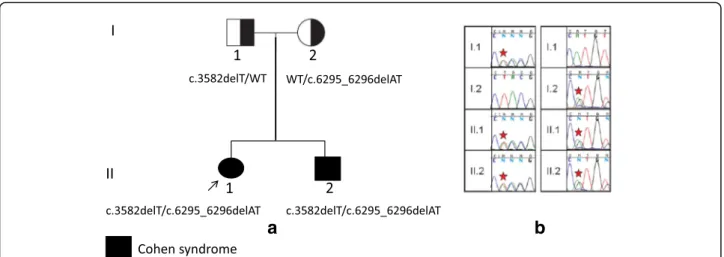
Fig. 3 a Pedigree of the Tunisian family and compound heterozygosity of the proband, formed by the c.3582delT mutation, inherited from the father of the proband and also present in her brother, and the c.6295_6296delAT mutation inherited from the mother. The 2 mutations are shown in black, and in white the wild type alleles (WT). b Sanger validation shows segregation of the VPS13B mutations in the four family members
a presumed role in vesicle-mediated sorting and intracellular protein transport. Am J Hum Genet. 2003;72:1359–69.
7. Evans RJ, Schwarz N, Nagel-Wolfrum K, Wolfrum U, Hardcastle AJ, Cheetham ME. The retinitis pigmentosa protein RP2 links pericentriolar vesicle transport between the Golgi and the primary cilium. Hum Mol Genet. 2010; 19:1358–67.
8. Yan D, Swain PK, Breuer D, et al. Biochemical characterization and subcellular localization of the mouse retinitis pigmentosa GTPase regulator (mRpgr). J Biol Chem. 1998;273:19656–63.
9. Duplomb L, Duvet S, Picot D, et al. Cohen syndrome is associated with major glycosylation defects. Hum Mol Genet. 2014;23(9):2391–9. 10. Metzker ML. Sequencing technologies the next generation. Nat Rev Genet.
2010;11:31–46.
11. Liu L, Li Y, Li S, et al. Comparison of next generation sequencing systems. J Biomed Biotechnol. 2012;2012:251364.
• We accept pre-submission inquiries
• Our selector tool helps you to find the most relevant journal • We provide round the clock customer support
• Convenient online submission • Thorough peer review
• Inclusion in PubMed and all major indexing services • Maximum visibility for your research
Submit your manuscript at www.biomedcentral.com/submit