HAL Id: inserm-00628487
https://www.hal.inserm.fr/inserm-00628487
Submitted on 4 Oct 2011
HAL is a multi-disciplinary open access
archive for the deposit and dissemination of
sci-entific research documents, whether they are
pub-lished or not. The documents may come from
teaching and research institutions in France or
abroad, or from public or private research centers.
L’archive ouverte pluridisciplinaire HAL, est
destinée au dépôt et à la diffusion de documents
scientifiques de niveau recherche, publiés ou non,
émanant des établissements d’enseignement et de
recherche français ou étrangers, des laboratoires
publics ou privés.
Ubiquinone analogs: a mitochondrial permeability
transition pore-dependent pathway to selective cell
death.
Flavien Devun, Ludivine Walter, Julie Belliere, Cécile Cottet-Rousselle,
Xavier Leverve, Eric Fontaine
To cite this version:
Flavien Devun, Ludivine Walter, Julie Belliere, Cécile Cottet-Rousselle, Xavier Leverve, et al..
Ubiquinone analogs: a mitochondrial permeability transition pore-dependent pathway to selective cell
death.. PLoS ONE, Public Library of Science, 2010, 5 (7), pp.e11792. �10.1371/journal.pone.0011792�.
�inserm-00628487�
Transition Pore-Dependent Pathway to Selective Cell
Death
Flavien Devun1,2, Ludivine Walter1,2, Julie Belliere1,2, Ce´cile Cottet-Rousselle1,2, Xavier Leverve1,2, Eric Fontaine1,2*
1 INSERM, U884, F-38041, Grenoble, France, 2 Universite´ Joseph Fourier, Laboratoire de Bioe´nerge´tique Fondamentale et Applique´e, F-38041, Grenoble, France
Abstract
Background: Prolonged opening of the mitochondrial permeability transition pore (PTP) leads to cell death. Various ubiquinone analogs have been shown to regulate PTP opening but the outcome of PTP regulation by ubiquinone analogs on cell fate has not been studied yet.
Methodology/Principal Findings:The effects of ubiquinone 0 (Ub0), ubiquinone 5 (Ub5), ubiquinone 10 (Ub10) and
decyl-ubiquinone (DUb) were studied in freshly isolated rat hepatocytes, cultured rat liver Clone-9 cells and cancerous rat liver MH1C1 cells. PTP regulation by ubiquinones differed significantly in permeabilized Clone-9 and MH1C1 cells from that previously reported in liver mitochondria. Ub0inhibited PTP opening in isolated hepatocytes and Clone-9 cells, whereas it
induced PTP opening in MH1C1 cells. Ub5 did not affect PTP opening in isolated hepatocytes and MH1C1 cells, but it
induced PTP opening in Clone-9 cells. Ub10regulated PTP in isolated hepatocytes, whereas it did not affect PTP opening in
Clone-9 and MH1C1 cells. Only DUb displayed the same effect on PTP regulation in the three hepatocyte lines tested. Despite such modifications in PTP regulation, competition between ubiquinones still occurred in Clone-9 and MH1C1 cells. As expected, Ub5induced a PTP-dependent cell death in Clone-9, while it did not affect MH1C1 cell viability. Ub0induced a
PTP-dependent cell death in MH1C1 cells, but was also slightly cytotoxic in Clone-9 by an oxidative stress-dependent mechanism.
Conclusions/Significance:We found that various ubiquinone analogs regulate PTP in different ways depending on the cell studied. We took advantage of this unique property to develop a PTP opening-targeted strategy that leads to cell death specifically in cells where the ubiquinone analog used induces PTP opening, while sparing the cells in which it does not induce PTP opening.
Citation: Devun F, Walter L, Belliere J, Cottet-Rousselle C, Leverve X, et al. (2010) Ubiquinone Analogs: A Mitochondrial Permeability Transition Pore-Dependent Pathway to Selective Cell Death. PLoS ONE 5(7): e11792. doi:10.1371/journal.pone.0011792
Editor: Alfred Lewin, University of Florida, United States of America Received January 6, 2010; Accepted July 2, 2010; Published July 26, 2010
Copyright: ß 2010 Devun et al. This is an open-access article distributed under the terms of the Creative Commons Attribution License, which permits unrestricted use, distribution, and reproduction in any medium, provided the original author and source are credited.
Funding: This work was supported by grants from INSERM, Agence Nationale de la Recherche (QuinoMitEAO) and the Ministe`re de l’Enseignement de la Recherche et de la Technologie (MERT). FD was supported by fellowship from the Ligue Nationale contre le Cancer. LW was supported by fellowship from the Re´gion Rhoˆne-Alpes (programme Emergence). The funders had no role in study design, data collection and analysis, decision to publish, or preparation of the manuscript.
Competing Interests: The authors have declared that no competing interests exist. * E-mail: [email protected]
Introduction
Mitochondria are involved in several physiological processes including energy metabolism, calcium homeostasis and pro-grammed cell death [1,2,3]. Numerous mitochondrial proteins, which have no pro-apoptotic activity when they remain inside mitochondria, promote cell death once released into the cytosol [4]. Both extra-mitochondrial and intra-mitochondrial signaling pathways can trigger the release of the mitochondrial pro-apoptotic proteins [2].
The mitochondrial permeability transition consists of a sudden non-specific increase in the permeability of the inner membrane [5,6]. A prolonged mitochondrial permeability transition results in a drastic ATP synthesis inhibition through the collapse of the proton-motive force, a dramatic increase in ROS production and the release of the mitochondrial pro-apoptotic proteins [7,8,9].
Permeability transition is due to the opening of an inner membrane channel [10]: the Permeability Transition Pore (PTP). Matrix Ca2+ is the single most important factor for PTP opening. The amount of matrix Ca2+required to open the pore is modulated by a number of factors. The ‘‘PTP-inhibitors’’ and the other so-called ‘‘PTP-inducers’’ designate factors that increase and decrease the amount of Ca2+required to induce PTP opening [6]. Cyclosporin A (CsA) is the reference PTP inhibitor. It inhibits PTP opening by detaching Cyclophilin D (CyP-D) from the other components of the pore [11].
In primary and cultured cells, several drugs known to inhibit PTP opening also decrease cell death in response to various cytotoxic insults [12,13,14]. In animal models, the inhibition of PTP opening by either CsA or genetic ablation of CyP-D provides strong protection from reperfusion injury [15,16,17]. In humans, the first clinical trial has recently shown that CsA treatment
reduces infarct size after reperfusion of a coronary thrombosis [18]. These data suggest that PTP inhibition can be beneficial in particular pathological conditions, most likely through its effect on cell death. On the other hand, resistance to Doxorubicin has been shown to be related to PTP inhibition in a human chronic myelogenous leukemia cell line [19], while hepatocarcinogenesis with 2-acetylaminofluorene is preceded by PTP inhibition [20]. Thus, PTP inhibition might in some cases hamper cancer treatments or eventually participate in carcinogenesis.
Because prolonged PTP opening leads to cell death, the PTP represents a cellular target for the commitment to cell death [21]. Indeed, pharmacological agents used in anti-cancer therapy have been reported to target the PTP and to induce cell death via PTP opening [1,21]. Ideally, drugs used for the treatment of malignancies would be far more toxic for cancer cells than for normal cells. However, a PTP-targeted drug able to selectively open the PTP in cancerous cells only remains to be developed.
In a series of experiments conducted with isolated rat liver mitochondria, we have shown that several ubiquinone analogs regulate PTP opening [22,23,24,25]. Three functional classes of quinones were defined, the inhibitory quinones, the PTP-inducing quinones and the PTP-inactive quinones that counteract the effects of both inhibitory and inducing quinones [24,25]. To date, few studies have reported a preventive effect of ubiquinone analogs in a model of cell death. DUb, a PTP-inhibitor quinone in the liver, has been shown to prevent PTP opening-induced cell death in HL 60 cells [26]. In contrast, although Ub0 is more
potent than CsA at PTP inhibition in liver and skeletal muscle mitochondria [27], Ub0 was ineffective in preventing PTP
opening-induced cell death in HL 60 cells [26].
To clarify this issue, and because PTP regulation can vary depending on the tissue studied [27], we have begun a comprehensive study of PTP regulation in different cell lines. This work presents the effect of four ubiquinone analogs on three different rat liver cell lines. We confirm that ubiquinone analogs regulate PTP opening in the different cells tested. However, we found that a number of ubiquinone analogs may regulate PTP in different ways depending on the cell studied. We took advantage of this unique property to develop a PTP opening-targeted strategy that leads to cell death specifically in cells where the ubiquinone analog used induces PTP opening.
Materials and Methods Cells
Clone-9 and MH1C1 cells are, respectively, non-cancerous and cancerous rat hepatocyte cell lines. Clone-9 and MH1C1 cells were maintained in exponential growth phase using Dulbecco’s modified Eagle’s medium supplemented with 10% fetal bovine serum for Clone-9, and Ham’s F12K medium supplemented with 2.5% fetal bovine serum and 15% horse serum for MH1C1. Both media were supplemented with 2 mM glutamine, 1mM sodium pyruvate, 1% non-essential amino acids, 50 units/ml penicillin, and 50mg/ml streptomycin. Hepatocytes were isolated according to Berry and Friend’s methodology, modified by Groen et al [28].
Ca2+retention capacity
Non confluent cultured cells, harvested by trypsinization, washed with PBS or freshly isolated hepatocytes were permeabi-lized immediately before use by incubation of 56106cells under agitation for 2 min at 25uC in a Ca2+
free medium (Chelex resin, overnight, 4uC) containing 250 mM sucrose, 1 mM Pi-Tris, 10 mM Tris-MOPS (pH 7.4) and 50mg/ml digitonin. Measure-ments of Ca2+were performed fluorimetrically at 25uC with a PTI
Quantamaster C61 spectrofluorometer equipped with magnetic stirring and thermostatic controls. Extra-mitochondrial Ca2+was measured in the presence of 1mM Calcium Green-5N with excitation and emission wavelengths set at 506 and 530 nm, respectively. The Ca2+ uptake and Ca2+ release of digitonin permeabilized cells were measured by loading cells with trains of Ca2+pulses at constant time intervals.
ROS production
Cells were incubated in a medium containing 250 mM sucrose, 1 mM Pi-Tris, 10 mM Tris-MOPS, and 5mM H2DCFDA.
Measurement of H2DCFDA oxidation were performed
fluorime-trically at 37uC (with excitation and emission wavelengths set at 506 and 521 nm, respectively) with a PTI Quantamaster C61 spectrofluorometer equipped with magnetic stirring and thermo-static controls.
Cells Staining
Clone-9 cells were labeled with the lipophilic dye PKH26 according to the manufacturer’s instructions. This non-toxic fluorescent dye binds irreversibly to the cell membrane without affecting cell growth. Therefore, upon cell division, the probe is partitioned equally between each daughter cell, but does not transfer to co-cultured cells [29].
Cells treatment and cell death analysis
Cells were exposed to the indicated concentrations of ubiquinone analogs for 30 min at 37uC in a serum-free medium. Cells were then harvested and incubated in a complete medium at 37uC for 24 h. Annexin V-positive cells were quantified by flow cytometry using a FACSCan flow cytometer (Becton-Dickinson). Cells (16106/ml) were exposed to 5% v/v annexin V-FluoProbes Alexa 488 for 15 min at room temperature. For each sample, a minimum of 10,000 events were analyzed.
Reagents
Ub0 (2,3-dimethoxy-5-methyl-1,4-benzoquinone or coenzyme
Q0), Ub5
(2,3-Dimethoxy-5-methyl-6-(3-methyl-2-butenyl)-1,4-benzoquinone or coenzyme Q1), Ub10
(2,3-Dimethoxy-5-methyl-6-geranyl-1,4-benzoquinone or coenzyme Q2), DUb
(2,3-Di-methoxy-5-methyl-6-decyl-1,4-benzoquinone) and CsA were pur-chased from Sigma. Calcium Green-5N, H2DCFDA and Annexin
V-FluoProbes Alexa 488 were purchased from Molecular Probes. Ham’s F12K medium was purchased from Gibco, Fetal Bovine Serum from Biotech and trypsin from Jacques Boy. The remaining reagents were purchased from Sigma.
Statistics
Stastistical analyses were performed using two-tailed unpaired Student’s t tests with equal variances.
Results
The Ca2+retention capacity (CRC) represents the minimum Ca2+load required to induce PTP opening in an entire population of mitochondria. Therefore, CRC measurement represents a suitable method to quantify and compare the potency of different PTP regulators. The CRC is measured by loading mitochondria with train of Ca2+pulses until a rapid Ca2+release occurs. This event is accompanied by mitochondrial swelling and membrane depolarization, and is prevented by CsA [27].
The CRC measurement can be performed equally well in isolated or in situ mitochondria (i.e. in digitonin permeabilized cells). However, because PTP regulation may be very sensitive to
the conditions of incubation used [27,30], we first checked whether PTP regulation by ubiquinone analogs in permeabilized rat hepatocytes was identical to that previously measured in isolated rat liver mitochondria.
As shown in Figure 1A, Ub0 and DUb increased the CRC
(i.e. inhibited PTP opening) in rat hepatocytes in a concentra-tion-dependent manner up to an optimal concentration beyond which Ub0 and DUb became less potent at PTP inhibition.
Ub10 inhibited PTP opening at low concentrations, but
activated PTP opening at high concentration in rat hepatocytes. Ub5 did not affect PTP regulation in rat hepatocytes. These
results are in total agreement with those previously found with
isolated rat liver mitochondria [23,24,25], indicating that the model used (i.e. isolated mitochondria or in situ mitochondria) did not influence the effect of the tested ubiquinone analogs at PTP regulation.
We next studied the effects of Ub0, Ub5, Ub10and DUb on the
CRC of two other digitonin permeabilized rat liver cells, namely cultured rat liver Clone-9 cells and cancerous rat liver MH1C1 cells. As shown in Figure 1, PTP opening in the absence of ubiquinone analogs occurred at approximately 60, 10 and 25 nmol Ca2+ per million cells in isolated hepatocytes, Clone-9 and MH1C1, respectively. Such differences in the basal CRC are expected [31] and may be at least partly related to differences in
Figure 1. The regulation of PTP opening by ubiquinone analogs depends on the cell lines. The incubation medium contained 250 mM sucrose, 1 mM Pi-Tris, 10 mM Tris-MOPS, 5 mM succinate-Tris, 50 mM digitonin and 1 mM Calcium Green-5N. The final volume was 2 ml, pH 7.4, 25uC. Experiments were begun by the addition of 5.106cells (isolated rat hepatocytes, MH1C1 or Clone-9) followed by the addition of ubiquinone 0 (Ub
0),
ubiquinone 5 (Ub5), ubiquinone 10 (Ub10) or decyl-ubiquinone (DUb) at the indicated concentrations. After 2 min of incubation, the Ca2+Retention
Capacity (CRC) was measured by adding Ca2+pulses every 90 s until PTP opening. Results are mean 6 S.D. of at least four independent experiments. doi:10.1371/journal.pone.0011792.g001
the number of mitochondria per cell. Note, however, that CsA inhibited PTP opening in the three different cell lines (data not shown).
In immortalized Clone-9 cells (Figure 1B), Ub0and DUb were
PTP-inhibitors as observed in permeabilized hepatocytes, al-though the optimal concentration varied with the cell line studied. Surprisingly, contrary to what occurred in permeabilized hepato-cytes, Ub5favored PTP opening, whereas Ub10was ineffective in
Clone-9 cells. In cancerous rat liver MH1C1 cells (Figure 1C), Ub0
favored PTP opening, Ub5 and Ub10 were ineffective, whereas
DUb inhibited PTP opening. These data indicate that the effects of ubiquinone analogs on PTP regulation dramatically differed according to the type of cell used.
In isolated rat liver mitochondria, we have shown that PTP-inactive quinones were able to counteract the effects of both inhibitory and inducing quinones, suggesting the existence of a common site of action for which the ubiquinone analogs compete [25]. In order to check whether these competitions persist in cells in which PTP regulation differs from that observed in liver mitochondria, permeabilized Clone-9 and MH1C1 cells were exposed to a combination of PTP-active plus PTP-inactive quinones. Results are presented in Figure 2. In Clone-9, the PTP-inactive quinone Ub10 was able to
counteract the effect of the PTP- inhibitory Ub0 and of the
PTP-inducing Ub5. Similarly, in MH1C1, the PTP-inactive
quinone Ub5was able to counteract the inducing effect of Ub0.
Therefore, despite the fact that PTP regulation by quinone changes according to the cell studied, this data suggests that the competition between 3 functionally classes of quinones is a ubiquitous phenomenon.
Because PTP opening is expected to induce cell death, we next verified whether the changes observed in PTP regulation were associated with consecutive changes in cell death regulation. In other words, we hypothesized that ubiquinone analogs were capable of inducing cell death selectively in cells in which they favored PTP opening.
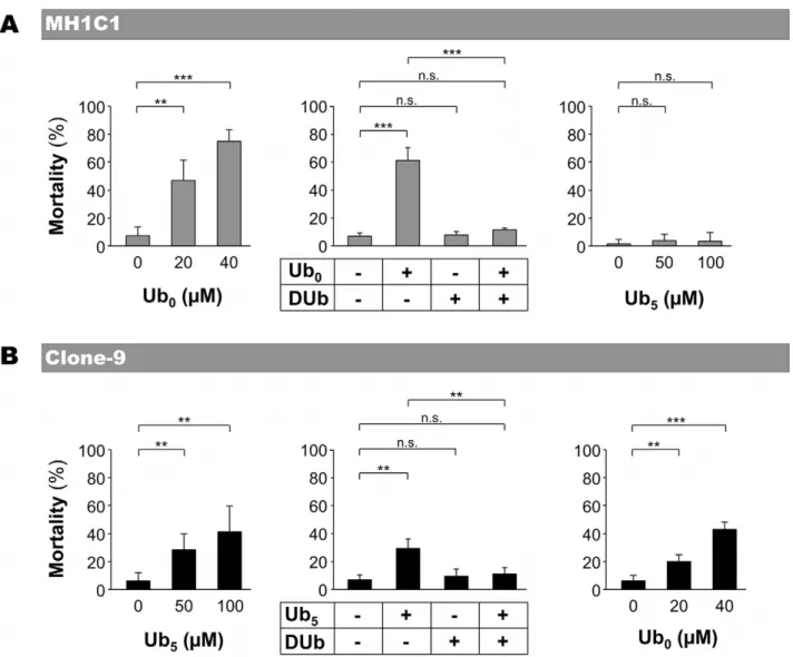
In MH1C1 cells (Figure 3A), PTP-inducer Ub0 induced cell
death in a concentration-dependent manner. Moreover, Ub0
-induced cell death was prevented by PTP-inhibitor DUb
(Figure 3A) and by CsA (not shown), confirming that Ub0-induced
cell death was due to PTP opening in that cell line. As expected, PTP-inactive Ub5 and PTP-inhibitor DUb did not induce any
significant toxicity.
In Clone-9 cells (Figure 3B), PTP-inducer Ub5 induced cell
death in a concentration-dependent manner. As expected, Ub5
-induced cell death was prevented by PTP-inhibitors DUb (Figure 3B) and by CsA (not shown). Surprisingly, Ub0 and
DUb, which both inhibited PTP opening in that cell line (see Figure 1), affected cell viability in a different manner. DUb did not induce any significant toxicity, whereas Ub0induced Clone-9 cell
death.
Ubiquinone analogs have been reported to either reduce or increase reactive oxygen species (ROS) formation [25,26]. In order to check whether Ub0-induced cell death in Clone-9 was
related to oxidative stress, we next measured H2DCFDA oxidation
(i.e., ROS production) in Clone-9 and MH1C1 cells before and after the addition of Ub0, Ub5, Ub10 or DUb. As shown in
Figure 4A, Ub0dramatically increased ROS production in
Clone-9, whereas it slightly decreased ROS production in MH1C1 cells. Ub5and Ub10also acted as pro-oxidants in Clone-9 (although they
were less potent than Ub0), whereas they acted as antioxidants in
MH1C1. Only DUb was antioxidant in the two cell lines. Confirming that Ub0 toxicity in Clone-9 was due to oxidative
stress, tocopherol or tiron hampered Ub0-induced cell death in
Clone-9 (Figure 4B).
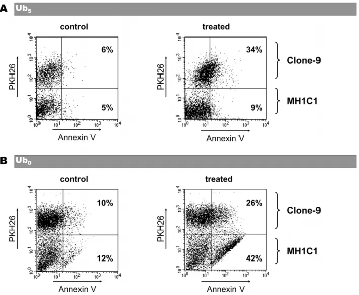
Because Ub5 induced cell death in Clone-9 but not in
MH1C1, we next measured the toxicity of Ub5in co-cultures of
MH1C1 plus Clone-9 cells. In order to distinguish the two populations of cells, Clone-9 were labeled with the fluorescent lipophilic dye PKH-26 before being co-cultured with MH1C1 in a complete F12K medium. In preliminary experiments, we first checked that PKH-26 did not affected PTP regulation or cell viability (data not shown). As shown in Figure 5A, Ub5induced
cell death in the Clone-9 population, but it spared the MH1C1 population.
Because a same concentration of Ub0 was more cytotoxic in
cells in which it induced PTP opening (i.e. MH1C1) than in cells where it inhibited PTP opening (i.e. Clone-9), we also measured
Figure 2. Competition between PTP-inactive and PTP-active ubiquinone analogs on PTP regulation in Clone-9 and MH1C1 cells. The Ca2+Retention Capacity was measured as in Fig. 1. When indicated, 20 mM Ub0, 100 mM Ub5or 100 mM Ub10were added. The results represent the
means 6 S.D. of three independent experiments. *, p#0.05; **, p#0.01, unpaired Student’s t test. doi:10.1371/journal.pone.0011792.g002
the toxicity of Ub0in co-cultures of MH1C1 plus Clone-9 cells.
As shown in Figure 5B, Ub0 induced cell death in the two
populations but with a higher percentage of cell death in MH1C1 cells.
Discussion
In this work we have shown that several ubiquinone analogs are able to regulate PTP opening according to the cell type. Therefore, an important and practical conclusion of this work is that PTP regulation by ubiquinones is an unpredictable phenomenon that cannot be extrapolated from results observed with isolated liver mitochondria. However, this remarkable property led us to develop a PTP-oriented strategy that allows a selective death in cells in which the ubiquinone induces PTP opening, while sparing totally (Ub5in MH1C1) or partially (Ub0
in Clone-9) the cells in which the ubiquinone does not induce PTP opening. If every PTP inhibiting ubiquinone did not
necessarily prevent cell death, all the PTP inducing ubiquinones induced cell death, confirming that PTP opening triggers the cell suicide program.
Another important finding of this work is that ubiquinone analogs are able to modulate ROS production in different way according to the cell type. Note however that within a given cell line, the way a ubiquinone analog modulates ROS production does not correlate with the way it regulates PTP opening (compare Figures 1 and 4). Indeed, ubiquinone analogs can (i) inhibit PTP opening and stimulate ROS production (Ub0in Clone-9), (ii) favor
PTP opening and inhibit ROS production (Ub0in MH1C1 cells),
(iii) favor both PTP opening and ROS production (Ub5in
Clone-9), (iv) inhibit both PTP opening and ROS production (DUb in Clone-9 and MH1C1 cells), (v) modify ROS production without obvious effect on PTP regulation (Ub5and Ub10in MH1C1 cells,
Ub10in Clone-9). Therefore, the effects of the ubiquinone analogs
on ROS production cannot account for the effects on PTP opening, and vice versa.
Figure 3. Effect of ubiquinone analogs on MH1C1 and Clone-9 viability. MH1C1 and Clone-9 cells were exposed for 30 min to serum-free culture medium supplemented or not with the indicated concentrations of Ub0or Ub5(left and right panels). When used in combination (middle
panels), the concentrations were 20 mM for Ub0, 50 mM Ub5and 100 mM for DUb. Cells were then incubated in normal medium for 24 h. The
percentage of mortality represents the proportion of Annexin V-positive cells measured by flow cytometry. The results represent the means 6 S.D of at least four independent experiments. **, p#0.01; ***, p#0.001, unpaired Student’s t test. In preliminary experiments, we found that Annexin V-positive cells were mostly Propidium iodide V-positive.
Despite the fact that Ub0 is the most potent PTP inhibitor
discovered so far in rat liver mitochondria [27], this work shows that Ub0favors PTP opening in MH1C1 cells or induces oxidative
stress in Clone-9 cells. It has been reported that Ub0 was
ineffective in preventing cell death in HL 60 cells [26]. This may be easily explained hypothesizing that Ub0may not inhibit PTP
opening or may induce oxidative stress in HL 60 cells.
Some tissue-specificities in PTP regulation have been previously reported. For example, CsA does not inhibit PTP opening in a particular line of K562 cell resistant to doxorubicin [19]. The inhibition of respiratory chain complex 1 inhibits PTP opening in U937, KB and HMEC cells [31,32,33], whereas it does not have this effect in rat liver or rat heart mitochondria [6]. However, to the best of our knowledge, this is the first piece of evidence showing that a drug can regulate PTP opening in contradictory ways which are dependent on the cell line.
In previous works conducted with isolated rat liver mitochon-dria, we proposed that ubiquinone analogs might regulate PTP via a common site. This hypothesis was sustained by the fact that inactive quinones were able to counteract the effects of both inhibitory and inducing quinones. At that time, the biphasic effect of some quinones (inhibitory at low concentration and inactive or even activating at high concentration) was explained by hypoth-esizing (i) that quinones formed aggregates at high concentrations and (ii) that these aggregates were either inactive or activating compounds able to compete with the monomeric-inhibiting quinone.
The observation that the same concentration of ubiquinone analog can inhibit or activate PTP opening according to the cell
line used no longer supports this model. These data are now more consistent with another proposed model [25] involving two regulatory sites: one responsible for inhibition and one for activation. The occupancy of a site by an active compound would, in turn, modulate PTP opening through secondary changes in the PTP Ca2+binding affinity, whereas binding by an inactive compound would not. In this model, the biphasic response observed with some quinones could easily be explained through the assumption that these quinones may bind the two sites: the inhibitory site with high affinity and the inducing site with a lower affinity.
Because the effect of some ubiquinone analogs depends on the cell line, we propose that the affinity of these sites for one particular quinone, as well as the secondary changes in the PTP Ca2+ binding affinity, may change according to the cell line. These changes would depend on genetic or metabolic differences. Note, however, that the observed changes were not due to differences in the growth media (Figure 4), and that the CRC experiments were performed in the same experimental condition (Figure 1). Therefore, the putative modifications may directly affect the PTP.
This study constitutes the first report of a PTP-targeted strategy able to selectively open the PTP and consecutively provoke cell death in some cells whilst sparing others. Therefore, this work should be viewed as the first proof of concept suggesting that the PTP is a key target for selecting cells that will commit the cell death program. Finding compounds that open PTP in one cancerous cell line (without any side effect on normal cells) needs further studies. This will benefit from an improved knowledge of
Figure 4. The effect of ubiquinone analogs on ROS production depends on the cell lines. A - The incubation medium contained 250 mM sucrose, 1 mM Pi-Tris, 10 mM Tris-MOPS, and 5 mM H2DCFDA. The final volume was 2 ml, pH 7.4, 37uC. Experiments were begun by the addition of
5.106cells (MH1C1 or Clone-9) followed by the addition of 20mM Ub0, 100mM Ub5, 100mM Ub10or 100mM DUb. FUb/F0represents the change in
fluorescence during 5 min after the addition of ubiquinone analog divided by the change in fluorescence during 5 min before the addition of ubiquinone analog. Results are mean 6 S.D. of at least three independent experiments.B - Clone-9 cells were exposed for 30 min to serum-free culture media supplemented or not with 20 mM Ub0, 200 mM tocopherol or 1 mM tiron. Cells were then incubated in normal medium for 24 h. The
percentage of mortality represents the proportion of Annexin V-positive cells measured by flow cytometry. The results represent the means 6 S.D of at least three independent experiments. *, p#0.05; **, p#0.01, unpaired Student’s t test.
doi:10.1371/journal.pone.0011792.g004
the molecular nature of the PTP, which in turn will deepen our understanding of why PTP-regulation by quinones (and probably other compounds) changes according to the cell line.
Acknowledgments
We thank Mr. Gareth Butt for the English corrections to this paper.
Author Contributions
Conceived and designed the experiments: EF. Performed the experiments: FD LW JB. Analyzed the data: FD LW JB XL EF. Contributed reagents/ materials/analysis tools: CCR. Wrote the paper: LW EF.
References
1. Bouchier-Hayes L, Lartigue L, Newmeyer DD (2005) Mitochondria: pharma-cological manipulation of cell death. J Clin Invest 115: 2640–2647. 2. Desagher S, Martinou JC (2000) Mitochondria as the central control point of
apoptosis. Trends Cell Biol 10: 369–377.
3. Duchen MR (2000) Mitochondria and calcium: from cell signalling to cell death. J Physiol 529 Pt 1: 57–68.
4. Saelens X, Festjens N, Vande Walle L, van Gurp M, van Loo G, et al. (2004) Toxic proteins released from mitochondria in cell death. Oncogene 23: 2861–2874.
5. Bernardi P, Krauskopf A, Basso E, Petronilli V, Blachly-Dyson E, et al. (2006) The mitochondrial permeability transition from in vitro artifact to disease target. Febs J 273: 2077–2099.
6. Zoratti M, Szabo I (1995) The mitochondrial permeability transition. Biochim Biophys Acta 1241: 139–176.
7. Batandier C, Leverve X, Fontaine E (2004) Opening of the mitochondrial permeability transition pore induces reactive oxygen species production at the level of the respiratory chain complex I. J Biol Chem 279: 17197– 17204.
8. Kantrow SP, Piantadosi CA (1997) Release of cytochrome c from liver mitochondria during permeability transition. Biochem Biophys Res Commun 232: 669–671.
9. Zorov DB, Filburn CR, Klotz LO, Zweier JL, Sollott SJ (2000) Reactive oxygen species (ROS)-induced ROS release: a new phenomenon accompanying induction of the mitochondrial permeability transition in cardiac myocytes. J Exp Med 192: 1001–1014.
10. Haworth RA, Hunter DR (1979) The Ca2+-induced membrane transition in mitochondria. II. Nature of the Ca2+ trigger site. Arch Biochem Biophys 195: 460–467.
Figure 5. Effect of Ub0or Ub5on coculture of MH1C1 plus Clone-9 cells. Clone-9 cells labeled with PKH26 lipophilic dye were cocultured
with MH1C1 cells for 24 h in complete F12K medium. Cocultures were exposed for 30 min to serum-free culture medium supplemented or not with 20 mM Ub0or 100 mM Ub5. Cocultures were then incubated in complete F12K medium for 24 h. Annexin V-positive cells and PKH-26-positive cells
were measured by flow cytometry. The indicated percentages represent the percentage of Annexin V-positive cells within each population in the shown experiment. Results are representative of six independent experiments.
11. Basso E, Fante L, Fowlkes J, Petronilli V, Forte MA, et al. (2005) Properties of the permeability transition pore in mitochondria devoid of Cyclophilin D. J Biol Chem 280: 18558–18561.
12. Crompton M (2000) Mitochondrial intermembrane junctional complexes and their role in cell death. J Physiol 529 Pt 1: 11–21.
13. Ichas F, Mazat JP (1998) From calcium signaling to cell death: two conformations for the mitochondrial permeability transition pore. Switching from low- to high-conductance state. Biochim Biophys Acta 1366: 33–50. 14. Kroemer G, Reed JC (2000) Mitochondrial control of cell death. Nat Med 6:
513–519.
15. Baines CP, Kaiser RA, Purcell NH, Blair NS, Osinska H, et al. (2005) Loss of cyclophilin D reveals a critical role for mitochondrial permeability transition in cell death. Nature 434: 658–662.
16. Nakagawa T, Shimizu S, Watanabe T, Yamaguchi O, Otsu K, et al. (2005) Cyclophilin D-dependent mitochondrial permeability transition regulates some necrotic but not apoptotic cell death. Nature 434: 652–658.
17. Schinzel AC, Takeuchi O, Huang Z, Fisher JK, Zhou Z, et al. (2005) Cyclophilin D is a component of mitochondrial permeability transition and mediates neuronal cell death after focal cerebral ischemia. Proc Natl Acad Sci U S A 102: 12005–12010.
18. Piot C, Croisille P, Staat P, Thibault H, Rioufol G, et al. (2008) Effect of cyclosporine on reperfusion injury in acute myocardial infarction. N Engl J Med 359: 473–481.
19. De Oliveira F, Chauvin C, Ronot X, Mousseau M, Leverve X, et al. (2006) Effects of permeability transition inhibition and decrease in cytochrome c content on doxorubicin toxicity in K562 cells. Oncogene 25: 2646–2655. 20. Klohn PC, Soriano ME, Irwin W, Penzo D, Scorrano L, et al. (2003) Early
resistance to cell death and to onset of the mitochondrial permeability transition during hepatocarcinogenesis with 2-acetylaminofluorene. Proc Natl Acad Sci U S A 100: 10014–10019.
21. Armstrong JS (2006) Mitochondria: a target for cancer therapy. Br J Pharmacol 147: 239–248.
22. Fontaine E, Bernardi P (1999) Progress on the mitochondrial permeability transition pore: regulation by complex I and ubiquinone analogs. J Bioenerg Biomembr 31: 335–345.
23. Fontaine E, Ichas F, Bernardi P (1998) A ubiquinone-binding site regulates the mitochondrial permeability transition pore. J Biol Chem 273: 25734–25740. 24. Walter L, Miyoshi H, Leverve X, Bernard P, Fontaine E (2002) Regulation of
the mitochondrial permeability transition pore by ubiquinone analogs. A progress report. Free Radic Res 36: 405–412.
25. Walter L, Nogueira V, Leverve X, Heitz MP, Bernardi P, et al. (2000) Three classes of ubiquinone analogs regulate the mitochondrial permeability transition pore through a common site. J Biol Chem 275: 29521–29527.
26. Armstrong JS, Whiteman M, Rose P, Jones DP (2003) The Coenzyme Q10 analog decylubiquinone inhibits the redox-activated mitochondrial permeability transition: role of mitcohondrial [correction mitochondrial] complex III. J Biol Chem 278: 49079–49084.
27. Fontaine E, Eriksson O, Ichas F, Bernardi P (1998) Regulation of the permeability transition pore in skeletal muscle mitochondria. Modulation By electron flow through the respiratory chain complex i. J Biol Chem 273: 12662–12668.
28. Groen AK, Sips HJ, Vervoorn RC, Tager JM (1982) Intracellular compart-mentation and control of alanine metabolism in rat liver parenchymal cells. Eur J Biochem 122: 87–93.
29. Rousselle C, Barbier M, Comte VV, Alcouffe C, Clement-Lacroix J, et al. (2001) Innocuousness and intracellular distribution of PKH67: a fluorescent probe for cell proliferation assessment. In Vitro Cell Dev Biol Anim 37: 646–655. 30. Nogueira V, Devin A, Walter L, Rigoulet M, Leverve X, et al. (2005) Effects of
decreasing mitochondrial volume on the regulation of the permeability transition pore. J Bioenerg Biomembr 37: 25–33.
31. Chauvin C, De Oliveira F, Ronot X, Mousseau M, Leverve X, et al. (2001) Rotenone inhibits the mitochondrial permeability transition-induced cell death in U937 and KB cells. J Biol Chem 276: 41394–41398.
32. Detaille D, Guigas B, Chauvin C, Batandier C, Fontaine E, et al. (2005) Metformin prevents high-glucose-induced endothelial cell death through a mitochondrial permeability transition-dependent process. Diabetes 54: 2179–2187.
33. Guigas B, Detaille D, Chauvin C, Batandier C, De Oliveira F, et al. (2004) Metformin inhibits mitochondrial permeability transition and cell death: a pharmacological in vitro study. Biochem J 382: 877–884.