HAL Id: hal-00646915
https://hal.archives-ouvertes.fr/hal-00646915
Submitted on 1 Dec 2011
HAL is a multi-disciplinary open access archive for the deposit and dissemination of sci-entific research documents, whether they are pub-lished or not. The documents may come from teaching and research institutions in France or abroad, or from public or private research centers.
L’archive ouverte pluridisciplinaire HAL, est destinée au dépôt et à la diffusion de documents scientifiques de niveau recherche, publiés ou non, émanant des établissements d’enseignement et de recherche français ou étrangers, des laboratoires publics ou privés.
Analysis of H19 methylation in control and abnormal
human embryos, sperm and oocytes
Samira Ibala-Romdhane, Mohamed Al-Khtib, Rita Khoueiry, Thierry
Blachère, Jean-François Guérin, Annick Lefèvre
To cite this version:
Samira Ibala-Romdhane, Mohamed Al-Khtib, Rita Khoueiry, Thierry Blachère, Jean-François Guérin, et al.. Analysis of H19 methylation in control and abnormal human embryos, sperm and oocytes. European Journal of Human Genetics, Nature Publishing Group, 2011, �10.1038/ejhg.2011.99�. �hal-00646915�
Analysis of H19 methylation in control and abnormal human embryos, sperm and 1
oocytes 2
3
Samira Ibala-Romdhane 1,3, Mohamed Al-Khtib 1, Rita Khoueiry 1, Thierry Blachère1,
4
Jean-François Guérin 2, Annick Lefèvre 1*.
5 6
1: INSERM U846, Institut Cellule Souche et Cerveau, 18 Av Doyen Lépine, 69500
7
Bron, France. 8
2: Service de Biologie de la Reproduction, Hôpital Femme Mère Enfants, 69500 Bron,
9
France. 10
3: Service Cytogénétique et Biologie de la Reproduction, Hôpital Farhat Hached, 4000
11
Sousse, Tunisie. 12
13
*: corresponding author: Annick Lefèvre, INSERM U846, Institut Cellule Souche et 14
Cerveau, 18 Av Doyen Lépine, 69500 Bron, France. E-mail : annick.lefevre@inserm.fr, 15
telephone : 33 4 78 77 28 79, fax : 33 4 78 77 72 64. 16
17 18
Running title: H19 methylation in human sperm, oocytes and embryos 19
20
Key words: ART, genomic imprinting , development, H19. 21
22 23 24
Abstract 1
ART is suspected to generate increased imprinting errors in the lineage. 2
Following an ICSI (Intra Cytoplamic Sperm Injection) procedure, a certain number of 3
embryos fail to develop normally and imprinting disorders may be associated to the 4
developmental failure. 5
To evaluate this hypothesis, we analysed the methylation profile of H19DMR, a 6
paternally imprinting control region, in high graded blastocysts, in embryos showing 7
developmental anomalies, in the matching sperm and in oocytes of the concerned 8
couples when they were available. 9
Significant hypomethylation of the paternal allele was observed in half the embryos, 10
independently of the stage at which they were arrested (morula, compacted morula, pre 11
blastocyst or BC graded blastocysts). Conversely, some embryos showed significant 12
methylation on the maternal allele, whereas few others showed both, hypomethylation 13
of the paternal allele and abnormal methylation of the maternal allele. The matching 14
sperm at the origin of the embryos exhibited normal methylated H19 patterns. Thus, 15
hypomethylation of the paternal allele in the embryos does not appear inherited from the 16
sperm but likely reflects instability of the imprint during the demethylating process 17
which occurred in the early embryo. Analysis of a few oocytes suggests that the defect 18
in erasure of the paternal imprint in the maternal germ line may be responsible for the 19
residual methylation of the maternal allele in some embryos. None of these imprinting 20
alterations could be related to a particular stage of developmental arrest; compared to
21
high grade blastocysts, embryos with developmental failure are more likely to have
22
abnormal imprinting at H19 (p<0.05).
23
Key words: ART, genomic imprinting, development, H19. 24
Introduction 1
Normal mammalian development requires that both paternal and maternal 2
genome should be expressed properly. Therefore, epigenetic marks acquired in the germ 3
line drive the monoallelic expression, according to parent of origin, of so called 4
“imprinted genes” in the embryo (1). Many imprinted genes are involved in the 5
regulation of fetal and/or placental growth (2). Imprinted genes are regulated through 6
DNA sequences known as imprinting control regions (ICRs) that are differentially 7
methylated, DNA methylation at CpG sequences resulting typically in gene repression. 8
Methylation of ICRs is erased early in life and reset in the germ line, according to sex 9
(2). Another wave of genome-wide demethylation followed later by de novo 10
methylation occurs during preimplantation development from which imprinted genes 11
are protected (3). 12
Several reports have supported the idea that artificial reproductive techniques 13
(ARTs) would favour the acquisition of imprinting errors (4-8). Epigenetic 14
abnormalities in ART could be related to parental infertility, corresponding to 15
imprinting errors in the gametes, transmitted at fertilisation, or to in vitro manipulation 16
of gametes and embryos (9). Therefore, looking for imprinting defects in embryos that 17
failed to develop normally and in the gametes would provide information on whether 18
unsuitable imprint in the gametes could be transmitted and associated with 19
developmental arrest. Thus, we have analysed the methylation profile of H19DMR (one 20
of the 3 imprinting control region that acquire methylation in the paternal germ line) in 21
control blastocyts, initially suitable for transfer, in preimplantation embryos arrested at 22
different stages of their development: morula, preblastocyst and blastocyst showing 23
poor morphology (graded B C), in the parental sperm and in the oocytes of the couples 1
when they were available. 2
H19DMR regulates the expression of two oppositely imprinted genes (10): H19 3
encodes an untranslated RNA with tumour suppressor activity (11) and it is expressed 4
from the maternal allele ; IGF2 (insulin growth factor 2) encodes a growth factor 5
essential for development (12) and it is expressed from the paternal allele. H19DMR 6
harbours several CTCF (CCCTC-binding factor) binding sites (13). CTCF binds to the 7
maternal unmethylated DMR and prevents IGF2 from access to the common enhancers. 8
Conversely, methylation on the paternal DMR prevents the binding of CTCF, 9
permitting IGF2 expression. Hypermethylation of H19 maternal allele is linked to 10
Beckwith-Wiedemann syndrome (BWS) in some patients (4), while hypomethylation of 11
the paternal allele is associated with Silver-Russell syndrome (14), both syndromes 12
showing opposed growth disorders. 13
Materials and Methods 1
Source of human embryos, oocytes and sperm:
2
A total of 33 embryos from 11 different couples, derived from fertilised ICSI 3
oocytes, were donated for research by patients of Laboratoire de Biologie de la 4
Reproduction at Femme Mère Enfant Hospital (Bron, France), after informed consent. 5
Women included in this study were stimulated prior to ICSI procedure with standard 6
long term stimulation protocol using FSH and HCG. The control group was constituted 7
of 5 high graded ICSI blastocysts; the abnormal embryos were distributed as follows: 5 8
pre-blastocysts, 8 abnormal BC blastocysts (according to Gardner et al. grading system: 9
the inner cell mass contained several cells, loosely grouped and the trophectoderm 10
contained very few large cells forming a loose epithelium), 13 compacted morula and 2 11
morula. Protocols were approved by the French legal institution for research on human 12
embryos, “Agence de la Biomédecine”. ICSI indications were heterogeneous as shown 13
in table I, and all embryos originated from super ovulated oocytes. Zona pellucida and 14
attached cumulus cells were removed by digestion with proteinase K (9 units/ml). 15
Denuded embryos were carefully examined under an inverted microscope with Hoffman 16
Modulation Contrast optics (Leica DM IRB) and only cumulus-free embryos were 17
selected for analysis and stored individually at -80°C. 18
After oocyte collection, the cumulus-oocyte complexes were partially denuded 19
of cumulus cells by repeated pipetting in a hyaluronidase solution (150 units, type VII; 20
Sigma) and the oocytes were evaluated for maturity; the immature partially denuded 21
oocytes, either at the germinal vesicle (GV) or at metaphase I (MI) stage, at the time of 22
retrieval, were used for experiments. A total of 15 oocytes from 2 patients were 23
included in this study: 1GV, 12 MI and 2 metaphase II (MII) that were retrieved 24
immature and spontaneously matured in culture medium. Zona pellucida and any 1
remaining somatic cells were removed by digestion with proteinase K (9 units/ml). 2
After careful examination under an inverted microscope with Hoffman Modulation 3
Contrast optics (Leica DM IRB), only cumulus-free oocytes were selected for analysis. 4
The ejaculated sperm samples were collected from the male partner the day 5
when the ICSI procedure was programmed and the methylation analysis was performed 6
on each individual sperm. Control sperm corresponds to sperm of normally fertile men. 7
Routine semen analysis was performed (volume, sperm concentration, motility, 8
morphology) and motile sperm cells were purified on density gradient, to eliminate 9
somatic cell contamination. The sperm was washed repeatedly and placed in phosphate-10
buffered saline, and DNA was extracted by a standard method. 11
² 12 13
DNA methylation analysis
14
The methylation profile of H19 DMR was determined by bisulphite mutagenesis 15
and sequencing as previously described (15). After treatment with bisulphite and 16
purification the DNA was immediately used for nested PCR. Five independent nested 17
PCRs were performed per embryo and per sperm sample. We analysed 18 CpG sites in 18
a 234 bp fragment of H19DMR (6097-6330 bp, AF087017) harbouring a single 19
nucleotide polymorphism (SNP) A/C (A/T following bisulphite treatment) at nucleotide 20
6236. This 234 bp fragment contains the 6th CTCF binding site. Primers specific for 21
bisulfite converted DNA were the followings: external forward: 5’-22
AATAATGAGGTGTTTTAGTTTTATGGATG3’; external reverse: 5' -23
ACTTAAATCCCAAACCATAACACTAAAAC- 3'; internal forward: 5’-24
TTGTATAGTATATGGGTATTTTTGGAGGTT-3’; internal reverse: 5’-1
ACTCCTATAAATATCCTATTCCCAAATAACCCC-3’. The PCR products were sub 2
cloned into pGEM-T plasmid (Promega, France). Three to six clones were sequenced 3
for each PCR product (Biofidal, Lyon, France). 4
5
Statistics 6
Statistical analysis were done using non parametric t-Test or ANOVA and a difference 7
was considered significant when p≤0.05. To compute the significance of altered 8
methylation in arrested embryos, we used Fisher’s exact test and a p value ≤0.05 was 9 considered significant. 10 11 Results 12
Fifteen to 30 clones were sequenced per embryo issued from five independent 13
PCR. Because of the limited starting material and as bisulfite treatment being 14
deleterious for DNA, identical sequences from separated PCRs are certain to represent 15
distinct chromosomes, but identical sequences obtained from the same PCR product are 16
counted only once, as previously discussed (15); twenty to 30 clones could be scored for 17
control embryos and 12 to 15 clones for embryos that failed to develop (see 18
supplementary information figure 1 bis and 2 bis). 19
20
Control blastocysts and control sperm
21
Control blastocyts were cryopreserved for 2 to 9 years before they were donated 22
for research. The SNP localised within the amplified sequence allow parental allele 23
discrimination in 4 out of the 5 analysed blastocyts as shown in figure 1. Thus, even 24
though the corresponding sperms were not available, all methylated sequences carrying 1
the same SNP are most likely from paternal origin while all unmethylated sequences 2
carrying the other SNP are most likely from maternal origin. Therefore, control 3
blastocyts appeared differentially methylated, as expected, with an average of 85.42% ± 4
2.76 methylated CpGs (615 CpGs being methylated out of 720 analysed) for the 5
presumed paternal allele and 1.08 % ± 0.95 methylation (8 methylated CpGs out of 738 6
analysed) for the presumed maternal allele. A pool of 5 sperms from fertile men 7
exhibited 93.65% methylation and showed both 6236-C and 6236-A alleles while the 8
sperm from a single fertile man was homozygous 6236-C and exhibited 96.49% 9
methylation. Thus, the highly methylated state of the paternal allele of H19 was 10
conserved in blastocysts, even though with 8.7% significant leakage. According to the 11
SNP depicted, the strands were distributed into two distinct profiles, 6236-C or 6236-A. 12
Embryos with developmental failure and matching sperms
13
Sperms that matched embryos showing developmental failure belonged to 14
various patients, presenting normal or altered sperm parameters, either severe sperm 15
morphology abnormalities (teratozoospermy), mobility defect (asthenozoospermy), or 16
associated sperm morphology, mobility and concentration defects (oligo-astheno-17
teratozoospermy), as shown in table 1. All exhibited normal high methylation at H19 as 18
shown in figure 1 and 2, with an average of 95.16% methylated CpGs ± 2.61, which is 19
comparable to that observed in sperms from fertile men. Six were homozygous (6236-C 20
or 6236-A) and five were heterozygous (6236-C and 6236-A) for the SNP described. 21
In 21 embryos out of 28, the observed SNP permitted parental allele 22
discrimination, as shown in figure 1 and 2. Embryos included in this study were 23
blocked at various stages, from morula to compacted morula and pre-blastocyst stage, 24
while 5 embryos reached the blastocyst stage but with poor morphology (graded BC). 1
Embryos from the same couple could be arrested at different stages of development, and 2
there was no correlation between the stage of blockage and any infertility factor (Table 3
1 and 2). We observed significant relaxation of H19 paternal imprint in 8 embryos out 4
of 21 (4.1; 4.3; 5.4; 26.2; 30.2; 30.3; 31.1; 33.3) where parental allele discrimination 5
was possible, the altered strands being partially to totally unmethylated (figure 1 and 2). 6
Moreover, 8 of these embryos (4.1; 30.1; 30.3; 30.4; 33.3; 52.1; 52.2; 52.5) exhibited 7
significant, even minor, methylation of the maternal allele, with 2 embryos, 33.3 and 8
52.2, showing up to 15.87% and 20.14 % methylation respectively. Altered metylation 9
patterns on the paternal and the maternal alleles were not correlated, nor were they 10
associated with a particular stage of developmental arrest (Table 2). 11
The methylation status of the oocytes could be analysed for 2 couples (figure 1). 12
In couple 5, the father was homozygous C while the mother was heterozygous A or C. 13
One GV oocyte, carrying the SNP 6236-C, exhibited hypermethylated alleles while a 14
pool of 2 mature MII oocytes, carrying the SNP 6236-A, showed normal 15
hypomethylation. As hypothesised in figure 3, the hypermethylated 6236-C alleles 16
would be inherited from the grand father and would correspond to alleles that escaped 17
erasure of paternal marks, early during oogenesis. Thus, embryo 5.4 inherited a paternal 18
6236-C allele, the corresponding sperm being homozygous 6236-C and a maternal 19
6236-A allele showing normal hypomethylation that is likely to be inherited from the 20
grand mother. The paternally inherited 6236-C allele exhibited significant 21
hypomethylation (p<0.001). In couple 33, both the father and the mother were 22
heterozygous 6236-C and 6236-A. Twelve immature MI oocytes could be collected and 23
analysed. They showed two different allelic profiles: 6236-A, normally hypomethylated 24
and presumably inherited from the mother or 6236-C, highly methylated (35 methylated 1
CpGs out of 36 analysed) and presumably paternally inherited, corresponding to alleles 2
that escaped erasure of paternal marks early during oogenesis, as previously discussed. 3
Thus, embryo 33.3 is likely to originate from a paternal spermatozoon carrying a 6236-4
A methylated allele, since the two methylated 6236-A strands carried an umethylated 5
6194-C nucleotide, as can be seen in all 6236-A sperm alleles, and an oocyte carrying a 6
6236-C methylated allele for H19, likely inherited from the grand father as speculated in 7
figure 3. The presumed paternal allele is significantly hypomethylated (39 methylated 8
CpGs out of 72 analysed p<0.001) while the presumed maternal allele is significantly 9
methylated (20 methylated CpGs out of 126 analysed, p<0.001). 10
Compared to high grade blastocysts, embryos with developmental failure are more
11
likely to have abnormal imprinting at H19 (13/21 versus 0/4, p < 0.05).
12 13 14 15 16 17 18 19 20 21 22 23
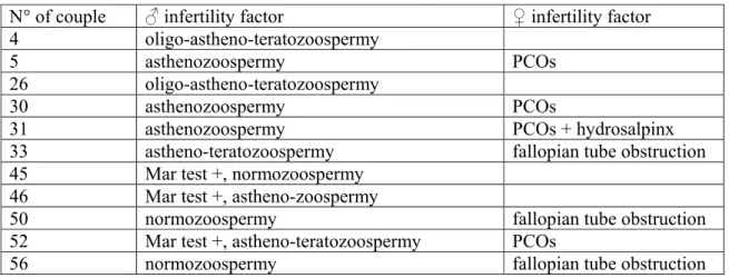
Table 1: ICSI indication per couple. PCOs: polycystic ovary syndrome. Mar test +: 1
presence of sperm antibody. 2
3
N° of couple ♂ infertility factor ♀ infertility factor 4 oligo-astheno-teratozoospermy
5 asthenozoospermy PCOs
26 oligo-astheno-teratozoospermy
30 asthenozoospermy PCOs
31 asthenozoospermy PCOs + hydrosalpinx
33 astheno-teratozoospermy fallopian tube
obstruction 45 Mar test +, normozoospermy
46 astheno-zoospermy, Mar test +
50 fallopian tube
obstruction 52 Mar test +, astheno-teratozoospermy PCOs
56 normozoospermy fallopian tube
obstruction 4 5 6 7 8 9
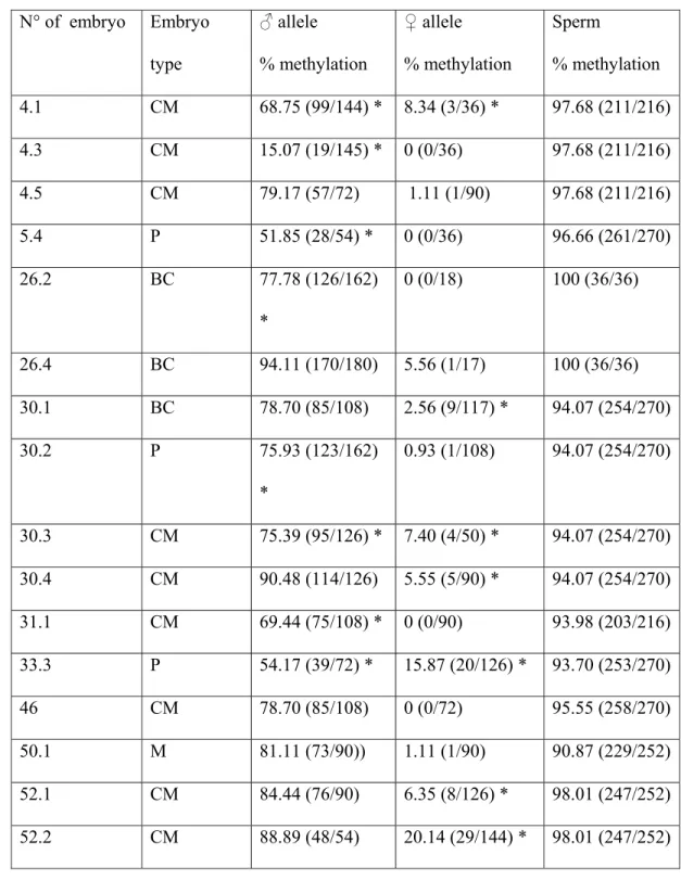
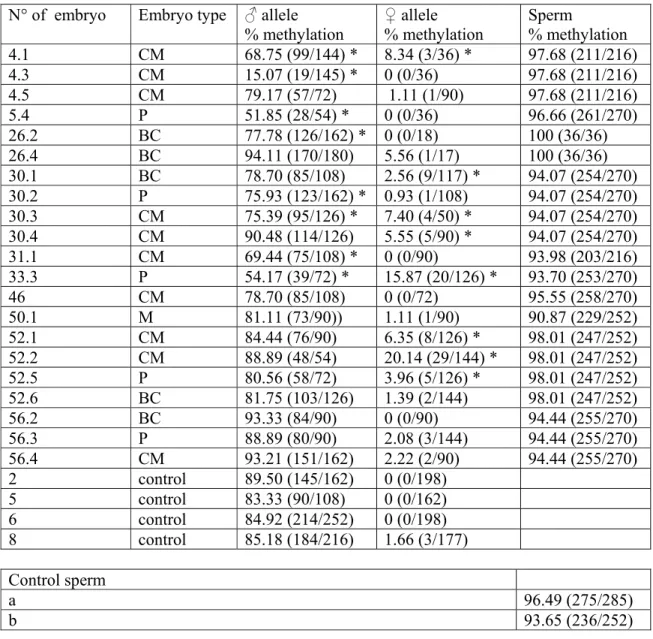
Table 2: Methylation of H19DMR in embryos in which parental allele discrimination 1
could be done and in sperms. 2 3 N° of embryo Embryo type ♂ allele % methylation ♀ allele % methylation Sperm % methylation 4.1 CM 68.75 (99/144) * 8.34 (3/36) * 97.68 (211/216) 4.3 CM 15.07 (19/145) * 0 (0/36) 97.68 (211/216) 4.5 CM 79.17 (57/72) 1.11 (1/90) 97.68 (211/216) 5.4 P 51.85 (28/54) * 0 (0/36) 96.66 (261/270) 26.2 BC 77.78 (126/162) * 0 (0/18) 100 (36/36) 26.4 BC 94.11 (170/180) 5.56 (1/17) 100 (36/36) 30.1 BC 78.70 (85/108) 2.56 (9/117) * 94.07 (254/270) 30.2 P 75.93 (123/162) * 0.93 (1/108) 94.07 (254/270) 30.3 CM 75.39 (95/126) * 7.40 (4/50) * 94.07 (254/270) 30.4 CM 90.48 (114/126) 5.55 (5/90) * 94.07 (254/270) 31.1 CM 69.44 (75/108) * 0 (0/90) 93.98 (203/216) 33.3 P 54.17 (39/72) * 15.87 (20/126) * 93.70 (253/270) 46 CM 78.70 (85/108) 0 (0/72) 95.55 (258/270) 50.1 M 81.11 (73/90)) 1.11 (1/90) 90.87 (229/252) 52.1 CM 84.44 (76/90) 6.35 (8/126) * 98.01 (247/252) 52.2 CM 88.89 (48/54) 20.14 (29/144) * 98.01 (247/252)
52.5 P 80.56 (58/72) 3.96 (5/126) * 98.01 (247/252) 52.6 BC 81.75 (103/126) 1.39 (2/144) 98.01 (247/252) 56.2 BC 93.33 (84/90) 0 (0/90) 94.44 (255/270) 56.3 P 88.89 (80/90) 2.08 (3/144) 94.44 (255/270) 56.4 CM 93.21 (151/162) 2.22 (2/90) 94.44 (255/270) 2 control 89.50 (145/162) 0 (0/198) 5 control 83.33 (90/108) 0 (0/162) 6 control 84.92 (214/252) 0 (0/198) 8 control 85.18 (184/216) 1.66 (3/177) 1 Control sperm a 96.49 (275/285) b 93.65 (236/252) 2 *: p≤0.005 3
M: morula; CM: compacted morula; BC: abnormal BC blastocyts; P: preblastocyts; 4
control: high graded blastocysts, suitable for transfer. a: sperm from a fertile man; b: 5
pool of sperms from 5 fertile men. 6
Discussion 1
Imprinting is both heritable and reversible; in the early embryo, epigenetic 2
information is erased from the genome on a large scale to permit the return of 3
developmental pluripotency to embryonic cells, and this limits the amount of epigenetic 4
information that can be inherited across generation (16). Imprinted genes that carry such 5
heritable epigenetic information must be protected against this powerful remodelling of 6
the epigenome proceeding during preimplantation development. Considering the sensor 7
gene H19, the paternal allele normally escapes the active demethylation of the male 8
genome that occurs in the pronucleus soon after fertilisation and the maternal allele is 9
protected against de novo methylation of the zygotic DNA from morula to blastocyst 10
stage (3). 11
Very little is known concerning the differential methylation of imprinted genes 12
in preimplantation human embryos. In the present work, using an SNP within 13
H19DMR, we can determine the differential methylation status of 4 out of 5 control 14
blastocysts and of 21 out of 28 abnormal embryos, the paternally inherited alleles being 15
generally methylated while the maternally inherited alleles were unmethylated. Our 16
results from control embryos confirmed that, as observed in the mouse (17), H19 is 17
differentially methylated in human blastocyts conceived via ART. In contrast, 38% of 18
the embryos that failed to develop exhibited significant hypomethylation of the paternal 19
allele, independently of the type of developmental failure: blockage at the compacted 20
morula or pre blastocyst stage, or poor morphology at the blastocyst stage. Since normal 21
methylation of the paternal allele was observed in the other abnormal embryos, 22
imprinting errors at H19/IGF2 are likely not to be primarily involved in the 23
developmental failure of human ICSI embryos. Culture conditions were identical for 24
control and developmentally failing embryos and thus could not be the cause of 1
abnormal methylation. 2
Embryos showing hypomethylated paternal alleles may originate from sperm 3
in which resetting of the imprint has been only partially accomplished. Recently, 4
Kobayashi et al. (18) showed that hypomethylation of H19DMR in aborted ART 5
concepti originated from the parental sperm. A case of Silver-Russel syndrome due to 6
paternal H19DMR hypomethylation in an ICSI patient has been likewise documented 7
(19). In fact, we found no alteration of the normal methylated pattern of H19DMR in all 8
the examined parental sperm, whatever their biological characteristics were, from 9
normozoospermic to oligo-astheno-teratospermic, demonstrating that the alterations in 10
DNA methylation seen in some arrested embryos did not pre-exist in the father's germ 11
line. It is interesting to note that, in sperm carrying the SNP A, the cytosine 12
corresponding to the 4th CpG of the 6th CTCF binding site (nucleotide 6194) commonly 13
appeared as a thymidine (in 84 out of 87 analysed alleles), presumably corresponding to 14
an unmethylated CpG in the original strand. Renda et al. (20) demonstrated that 15
methylation of the first and the second CpGs of the CTCF binding sequence was 16
sufficient to inhibit the binding of CTCF, the methylation of the first CpG being more 17
powerful. On the other hand, in most arrested embryos, hypomethylation of the paternal 18
allele did not particularly target the key CpGs, suggesting normal expression of IFG2. 19
However, the number of samples analysed remained limited, and sperm-transmitted 20
aberrant hypomethylation of the paternal allele of H19 in arrested embryos cannot be 21
totally excluded, since H19 hypomethylation in sperm has been associated with paternal 22
infertility in a number of articles (21-23). Thus, considering that the resetting of 23
methylation during gametogenesis has normally proceeded, it could be hypothesised 24
that the hypomethylation observed is due to an alteration of the methylation 1
maintenance in the early embryo. Maintenance of DNA methylation at imprinting 2
DMRs in preimplantation embryos has recently been assigned to the somatic and the 3
oocyte form of DNMT1, in cooperation. Maternal deletion of both isoforms, DNMT1s 4
and DNMT1o, caused the loss of methylation at multiple imprinted loci in mouse 5
blastocysts (24). Other trans-acting factors critical for the maintenance of methylation at 6
imprinted genes have been identified, such as MBD3 which is required to maintain the 7
methylation of the paternal H19 allele and its silencing (25). 8
Some embryos exhibited significant methylation of the maternal allele. Results 9
from couples 5 and 33 whose oocytes were available allow speculating. In both couples, 10
the paternal allele show some relaxation of the imprint, probably due to alterations in 11
DNA methylation maintenance as previously discussed. In both couples some immature 12
GV and MI oocytes were highly methylated. We previously evidenced such altered 13
pattern of methylation in human oocytes, particularly in oocytes that were immature on 14
the day of retrieval, but also in mature oocytes (15). While the maternally originating 15
H19 strands were not methylated in embryo 5.4, they were significantly methylated in 16
embryo 33.3. Both the father and the mother were heterozygous in couple 33. The allele 17
likely inherited from the father carried the A SNP; it was initially methylated since the 18
sperm appeared normally methylated, and was then partially demethylated in the course 19
of cell divisions. The allele likely inherited from the mother carried the C SNP and 20
showed abnormal methylation. Methylated H19 strands in the oocytes may correspond 21
to the acquisition of abnormal methylation on the maternal allele during oogenesis or 22
lack of erasure of the paternal imprint early in the gonocytes. Because the oocytes from 23
the mother which could be analysed showed either unmethylated A-SNP-H19 strands or 24
methylated C-SNP-H19 strands, methylation apposition during oogenesis is unlikely, 1
otherwise, it would also have randomly mark A-SNP-H19 strands. Lack of erasure of 2
the copies originating from the grand father in the gonocytes of the mother is a more 3
credible scenario. Thus, all methylated copies seen in the oocytes are likely originated 4
from paternal copies, the imprint of which has not been erased, as postulated in figure 3. 5
Such a gain of maternal methylation and a loss of paternal methylation at H19DMR 6
have been recently documented in mouse blastocysts and were attributed to 7
superovulation induced imprinting disorders (17). The authors did not give information 8
on the imprint in the oocytes themselves. On the other hand, we found almost no 9
residual methylation on the maternal allele of control human blastocysts while Market-10
Velker et al. (17) showed an average methylation of the maternal allele of H19 of up to 11
15% in mouse blastocysts obtained from spontaneously ovulated female, demonstrating 12
one more time the discrepancies existing between species at the level of epigenome 13
regulation. 14
In this work, we show that a significant number of arrested preimplantation 15
embryos have altered H19 imprints on both paternal and maternal alleles, while 16
blastocysts suitable for transfer were normally imprinted. Our results suggested both a 17
failure of erasure of the paternally inherited imprint in the maternal germ line and a lack 18
of methylation maintenance on the paternal allele. The question remains whether there 19
is a link between imprinted errors and parental infertility. No evident link appeared 20
between imprint anomalies and a particular stage of development in failing embryos. 21
However, the data presented show a significant association between developmental 22
failure of embryos and abnormal imprinting of H19. 23
Acknowledgments: we are very grateful to all the couples that donated embryos and
1
gametes for research and to the staff of “Service de Biologie de la Reproduction” at 2
FME Hospital, Bron, particularly to Jacqueline Lornage and Astrid Perret. This work 3
was supported by Université Claude Bernard Lyon 1. 4
Conflict of interest: The authors declare no conflict of interest. 5
6 7
References 8
1 Surani MA: Imprinting and the initiation of gene silencing in the germ line. Cell 9
1998; 93: 309-312. 10
2 Hajkova P, Erhardt S, Lane N et al.: Epigenetic reprogramming in mouse 11
primordial germ cells. Mech Dev 2002; 117: 15-23. 12
3 Reik W, Dean W, Walter J: Epigenetic reprogramming in mammalian 13
development. Science 2001; 293: 1089-1093. 14
4 DeBaun MR, Niemitz EL, Feinberg AP: Association of in vitro fertilization with 15
Beckwith-Wiedemann syndrome and epigenetic alterations of LIT1 and H19. 16
Am J Hum Genet 2003; 72: 156-160.
17
5 Gicquel C, Gaston V, Mandelbaum J, Siffroi JP, Flahault A, Le Bouc Y: In vitro 18
fertilization may increase the risk of Beckwith-Wiedemann syndrome related to 19
the abnormal imprinting of the KCN1OT gene. Am J Hum Genet 2003; 72: 20
1338-1341. 21
6 Maher ER, Brueton LA, Bowdin SC et al.: Beckwith-Wiedemann syndrome and 22
assisted reproduction technology (ART). J Med Genet 2003; 40: 62-64. 23
7 Orstavik KH, Eiklid K, van der Hagen CB et al.: Another case of imprinting 1
defect in a girl with Angelman syndrome who was conceived by 2
intracytoplasmic semen injection. Am J Hum Genet 2003; 72: 218-219. 3
8 Bowdin S, Allen C, Kirby G, et al.: A survey of assisted reproductive 4
technology births and imprinting disorders. Hum Reprod 2007; 22: 3237-3240. 5
9 Grace KS, Sinclair KD: Assisted reproductive technology, epigenetics, and 6
long-term health: a developmental time bomb still ticking. Semin Reprod Med 7
2009; 27: 409-416. 8
10 Zemel S, Bartolomei MS, Tilghman SM: Physical linkage of two mammalian 9
imprinted genes, H19 and insulin-like growth factor 2. Nat Genet 1992; 2: 61-10
65. 11
11 Yoshimizu T, Miroglio A, Ripoche MA et al.: The H19 locus acts in vivo as a 12
tumor suppressor. Proc Natl Acad Sci U S A 2008; 105: 12417-12422. 13
12 Reik W, Constancia M, Dean W et al.: Igf2 imprinting in development and 14
disease. Int J Dev Biol 2000; 44: 145-150. 15
13 Takai D, Gonzales FA, Tsai YC, Thayer MJ, Jones PA: Large scale mapping of 16
methylcytosines in CTCF-binding sites in the human H19 promoter and aberrant 17
hypomethylation in human bladder cancer. Hum Mol Genet 2001; 10: 2619-18
2626. 19
14 Gicquel C, Rossignol S, Cabrol S et al.: Epimutation of the telomeric imprinting 20
center region on chromosome 11p15 in Silver-Russell syndrome. Nat Genet 21
2005; 37: 1003-1007. 22
15 Borghol N, Lornage J, Blachere T, Garret AS, Lefèvre A: Epigenetic status of 1
the H19 locus in human oocytes following in vitro maturation. Genomics 2006; 2
87: 417-426. 3
16 Santos F, Hendrich B, Reik W, Dean W: Dynamic reprogramming of DNA 4
methylation in the early mouse embryo. Dev Biol 2002; 241: 172-182. 5
17 Market-Velker BA, Zhang L, Magri LS, Bonvissuto AC, Mann MR: Dual 6
effects of superovulation: loss of maternal and paternal imprinted methylation in 7
a dose-dependent manner. Hum Mol Genet; 19: 36-51. 8
18 Kobayashi H, Hiura H, John RM et al.: DNA methylation errors at imprinted 9
loci after assisted conception originate in the parental sperm. Eur J Hum Genet 10
2009; 17: 1582-1591. 11
19 Chopra M, Amor DJ, Sutton L, Algar E, Mowat D: Russell-Silver syndrome due 12
to paternal H19/IGF2 hypomethylation in a patient conceived using 13
intracytoplasmic sperm injection. Reprod Biomed Online; 20: 843-847. 14
20 Renda M, Baglivo I, Burgess-Beusse B et al.: Critical DNA binding interactions 15
of the insulator protein CTCF: a small number of zinc fingers mediate strong 16
binding, and a single finger-DNA interaction controls binding at imprinted loci. 17
J Biol Chem 2007; 282: 33336-33345.
18
21 Boissonnas CC, Abdalaoui HE, Haelewyn V et al.: Specific epigenetic 19
alterations of IGF2-H19 locus in spermatozoa from infertile men. Eur J Hum 20
Genet; 18: 73-80.
21
22 Marques CJ, Costa P, Vaz B et al.: Abnormal methylation of imprinted genes in 22
human sperm is associated with oligozoospermia. Mol Hum Reprod 2008; 14: 23
67-74. 24
23 Poplinski A, Tuttelmann F, Kanber D, Horsthemke B Gronoll J: Idiopathic male 1
infertility is strongly associated with aberrant methylation of MEST and 2
IGF2/H19 ICR1. Int J Androl 2010; 33: 642-649. 3
24 Hirasawa R, Chiba H, Kaneda M et al.: Maternal and zygotic Dnmt1 are 4
necessary and sufficient for the maintenance of DNA methylation imprints 5
during preimplantation development. Genes Dev 2008; 22: 1607-1616. 6
25 Reese KJ, Lin S, Verona RI, Schultz RM Bartolomei MS: Maintenance of 7
paternal methylation and repression of the imprinted H19 gene requires MBD3. 8
PLoS Genet 2007; 3: e137.
9 10 11 12
1
Titles and legends to figures 2
Figure1 and 2: bisulphite sequencing analysis of H19DMR in control blastocysts and 3
control sperm (a: sperm from a fertile man; b: pool of sperm from 5 fertile men), and in 4
arrested embryos and their matching sperm and oocytes (when available, couples 5 and 5
33). Each line represents a single allele. Black squares indicate a methylated CpG and 6
an open square denotes an unmethylated CpG. Blue lines correspond to alleles 7
exhibiting a nucleotide 6236 A, while black lines correspond to alleles carrying a 8
nucleotide 6237 C. In H19DMR, sequence from 6097bp to 6330bp, the SNP A/C, bp 9
6236, is indicated in blue while the 6th CTCF binding site is indicated in red. 10
11
Figure 3: Proposed origin of the methylation alterations observed in embryos 5.4 and 12
33.3. 13
Figure 1
H19DMR (6097-6330 bp, Genbank AF087017)
ttgc atagcacatg ggtatttctg gaggcttctc cttcggtctc accgcctgga tggcacggaa ttggttgtag ttgtggaatc ggaagtggcc gcgcggcggc agtgcaggct cacacatcac agcccgagcc cgcccc
A/
C
act ggggttcgcc cgtggaaacg tcccgggtca cccaagccac gcgtcgcagg gttcacgggg gtcatctggg■■■■■■□■■■■■■■■■■■ ■■■■■■□■■■■■■■■■■■ ■■■■■■□■■■■■■■■■■■ ■■■■■■□■■■■■■□■■■■ ■■■■■■□□■■■■■■■■■■ ■■■■■■□■■■■■■□■■■■ ■■■■■■□■■■■■■■■■■■ C6 C2 ■■■■■■■■■■■■■■■■■■ ■■■■■■■■■■■■■■■■■■ ■■■■■■■■■■■■■■■■■■ ■■■■■■■■■■□■■■■■■■ ■■■■■■■■■■□■■■■■■■ □■■■■■■■■■■■■■■■■■ ■■■■■□■■■■■■■■□□■■ C8 ■■■■■■□■■■■■■■■■■■ ■■■■■■□■■■■■■■■■■■ ■■■■■■□■□■■■■■■■■■ ■■■■■■□■■■■■■■■■■■ ■■■□■■□□■■■■■■■■■■ ■■□■■■□■■■■■■■■■■■ ■■■■□□□□■■■■■■■■■■ C5 ■■■■■■□■■■■■■■■■■■ ■■■□■■□■■■■■■■■■■■ ■■■■■■□■■■■■■■■■■■ ■■■■■■□■■■■■■■■■■■ ■■■■■■□■■■■■■■■■□□ □□■■□□□□■■□■■■□■□□ ■■□■■■■■■■■■■■■■■■ ■■□■■■■■■■□■■■■■■■ ■■■■■■■■■■□■■■■■■■ □■■■■■■■■■■■■■■■■■ ■■□■■■■■■■□■■■■■■■ ■■■■□□■■■■□■□■■■■□ ■■■■■■■■□■■□□□■■■□ C9 Control blastocysts
ggggttcgcc cgtggaaacg tcccgggtca cccaagccac gcgtcgcagg gttcacgggg gtcatctggg aataggacac tcataggagc ■■■■■■□■■■■■■■■■■■ ■■□□■■□□■■■■■■■■■■ ■■■■■■□■■■□□■■■■■□ ■■■■■■□■■■□□■■■■■□ ■■■■■■□■■■□□■■■■■□ ■■■□■■□■■■■■■■□□□□ ■■■■■■□■■■□□■■■■■□ ■■■■■■□■■■□□■■■■■■ □□□□□□□□□□□□□□■■■■ □□□□□□□□□□□□□□□□□□ □□□□□□□□□□□□□□□□□□ □□□□□□□□□□□□□□□□□□ □□□□□□□□□□□□□□□□□□ □□□□□□□□□□□□□□□□□□ □□□□□□□□□□□□□□□□□□ □□□□□□□□□□□□□□□□□□ □□□□□□□□□□□□□□□□□□ ■■■■■□■■■■■■■■□□■■ ■■■■■■■■■■□■□□□□□□ □□■■■■■■■■■■■■■■□□ □□□□□□□□□□■□□□□□□□ □□□□□□□□□□□□□□□□□□ □□□□□□□□□□□□□□□□□□ □□□□□□□□□□□□□□□□□□ □□□□□□□□□□□□□□□□□□ □□□□□□□□□□□□□□□□□□ □□□□□□□□□□□□□□□□□□ □□□□□□□□□□□□□□□□□□ □□□□□□□□□□□□□□□□□□ □□□□□□□□□□□□□□□□□□ □□□□□□□□□□□□□□□□□□ ■■■■□□□□■■■■■■■■■■ ■□■■■■□■■■■■■■■■■■ ■■■■■□□■■■■■■□■■■■ ■■■■■■□■■■■■■□■■■■ □■□■□□□□□□■■□□■■■■ □□□□□□□□□□□□□□■■■□ □□□□□□□□□□□□□□□□□□ □□□□□□□□□□□□□□□□□□ □□□□□□□□□□□□□□□□□□ □□□□□□□□□□□□□□□□□□ □□□□□□□□□□□□□□□□□□ □□□□□□□□□□□□□□□□□□ □□□□□□□□□□□□□□□□□□ □□□□□□□□□□□□□□□□□□ □□□□□□□□□□□□□□□□□□ □□□□□□□□□□□□□□□□□□ □□□□□□□□□□□□□□□□□□ □□□□□□□□□□□□□□□□□□ □□□□□□□□□□□□□□□□□□ □□□□□□□□□□□□□□□□□□ □□□□□□□□□□□□□□□□□□ □□□□□□□□□□□□□□□□□□ □□□□□□□□□□□□□□□□□□ □□□□□□□□□□□□□□□□□□ ■■■■■■■■□■■□□□■■■□ ■■■■■■■■□■■□□□■■■□ ■■■■■■■■□■■□□□□□□□ ■■■■■■■■□□□□□□□□□□ □□□□□□□□□□□□□□□□□□ □□□□□□□□□□□□□□□□□□ □□□□□□□□□□□□□□□□□□ □□□□□□□□□□□□□□□□□□ □□□□□□□□□□□□□□□□□□ ■□□□□□□□□□□□□□■■■□ □□□□□□□□□□□□□□□□□□ □□□□□□□□□□□□□□□□□□ □□□□□□□□□□□□□□□□□□ ■■■■■■■■■■■■■■■■■■ ■■■■■□■■■■□■■■■■■■ ■■■■■□■■■■□■■■■■■■ ■■■□■□■■□■■■■■■■■■ ■■□■■□■■■■■□■■■■■■ ■■■■■□■■■■■■■■■■■■ ■■■■■□■■■■□■■■■■■■ □□□□□□□□□□□□□□□□□□ □□□□□□□□□□□□□□□□□□ □□□□□□□□□□□□□□□□□□ 5.5 BC ♂ ♀ ■□■■■■■■■■■■■□■■■□ ■□■■■■□■□■□■■■■■■□ □□□□□□□□□□□□□□□□□□ □□□□□□□□□□□□□□□□□□ □□□□□□□□□□□□□□□□□□ 5.4 P 5 sperm ■■■■■■■■■■■■■■■■■■ ■■■■■■■■■■■■■■■■■■ ■■■■■■■■■■■■■■■■■■ ■■■■■■■■■■■■■■■■■■ ■■■■■■■■■■■■■■■■■■ ■■■■■■■■■■■■■■■■■■ ■■■■■■■■■■■■■■■□■■ ■■■■■□■■■■■■■■■■■■ ■■■■■□■■■■■■■■■■■■ ■■■■■□■■■■■■■■■■■■ ■■■■□■■■■■■■■■■■■■ ■■■■■■■■■■■■■■■■■■ 5: 1GV oocyte □□□□□□□□□□□□□□□□□□ □□□□□□□□□□□□□□□□□□ □□□□□□□□□□□□□□□□□□ 5: 2MII oocytes
Control sperm cells
■■■■■■■■■■■■■■■■■■ ■■■■■■■■■■■■■■■■■■ ■■■■■■■■■■■■■■■■■■ ■■■■■■■■■■■■■■■■■■ ■■■■■■■■■■■■■■■■■■ ■■■■■■■■■■■■■■■■■■ ■■■■■■■■■■■■■■■■■■ ■■■■■■■■■■■■■■■■■■ ■■■■■■■■■■■■■■■■■■ ■■■■■■■■■■■■■■■■■■
a
□□□□□□□□□□□□□□□□□□ ■■■■■□■■■■■■■■■■■■ ■■■■■■■■■■■■■■■■■■ ■■■■■□■■■■■■■■■■■■ ■■■■■■□■■■■■□■■■■■ ■■■■■■■■■■□■■■■■□■ ■■■■■■■■■■■■■■■■■■ 26.1 BC ■■■■■■■■■■■■■■■■■■ ■■□■■■■■■■■■■■■■■■ ■■■■■■■■■■■■■■■■■■ ■■■■■■■■■□■■■■■■□■ □■■■■■■■■■■■■■■■■■ 26.2 BC ■■■■■■■■■■■■■■■■■■ ■■■■■■■■■■■■■■■■■■ ■■■■■■■■■■■■■■■■■■ ■■■■■■■■■■■■■■■■■■ ■■■■■■■■■■■■■■■■■■♂ ♂ ■■■■■■■■■■■■■■■■■■ ■■■■■■■■■■■■■■■■■■ ■■■■■■■■■■■■■■■■■■ ■■■■■■■■■■■■■■■■■■ ■■■■■■■■■■■■■■■■■□ ■■■■■■■■■■■■■■■□■■ 26.4 BC ■■■■■■■■■■■■■■■■■■ ■■■■■■■■■■■■■■■■■■ 26 sperm ■■■■■■□■■■■■■■■■■■ ■■■■■■□■■■■■■■■■■■ ■■■■■■□■■■■■■■■■■■ ■■■■■■□■■■■■■■■■■■ ■■■■■■□■■■■■■■■■■■ ■■■■■■□■■■■■■■■■■■ ■■■■■■■■■■■■■■■■■■ ■■■■■■■■■■■■■■■■■■ ■■■■■■■■■■■■■■□■■■ ■■■■■■■■■■■■■■■□■■ ■■■■■■■■■■■■■■■□■■ ■■■■□■■■■■■■■■■■■■ ■■■■■■■■■■■■■■■■■■ ■■■■■■■■■■■■■■■■■■ □■■□■■■■■■■■■■■■□■ ■□■□■■■■■■■□■■■■■■b
45.2 CM □■■■■■□■■■■■■■□□■□ □■□■■■□■■□□□■■■■■■ ■□□■■■□■■■□□□■■■■□ ■■■■■■□■■□■■□■■■■■ ■■■■■■□■■■■■■■■■■■ ■■■■■■□■■■■■■■■■■■ ■■■■■■□■■■■■■■■■■■ ■■■■■■□■■■■■■■■■■■ 45 sperm □□□□□□□□□□□□□□□□□□ □□□□□□□□□□□□□□□□□□ □□□□□□□□□□□□□□□□□□ □□□□□□□□□□□□□□□□□□ □□□□□□□□□□□□□□□□□□ ■□□□□□□□□□□□□□□□□□ □□□□□□□□□□□□□□□□□□ ■■■■■■■■■■■■■■■■■■ ■■■■■■■■■■■■■■■■■■ □□□□□□□□□□□□□□□□□□ □□□□□□□□□□□□□□□□□□ □□□□□□□□□□□□□□□□□□ ♂ ♀ ♂ ♀ ■■■■■■■■■■■■■■■□■■ ■■□■■■■■■■■■■■■■■■ ■■■■■■■■■□□■■■■■■■ ■■□■■■■■■■□□■■■■■■ ■■□□■■■■■■■■■■■■■■ ■□□□□□□□□□□□□□□□□□ ■■■■■■■■■■■■■■■■■■ ■■■■■■■■■■■■■■■■■■ ■■■■■■■■■■■■■■■□■■ ■■■■■■■■■■■■■■■□■■ 33 sperm 33.3 P ■■■■■■□■■■■■■□■■■■ ■■■■■■□■■□■■■■■■■■ ■■□□□□□□□□□□□□■■■□ □■■□□□□□□□□□□□□□□□♂ 33: 12 MI oocytes ■■■■■■□■■■■■■■■■■■ ■■■■■■■■■■■■■■■■■■ □□□□□□□□□□□□□□□□□□ ■■■■■■□■■■■■■■■■■■ ■■■■■■□■■■■■■■■■■■ ■■■■■■□■■■■■■■■■■■ ■■■■■■□■■■■■■■■■■■ ■■■■■■□■■■■■■■■■□■ ■■■■■■□■■■■■□■■■■■ ■■■■■■□■□■■■■■■■■■ ■■■■■■■■■■■■■■■■■■ ■■■■■■■■■■■■■■■■□■ 31 1 CM 31 sperm ■■■■■■□■■□□■■■■■■■ □□□□□□□□□□□□□□■■■□ □□□□□□□□□□□□■□■■■■ □□□□□□□□□□□□□□□□□□ □□□□□□□□□□□□□□□□□□ □□□□□□□□□□□□□□□□□□ □□□□□□□□□□□□□□□□□□ □□□□□□□□□□□□□□□□□□ □□□□□□□□□□□□□□□□□□ ■■■■■■□■■■■■■■■■■■ ■■■■■■□■■■■■■■■■■■ ■■■■■■■■■■■■■■□■■■ ■■■■■■■■■■□□■■■■■■ ■■■■■■■■■■■■■■■■■■ ■■■■■■■■■■■■■■■■■■ ■■■■■■■■■■■■■■■■■■ ■■■■■■■■■■■■■■■□■■ ■■■■■■■■■■■■■■■■■■ ■■■■■■■■■■■■■■■■■■ ■■■■■□■■■■■■■■■■■■ 50 1 M 50 3 M 50 sperm ■■■■■■■■■■■■■■■□■■ ■■■■■■■■■■■■■■■■■■ ■■■□■■□■■■□■■■■□□■ ■■■■■■■■■■■■■■■■■■ ■■■■■□■■■■■■■■■■■■ ■■■■■■□■■■■■■■■■■■ ■■■■■■□■■■■■■■■■■■ ■■■■■■□■■■■■■■■■■■ ■■■■■■□■■□■■■■■■■■ ■■■■■■□■■■■■■■■■■■ ■■■□■■□■■■■■■■■■■■ ■■■■■■□■■■■■■■■■■■ □■■□□□□□□□□□□□□□□□ ■■■■■□□□□□□□□□□□□□ ■■■□□□□■□■□□□■□□■□ ■■■□□□□■□■□□□■□□■□ □□□□□□□□□□□□□□□□□□ ■□□□□□□□□□□□□□□□□□ □□□□□□□□□□□□□□□□□□ □□□□□□□□□□□□□□□□□□ ♀ □□□□□□□□□□□□□□□□□□ □□□■□□□□□□□□□□□□□□ □□□□□□□□□□□□□□□□□□ □□□□□□□□□□□□□□□□□□ ♂ ♀ 31.1 CM 31 sperm ■■■■■■□■■■■■■■■■■■ ■■■■■■□■■■■■■■■■■■ ■■■■■■□■■■■■■■■■■■ ■■■■■■□■■■■■■■■■■■ ■■■■■■□■■■■■■■■■■■ ■■■■■■□■■■■■■■■■■■ ■■■■■■□■■■■■■■■■■■ ■■■■■■□■■■■■■■■■■■ ■■■■■■□■■■■■■■■■■■ ■■■■■■□■■□■■■■■■■■ ■■■■■■□■■■■■■■■■■■ ■■■■■■□■■■■■■■■■■■ ■■■■■■□■■■■■■■■■■■ ■■■■■■□■■■■■■□■■■■ ■■■■■■□■■■■■■■■■□□ □□□■■■□□■□□□□□□□□□ ■■■■□□□□□□□□□□■■□□ □□□□□□□□□□□□□□□□□□ □□□□□□□□□□□□□□□□□□ □□□□□□□□□□□□□□□□□□ □□□□□□□□□□□□□□□□□□ □□□□□□□□□□□□□□□□□□ 50.1 M ♂ ♀ ■■■■■■□■■■■■■□■■■■ ■■■■■■□■□■■■■■■■■■ ■■■■■■□■□■□■□■■■■■ ■■■■■■□■■■■■■■□□□□ ■■■■■■□■□■□■□■■■■■ □□□□□□□□□□□□□□□□□□ □□□□□□□□□□□□□□□□□□ □□□□□□□□□□□□□□□□□□ □□□□□□□□□□□□□□□□□□ ■□□□□□□□□□□□□□□□□□ 50.3 M ■■■■■■□■■■■■■■■□■■ ■■■■■■□■■■□■■■■□■■ ■■■■■■□■■■□■■■■□■■ ■■■■■■□■■■□□■□■□■■ ■■■■■■□■■■■■■■□□□■ ■■■■■■□■■■■■■■■□■■ ■■□□□□□■■□■■■□□□■□ □□□□■■□■■■□□■□■□■■ □□□□□□□□□□□□□□□□□□ □□□□□□□□□□□□□□□□□□ ■■■■■■□■■■■■■■■■■■ ■■■■■■□■■■■■■■■■■■ ■■■■■■□■■■■■■■■■■■ ■■■■■■□■■■■■■■■■■■ ■■■■■■□■■■■■■■■■■■ ■■■■■■□■■■■■■■■■■■ ■■■■■■□■■■■■■■□■■■ ■■■■■■□■■■■■■■□■■■ ■■■■■■□■■■■■■■■■■■ ■■■■■■□■■■■□■■■■■■ ■■■■■□□■■■■■■■□■■■ ■□■■■□□■■■■■■■■■■■ ■■■■■■□■■■■■■■■■■■ ■■■■■■□■■■■□■□■■■■ 50 sperm♂ ■■■■■■■■■■■□■■■■■■ ■■■■■■■■■■■□■■■■■■ ■□■■■■■■■■■□■■■■■■ ■□■■■■■■■■■□■■■■■■ ■□■■■■■■■■■□■■■■■■ □□■■■■■■■■■□■■■■■■ ■□■■■■■■■■■□■□■□■■ ■□■■■■■■■■■□■□■□■■ ■□■■■■■■■■□□□■■■■■ 4.2 CM 4.3 CM ♂ ♀ 4.5 CM ♂ ♀
Figure 2
□□□□□□□□□□□□□□□□□□ □□□□□□□□□□□□□□□□□□ □□□□□□□□□□□□□□□□□□ □□□□□□□□□□□□□□□□□□ □□□□□□□□□□□□□□□□□□ □□□□□□□□□□□□□■□□□□ ■■■■■■■■■■■■■■■■■■ □□□□□□□□□□□□□□□□□□ ■□□■■■■■■■■■■■■■■■ ■■■■□■■■■■■■□□■■■■ ■■□■■■■□■■■■■■□■■■ □■■□■■■■■■■■□■□□□□ □□□□□□□□□□□□□□□□□□ □□□□□□□□□□□□□□□□□□ □□□□□□□□□□□□□□□□□□ □■□□□□□□□□□□□□□□□□ 4.1 CM ■■■■■■□■■■■■■■■■■■ ■■■■■■□■■■■■■■■■■■ ■□■■■■□■■■■■■■■■■■ □■■■■■□■■■■■■■■■■■ ■■■■■■□■■■■■■□■■■□ ■■■■■■□■■■■■□■■■■■ □■□□□□□□□□□□□□□□□□ □□□□□□□□□□□□□□■□□□ ♀ □□■■■■■■■■■□■□■■■■□□□□□□□□□□□□□□□□□□ □□□□□□□□□□□□□□□□□□ ■■■■■■■■■■■■■■■■■■ ■■■■■■□■■■■■■■■■■■ ■■■■■■□■■■■■■■■■■■ ■■■■■□■■■■■■■■■■■■ ■■■■■■■■■■■■■■■■■■ □□■■■□■■■■■■■■■■■■ ■■■■■■□■■■■■■■■■■■ ■■■■■■□■■■■■■■■■■■ ■■■■■■□■■■■■■■■■■■ ■□■■■■□■■■■■■■■■■■ ■■■■■□□■■■■■■■■■■■ ■■■■■■□■■■■□■■■■■■ 4 sperm ♀ 4.8 BC ■■■■□■□■■■■□■■■■■■ ■■■■□□□■■■■■■□■■■■ ■■■■□□□■■■■■■□□□□□ □□□□□□□□□□□□□□□□□□ □□□□□□□□□□□□□□□□□□ □□□□□□□□□□□□□□□□□□ □□□□□□□□□□□□□□□□□□ □□□□□□□□□□□□□□□□□□ □□□□□□□□□□□□□□□□□□ □□□□□□□□□□□□□□□□□□ 4.7 CM ■■■■■■□■■■■■■■■■■■ ■■■■■■□■■■■■■□■■■■ ■□□□□□□□□□■■■□■■■■ □□□□□□□□□□□□□□□□□□ □□□□□□□□□□□□□□□□□□ □□□□□□□□□□□□□□□□□□ □□□□□□■□□□□□□□□□□□ □□□□□□□□□□□□□□■■■□ □□□□□□□□□□□□□□□□□□ □□□■□□□□□□□□□□□□□□ □□□□□□□□□□□□□□□□□□ □□□□□□□□□□□□□□□□□□ □□□□□□□□□□□□□□□□□□ □□□□□□□□□□□□□□□□□□ □□□□□□□□□□□■□□□□□□ □□□□■□□□□□□■□□□□□□ ♂ ♀ ■□■■□■□■■■■■■■■■■■ ■□■■■■□■■■■■■■■■■■ ■□□■■■□■■■■■■■■■■■ ■■■■■■□■■■■■■■■□■□ ■□□■■■□■□□□■■■■■■■ ■□■■■■□■■■□□□■■■□■ □□■□□□□□□□□□□□□□□□ □□□□□□□□□□□□□□□□□□ □□□□□□□□□□□□□□□□□□ □□□□□□□□□□□□□□□□□□ 30.1 BC ■■■■■■□□■■■■■■■■■■ ■■■■■■□■■■■■■■■■■■ ■■■■■■□■■■■■■■■■■■ ■■■■■■□■■■□■■■■□■■ ■■□■■■□■■□□■■■■■■□ ■□■□■■□■■■■□■■■■■■ □□■■■■□■■■■■■■■■■■ □□□□■■□■■■■■■■■■■■ □□■□□□□□□□□■■□□□□□ □□□□□□□□□□□□□□□□□□ 30.2 P ♂ ♂ ♀ ♂ ♀ ■■■■■■□■■■■■■■■■■■ ■■■■■■□■■■■■■■■■■■ ■■■■■■□■■■■■■■■■■■ ■■■■■■□■□■■■■■■■■■ ■■■■■■□■■■■■■■■■■□ ■■■■□■□■■□■■■■■■■■ □■■■■■□■■■■■■■■■■■ ■□■□□□□□□□□□□□□□□□ ■□□□□□□□□□□□□□□□□□ □□□□□□□□■□□□□□□□□□ 30.4 CM 30 sperm ■■■■■■□■■■■■■■■■■■ ■■■■■■□■■■■■■■■■■■ ■■■■■■□■■■■■■■■■■■ ■■■■■■□■■■■■■■■■■■ ■■■■■■□■■■■■■■■■■■ ■■■■■■□■■■■■■■■■■■ ■■■■■■□■■■■■■■■■■■ ■■■■■■□■■■■■■■■■■■ ■■■■■■□■■■■■■■■■■■ ■■■■■■□■■■■■■■■■■■ ■■■■■■□■■■■■■■■■■■ 30.3 CM ■■■■■□□■■■■■■■■■■■ ■■■■■■□■■■■■■■■■■■ ■■■■■□□■■□■■■■■■■■ ■■□□■■□■■■■■■■■■■■ ■□■■■□□□■□□■■■■■■■ ■■■■■■□■■■■■□■■■■■ ■■■□□□□□□□□□□□□■□□ □□□□□□□□□□□□□□□□□■ □□■□□□□□□□□□□□□□□□ □□□□□□□□■□■□□□□□□□ ♂ ■■■■■■■■■■■■■■■■■■ ■■■■■■■■■■■■■□■■■□ ■■■□■■■■□□□■■□■■■■ ■□■■□□□■■■■■■■■□■■ ■■■■■□■□■■■■■■■■■■ 52.1 CM ♂ ■■■■■■■■■■■■■■■■■■ ■■■■■■■■■■□■■■■■■■ □□□■■■■■■■■■■■■■□□ ■■■■■■□■■■□■■■■□■■ 52.2 CM ♂ ■■■■■■■■■■■■■■■■■■ ■■□□■■■□□■■■■■■■■□ ■■□□■■■■□■□■■■■■■■ ■■□□■■■■□□■□■■■■■■ 52.5 P 52 sperm ■■■■■■■■■■■■■■■■■■ ■■■■■■■■■■■■■■■■■■ ■■■■■■■■■■■■■■■■■■ ■■■■■■■■■■■■■■■■■■ ■■■■■■■■■■■■■■■■■■ 52.6 BC □□■□■■■■■■■■■■■■■□ □□■■■■■■■■■■■■■■■■ □■■■■■■■■■■■■□■■■■ □■■■■■■■■■■■■■□□■■ □□■■■■■■■■■■■■□□■■♂ ♀ □□□□□□□□□□□□□□□□□□ ■□□□□□□□□□□□□□□□□■ □□□□□□□□□□□□□□□□□□ □□□□□□□□□□□□■■■■■■ □□□□□□□□□□□□□□□□□□ □□□□□□□□□□□□□□□□□□ □□□□□□□□□□□□□□□□□□ □□□□□□□□□□□□□□□□□□ □□□□□□□□□□□□□□□□□□ □□□□□□□□□□□□□□□■□□ ♀ ♀ □□□□□□□□■□□□□□□□□□ □□□□□□□□□□□□□□□□□□ □□□□□□□□□□□□□□■□□□ ■■■■■■□■■■■■■■■■■■ ■■■■■■□■■■■■■■■■■■ ■■■■■□□■■■■■■■■■■■ ■■■■■■□■■■■■■■■■■■ ■■■■■■□■■■■■■■■■■■ □□□□□□□□■□■□□□□□□□ ♀ ■■■■■□■□■■■■■■■■■■ ■■■□■■■■□□□□□□□□□□ ■□□□□□□□□□□□□□□□□□ □□□□□□□□□□□□□□□□□□ □□□□□□□□□□□□□□□□□□ □□□□□□□□□□□□□□□□□□ □□□□□□□□□□□□□□□□□□ □□□□□□□□□□□□□□□□□□ ♀ ■■■■■■□■■■□■■■■□■■ ■■■■■■■■■■□□□□□□□□ □□□□□□□□□□□□□□□□□□ □□□■□□□□□□□□□□□□□□ □□□□□□□□□□□□□□□□□□ □□□□□□□□□□□□□□□□□□ □□□□□□□□□□□□□□□□□□ □□□□□□□□□□□□□□■■■□ ♀ □□□□□□□□□□□□□□□□□□ □□□□□□□□□□□□□□□□□□ □□□□□□□□□□□□□□□□□□ ■■□□□□□■□□□□□□□□□□ ■■□□□□□□□□□□□□□□□□ □□□□□□□□□□□□□□□□□□ □□□□□□□□□□□□□□□□□□ ■■■■■■■■■■■■■■■■■■ ■■■■■■■■■■■■■■■■■■ ■■■■■□■■■■■■■■■■■■ ■■■■■□■■■■■■■■■■■■ ■■■■■■■■■□■■■■■■■■ ■■■■■■■■■■■■■■■□■■ ■■■■■■■■■■■■■■■■■■ ■■■■■■■■■■■■■■■■■□ ■■■■■■■■■■■■■■■■■■ ■■■■■■■■■■■■■■■■■■ □□□■■■■■■■■■■■□■■■ □□■■■■■■■■■■■■□□■■ □□□□□□□□□□□□□□□□□□ □□□□□□□□□□□□□□□□□□ □□□□□□□□□□□□□□□□□□ ■□□□□□□□□□□□□□□□□□ □□□□□□□□□□□□□□□□□□ ■□□□□□□□□□□□□□□□□□ □□□□□□□□□□□□□□□□□□ □□□□□□□□□□□□□□□□□□ ♀ 56.2 BC 56.3 P 56.4 CM 56 sperm ♂ ♀ ■■■■■■■■■■■■■■■■■■ □■□■■■■■■■■■■■■■■■ ■□■■■■■■■■□■■■■■■■ ■■■■■■■■■■■■■■■■■■ □■□■■■■■■■■■■■■■■■ □□□□□□□□□□□□□□□□□□ □□□□□□□□□□□□□□□□□□ □□□□□□□□□□□□□□□□□□ □□□□□□□□□□□□□□□□□□ □□□□□□□□□□□□□□□□□□ ♂ ♀ ■■□■■■■■■■■■■■□■■□ ■■□□■■■□■■■■■■■■■■ ■■□□■■■□■■■■■■■■■■ ■■■■■■■■■■■■■■■■■■ ■■■■■■□■■■■■■■■■■■ ■□□□□□□□□□□□□■□□□□ ■□□□□□□□□□□□□□□□□□ □□□□□□□□□□□□□□□□□□ □□□□□□□□□□□□□□□□□□ □□□□□□□□□□□□□□□□□□ □□□□□□□□□□□□□□□□□□ □□□□□□□□□□□□□□□□□□ □□□□□□□□□□□□□□□□□□ ■■■■■■■■■■■■■■■■■■ ■■■■■■□■■■■■■■■■■■ ■■□■■■■■■■■■■■■■■■ ■■■■■■□■■■■■■■■■■■ ■■■■■■□■■■■■■■■■■□ ■■■■■■■■■■■■■■■■■■ ■■■■■■■■■■■■■■□■■■ ■■■■■■■■□■■■■■■■■■ ■■■■■■■■■■■■■■□□□□ □□□□□□□□□□□□□□□□□□ □□□□□□□□□□□□□□□□□□ □□□□□□□□□□□□□□□□□■ □□□□□□□□□□□□□□□□□□ ■□□□□□□□□□□□□□□□□□ ♂ ♀ ■■■■■■■■■■■■■■■■■■ ■■■■■■■■■■■■■■■■■■ ■■■■■■■■■■■■■■■■■■ ■■■■■■■■■■■■■■■■■■ ■■■■■■■■■■■■■■■■■■ ■■■■■□■■■■■■■■■■■■ ■□■□■■■■■■■■■■■■■■ ■■■□■■■■■■■□■■■■■■ ■■■■■■□■■■■■■■■■■■ ■■■■■■□■■■■■■■■■■■ ■■■■■■□■■■■■■■■■■■ ■■■■■■□■■■■■■■■■■■ ■■■■■■□■■□■■■■■■■■ ■■■■■■□□■■■■■■■■■■ □■■■■■□■■■■■■■■■■■ p ♂ ♀ 46.1 M ■■■□■■■■■■■■■■■■■■ ■■■□■□■■■■□□■■■■■■ ■■■□□□■■■■■■■■■■■■ □■■□□□■■■■■■■■■■■■ ■■■□■□■■■■■■■■■■■■ □□□□□□□□□■■■■■■■■■ □□□□□□□□□□□□□□□□□□ □□□□□□□□□□□□□□□□□□ □□□□□□□□□□□□□□□□□□ ■■■■■■■■■■■■■■■■■■ ■■■■■■□■■■■■■■■■■■ ■■■■■■□■■■■■■■■■■■ ■■■■■■□■■■■■■■■■■■ ■■■■■■□■■■■■■■■■■■ ■■■■■■□■■■■■■■■■■■ □■■■■■□■■■■■■■■■■■ ■■■■■■□■■■■■■■■■■■ ■■■■■■□■■■■■■■■■■■ 46 sperm P: pre-blastocyst CM: compacted morula M: morula BC: atypic BC blastocyst SNP A, bp 6236 SNP C, bp 6236 ♀ □□□□□□□□□□□□□□□□□□ □□□□□□□□□□□□□□□□□□ ■■■■■■■■■■■■■■■■■■□■■■■□■■■■■■■□■■■■ ■■■■■■■■■■■■■■■■■■ ■■■■■■■■■■■■■■■■■■ ■■■■■■■■■■■■■■■■■■ ■■■■■■■■■■■■■■■■■■Couple 5
Figure 3
mother
oocytes: T■/A□ sperm: T■father
b 5 4 ♀A /♂T
x
Couple 5
2dgeneration
d
1stgeneration grand mother oocyte: A□
x
grand fathersperm: T■embryos 5.4:♀A□/♂T■ 3dgeneration
(alleles inherited from the parents)
In embryo 5.4, the maternal allele was inherited from the grand-mother
Couple 33
d h d f h embryo 33.3: ♀T■/♂A■x
2dgeneration 3dgeneration(alleles inherited from the parents)
1stgeneration grand mother oocyte: A□
x
mother
oocytes: T■/A□ sperm: T■/father A■
grand father sperm: T■
■■ methylated H19 alleles
□□ unmethylated H19 alleles
A: snp A base 6236 (alleles inherited from the parents)
In embryo 33.3, the maternal allele was inherited from the grand-father.
Table 1: ICSI indication per couple. PCOs: polycystic ovary syndrome. Mar test +: presence of sperm antibody.
N° of couple ♂ infertility factor ♀ infertility factor 4 oligo-astheno-teratozoospermy
5 asthenozoospermy PCOs
26 oligo-astheno-teratozoospermy
30 asthenozoospermy PCOs
31 asthenozoospermy PCOs + hydrosalpinx
33 astheno-teratozoospermy fallopian tube obstruction 45 Mar test +, normozoospermy
46 Mar test +, astheno-zoospermy
50 normozoospermy fallopian tube obstruction
52 Mar test +, astheno-teratozoospermy PCOs
Table 2: Methylation of H19DMR in embryos in which parental allele discrimination could
be done and in sperms.
N° of embryo Embryo type ♂ allele
% methylation ♀ allele % methylation Sperm % methylation 4.1 CM 68.75 (99/144) * 8.34 (3/36) * 97.68 (211/216) 4.3 CM 15.07 (19/145) * 0 (0/36) 97.68 (211/216) 4.5 CM 79.17 (57/72) 1.11 (1/90) 97.68 (211/216) 5.4 P 51.85 (28/54) * 0 (0/36) 96.66 (261/270) 26.2 BC 77.78 (126/162) * 0 (0/18) 100 (36/36) 26.4 BC 94.11 (170/180) 5.56 (1/17) 100 (36/36) 30.1 BC 78.70 (85/108) 2.56 (9/117) * 94.07 (254/270) 30.2 P 75.93 (123/162) * 0.93 (1/108) 94.07 (254/270) 30.3 CM 75.39 (95/126) * 7.40 (4/50) * 94.07 (254/270) 30.4 CM 90.48 (114/126) 5.55 (5/90) * 94.07 (254/270) 31.1 CM 69.44 (75/108) * 0 (0/90) 93.98 (203/216) 33.3 P 54.17 (39/72) * 15.87 (20/126) * 93.70 (253/270) 46 CM 78.70 (85/108) 0 (0/72) 95.55 (258/270) 50.1 M 81.11 (73/90)) 1.11 (1/90) 90.87 (229/252) 52.1 CM 84.44 (76/90) 6.35 (8/126) * 98.01 (247/252) 52.2 CM 88.89 (48/54) 20.14 (29/144) * 98.01 (247/252) 52.5 P 80.56 (58/72) 3.96 (5/126) * 98.01 (247/252) 52.6 BC 81.75 (103/126) 1.39 (2/144) 98.01 (247/252) 56.2 BC 93.33 (84/90) 0 (0/90) 94.44 (255/270) 56.3 P 88.89 (80/90) 2.08 (3/144) 94.44 (255/270) 56.4 CM 93.21 (151/162) 2.22 (2/90) 94.44 (255/270) 2 control 89.50 (145/162) 0 (0/198) 5 control 83.33 (90/108) 0 (0/162) 6 control 84.92 (214/252) 0 (0/198) 8 control 85.18 (184/216) 1.66 (3/177) Control sperm a 96.49 (275/285) b 93.65 (236/252) *: p≤0.005
M: morula; CM: compacted morula; BC: abnormal BC blastocyts; P: preblastocyts; control: high graded blastocysts, suitable for transfer. a: sperm from a fertile man; b: pool of sperms from 5 fertile men.


