1
Defining sources of nutrient limitation for tumors
by
Mark Robert Sullivan
B.S. Molecular Biology and Chemistry University of Pittsburgh (2013) Submitted to the Department of Biology
in Partial Fulfillment of the Requirements for the Degree of DOCTOR OF PHILOSOPHY
at the
MASSACHUSETTS INSTITUTE OF TECHNOLOGY June 2019
© 2019 Mark R. Sullivan. All rights reserved.
The author hereby grants to MIT permission to reproduce and to distribute publicly paper and electronic copies of this thesis document in whole or in part in any medium now known or
hereafter created.
Signature of Author... Department of Biology
April 30, 2019
Certified by ... Matthew G. Vander Heiden Associate Professor of Biology Thesis Supervisor Accepted by...
Amy E. Keating Professor of Biology Co-Director, Biology Graduate Committee
3
Defining sources of nutrient limitation for tumors
by
Mark Robert Sullivan
Submitted to the Department of Biology on April 30, 2019 in Partial Fulfillment of the Requirements for the Degree of Doctor of Philosophy in Biology
ABSTRACT
Tumor growth requires that cancer cells accumulate sufficient biomass to grow and divide. To accomplish this, tumor cells must acquire various nutrients, and growth slows if these
metabolites are not obtained in sufficient quantities. Importantly, the metabolic demands of cancer cells can be different from those of untransformed cells, and nutrient accessibility in tumors is different than in normal tissues. Thus, tumor survival and growth may be limited by different metabolic factors than those that are necessary to maintain non-cancerous cells. This dissertation examines sources of nutrient limitation in tumors. We study the role of the amino acid serine in tumor growth and show that endogenous serine availability restrains growth of breast tumors. We also demonstrate that breast cancer and melanoma can overcome
physiological serine limitation by upregulating expression of the serine synthesis pathway enzyme phosphoglycerate dehydrogenase (PHGDH). To further study amino acid and nucleotide metabolism in tumor growth, we examine the role of the enzyme methionine synthase (MTR) in tumor progression. MTR is involved in both methionine synthesis and folate metabolism and may be important for tumor progression. We find that MTR is required to maintain intracellular levels of both S-adenosyl methionine and nucleotides, but not
methionine. We observe that MTR is dispensable for growth in standard culture media, but essential in media containing the folate source available in blood. Further, MTR is essential for folate metabolism and tumor growth in vivo. The conditional requirement for MTR depending on the source of extracellular folate highlights the importance of understanding which nutrients are available to tumors in vivo, as nutrient accessibility can determine whether a given
metabolite or pathway is limiting for tumor growth. To define the nutrient environment present in tumors, we quantitatively profile the metabolites present in tumor interstitial fluid (TIF). We find that the nutrients available to tumors in TIF differ from those present in circulation. Further, by comparing TIF nutrient levels between murine cancer models, we find that tumor type, anatomical location and animal diet affect local nutrient availability. Together, these studies provide new insight into sources of nutrient limitation in tumors.
Thesis supervisor: Matthew G. Vander Heiden Title: Associate Professor of Biology
4
ACKNOWLEDGEMENTS
This dissertation was only made possible by the constant advice and support that I received from many people over the last few years. I first want to thank my advisor Matthew Vander Heiden for his enthusiasm and unconditional support. Matt gave me great license to explore the areas of biology that I found most exciting and encouraged me to build my own approach to experimentation and scientific thought. His willingness to allow me that freedom but to also express incredible enthusiasm and always offer his help and support has provided me with an ideal environment to generate ideas and has been invaluable to my growth as a scientist. I feel fortunate to have joined Matt’s lab, and I will always be grateful to him for the positive and productive experience I have had there.
I would like to thank my thesis committee, Jacqueline Lees and Jing-Ke Weng. Jackie and Jing-Ke have provided me with many useful discussions about both my project and my career and have always been willing to give me a different perspective to consider. I’d also like to thank Marcia Haigis for taking the time to serve as my outside committee member.
I would like to acknowledge all of the members of the Vander Heiden lab past and present for helping to foster such a friendly and collaborative environment. I’ve been lucky to overlap with almost every person who has been a part of the Vander Heiden lab to this point, and it has been incredible to have such thoughtful, helpful people surrounding me as I’ve progressed through my PhD. I feel that I was able to learn something from nearly every person that I’ve shared time with in the lab, and I’ve constantly enjoyed all of their company. I particularly want to thank Katie Mattaini; Katie recruited me to the lab, served as my first mentor, and handed off an amazing project for me to work on. Most importantly, Katie is a lovely human being who has consistently served as a role model for me. I’d like to thank Aaron Hosios for teaching me about essentially every topic imaginable, from how to approach science to how to be an effective teacher. Aaron is an incredible font of knowledge and has always been willing to give his time to help me learn. Much of the work described in this dissertation was derived from ideas generated by Aaron, and I could not be more grateful for his constant support and friendship. I also owe a lot to Alex Muir. For the last few years of my PhD, Alex and I have discussed at length just about every idea that either of us has generated, scientifically or philosophically. Those discussions have been essential to my growth as a scientist and as a person. Alex’s thoughtfulness and wit have enriched my lab experience greatly, and I’m very thankful for his friendship. I want to thank Dan Gui for his scientific insight, but also for countless games of basketball together. I’d like to thank Laura Danai; nobody gives better advice, scientifically or personally, and nobody else makes lab as enjoyable as Laura. I’d like to thank Anna Nguyen for always brightening everyone’s day in lab and for her help maintaining mice, no matter how much dander they had. I also want to thank Emily Dennstedt; Emily’s enthusiasm is infectious and her technical skills were vital for much of the work described in this dissertation. I want to thank Allison Lau for being the best baymate I could have asked for, for her scientific and career advice, and for always stepping up her cake-making game on my birthday. I’d like to thank Zhaoqi Li for always having creative ideas and for many helpful
5
conversations. I want to thank Caroline Lewis for being an original part of team serine, but also for her constant help with mass spectrometry. I’d also like to thank Alicia Darnell, who has helped me with her thoughtful approach to science and editing. All of the members of the Vander Heiden lab, not just those that I have mentioned here, have taught me so much, and I truly could not have asked for a better set of people to work with.
I had the opportunity to work with two incredible undergraduates during my PhD. Emma Gong worked in the lab throughout most of her time as an undergraduate, and it was such a
rewarding experience to watch her develop. Emma’s ability to cut to the heart of a topic and identify the important questions always pushed me to be the best mentor and scientist that I could be. I was also fortunate to work with Montana Reilly, who is one of the most intelligent, hard-working people that I know. Much of the work in this dissertation would not have been possible without Montana’s contributions. Beyond their abilities as scientists, Emma and Montana are both a joy to work with and I always look forward to talking with them. They are two of the brightest people that I know, and I am excited to see what they do next.
I would like to thank my undergraduate advisor Karen Arndt, as well as Kristin Klucevsek, the graduate student who served as my first scientific mentor. I learned so much in the Arndt lab about how to approach science, and the lessons that I learned in that environment have been critical throughout my time in graduate school.
Finally, I’d like to thank my parents, Katie, Lauren, and my friends both in and out of lab for their constant love and support. I am very fortunate to have all of you.
6
TABLE OF CONTENTS
ABSTRACT ... 3
ACKNOWLEDGEMENTS ... 4
TABLE OF CONTENTS ... 6
CHAPTER ONE: Introduction ... 10
Introduction to Cancer Metabolism ... 10
Nutrient demand ... 11
Demands imposed by mutations that drive tumor progression ... 12
MYC and its upstream activators ... 13
TP53 ... 14
KEAP1/NFE2L2 axis ... 14
Metabolic demands driven by chromosomal abnormalities ... 15
Collateral mutation of metabolic genes: CDKN2A and MTAP ... 16
Collateral mutation of metabolic genes: SMAD4 and ME2 ... 17
Aneuploidy ... 18
Nutrient demands determined by cellular programs that are important in some cancers ... 19
Metabolic demands dictated by tumor tissue of origin ... 20
Nutrient accessibility ... 20
Circulating nutrient levels ... 21
Diet ... 21
Hormonal control of metabolism ... 23
Influence of the microbiome on systemic metabolite levels ... 24
Fluctuating metabolite levels due to circadian rhythms ... 24
Nutrient levels in the tumor microenvironment ... 25
Tumor vascularization and lymphatics ... 25
Competition for nutrients between cell types in the tumor microenvironment ... 26
Nutrient sharing between cell types in the tumor microenvironment ... 26
Uptake of nutrients ... 27
Cell surface transporters ... 27
Nutrient uptake from extracellular polymers and macromolecules ... 29
Nutrient recycling ... 30
Altered nutrient biosynthesis ... 31
Conclusions ... 32
References ... 33
CHAPTER TWO: Increased serine synthesis provides an advantage for tumors arising in tissues where serine levels are limiting ... 44
Abstract ... 45
Introduction ... 45
Results ... 48
PHGDH expression cooperates with mutant Braf to promote melanoma formation ... 48
PHGDH expression accelerates melanoma growth... 49
PHGDH expression accelerates breast cancer ... 53
Breast cancer cell lines are dependent on PHGDH to produce serine ... 54
Serine levels fluctuate in fed mice ... 56
Diet can modulate plasma serine availability ... 59
7
PHGDH supports serine-dependent biosynthetic processes ... 70
Discussion ... 73
Materials and Methods ... 76
Acknowledgements ... 86
Author Contributions ... 86
References ... 87
CHAPTER THREE: Methionine synthase is essential for cell proliferation in environments with physiological folates ... 92
Abstract ... 93
Introduction ... 93
Results ... 96
Cancer cells can be cultured in 5-methyl THF ... 96
MTR is necessary for growth in a physiological folate source ... 101
MTR expression is not necessary to maintain methionine levels ... 103
MTR deletion perturbs SAM metabolism... 103
MTR is required for nucleotide synthesis ... 107
MTR is necessary for tumor growth in order to maintain folate metabolism ... 111
Discussion ... 114
Materials and Methods ... 117
Acknowledgements ... 123
Author Contributions ... 123
References ... 124
CHAPTER FOUR: Quantification of microenvironmental metabolites in murine cancers reveals determinants of tumor nutrient availability ... 127
Abstract ... 128
Introduction ... 128
Results ... 131
Isolation of TIF from murine PDAC tumors ... 131
Quantification of TIF metabolites ... 133
Pancreatic tumor TIF differs from plasma ... 137
Tumor size does not dictate PDAC TIF composition ... 142
Tumor location affects the composition of TIF ... 143
Dietary changes alter TIF composition ... 145
Tumor tissue of origin affects TIF makeup ... 147
Genetic loss of the tumor suppressor Keap1 has a moderate effect on TIF composition ... 150
Discussion ... 152
Materials and Methods ... 159
Acknowledgements ... 166
Author Contributions ... 167
References ... 168
CHAPTER FIVE: Discussion and Future Directions ... 176
Summary ... 176
Discussion ... 178
Limiting nutrients may be variable across cancers ... 178
Studies of cancer metabolism require biological context ... 180
Examination of conditionally essential metabolic pathways can yield therapeutic targets ... 180
8
References ... 184
APPENDIX A: Increased PHGDH expression uncouples hair follicle cycle progression and promotes inappropriate melanin accumulation ... 187
Abstract ... 188
Introduction ... 188
Results ... 190
Generation of a PHGDHtetO allele ... 190
Characterization of PHGDHtetO mice ... 193
Mice with long-term PHGDH overexpression are grossly normal ... 196
Pre-anagen IIIa hair follicles in PHGDHtetO mice inappropriately contain melanin granules ... 197
PHGDH expression does not globally affect timing of the hair follicle cycle ... 198
Anagen II hair follicles in synchronized PHGDHtetO skin contain melanin granules ... 203
Melanin accumulation in PHGDHtetO mice is caused by cell autonomous PHGDH expression . 207 Melanin accumulation can be caused by acute PHGDH overexpression ... 207
Increased PHGDH expression in melanocytes increases melanocyte abundance in anagen II skin ... 210
Discussion ... 212
Materials and Methods ... 213
Acknowledgements ... 219
Author Contributions ... 220
10
CHAPTER ONE: Introduction
A version of this chapter has been submitted for publication at Critical Reviews in Biochemistry
and Molecular Biology.
INTRODUCTION TO CANCER METABOLISM
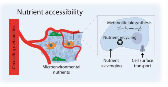
Proliferation requires that cells accumulate sufficient biomass to grow and divide. Cancer cells within tumors must acquire a variety of nutrients, and tumor growth slows or stops if necessary metabolites are not obtained in sufficient quantities. For most proliferating cells, survival and proliferation rate can be dictated by nutrient availability (Vander Heiden and DeBerardinis 2017). This effect can be mediated by a lack of substrate availability necessary to produce the macromolecules needed for biomass accumulation, or may affect critical signaling pathways that respond to nutrient levels and are required to orchestrate the processes needed for cell growth and proliferation (Torrence and Manning 2018). Regardless of mechanism, proliferation is reduced when the intracellular levels of some nutrients fall below a certain threshold. This threshold is dictated by two terms: cellular demand for that nutrient and the ability of the cell to access that nutrient or its precursors from the environment (Figure 1). Both of these terms are affected by a multitude of cell-intrinsic and cell-extrinsic factors. As a result, proliferating cells in different tumors and tissues are not universally limited by the availability of the same nutrients. Understanding which nutrients are most limiting for specific cells and determining the contexts that dictate those limitations is critical to find metabolic treatments for cancer that
11
take advantage of tumor-specific nutrient requirements and effectively starve malignancies without substantially damaging normal tissues.
NUTRIENT DEMAND
The demand for specific nutrients by cancer cells in tumors is determined by the complex interplay of many factors that influence metabolic pathway use (Figure 2). Here we will examine key variables that affect nutrient demand, including tumor-promoting mutations, chromosomal abnormalities, cancer-specific phenotypic programs, and tissue of origin.
Figure 1. Nutrient limitation is determined by the combined effects of demand and accessibility. Tumor
survival and proliferation is influenced by the intracellular concentrations of many metabolites. This intracellular concentration is dictated by the rate of consumption of the metabolite (demand) as well as the rate of metabolite acquisition (accessibility).
12
Demands imposed by mutations that drive tumor progression
Tumorigenesis is driven by genetic alterations (Hanahan and Weinberg 2011). Many of these genetic changes occur in growth-promoting signaling pathways that also activate metabolic pathways to enable biomass production. Genetic changes in cancer can also occur directly in the metabolic pathways that carry out reactions important for biomass accumulation and can alter the metabolic demands of a cell. Comprehensive descriptions of the metabolic changes that occur due to specific oncogenic mutations are explored in-depth elsewhere (Cairns et al. 2011; Nagarajan et al. 2016); here, we discuss representative examples of tumor-promoting genetic changes found in many cancers and highlight how these genetic changes impact nutrient demand in the tumor.
Figure 2. Metabolite demand of cancer cells is determined by several cell-intrinsic factors. These variables
include the presence of specific tumor-promoting mutations, chromosomal abnormalities, phenotypic states, and the tissue of origin of the tumor.
13
MYC and its upstream activators
MYC is a transcription factor that regulates the expression of a broad range of genes required
for proliferation; when dysregulated, MYC can thus act as an oncogene (Wolpaw and Dang 2018). Alterations leading to constitutive MYC expression occur frequently in cancer, and MYC is the third-most commonly amplified gene across all cancers studied in The Cancer Genome Atlas (Zack et al. 2013). Constitutive MYC expression can occur through somatic gene
amplification (Zack et al. 2013) or as a result of mutations in upstream signaling pathways such as the mitogen activated protein kinase (MAPK) pathway (Wolpaw and Dang 2018). Thus, common mutations in proto-oncogenes that are a part of the MAPK pathway, such as KRAS and
BRAF, yield similar metabolic effects as MYC activation (Dang et al. 2009; Bryant et al. 2014;
Santana-Codina et al. 2018). When active, MYC serves to stimulate broad metabolic remodeling (Nikiforov et al. 2002; Liu YC et al. 2008; Dang et al. 2009) that can alter the metabolic demands of the tumor. One prominent example is that constitutive MYC expression generates a higher requirement for consumption of the amino acid glutamine in cultured cells (Yuneva et al. 2007). This effect may be driven by MYC-associated expression of xCT, a cell-surface transporter that takes cystine into the cell while exporting glutamate (Ji et al. 2018). In the presence of
environmental cystine, high expression of xCT leads to rapid export of glutamate, which imposes a need for increased glutamine consumption in order to replenish glutamate levels (Muir et al. 2017; Sayin et al. 2017). Thus, altered metabolism in tumors with constitutive MYC activity can create new demands for certain metabolites, such as glutamine.
14
TP53
The most commonly mutated tumor suppressor gene in cancer is TP53; at least 50% of tumors display some sort of alteration in the TP53 gene (Ciriello et al. 2013). The TP53 gene product, p53, is a protein with myriad functions as a transcription factor and as a cytosolic protein (Kastenhuber and Lowe 2017). Among its many functions, p53 allows cells to adapt to nutrient deprivation (Kruiswijk et al. 2015). For instance, in response to stress conditions, p53
downregulates glycolysis through multiple mechanisms including direct inhibition of glucose transporters (Schwartzenberg-Bar-Yoseph et al. 2004) and induction of glycolysis inhibitors such as TIGAR (Bensaad et al. 2006). Metabolic genes downstream of p53 can also play a role in triggering p53-induced cell death (Jiang L et al. 2015). p53 can directly or indirectly influence expression of genes involved in lipid metabolism, amino acid transport and synthesis, and other metabolic pathways (Puzio-Kuter 2011), making it difficult to predict a priori exactly how
nutrient demands are altered by p53 loss. Further complicating the effect of p53 on nutrient demand, specific mutations of TP53 can have different effects on tumor metabolism (Humpton et al. 2018; Schofield et al. 2018). Additional study of the complex changes caused by loss of
TP53 will shed light the specific metabolic demands that are altered by this critical tumor
suppressor.
KEAP1/NFE2L2 axis
Another common alteration that occurs in cancer with implications for nutrient demand is the activation of NFE2L2, which encodes the transcription factor NRF2 that is involved in the
15
cellular response to oxidative stress (Venugopal and Jaiswal 1996; Itoh et al. 1997; Raghunath et al. 2018). NRF2 activity can also be induced by loss of function mutations in KEAP1, which encodes a ubiquitin ligase that regulates NRF2 levels by targeting it for proteasomal
degradation (Itoh et al. 1999; Kobayashi et al. 2004). NRF2 activation leads to increased
expression of genes involved in the response to oxidative stress, which includes such processes as xenobiotic detoxification and glutathione synthesis (Raghunath et al. 2018). Further, NRF2 activation leads to induction of ATF4, a transcription factor involved in the response to both nutrient deprivation and endoplasmic reticulum stress (He et al. 2001). As a result, NRF2 activation yields ATF4-dependent metabolic remodeling, including induction of de novo serine synthesis (DeNicola et al. 2015) and increased expression of xCT (Romero et al. 2017; Sayin et al. 2017) resulting in an increased dependence on glutamine metabolism as described above. Thus, alteration of the KEAP1/NRF2 signaling axis leads to metabolic changes that modify cellular demands for some amino acids. These examples typify characteristic alterations to metabolic demand created by oncogenic mutations. Given the pleiotropic, complex effects of tumor-promoting mutations, further work to develop a more thorough understanding of the metabolic consequences of these mutations is warranted.
Metabolic demands driven by chromosomal abnormalities
The tumor-promoting mutations described above activate pathways that coopt normal
16
cells and some untransformed, proliferating cells may share metabolic alterations that allow them to adapt to the metabolic demands imposed by growth signaling pathway activation (Fendt 2017). However, some tumor-promoting mutations occur through loss of large chromosomal segments, which can result in deletion of genes in regions adjacent to tumor suppressors. These large deletions sometimes include metabolic genes, which can affect metabolic pathway use (Muller et al. 2015). Beyond specific focal deletions of chromosomal regions, many cancers exhibit large-scale changes in chromosome number, known as
aneuploidy (Sansregret and Swanton 2017), that can create tumor cell characteristics that are not recapitulated in normal tissues (Knouse et al. 2014). Because these events are not
associated with a physiological metabolic program, they may create unique nutrient demands for cancer that differ from those found in all other normal cells.
Collateral mutation of metabolic genes: CDKN2A and MTAP
The most commonly deleted chromosomal locus across cancers is 9p21, due to the presence of the tumor suppressor gene CDKN2A in that region (Beroukhim et al. 2010; Zack et al. 2013).
CDKN2A codes for two proteins, p16INK4A and p14ARF (Duro et al. 1995; Mao et al. 1995; Quelle
et al. 1995; Stone et al. 1995), each of which is a tumor suppressor (Serrano et al. 1993; Serrano et al. 1995; Stott et al. 1998). CDKN2A deletions of are often accompanied by deletion of
surrounding genes (Zhang H et al. 1996), including the enzyme methylthioadenosine phosphorylase (MTAP), which is responsible for metabolizing methylthioadenosine that is produced as a byproduct of polyamine synthesis (Pegg and Williams-Ashman 1969a, 1969b; Carrera et al. 1984; Pegg 2009). Deletion of MTAP as a consequence of CDKN2A loss leads to
17
dysfunctional salvage of methylthioadenosine. As methylthioadenosine accumulates, it inhibits the protein arginine methyltransferase PRMT5, rendering cancer cells particularly sensitive to knockdown of PRMT5 and related proteins (Marjon et al. 2016; Mavrakis et al. 2016). MTAP deletion also causes global changes in metabolism that may be caused by altered epigenetic state or by perturbed methionine metabolism (Sanderson et al. 2018); in either case, MTAP deleted cells may exhibit differential nutritional demands that they must meet through adaptation of other metabolic pathways.
Collateral mutation of metabolic genes: SMAD4 and ME2
Another tumor suppressor that is commonly deleted in cancer is SMAD4 (Hahn et al. 1996). SMAD4 is a part of the TGF-β signaling pathway, and SMAD4 deletion can promote tumor progression in a variety of cancers, particularly pancreatic ductal adenocarcinoma (Bardeesy et al. 2006; Zhao et al. 2018). Among the genes located proximal to SMAD4 is malic enzyme 2 (ME2) (Dey et al. 2017), a mitochondrial enzyme that is one of three isoforms responsible for interconversion of malate and NAD(P)+ with pyruvate, NAD(P)H, and CO
2 (Moulder et al. 1945;
Hsu 1982; Taroni et al. 1987). Loss of ME2 has been suggested to limit both NADPH production and lipid synthesis in cancer (Jiang P et al. 2013), and cancer cells with ME2 deletion become sensitive to depletion of malic enzyme 3 (ME3) (Dey et al. 2017). This suggests that the demand to produce NADPH through other metabolic pathways must be increased in SMAD4/ME2 deleted tumors. Understanding how cancers cope with ME2 loss could identify metabolic liabilities to target therapeutically in SMAD4-deleted cancers.
18
Beyond focal deletions involving tumor suppressors, some larger chromosomal regions are consistently lost in specific cancers (Beroukhim et al. 2010; Zack et al. 2013; Cai et al. 2016). These losses can also eliminate expression of metabolic genes (Boots-Sprenger et al. 2013; Muller et al. 2015; Branzoli et al. 2019) and create potential therapeutic targets (Muller et al. 2012; Lin et al. 2018). Further examination of how metabolic pathways are affected by chromosomal segment deletions has the potential to uncover novel metabolic demands for certain cancers, and may uncover cancer-specific auxotrophies.
Aneuploidy
Aneuploidy produces a broad range of stresses affecting nearly every facet of biology (Zhu et al. 2018). One of the changes that occurs in aneuploid cells is widespread remodeling of
metabolism (Sheltzer 2013; Zhu et al. 2018) For instance, aneuploidy results in increased levels of ceramide lipid species, rendering aneuploid cells more dependent on pathways that
normalize ceramide levels (Tang et al. 2017). Similarly, in yeast, aneuploidy imposes an increased demand for certain sphingolipid species, and increases demand for the amino acid serine, which is required for de novo sphingolipid synthesis (Hwang et al. 2017). These findings illustrate how abnormal chromosome number and chromosomal rearrangements can alter nutrient demand, and may represent a targetable type of metabolic remodeling that is specific to cancer cells.
19
Nutrient demands determined by cellular programs that are important in some cancers
In addition to genetic changes that can alter metabolic demands, cancer cells often exhibit phenotypic changes that impact metabolism. Some prominent examples of phenotypes
observed across cancers include the epithelial to mesenchymal transition (EMT) (Brabletz et al. 2018), adoption of stem-cell like properties (Batlle and Clevers 2017), and the development of drug resistance (Brown et al. 2014; Mansoori et al. 2017). Cells that have undergone EMT, cancer cells with stem-like properties, and drug resistant cancer cells all exhibit altered metabolism, frequently driven by expression of the same genes (Morandi et al. 2017). For example, each of these states is characterized by high expression of the enzyme
dihydropyrimidine dehydrogenase (DPYD), which degrades the nucleobases uracil and thymine into dihydropyrimidines (Mani et al. 2008; Li et al. 2013; Shaul et al. 2014). Increased activity of DPYD alters levels of dihydropyrimidines relative to uracil and thymine (Shaul et al. 2014), and suggests that these cells may have an increased demand for consumption of these nucleobases. Some cellular programs alter nutrient demand in ways that impose increased requirements for certain enzymes. For instance, drug resistant cells have an increased demand for the amino acid cysteine and the tripeptide glutathione; as a result, these cells are highly dependent on
pathways that prevent lipid peroxidation and ferroptotic cell death (Hangauer et al. 2017). Broadly, the various phenotypic states adopted by tumors can result in different metabolic demands that affect which nutrients are limiting for tumor growth or survival.
20
Metabolic demands dictated by tumor tissue of origin
Beyond genetic changes and phenotypic states of tumors, some nutrient demands are shaped by the cell or tissue type from which a tumor arose (Hu et al. 2013; Gaude and Frezza 2016). Thus, these characteristics may not be shared across all tumor types, even those driven by the same oncogenes. For instance, tissue of origin can determine the extent to which tumors are dependent on particular amino acids, including non-essential amino acids such as glutamine (Yuneva et al. 2012), as well as essential nutrients such as branched chain amino acids (Mayers et al. 2016). In the case of branched chain amino acids, tumors arising from lung require the enzyme required to catabolize branched chain amino acids, while tumors arising from the pancreas do not (Mayers et al. 2016). This discrepancy in the requirement for branched chain amino acid transamination may be to fulfill differential demands for products of branched chain amino acid breakdown, such as acquisition of nitrogen or production of the amino acid
glutamate. Thus, tissue of origin can be an important determinant of nutrient demand in tumors.
NUTRIENT ACCESSIBILITY
In order to meet varying metabolic demands, tumor cells must be able to acquire relevant nutrients from their environment. Thus, as alluded to above, the second factor that determines which nutrients are limiting for cancer cell proliferation is the accessibility of nutrients in the tumor. Accessibility of nutrients is itself affected by two variables: cell-extrinsic metabolite availability in the environment and cell-intrinsic ability to obtain and effectively use those metabolites (Figure 3).
21
Circulating nutrient levels
Diet
Nutrient availability to the tumor is dependent on the abundance of circulating metabolites in the blood. The macronutrient content of diet can lead to complex changes in circulating nutrient levels. For instance, consuming diets with varying caloric content and different calorie sources can alter the lipid profile in blood (Raeini-Sarjaz et al. 2001; Appel et al. 2005; Ma et al. 2006). Further, depletion or supplementation of certain nutrients in the diet can lead to a concomitant change in circulating nutrient abundance. In the most extreme example, the circulating levels of nutrients that cannot be synthesized by humans are strongly influenced by diet (Fitzpatrick et al. 2012). As a result, dietary vitamin levels can impact tumor growth,
Figure 3. Nutrient accessibility to cells within tumors is driven by both cell-extrinsic and cell-intrinsic variables. Circulating metabolite levels and local microenvironmental nutrient levels determine nutrient
accessibility to cells within the tumor, and cell surface transport, scavenging, recycling, and de novo metabolite synthesis influence the intracellular levels of metabolites that can be used by the cells.
22
because although vitamins are typically not consumed by enzymatic reactions, each newly formed cancer cell must be able to obtain a sufficient supply of vitamins to support its
enzymatic reactions. For example, dietary folic acid supplementation was noted to exacerbate childhood leukemia in the 1940s (Farber et al. 1947; Farber et al. 1948), and can accelerate the development of murine breast tumors (Hansen et al. 2017). However, given the pleiotropic effects of vitamin deprivation on overall animal health, dietary levels of folic acid have been reported to both positively and negatively affect the risk of developing tumors in humans (Ulrich 2007; Lamm et al. 2015; Ashkavand et al. 2017).
The dietary content of some non-essential nutrients can also influence circulating levels of those metabolites. Feeding mice a diet lacking the amino acids serine and glycine results in lower serine levels in circulation (Maddocks et al. 2013). Reduced serine accessibility in this setting can slow tumor growth without grossly affecting animal health (Maddocks et al. 2013; Maddocks et al. 2017), consistent with tumors having a high demand for serine that they are not able to meet given a diminished accessibility of serine in the circulation. This principle can also be applied more generally in fasted animals. Long-term fasting broadly alters circulating metabolite levels (Broer S and Broer 2017), shifting nutrient accessibility in a manner that decreases tumor proliferation (Lee et al. 2012; Sun et al. 2017). Thus, both broad and specific dietary manipulations can alter nutrient accessibility in tumors.
23
Hormonal control of metabolism
Circulating nutrient levels are not solely determined by the diet. Instead, complex hormonal mechanisms influence the levels of some nutrients in blood. Perhaps the most well-studied example of hormonal regulation of metabolism is in the control of circulating glucose levels. Glucose levels in the blood are tightly regulated by the action of the hormones insulin and glucagon. Insulin stimulates glucose uptake in many cell types and broadly functions to clear glucose from circulation (Wilcox 2005). Conversely, glucagon stimulates glucose release into the bloodstream from hepatic glycogen stores and de novo glucose synthesis through the process of gluconeogenesis (Han et al. 2016). Glucose is not the only metabolite regulated by
hormones. In fact, it has long been recognized that endocrine signaling influences the
concentrations of amino acids in blood (Friedberg and Greenberg 1947). As a result, circulating amino acid levels are largely held within a certain range of concentrations independent of the composition of diet. Systemic metabolism does not fully control circulating amino acid levels, as within this normal range, amino acid levels can fluctuate over the course of the day in response to normal feeding and fasting, and certain amino acids such as serine, glycine, and alanine vary over a wider range than other amino acids (Broer S and Broer 2017). However, consistent with the importance of hormonal regulation of amino acid levels, derangement of whole-body metabolism in obesity or cancer alters circulating levels of branched chain amino acids (Newgard et al. 2009; Mayers et al. 2014). Beyond regulation of metabolite levels, hormonal processes mediate the effects of dietary and environmental perturbations on circulating
nutrient levels. For instance, fasting has been shown to inhibit leukemia progression by altering expression of leptin receptor (Lu et al. 2017), a protein which binds to the hormone leptin and
24
is involved in maintenance of whole-body energy homeostasis (Kelesidis et al. 2010). Thus, in addition to directly setting the circulating levels of many metabolites, hormonal mechanisms may also mediate the effects of diet and other environmental factors on nutrient accessibility in tumors.
Influence of the microbiome on systemic metabolite levels
Systemic metabolism can be influenced by the actions of the gut microbiome. The microbiome carries out many metabolic reactions, and can affect which nutrients in the diet end up in circulation, or produce metabolites that do not directly reflect the content of diet (LeBlanc et al. 2013; Fujisaka et al. 2018). Microbiome composition may also affect systemic metabolism, as fecal microbiota transplants are sufficient to predictably alter non-fasting glucose levels in mice (Ussar et al. 2015). Thus, the behavior of the microbiome appears to also influence tumor nutrient accessibility.
Fluctuating metabolite levels due to circadian rhythms
Nutrient availability in circulation is not constant throughout the day. In fact, circadian rhythms exhibit a profound effect on the plasma levels of metabolites, with some nutrients displaying greater than 2.5 fold differences in circulating concentration throughout the day (Dallmann et al. 2012; Masri and Sassone-Corsi 2018). In contrast, some metabolites largely do not fluctuate throughout the day (Dallmann et al. 2012). Given the variability in circadian fluctuation
between nutrients, certain metabolites could be less accessible and thus more limiting for tumor growth at particular times during the day. For this reason, it may be advantageous for
25
tumors to reprogram circadian metabolism to promote more favorable nutrient accessibility. Indeed, lung adenocarcinoma has been observed to alter hepatic circadian rhythms in a way that alters whole-body metabolism (Masri et al. 2016). For these reasons, when examining nutrient accessibility with the goal of understanding metabolic limitations on tumors, it is important to consider the effects that circadian rhythms have on circulating metabolite levels.
Nutrient levels in the tumor microenvironment
Though systemic metabolism can impact blood nutrient levels, tumors do not have
straightforward access to all of the nutrients present in circulation. The delivery of nutrients to tumor cells is complicated by altered vasculature and competition for metabolites between various cells within the tumor microenvironment. Here we examine some of the factors in the local tumor microenvironment that alter nutrient accessibility.
Tumor vascularization and lymphatics
In contrast to normal tissues, tumors have irregularly spaced, poorly functioning blood vessels (Fukumura et al. 2010). As a result, tumors may not be able to efficiently exchange nutrients and waste products with the circulation. Further compounding the abnormal vasculature is the presence of dysfunctional lymphatics in tumors. Lymphatic ducts are responsible for returning fluid and metabolites that drain from a tissue into the blood (Wiig and Swartz 2012). Solid tumors are typically highly compressed and, as a result, can be deficient in functional
lymphatics. This further increases tumoral interstitial pressure, which inhibits nutrient uptake from the blood (Fukumura et al. 2010; Wiig and Swartz 2012). As a result, the accessibility of
26
nutrients to the tumor depends both on the extent of vascularization and the effectiveness of the blood vessels present in the tumor.
Competition for nutrients between cell types in the tumor microenvironment
Once nutrients are delivered to the local tumor microenvironment, all cells within the tissue, including stromal and immune cells compete for nutrients within the tumor. For example, some immune cells important for restricting tumor growth, such as activated T cells, acquire
metabolic characteristics that are similar to tumor cells and are often driven by the same transcriptional programs that exist in cancer (Wang et al. 2011; Le Bourgeois et al. 2018). Thus, these cells compete with tumor cells for the same nutrients. Beyond competition, some
immune cells degrade or sequester critical nutrients in the tumor microenvironment (Lyssiotis and Kimmelman 2017). For instance, myeloid derived suppressor cells (MDSCs) deplete the amino acids arginine, tryptophan, and cystine from the tumor microenvironment beyond their own metabolic needs (Kumar et al. 2016). Degradation of these nutrients has an
immunosuppressive effect that can favor tumor progression; however, tumors must also cope with this altered nutrient availability.
Nutrient sharing between cell types in the tumor microenvironment
Stromal cells in the tumor microenvironment can also alter nutrient accessibility in a way that is favorable to cancer cells (Lyssiotis and Kimmelman 2017). For example, primary chronic
lymphocytic leukemia (CLL) cells have a limited ability to uptake the amino acid cystine due to low expression of the cystine-glutamate antiporter xCT. Bone marrow stromal cells that are
27
present in the CLL niche, in contrast, are capable of importing cystine using xCT and then excreting cysteine, the reduced form of cystine that can be transported using the ASC family of amino acid transporters (Barker and Ellory 1990; Zhang W et al. 2012). This allows the CLL cells to take up cysteine, providing increased access to a crucial amino acid. Further examples of how cell populations within a tumor might exchange nutrients in a symbiotic relationship are also prominent in the literature (Sonveaux et al. 2008; Tardito et al. 2015; Sousa et al. 2016; Yang et al. 2016).
Uptake of nutrients
Rather than relying upon local delivery or microenvironmental production of specific
metabolites, tumor cells can increase the accessibility of nutrients by more effectively taking up metabolites from their environment. For instance, xCT deficient CLL cells mentioned above would not have to rely upon stromal cells to produce reduced cysteine if they were more capable of oxidized cystine uptake. To more effectively acquire metabolites, cancer cells can modulate nutrient uptake using a variety of mechanisms; here we examine some of the adaptations that tumors utilize to better obtain nutrients and therefore increase the accessibility of metabolites from the environment.
Cell surface transporters
Most nutrients are transported into cells through transmembrane proteins, and transport can be either equilibrative or coupled to an energy consuming process to concentrate the nutrient in cells (Glatz et al. 2010; Hediger et al. 2013; Szablewski 2013; Perez-Escuredo et al. 2016;
28
Young 2016; Inoue 2017). Many nutrient transport proteins are upregulated in cancer, and there has been a renewed interest in understanding nutrient transport phenomena in cancer (Cesar-Razquin et al. 2015; Krall et al. 2016; Broer A et al. 2018; Cha et al. 2018; Ladanyi et al. 2018; Tajan et al. 2018; Todenhofer et al. 2018). One of the most well-known cancer
phenotypes is the avid uptake of glucose, a phenomenon that is at least partially driven by increased expression of GLUT family glucose transporters (Szablewski 2013). This increased capacity for glucose transport may increase glucose accessibility to tumors.
In some cases, transporter expression has less predictable effects on nutrient uptake, and therefore availability. The uptake of amino acids occurs through a series of transmembrane transporters with overlapping amino acid specificities (Hediger et al. 2013; Kandasamy et al. 2018). Many of these transporters are obligate amino acid exchangers, which take up one amino acid and excrete a second. These amino acid exchangers are unable to facilitate net uptake of amino acids, but are instead able to shift the relative ratios of various intracellular amino acids. Multiple studies have shown that knocking down expression of amino acid exchangers such as ASCT2 can hinder cancer growth (van Geldermalsen et al. 2016; Cormerais et al. 2018), suggesting that the redistribution of intracellular and extracellular amino acids can affect tumor proliferation. However, the specific effects of increased expression of amino acid exchangers on nutrient accessibility are difficult to predict due to the complex and redundant interactions between amino acid transporters. Further, amino acid transporter activity will be affected by the intracellular and extracellular levels of multiple amino acids. Many studies of amino acid exchange involve non-physiological conditions wherein cells are loaded with high levels of an amino acid of interest and then efflux of that amino acid and uptake of others are
29
observed. Thus, further work to understand the functions of each transporter when physiological concentrations of amino acids are present is warranted.
Nutrient uptake from extracellular polymers and macromolecules
Not all nutrient acquisition is mediated by the uptake of free metabolites through cell-surface transporters; instead, some metabolites are scavenged from extracellular polymers. One such nutrient is glutathione, a tripeptide that is present at ~10 µM in circulation (Pastore et al. 1998). Glutathione contains the amino acids cysteine, glutamate, and glycine, and can be a source of these amino acids (Orlowski and Meister 1970); however, tumor cells must first degrade glutathione into its amino acid components for uptake through cell-surface transporters. This process requires the expression of γ-glutamyl transferases (GGTs), extracellular membrane proteins that degrade glutathione in the environment. GGT expression is upregulated in some tumors (Hanigan et al. 1999) and downregulated in others (Priolo et al. 2018), suggesting that tumors may differ in their ability to degrade and utilize glutathione as a source of amino acids. Recent work has suggested that glutathione breakdown may serve additional functions (Boysen 2017), but the ability of cells to express GGT and utilize extracellular glutathione as a nutrient source likely plays a role in determining the accessibility of certain amino acids to tumors.
Some cancer cells are able to take up larger macromolecules. For instance, certain tumors are able to consume whole protein from the environment through integrin-mediated scavenging (Finicle et al. 2018), receptor-mediated endocytosis (Merlot et al. 2014; Finicle et al. 2018) or by non-specific uptake involving macropinocytosis (Recouvreux and Commisso 2017; Finicle et al. 2018). The ability to effectively utilize extracellular protein as an amino acid source
30
can increase the effective accessibility of amino acids for cells within tumors (Finicle et al. 2018). Nutrient scavenging can also be used to take up molecules other than protein to support cellular metabolic processes (Kim et al. 2018). Beyond taking up macromolecules from the environment, some cells can even invade and consume neighboring cells through the process of entosis, allowing for replenishment of nutrients (Overholtzer et al. 2007; Krajcovic et al. 2013; Hamann et al. 2017). Though this process is likely a response to low nutrient levels rather than an active strategy of obtaining nutrients under basal conditions, it provides another path for cancer cells to increase nutrient accessibility.
Nutrient recycling
In addition to altered nutrient uptake, some tumors are able to modulate metabolite
accessibility by breaking down macromolecules into their constituent parts through the process of autophagy (Kimmelman and White 2017; Wyant et al. 2017; An and Harper 2018).
Autophagy can alter intracellular metabolite levels in response to starvation (Guo et al. 2016), but recycling of nutrients in this way cannot provide a net source of new metabolites for tumor cells. Recycling processes do not lead to net metabolite consumption, and are therefore unable to directly fuel cell growth; however, nutrient recycling can be important as a mechanism to alter nutrient accessibility to allow tumors to preserve levels of critical nutrients during
transient periods of deprivation, and thus can affect cancer cell survival. This effect on nutrient availability may explain, in part, why impairment of autophagy can hinder tumor growth (Kimmelman and White 2017).
31
Altered nutrient biosynthesis
For those metabolites that can be produced by tumors, de novo synthesis represents an important method to modulate nutrient availability. The synthesis of many classes of
metabolites, including amino acids (Liu W et al. 2012; Mattaini et al. 2016) and lipids (Long et al. 2018), is upregulated in tumors and may represent a method for tumors to bypass low
environmental nutrient accessibility in some tissue contexts. Often, the increased rate of de
novo metabolite synthesis is necessary to maintain tumor proliferation, suggesting that the
improved nutrient availability resulting from increased biosynthesis is required to meet the nutrient demands of the cell. For instance, many tumors upregulate the enzymes of the serine synthesis pathway, which converts the glycolytic intermediate 3-phosphoglycerate into serine through a three-step process (Adams 2007; Locasale et al. 2011; Possemato et al. 2011; Nilsson et al. 2012; DeNicola et al. 2015; Ben-Sahra et al. 2016; Samanta et al. 2016). The altered availability of nutrients due to upregulation of serine synthesis can promote tumor growth (Possemato et al. 2011; DeNicola et al. 2015; Pacold et al. 2016). However, biosynthesis of one nutrient inevitably generates a metabolic cost elsewhere. For instance, for each molecule of serine synthesized by a cancer cell, the cell must consume ½ glucose, 2 NAD+, and convert 1
glutamate into 1 α-ketoglutarate. Thus, altered nutrient biosynthesis can affect the allocation of metabolic resources to other pathways, not just the end product of a biosynthetic pathway. As a result, variability in biosynthetic rates can be an important determinant of nutrient availability for tumors.
32
CONCLUSIONS
Restricting tumor growth by targeting metabolic pathways or nutrients that are limiting for proliferation remains an attractive therapeutic strategy. However, to successfully target metabolism in this fashion, we must continue to develop a better understanding of what is limiting for tumor growth and the factors that determine these limitations. Both the demand for a metabolite and its accessibility to cancer cells within a tumor will define what is limiting, and each of these terms is determined by a complex mix of tumor-intrinsic and tumor-extrinsic factors that must be considered when studying cancer metabolism. Critically, these variables often represent factors that are unique to specific tumor types. Thus, the altered demand and accessibility of nutrients in tumors may render them specifically vulnerable to inhibition of certain metabolic pathways or deprivation of particular nutrients. Better understanding the complex interplay between these factors will be essential to turn these unique characteristics of cancers into specific, effective therapies.
33
REFERENCES
Adams CM. 2007. Role of the transcription factor ATF4 in the anabolic actions of insulin and the anti-anabolic actions of glucocorticoids. J Biol Chem. 282(23):16744-16753.
An H, Harper JW. 2018. Systematic analysis of ribophagy in human cells reveals bystander flux during selective autophagy. Nat Cell Biol. 20(2):135-143.
Appel LJ, Sacks FM, Carey VJ, Obarzanek E, Swain JF, Miller ER, 3rd, Conlin PR, Erlinger TP, Rosner BA, Laranjo NM et al. 2005. Effects of protein, monounsaturated fat, and carbohydrate intake on blood pressure and serum lipids: results of the OmniHeart randomized trial. JAMA. 294(19):2455-2464.
Ashkavand Z, O'Flanagan C, Hennig M, Du X, Hursting SD, Krupenko SA. 2017. Metabolic
Reprogramming by Folate Restriction Leads to a Less Aggressive Cancer Phenotype. Mol Cancer Res. 15(2):189-200.
Bardeesy N, Cheng KH, Berger JH, Chu GC, Pahler J, Olson P, Hezel AF, Horner J, Lauwers GY, Hanahan D et al. 2006. Smad4 is dispensable for normal pancreas development yet critical in progression and tumor biology of pancreas cancer. Genes Dev. 20(22):3130-3146.
Barker GA, Ellory JC. 1990. The Identification of Neutral Amino-Acid-Transport Systems. Exp Physiol. 75(1):3-26. English.
Batlle E, Clevers H. 2017. Cancer stem cells revisited. Nat Med. 23(10):1124-1134.
Ben-Sahra I, Hoxhaj G, Ricoult SJH, Asara JM, Manning BD. 2016. mTORC1 induces purine synthesis through control of the mitochondrial tetrahydrofolate cycle. Science. 351(6274):728-733. English.
Bensaad K, Tsuruta A, Selak MA, Vidal MN, Nakano K, Bartrons R, Gottlieb E, Vousden KH. 2006. TIGAR, a p53-inducible regulator of glycolysis and apoptosis. Cell. 126(1):107-120. Beroukhim R, Mermel CH, Porter D, Wei G, Raychaudhuri S, Donovan J, Barretina J, Boehm JS,
Dobson J, Urashima M et al. 2010. The landscape of somatic copy-number alteration across human cancers. Nature. 463(7283):899-905.
Boots-Sprenger SH, Sijben A, Rijntjes J, Tops BB, Idema AJ, Rivera AL, Bleeker FE, Gijtenbeek AM, Diefes K, Heathcock L et al. 2013. Significance of complete 1p/19q co-deletion, IDH1 mutation and MGMT promoter methylation in gliomas: use with caution. Mod Pathol. 26(7):922-929.
Boysen G. 2017. The Glutathione Conundrum: Stoichiometric Disconnect between Its Formation and Oxidative Stress. Chem Res Toxicol. 30(5):1113-1116.
Brabletz T, Kalluri R, Nieto MA, Weinberg RA. 2018. EMT in cancer. Nat Rev Cancer. 18(2):128-134.
Branzoli F, Pontoizeau C, Tchara L, Di Stefano AL, Kamoun A, Deelchand DK, Valabregue R, Lehericy S, Sanson M, Ottolenghi C et al. 2019. Cystathionine as a marker for 1p/19q codeleted gliomas by in vivo magnetic resonance spectroscopy. Neuro Oncol.
Broer A, Fairweather S, Broer S. 2018. Disruption of Amino Acid Homeostasis by Novel ASCT2 Inhibitors Involves Multiple Targets. Front Pharmacol. 9:785.
Broer S, Broer A. 2017. Amino acid homeostasis and signalling in mammalian cells and organisms. Biochem J. 474(12):1935-1963.
34
Brown R, Curry E, Magnani L, Wilhelm-Benartzi CS, Borley J. 2014. Poised epigenetic states and acquired drug resistance in cancer. Nat Rev Cancer. 14(11):747-753.
Bryant KL, Mancias JD, Kimmelman AC, Der CJ. 2014. KRAS: feeding pancreatic cancer proliferation. Trends Biochem Sci. 39(2):91-100.
Cai Y, Crowther J, Pastor T, Abbasi Asbagh L, Baietti MF, De Troyer M, Vazquez I, Talebi A, Renzi F, Dehairs J et al. 2016. Loss of Chromosome 8p Governs Tumor Progression and Drug Response by Altering Lipid Metabolism. Cancer Cell. 29(5):751-766.
Cairns RA, Harris IS, Mak TW. 2011. Regulation of cancer cell metabolism. Nat Rev Cancer. 11(2):85-95.
Carrera CJ, Eddy RL, Shows TB, Carson DA. 1984. Assignment of the gene for
methylthioadenosine phosphorylase to human chromosome 9 by mouse-human somatic cell hybridization. Proc Natl Acad Sci U S A. 81(9):2665-2668.
Cesar-Razquin A, Snijder B, Frappier-Brinton T, Isserlin R, Gyimesi G, Bai X, Reithmeier RA, Hepworth D, Hediger MA, Edwards AM et al. 2015. A Call for Systematic Research on Solute Carriers. Cell. 162(3):478-487.
Cha YJ, Kim ES, Koo JS. 2018. Amino Acid Transporters and Glutamine Metabolism in Breast Cancer. Int J Mol Sci. 19(3).
Ciriello G, Miller ML, Aksoy BA, Senbabaoglu Y, Schultz N, Sander C. 2013. Emerging landscape of oncogenic signatures across human cancers. Nat Genet. 45(10):1127-1133.
Cormerais Y, Massard PA, Vucetic M, Giuliano S, Tambutte E, Durivault J, Vial V, Endou H, Wempe MF, Parks SK et al. 2018. The glutamine transporter ASCT2 (SLC1A5) promotes tumor growth independently of the amino acid transporter LAT1 (SLC7A5). J Biol Chem. 293(8):2877-2887.
Dallmann R, Viola AU, Tarokh L, Cajochen C, Brown SA. 2012. The human circadian metabolome. Proc Natl Acad Sci U S A. 109(7):2625-2629.
Dang CV, Le A, Gao P. 2009. MYC-induced cancer cell energy metabolism and therapeutic opportunities. Clin Cancer Res. 15(21):6479-6483.
DeNicola GM, Chen PH, Mullarky E, Sudderth JA, Hu Z, Wu D, Tang H, Xie Y, Asara JM, Huffman KE et al. 2015. NRF2 regulates serine biosynthesis in non-small cell lung cancer. Nat Genet. 47(12):1475-1481.
Dey P, Baddour J, Muller F, Wu CC, Wang H, Liao WT, Lan Z, Chen A, Gutschner T, Kang Y et al. 2017. Genomic deletion of malic enzyme 2 confers collateral lethality in pancreatic cancer. Nature. 542(7639):119-123.
Duro D, Bernard O, Della Valle V, Berger R, Larsen CJ. 1995. A new type of p16INK4/MTS1 gene transcript expressed in B-cell malignancies. Oncogene. 11(1):21-29.
Farber S, Cutler EC, Hawkins JW, Harrison JH, Peirce EC, 2nd, Lenz GG. 1947. The Action of Pteroylglutamic Conjugates on Man. Science. 106(2764):619-621.
Farber S, Diamond LK, Mercer RD, Sylvester RF, Wolff JA. 1948. Temporary remissions in acute leukemia in children produced by folic acid antagonist, 4-aminopteryl-glutamic acid (aminopterin). New England Journal of Medicine. 238(23):787-793.
Fendt SM. 2017. Is There a Therapeutic Window for Metabolism-Based Cancer Therapies? Front Endocrinol (Lausanne). 8:150.
35
Finicle BT, Jayashankar V, Edinger AL. 2018. Nutrient scavenging in cancer. Nat Rev Cancer. 18(10):619-633.
Fitzpatrick TB, Basset GJ, Borel P, Carrari F, DellaPenna D, Fraser PD, Hellmann H, Osorio S, Rothan C, Valpuesta V et al. 2012. Vitamin deficiencies in humans: can plant science help? Plant Cell. 24(2):395-414.
Friedberg F, Greenberg DM. 1947. Endocrine regulation of amino acid levels in blood and tissues. J Biol Chem. 168(2):405-409.
Fujisaka S, Avila-Pacheco J, Soto M, Kostic A, Dreyfuss JM, Pan H, Ussar S, Altindis E, Li N, Bry L et al. 2018. Diet, Genetics, and the Gut Microbiome Drive Dynamic Changes in Plasma Metabolites. Cell Rep. 22(11):3072-3086.
Fukumura D, Duda DG, Munn LL, Jain RK. 2010. Tumor microvasculature and
microenvironment: novel insights through intravital imaging in pre-clinical models. Microcirculation. 17(3):206-225.
Gaude E, Frezza C. 2016. Tissue-specific and convergent metabolic transformation of cancer correlates with metastatic potential and patient survival. Nat Commun. 7:13041. Glatz JF, Luiken JJ, Bonen A. 2010. Membrane fatty acid transporters as regulators of lipid
metabolism: implications for metabolic disease. Physiol Rev. 90(1):367-417.
Guo JY, Teng X, Laddha SV, Ma S, Van Nostrand SC, Yang Y, Khor S, Chan CS, Rabinowitz JD, White E. 2016. Autophagy provides metabolic substrates to maintain energy charge and nucleotide pools in Ras-driven lung cancer cells. Genes Dev. 30(15):1704-1717.
Hahn SA, Schutte M, Hoque ATMS, Moskaluk CA, daCosta LT, Rozenblum E, Weinstein CL, Fischer A, Yeo CJ, Hruban RH et al. 1996. DPC4, a candidate tumor suppressor gene at human chromosome 18q21.1. Science. 271(5247):350-353. English.
Hamann JC, Surcel A, Chen R, Teragawa C, Albeck JG, Robinson DN, Overholtzer M. 2017. Entosis Is Induced by Glucose Starvation. Cell Rep. 20(1):201-210.
Han HS, Kang G, Kim JS, Choi BH, Koo SH. 2016. Regulation of glucose metabolism from a liver-centric perspective. Exp Mol Med. 48:e218.
Hanahan D, Weinberg RA. 2011. Hallmarks of cancer: the next generation. Cell. 144(5):646-674. Hangauer MJ, Viswanathan VS, Ryan MJ, Bole D, Eaton JK, Matov A, Galeas J, Dhruv HD, Berens
ME, Schreiber SL et al. 2017. Drug-tolerant persister cancer cells are vulnerable to GPX4 inhibition. Nature. 551(7679):247-250.
Hanigan MH, Gallagher BC, Townsend DM, Gabarra V. 1999. Gamma-glutamyl transpeptidase accelerates tumor growth and increases the resistance of tumors to cisplatin in vivo. Carcinogenesis. 20(4):553-559.
Hansen MF, Jensen SO, Fuchtbauer EM, Martensen PM. 2017. High folic acid diet enhances tumour growth in PyMT-induced breast cancer. Br J Cancer. 116(6):752-761.
He CH, Gong P, Hu B, Stewart D, Choi ME, Choi AM, Alam J. 2001. Identification of activating transcription factor 4 (ATF4) as an Nrf2-interacting protein. Implication for heme oxygenase-1 gene regulation. J Biol Chem. 276(24):20858-20865.
Hediger MA, Clemencon B, Burrier RE, Bruford EA. 2013. The ABCs of membrane transporters in health and disease (SLC series): introduction. Mol Aspects Med. 34(2-3):95-107.
36
Hu J, Locasale JW, Bielas JH, O'Sullivan J, Sheahan K, Cantley LC, Vander Heiden MG, Vitkup D. 2013. Heterogeneity of tumor-induced gene expression changes in the human metabolic network. Nat Biotechnol. 31(6):522-529.
Humpton TJ, Hock AK, Maddocks ODK, Vousden KH. 2018. p53-mediated adaptation to serine starvation is retained by a common tumour-derived mutant. Cancer Metab. 6:18.
Hwang S, Gustafsson HT, O'Sullivan C, Bisceglia G, Huang X, Klose C, Schevchenko A, Dickson RC, Cavaliere P, Dephoure N et al. 2017. Serine-Dependent Sphingolipid Synthesis Is a Metabolic Liability of Aneuploid Cells. Cell Rep. 21(13):3807-3818.
Inoue K. 2017. Molecular Basis of Nucleobase Transport Systems in Mammals. Biol Pharm Bull. 40(8):1130-1138.
Itoh K, Chiba T, Takahashi S, Ishii T, Igarashi K, Katoh Y, Oyake T, Hayashi N, Satoh K, Hatayama I et al. 1997. An Nrf2 small Maf heterodimer mediates the induction of phase II
detoxifying enzyme genes through antioxidant response elements. Biochemical and Biophysical Research Communications. 236(2):313-322. English.
Itoh K, Wakabayashi N, Katoh Y, Ishii T, Igarashi K, Engel JD, Yamamoto M. 1999. Keap1
represses nuclear activation of antioxidant responsive elements by Nrf2 through binding to the amino-terminal Neh2 domain. Gene Dev. 13(1):76-86. English.
Ji X, Qian J, Rahman SMJ, Siska PJ, Zou Y, Harris BK, Hoeksema MD, Trenary IA, Heidi C, Eisenberg R et al. 2018. xCT (SLC7A11)-mediated metabolic reprogramming promotes non-small cell lung cancer progression. Oncogene. 37(36):5007-5019.
Jiang L, Kon N, Li T, Wang SJ, Su T, Hibshoosh H, Baer R, Gu W. 2015. Ferroptosis as a p53-mediated activity during tumour suppression. Nature. 520(7545):57-62.
Jiang P, Du W, Mancuso A, Wellen KE, Yang X. 2013. Reciprocal regulation of p53 and malic enzymes modulates metabolism and senescence. Nature. 493(7434):689-693.
Kandasamy P, Gyimesi G, Kanai Y, Hediger MA. 2018. Amino acid transporters revisited: New views in health and disease. Trends Biochem Sci. 43(10):752-789.
Kastenhuber ER, Lowe SW. 2017. Putting p53 in Context. Cell. 170(6):1062-1078.
Kelesidis T, Kelesidis I, Chou S, Mantzoros CS. 2010. Narrative review: the role of leptin in human physiology: emerging clinical applications. Ann Intern Med. 152(2):93-100. Kim SM, Nguyen TT, Ravi A, Kubiniok P, Finicle BT, Jayashankar V, Malacrida L, Hou J, Robertson
J, Gao D et al. 2018. PTEN Deficiency and AMPK Activation Promote Nutrient Scavenging and Anabolism in Prostate Cancer Cells. Cancer Discov. 8(7):866-883.
Kimmelman AC, White E. 2017. Autophagy and Tumor Metabolism. Cell Metab. 25(5):1037-1043.
Knouse KA, Wu J, Whittaker CA, Amon A. 2014. Single cell sequencing reveals low levels of aneuploidy across mammalian tissues. Proc Natl Acad Sci U S A. 111(37):13409-13414. Kobayashi A, Kang MI, Okawa H, Ohtsuji M, Zenke Y, Chiba T, Igarashi K, Yamamoto M. 2004.
Oxidative stress sensor Keap1 functions as an adaptor for Cul3-based E3 ligase to regulate proteasomal degradation of Nrf2. Mol Cell Biol. 24(16):7130-7139.
Krajcovic M, Krishna S, Akkari L, Joyce JA, Overholtzer M. 2013. mTOR regulates phagosome and entotic vacuole fission. Mol Biol Cell. 24(23):3736-3745.
Krall AS, Xu S, Graeber TG, Braas D, Christofk HR. 2016. Asparagine promotes cancer cell proliferation through use as an amino acid exchange factor. Nat Commun. 7:11457.
37
Kruiswijk F, Labuschagne CF, Vousden KH. 2015. p53 in survival, death and metabolic health: a lifeguard with a licence to kill. Nat Rev Mol Cell Biol. 16(7):393-405.
Kumar V, Patel S, Tcyganov E, Gabrilovich DI. 2016. The Nature of Myeloid-Derived Suppressor Cells in the Tumor Microenvironment. Trends Immunol. 37(3):208-220.
Ladanyi A, Mukherjee A, Kenny HA, Johnson A, Mitra AK, Sundaresan S, Nieman KM, Pascual G, Benitah SA, Montag A et al. 2018. Adipocyte-induced CD36 expression drives ovarian cancer progression and metastasis. Oncogene. 37(17):2285-2301.
Lamm N, Maoz K, Bester AC, Im MM, Shewach DS, Karni R, Kerem B. 2015. Folate levels modulate oncogene-induced replication stress and tumorigenicity. EMBO Mol Med. 7(9):1138-1152.
Le Bourgeois T, Strauss L, Aksoylar HI, Daneshmandi S, Seth P, Patsoukis N, Boussiotis VA. 2018. Targeting T Cell Metabolism for Improvement of Cancer Immunotherapy. Front Oncol. 8:237.
LeBlanc JG, Milani C, de Giori GS, Sesma F, van Sinderen D, Ventura M. 2013. Bacteria as vitamin suppliers to their host: a gut microbiota perspective. Curr Opin Biotechnol. 24(2):160-168.
Lee C, Raffaghello L, Brandhorst S, Safdie FM, Bianchi G, Martin-Montalvo A, Pistoia V, Wei M, Hwang S, Merlino A et al. 2012. Fasting cycles retard growth of tumors and sensitize a range of cancer cell types to chemotherapy. Sci Transl Med. 4(124):124ra127.
Li LH, Dong H, Zhao F, Tang J, Chen X, Ding J, Men HT, Luo WX, Du Y, Ge J et al. 2013. The upregulation of dihydropyrimidine dehydrogenase in liver is involved in acquired resistance to 5-fluorouracil. Eur J Cancer. 49(7):1752-1760.
Lin Y-H, Satani N, Hammoudi N, Ackroyd JJ, Khadka S, Yan VC, Georgiou DK, Sun Y, Zielinski R, Tran T et al. 2018. Eradication of ENO1-deleted Glioblastoma through Collateral Lethality. bioRxiv. doi: http://dx.doi.org/10.1101/331538.
Liu W, Le A, Hancock C, Lane AN, Dang CV, Fan TW, Phang JM. 2012. Reprogramming of proline and glutamine metabolism contributes to the proliferative and metabolic responses regulated by oncogenic transcription factor c-MYC. Proc Natl Acad Sci U S A.
109(23):8983-8988.
Liu YC, Li F, Handler J, Huang CR, Xiang Y, Neretti N, Sedivy JM, Zeller KI, Dang CV. 2008. Global regulation of nucleotide biosynthetic genes by c-Myc. PLoS One. 3(7):e2722.
Locasale JW, Grassian AR, Melman T, Lyssiotis CA, Mattaini KR, Bass AJ, Heffron G, Metallo CM, Muranen T, Sharfi H et al. 2011. Phosphoglycerate dehydrogenase diverts glycolytic flux and contributes to oncogenesis. Nat Genet. 43(9):869-874.
Long J, Zhang CJ, Zhu N, Du K, Yin YF, Tan X, Liao DF, Qin L. 2018. Lipid metabolism and carcinogenesis, cancer development. Am J Cancer Res. 8(5):778-791.
Lu Z, Xie J, Wu G, Shen J, Collins R, Chen W, Kang X, Luo M, Zou Y, Huang LJ et al. 2017. Fasting selectively blocks development of acute lymphoblastic leukemia via leptin-receptor upregulation. Nat Med. 23(1):79-90.
Lyssiotis CA, Kimmelman AC. 2017. Metabolic Interactions in the Tumor Microenvironment. Trends Cell Biol. 27(11):863-875.
38
Ma YS, Li YF, Chiriboga DE, Olendzki BC, Hebert JR, Li WJ, Leung K, Hafner AR, Ockene IS. 2006. Association between carbohydrate intake and serum lipids. Journal of the American College of Nutrition. 25(2):155-163. English.
Maddocks ODK, Athineos D, Cheung EC, Lee P, Zhang T, van den Broek NJF, Mackay GM, Labuschagne CF, Gay D, Kruiswijk F et al. 2017. Modulating the therapeutic response of tumours to dietary serine and glycine starvation. Nature. 544(7650):372-376.
Maddocks ODK, Berkers CR, Mason SM, Zheng L, Blyth K, Gottlieb E, Vousden KH. 2013. Serine starvation induces stress and p53-dependent metabolic remodelling in cancer cells. Nature. 493(7433):542-546.
Mani SA, Guo W, Liao MJ, Eaton EN, Ayyanan A, Zhou AY, Brooks M, Reinhard F, Zhang CC, Shipitsin M et al. 2008. The epithelial-mesenchymal transition generates cells with properties of stem cells. Cell. 133(4):704-715.
Mansoori B, Mohammadi A, Davudian S, Shirjang S, Baradaran B. 2017. The Different
Mechanisms of Cancer Drug Resistance: A Brief Review. Adv Pharm Bull. 7(3):339-348. Mao L, Merlo A, Bedi G, Shapiro GI, Edwards CD, Rollins BJ, Sidransky D. 1995. A novel
p16INK4A transcript. Cancer Res. 55(14):2995-2997.
Marjon K, Cameron MJ, Quang P, Clasquin MF, Mandley E, Kunii K, McVay M, Choe S, Kernytsky A, Gross S et al. 2016. MTAP Deletions in Cancer Create Vulnerability to Targeting of the MAT2A/PRMT5/RIOK1 Axis. Cell Rep. 15(3):574-587.
Masri S, Papagiannakopoulos T, Kinouchi K, Liu Y, Cervantes M, Baldi P, Jacks T, Sassone-Corsi P. 2016. Lung Adenocarcinoma Distally Rewires Hepatic Circadian Homeostasis. Cell. 165(4):896-909.
Masri S, Sassone-Corsi P. 2018. The emerging link between cancer, metabolism, and circadian rhythms. Nat Med. 24(12):1795-1803.
Mattaini KR, Sullivan MR, Vander Heiden MG. 2016. The importance of serine metabolism in cancer. J Cell Biol. 214(3):249-257.
Mavrakis KJ, McDonald ER, 3rd, Schlabach MR, Billy E, Hoffman GR, deWeck A, Ruddy DA, Venkatesan K, Yu J, McAllister G et al. 2016. Disordered methionine metabolism in MTAP/CDKN2A-deleted cancers leads to dependence on PRMT5. Science.
351(6278):1208-1213.
Mayers JR, Torrence ME, Danai LV, Papagiannakopoulos T, Davidson SM, Bauer MR, Lau AN, Ji BW, Dixit PD, Hosios AM et al. 2016. Tissue of origin dictates branched-chain amino acid metabolism in mutant Kras-driven cancers. Science. 353(6304):1161-1165.
Mayers JR, Wu C, Clish CB, Kraft P, Torrence ME, Fiske BP, Yuan C, Bao Y, Townsend MK, Tworoger SS et al. 2014. Elevation of circulating branched-chain amino acids is an early event in human pancreatic adenocarcinoma development. Nat Med. 20(10):1193-1198. Merlot AM, Kalinowski DS, Richardson DR. 2014. Unraveling the mysteries of serum
albumin-more than just a serum protein. Front Physiol. 5:299.
Morandi A, Taddei ML, Chiarugi P, Giannoni E. 2017. Targeting the Metabolic Reprogramming That Controls Epithelial-to-Mesenchymal Transition in Aggressive Tumors. Front Oncol. 7:40.
Moulder JW, Vennesland B, Evans EA. 1945. A study of enzymatic reactions catalyzed by pigeon liver extracts. J Biol Chem. 160:305-325.