HAL Id: hal-02652291
https://hal.inrae.fr/hal-02652291
Submitted on 29 May 2020
HAL is a multi-disciplinary open access
archive for the deposit and dissemination of
sci-entific research documents, whether they are
pub-lished or not. The documents may come from
teaching and research institutions in France or
abroad, or from public or private research centers.
L’archive ouverte pluridisciplinaire HAL, est
destinée au dépôt et à la diffusion de documents
scientifiques de niveau recherche, publiés ou non,
émanant des établissements d’enseignement et de
recherche français ou étrangers, des laboratoires
publics ou privés.
Distributed under a Creative Commons Attribution - ShareAlike| 4.0 International
License
LipA, a tyrosine and lipid phosphatase involved in the
virulence of Listeria monocytogenes
Renate Kastner, Olivier Dussurget, Cristel Archambaud, Elisabeth
Kernbauer, Didier Soulat, Pascale Cossart, Thomas Decker
To cite this version:
Renate Kastner, Olivier Dussurget, Cristel Archambaud, Elisabeth Kernbauer, Didier Soulat, et al..
LipA, a tyrosine and lipid phosphatase involved in the virulence of Listeria monocytogenes. Infection
and Immunity, American Society for Microbiology, 2011, 79 (6), pp.2489 - 2498.
�10.1128/IAI.05073-11�. �hal-02652291�
INFECTION AND
IMMUNITY, June 2011, p. 2489–2498
Vol. 79, No. 6
0019-9567/11/$12.00
doi:10.1128/IAI.05073-11
Copyright © 2011, American Society for Microbiology. All Rights Reserved.
LipA, a Tyrosine and Lipid Phosphatase Involved in the Virulence of
Listeria monocytogenes
䌤
†
Renate Kastner,
1Olivier Dussurget,
2,3,4,5Cristel Archambaud,
2,3,4Elisabeth Kernbauer,
1Didier Soulat,
1ठPascale Cossart,
2,3,4* and Thomas Decker
1‡*
Max F. Perutz Laboratories, Department of Microbiology, Immunobiology and Genetics, University of Vienna, Dr. Bohr-Gasse 9/4,
1030 Vienna, Austria
1; Unite
´ des Interactions Bacte
´ries-Cellules, Institut Pasteur, F-75015 Paris, France
2; Inserm U604,
F-75015 Paris, France
3; INRA USC2020, F-75015 Paris, France
4; and University Paris 7, F-75013 Paris, France
5Received 5 August 2010/Returned for modification 26 August 2010/Accepted 17 March 2011
Intracellular bacterial pathogens manipulate host cell functions by producing enzymes that stimulate or
antagonize signal transduction. The Listeria monocytogenes genome contains a gene, lmo1800, encoding a
protein with a conserved motif of conventional tyrosine phosphatases. Here, we report that the
lmo1800-encoded protein LipA is secreted by Listeria and displays tyrosine as well as lipid phosphatase activity in vitro.
Bacteria lacking LipA are severely attenuated in virulence in vivo, thus revealing a so-far-undescribed
enzy-matic activity involved in Listeria infection.
Listeria monocytogenes is a Gram-positive facultative
intra-cellular pathogen and the causative agent of listeriosis. This
food-borne disease is rare but can cause clinically serious
symptoms with a high case fatality rate in
immunocompro-mised individuals (37). After ingestion of contaminated food,
bacteria cross the intestinal barrier and disseminate via blood
and lymph nodes to the spleen and liver and the main target
organs, the brain and the placenta. Listeria can either induce
their uptake into nonphagocytic cells or infect macrophages via
phagocytosis. Once ingested, bacteria evade the hostile
envi-ronment of the endosomal or phagosomal compartment by
lysis of the vacuole and escape to the cytoplasm, where they
rapidly multiply and move within the cell by exploiting the
actin cytoskeleton of the host cell. The actin polymerization
also allows Listeria to spread into neighboring cells by forming
a protrusion that is internalized in a double-membrane vacuole
which is ultimately lysed, releasing bacteria in the cytosol (46,
56). These processes require the expression of a set of
viru-lence genes, many of which are under the control of the
tran-scription factor PrfA (positive regulatory factor A). Six of the
PrfA-regulated genes are clustered on Listeria pathogenicity
island 1 (13, 21, 50). Some genes, such as inlA, inlB, inlC, mpl,
and hly, can be expressed from both dependent and
PrfA-independent promoters (20, 32–34). Other genes expressed
independently of PrfA, such as iap and aut, also contribute to
Listeria virulence (11, 32).
Phosphorylation of proteins as well as inositides is a
com-mon means of regulating cellular pathways in eukaryotic cells.
However, it was not until the mid-1990s that bacterial
inter-ference with host protein and lipid metabolism by secretion of
phosphatases was discovered (27). The secretion of kinases or
phosphatases enables pathogens to directly interfere with host
signal transduction and is employed by bacteria such as
Myco-bacterium, Salmonella, Shigella, and Yersinia. These bacteria
target host pathways regulating survival, vesicle movement, or
cytoskeletal rearrangements (10, 16, 24, 28, 39, 44, 45, 57).
The first phosphatase identified in L. monocytogenes is Stp,
which regulates bacterial translation by acting on EF-Tu.
Ad-ditionally, the enzyme controls the activity of L. monocytogenes
superoxide dismutase. Accordingly, Stp adapts the bacteria to
the stress imposed by their mammalian host, thus increasing
intracellular multiplication and virulence (2, 3). In contrast, L.
monocytogenes genes for tyrosine or lipid phosphatases have
not been identified. Here we characterize the protein Listeria
phosphatase A (LipA), encoded by the lmo1800 open reading
frame of the Listeria monocytogenes genome. We show that this
protein displays phosphatase activity on both protein and lipid
substrates and plays a major role in virulence in mice.
MATERIALS AND METHODS
Bacterial strains, media, and growth conditions. L. monocytogenes EGDe
(serovar 1/2a, BUG1600) (26) and L. monocytogenes LO28 (29) were used as parental strains. L. monocytogenes EGDe⌬prfA (BUG2214), ⌬sigB (BUG2215), ⌬plcAB (17), and ⌬lipA (BUG1959) strains as well as the L. monocytogenes LO28 ⌬lipA strain were the isogenic deletion mutant strains used in this study. The last strain was used as a parental strain to construct the complemented L.
monocy-togenes LO28⌬lipA::lipA strain. L. monocytogenes strains were routinely grown in brain heart infusion (BHI) broth (Difco) at 37°C and 200 rpm or on Oxford agar plates (Merck) at 37°C. When appropriate, L. monocytogenes strains were grown in a chemically defined improved minimal medium (IMM) described previously (43) or in LB medium. Growth conditions for L. monocytogenes gene expression analysis were described in our previous study (54). Escherichia coli DH5␣, XL1-BlueMRF⬘, or Top10 was used as the host for plasmid constructions. E. coli strains were grown in LB medium at 37°C with shaking. When required, LB medium for growth of E. coli was supplemented with antibiotics to the following final concentrations: ampicillin (Amp), 100g/ml; chloramphenicol (Chl), 15 g/ml; tetracycline (Tet), 15 g/ml; erythromycin (Ery), 500 g/ml; and kana-mycin (Kan), 25g/ml. When required, BHI medium for growth of L.
monocy-* Corresponding author. Mailing address for Pascale Cossart: Unite
´
des Interactions Bacte
´ries-Cellules, INSERM 4604, Institut Pasteur,
INRA USC2020, 25 rue du Dr. Roux, F-75015 Paris, France. Phone: 331
45 68 88 41. Fax: 331 45 68 87 06. E-mail: pcossart@pasteur.fr. Mailing
address for Thomas Decker: University of Vienna, Max F. Perutz
Laboratories,
Department
of
Microbiology,
Immunobiology
and
Genetics, Dr. Bohr-Gasse 9/4, 1030 Vienna, Austria. Phone: 43 0 1 4277
54605. Fax: 43 0 1 4277 9546. E-mail: thomas.decker@univie.ac.at.
† Supplemental material for this article may be found at http://iai
.asm.org/.
‡ D.S. and T.D. share senior authorship.
§ Present address: Centro Nacional de Biotecnología-CSIC, Madrid,
Spain.
䌤
Published ahead of print on 28 March 2011.
2489
on September 1, 2017 by INRA - old
http://iai.asm.org/
togenes was supplemented with chloramphenicol at 3g/ml, erythromycin at 3
g/ml, and kanamycin at 300 g/ml.
Sensitivity to hydrogen peroxide, plumbagin, hydrochloric acid, or sodium chloride.L. monocytogenes LO28 strains were grown overnight in BHI broth at
37°C and used to inoculate BHI at pH 5.5 (set with HCl) or BHI containing 5% NaCl at an optical density at 600 nm (OD600) of 0.05, and growth was monitored
until stationary phase. L. monocytogenes EGDe strains were grown overnight in BHI broth at 37°C and used to inoculate BHI at an OD600of 0.02. When the
OD600reached 0.5 to 1, 2 mM hydrogen peroxide or 15 M plumbagin or
hydrochloric acid was added to the cultures to set the pH at 6.5. Growth was monitored until stationary phase. Experiments were repeated at least twice independently.
Overexpression and purification of LipA.For high-level expression of an N-terminally His6-tagged version, the lipA gene was cloned into pQE31
(Qiagen). PCR with genomic DNA of L. monocytogenes (LO28) was performed using primers lipA1800_5⬘ and lipA1800_3⬘, including the stop codon of lipA (see Table S1 in the supplemental material). After restriction with the appropriate enzymes, the PCR fragment was inserted into pQE31, resulting in plasmid pQE31-LipA. pQE31-LipA was transformed into E. coli XL1-BlueMRF⬘, creat-ing the strain XL1-BlueMRF⬘(pQE31-LipA).
XL1-BlueMRF⬘(pQE31-LipA) bacteria were grown overnight in LB supple-mented with Amp and Tet and reinoculated 1:100 in 1 ml LB with Amp and Tet the following day. At an OD600of 0.5, LipA overexpression was induced by
addition of 1 mM IPTG (isopropyl--D-thiogalactopyranoside). Bacteria were centrifuged for 15 min at 8,000 rpm and washed once with purification buffer. Purification with Ni-nitrilotriacetic acid (Ni-NTA) beads was performed accord-ing to the manufacturer’s protocol.
Phosphatase activity assay.To measure the amount of free phosphate re-leased during dephosphorylation assays, a malachite green phosphate assay kit (Cayman Chemicals) was used with a range of substrates: diC8-PtdIns(3)P, diC8-PtdIns(4)P, diC8-PtdIns(5)P, diC8-PtdIns(3,4)P2, diC8-PtdIns(3,5)P2,
diC8-PtdIns(4,5)P2, diC8-PtdIns(3,4,5)P3(Echelon), O-phospho-DL-serine,
O-phospho-DL-threonine, and O-phospho-L-tyrosine (Sigma). To measure phos-phatase activity, 0.02g of purified His6-LipA or calf intestinal alkaline
phos-phatase (CIAP; Promega) was incubated with 20 mM phosphorylated amino acids or 25M phosphatidylinositol phosphate substrates for 10 min at 30°C.
For measurement of phosphatase activity in bacterial extracts, 10-ml station-ary-phase cultures of L. monocytogenes LO28 wild-type (wt), ⌬lipA, and ⌬lipA::lipA strains were washed with 50 mM bis-Tris (pH 6.0) and incubated for 20 min at 4°C in the same buffer supplemented with 10 mg/ml of lysozyme and 1 unit of DNase I. Cells were sonicated and centrifuged for 30 min at 20,000⫻
g at 4°C. Supernatants (90l) were incubated with 185 g of p-nitrophenylphos-phate (pNPP) and incubated at 37°C for 90 min. The reaction was stopped by addition of 50l 4 N NaOH, and OD595 was measured. PNP release was
normalized to the amount of total protein.
Expression of the GFP fusion protein.For the expression of a LipA-green fluorescent protein (LipA-GFP) fusion, an in-frame fusion of lipA with the gfp gene was expressed under the control of the strong Listeria promoter Pdlt in the pAM401 backbone (23). To amplify the Pdlt promoter as described in reference 1, primers PDLT5 and PDLT3, adapted from oligonucleotides EA20 and EA21, respectively (see Table S1 in the supplemental material), were used. The lipA gene, including the ribosome binding site (RBS), was amplified by PCR from genomic L. monocytogenes LO28 DNA, using oligonucleotides RBS59-5 and RBSFULL-3 (see Table S1). The GFPmut2 gene was amplified using oligo-nucleotides GFP2-5 and GFP2-3 (see Table S1). Fragments were ligated and inserted into pAM401, resulting in plasmid pAM401-LipA-GFP. As a control, GFP was cloned into pAM401, controlled by the same promoter region. The resulting plasmid was designated pAM401-GFP.
Fractionation of bacterial cells.Stationary-phase cultures of L. monocytogenes LO28(pAM401), LO28(pAM401-GFP), or LO28(pAM401-LipA-GFP) were grown overnight. The same numbers of bacteria of each strain (4⫻ 1010
) were centrifuged at 6,000⫻ g for 15 min. On one hand, the supernatant was passed through a 0.22-m filter, and protein was precipitated using 0.5% trichloroacetic acid (TCA) and 0.02% sodium deoxycholate (DOC) and washed with acetone. The precipitated proteins were resuspended in SDS sample buffer. On the other hand, the bacterial pellet was washed with phosphate-buffered saline (PBS), resuspended in PBS adjusted to a final concentration of 10 mM vanadate, 1 mM dithiothreitol (DTT), and 1⫻ proteinase inhibitor mix (Roche Diagnostics), and lysed by sonication. The lysate was centrifuged at 15,000⫻ g for 30 min at 4°C. Both the supernatant, containing cytosolic protein, and the pellet, containing membrane proteins, were resuspended in SDS sample buffer and used for West-ern blot analysis.
Mutagenesis of L. monocytogenes.The temperature-sensitive plasmid pEC64 was used to delete lipA from the L. monocytogenes LO28 genome. Plasmid pEC64 was kindly provided by Emmanuelle Charpentier (Umea University, Sweden). A 1,002-bp region located upstream of the lipA gene and a 1,001-bp region located downstream of the lipA gene were amplified by PCR using prim-ers KOUP1 and KOUP2 and primprim-ers KODW1 and KODW2, respectively, from
L. monocytogenes genomic DNA (see Table S1 in the supplemental material).
After restriction with the appropriate enzymes, the two PCR fragments were ligated and cloned into the vector pEC64, resulting in plasmid pEC64-koLipA. The resulting plasmid was introduced into L. monocytogenes LO28 by electro-poration (1,500 V, 400⍀, and 25 F), and chromosomal integration of the plasmid was achieved by growing the bacteria at 37°C in the presence of kana-mycin. Excision of plasmid sequences occurred after repeated passage of bacteria at the replication-permissive temperature 28°C in the absence of kanamycin. The chromosomal deletion of the lipA gene in L. monocytogenes LO28, resulting in the L. monocytogenes LO28⌬lipA strain, was verified by PCR. For mutagenesis of L. monocytogenes EGDe, 992-bp upstream and 971-bp downstream DNA regions flanking the lipA gene were amplified by PCR using genomic DNA from
L. monocytogenes EGDe and oligonucleotides U155/L156 and U157/L158,
re-spectively (see Table S1). The upstream BamHI-MluI and downstream MluI-BglII fragments were cloned sequentially into the thermosensitive vector pMAD (4), resulting in plasmid pOD57, which was maintained in E. coli TOP10, and creating the strain OD103. The pOD57 plasmid was electroporated into L.
monocytogenes EGDe at 2,500 V, 250⍀, and 25 F. Deletion of the lipA gene by
allelic exchange was carried out as previously described (4). Deletion mutants were identified by PCR with white colonies plated on BHI–X-Gal (BHI–5-bromo-4-chloro-3-indolyl--D-galactopyranoside) agar using oligonucleotides U185 and L186 (see Table S1). The lipA deletion was analyzed by Southern blotting according to standard protocols. Briefly, genomic DNAs from L.
mono-cytogenes EGDe wild-type and⌬lipA strains were digested by EcoRI. Fragments were separated on a 1% agarose gel and transferred onto a Hybond N⫹nylon membrane (Amersham). The probe corresponding to the lipA gene was 5⬘ end labeled with [␣-32
P]dCTP using the Klenow fragment and a Megaprime DNA labeling system (Amersham). The membrane was incubated with the radiola-beled probe in Rapid-hyb buffer (Amersham) at 65°C overnight. Fragments that hybridized specifically with the radiolabeled probe were detected by autoradiog-raphy.
To complement the L. monocytogenes LO28⌬lipA strain, the site-specific integrative bacteriophage vector pIMK2 (38) was used. The lipA gene was am-plified from L. monocytogenes LO28 genomic DNA using primers LipA5⬘ and LipA3⬘ (see Table S1 in the supplemental material). After restriction with the appropriate enzymes, the PCR fragment was cloned into the overexpression vector pIMK2, resulting in plasmid pIMK2-LipA. Here, pIMK2-LipA was intro-duced into the L. monocytogenes LO28⌬lipA strain by electroporation (1,500 V, 400⍀, and 25 F), followed by static incubation at 37°C for 6 h in BHI. Regenerated cells were plated on BHI agar containing kanamycin. The chromo-somal integration of pIMK2, resulting in the Listeria monocytogenes LO28 ⌬lipA::lipA strain, was verified by PCR with genomic DNA.
Mammalian cell culture.RAW 264.7 macrophages, CMT-93 colon carcinoma cells, and L2 fibroblasts were cultured in Dulbecco’s modified Eagle’s medium (DMEM; Gibco, Invitrogen) supplemented with 10% fetal calf serum (FCS; Gibco, Invitrogen). Ptk-2 kangaroo rat kidney cells were cultured in MEM (Gibco, Invitrogen) supplemented with 20% FCS (Gibco, Invitrogen) and 1%L -glutamine–GlutaMAX-I (Gibco). Bone marrow-derived macrophages (BMMs) were obtained by culture of bone marrow of 7- to 10-week-old C57BL/6 mice and cultured in DMEM (Gibco, Invitrogen) supplemented with 10% FCS (Gibco, Invitrogen) and L929 fibroblast-derived colony-stimulating factor 1 (CSF-1) as described previously (5). The murine macrophage-like cell line J774 was cultured in DMEM (Gibco) supplemented with 4 mML-glutamine (Gibco), 1 mM sodium pyruvate (Gibco), and 10% FCS (Biovalley). Cells were cultured at 37°C in a 5% CO2atmosphere.
Infection of cells.Cells were seeded in antibiotic-free medium and grown overnight. The OD600of Listeria overnight cultures was measured, and the
number of bacteria was determined by use of the following equation: OD600of
1⫽ 2 ⫻ 109
bacteria/ml. Cells were infected with L. monocytogenes bacteria at the indicated multiplicity of infection (MOI). Using BMMs or J774 macrophage-like cells, extracellular bacteria were killed 1 h after infection by addition of gentamicin-containing medium (final concentration, 50g/ml). An additional hour later, the concentration of gentamicin in the medium was changed to 10 g/ml. Two hours after infection of CMT-93, L2, or Ptk-2 cells, the medium was changed to medium containing 50g/ml gentamicin and 1 h later to medium containing 10g/ml gentamicin.
on September 1, 2017 by INRA - old
http://iai.asm.org/
Determination of CFU in vitro.Cells were infected as described above. At different time points, extracellular bacteria were removed by washing twice with PBS, and cells were lysed with distilled water. Serial dilutions of lysates were plated on BHI agar plates, and numbers of intracellular bacteria were deter-mined by counting CFU.
Localization of Akt-PH-GFP in infected Ptk-2 cells. Localization of Akt-pleckstrin homology-GFP (Akt-PH-GFP) in infected Ptk-2 cells was performed as described previously (51). Ptk-2 cells (3⫻ 106
) were seeded on coverslips in six-well dishes and incubated for 40 to 48 h. Cells were transfected with 1.2g plasmid encoding Akt-PH-GFP, using 10g/ml Lipofectamine reagent (Invitro-gen) and 11g/ml Plus reagent (Invitrogen) for 6 h. Forty-eight hours after transfection, cells were infected. Six hours after infection, cells were fixed with 4% paraformaldehyde and permeabilized with 0.2% Triton X-100 in PBS. Bac-teria were stained with rabbit antiserum to L. monocytogenes (Abcam) in anti-body diluent (Dako, Glostrup, Denmark) for 1 h at room temperature. A sec-ondary Alexa Fluor 594 chicken anti-rabbit IgG antibody (Molecular Probes) was applied for 1 h. Coverslips were mounted on slides using fluorescent mount-ing medium (Dako, Glostrup, Denmark).
Bacterial uptake and phagosomal escape assays.Bacterial uptake and pha-gosomal escape assays were performed as described previously (47). For mea-suring bacterial uptake, Listeria bacteria were stained with 10M carboxyfluo-rescein diacetate succinimidyl ester (CFSE) in PBS for 20 min at 37°C. At the indicated time point, cells were harvested in prewarmed citrate buffer (135 mM KCl, 15 mM sodium citrate), washed with PBS, and resuspended in fluorescein isothiocyanate (FACS) buffer (0.2% bovine serum albumin [BSA], 0.1% sodium azide in PBS). Extracellular signals were quenched by addition of trypan blue solution (1 mg/ml in citrate buffer, pH 4).
For determining phagosomal escape, 3⫻ 105
RAW 264.7 cells were seeded on coverslips and infected with CFSE-labeled bacteria at an MOI of 10. Three hours after infection, cells were washed twice with PBS and fixed with 4% paraformal-dehyde. After two PBS washing steps, cells were permeabilized with 0.2% Triton X-100 in PBS. Cells were again washed twice with PBS and incubated in 6.6M phalloidin-Alexa Fluor 594 (Molecular Probes, Invitrogen) in PBS for 40 min at room temperature. After two PBS washing steps, cells were counterstained with DAPI (4⬘,6-diamidino-2-phenylindole) (final concentration, 250 ng/ml).
Plaque assay.A plaque assay with the L2 mouse fibroblast cell line was performed as described previously (36). In brief, 3⫻ 106
L2 cells were seeded in six-well plates and incubated for 40 to 48 h. Cells were infected at an MOI of 0.5, and 2 h after infection, the cells were washed three times with sterile PBS and overlaid with L2 cell overlay (2⫻ DMEM containing 20% FCS and 20 g/ml gentamicin, mixed 1:1 with 1.4% agarose). At 3 days postinfection, plaques were visualized by overlay with L2 staining overlay (L2 cell overlay, containing 0.35% 1 N HCl and 6% neutral red solution [Sigma]). Plates were scanned with Al-phaImager (Bio-Rad) using white transillumination light. The plaque diameter (in pixels) was measured using the measure tool of GIMP 2.6 software. Due to the irregular shape of some plaques, the diameter was taken from left to right as well as from top to bottom, and the mean diameter was used for calculating the relative plaque area.
Western blot analysis.Western blot analysis was performed by following the method described in reference 31, modified using fluorophore-linked secondary antibodies detected by the Odyssey infrared imaging system (Li-Cor Biosciences, Lincoln, NE). All of the following primary antibodies were stored in PBS sup-plemented with 2% BSA and 0.05% sodium azide: rabbit polyclonal listeriolysin O (LLO) antibody (1:1,500; Diatheva), mouse monoclonal GFP (B-2) antibody (1:200; Santa Cruz), mouse monoclonal pan-extracellular signal-regulated kinase (panERK) antibody (1:2,000; Becton Dickinson), rabbit polyclonal phospho-AKT (Ser473) antibody (1:1,000; Cell Signaling), and phospho-Stat1 (Tyr701) antibody (1:1,000; Cell Signaling). Antibody recognizing the bacterial ribosomal protein S9 was kindly provided by Isabella Moll (Max F. Perutz Laboratories, University of Vienna, Austria). The following secondary antibodies were diluted 1:20,000 in PBS: conjugated anti-rabbit IgG (Rockland), IRDye800-conjugated anti-mouse IgG (Rockland), IRDye700-IRDye800-conjugated anti-rabbit IgG (Rockland), and Alexa Fluor 680 anti-mouse IgG (Molecular Probes).
Animal studies.Animal experiments were discussed and approved by the University of Veterinary Medicine Vienna institutional ethics committee and carried out in accordance with protocols approved by the Austrian law (GZ 680 205/67-BrGt/2003). Bacteria were prepared for infection as described previously (52). For infection with the L. monocytogenes LO28,⌬lipA, or ⌬lipA::lipA strain, bacteria were washed with PBS (endotoxin free; Sigma) and injected into the peritoneum of 8- to 10-week-old C57BL/6 mice (Charles River). Alternatively, mice were infected with 1⫻ 109
CFU via intragastric gavage. The infectious dose was controlled by plating serial dilutions on Oxford agar plates. The survival of mice was monitored for 10 days, and data were displayed as Kaplan-Meier plots.
For determination of bacterial loads of liver, spleen, and intestine, mice were killed at the indicated time points. The respective organs were isolated and homogenized in PBS (Polytron system PT 2100 and dispersing aggregate PT-DA 12/2 EC-D; Kinematica, Switzerland). Serial dilutions of the homogenates were plated on Oxford agar plates or BHI plates and incubated at 37°C for 48 h. Intestines were thoroughly flushed and washed with PBS before homogenization.
L. monocytogenes EGDe growth in vivo was studied after intravenous injection of
8⫻ 103
CFU into 8-week-old female BALB/c mice (Charles River). At the indicated time points, the liver and spleen were dissected aseptically and the number of CFU was determined by plating serial dilutions of organ homogenates on BHI agar.
ELISA.Blood was collected from the retro-orbital sinus of mice. Blood was incubated at room temperature for approximately 10 to 15 min to allow coagu-lation, followed by centrifugation for 10 min at 10,000⫻ g. Serum cytokine levels were measured by enzyme-linked immunosorbent assay (ELISA) using mouse tumor necrosis factor alpha (TNF-␣) DuoSet, mouse interleukin-6 (IL-6) Duo-Set, or mouse gamma interferon (IFN-␥) DuoSet by following the manufactur-er’s recommendations (R&D Systems).
RESULTS
LipA is homologous to known bacterial protein tyrosine
phosphatases.
The genome of L. monocytogenes EGDe
en-codes the ORF lmo1800, with a putative protein tyrosine
phos-phatase (PTP) domain (COG2365) (53). The lmo1800 gene,
from now on designated lipA (Listeria phosphatase A), is
lo-cated at positions 1872724 to 1873620 of the chromosome and
is surrounded by ffh, encoding the signal recognition particle
protein, and lmo1799, a gene encoding a putative LPXTG
protein (Fig. 1A). Genes orthologous to lipA are present in the
genomes of Listeria innocua (lin1914 encodes a protein with
92% homology to LipA), Listeria welshimeri (lwe1819 encodes
a protein with 86% homology to LipA), Listeria seeligeri
(YP_003465015 encodes a protein with 88% identity to LipA),
and Listeria grayi (ZP_04443093 encodes a protein with 31%
identity to LipA). Orthologs of LipA are present in the
ge-nomes of other bacteria, such as those of Lactobacillus brevis
(42% identity), Bacillus cereus and Bacillus thuringiensis (36%
identity), Enterococcus faecalis (33% identity), Clostridium
dif-ficile (25% identity), and Streptococcus pneumoniae (32%
iden-tity). Analysis of the LipA amino acid sequence using SignalP
3.0 software (http://www.cbs.dtu.dk/services/SignalP/) showed
that the 298-amino-acid LipA protein contains a putative
30-amino-acid N-terminal signal sequence for secretion. Further
examination revealed a C-terminal conserved domain (amino
acids 46 to 294) belonging to the PTP superfamily (COG2365)
(Fig. 1B and C). Amino acid sequence alignment with another
homologous phosphatase known to be secreted, MPtpB from
Mycobacterium tuberculosis, shows several conserved motifs
(Fig. 1C) (8, 35). The C-terminal HCXXGKDRXG stretch
corresponds to the distinctive active site CX5R signature
motif of tyrosine phosphatases (53). The conformation of
this motif, known as the P loop or PTP loop, is highly
conserved and maintains close proximity between the
cata-lytic cysteine and the arginine, which is important for
phos-phate binding. The predicted protein structure of LipA
(http://toolkit.tuebingen.mpg.de/hhpred) shows strong
ho-mology to the M. tuberculosis phosphatase MPtpB (Fig. 1D)
(35). This enzyme dephosphorylates phosphoserine (P-Ser),
phosphothreonine (P-Thr), and phosphotyrosine (P-Tyr) as
well as phosphoinositides (8). It promotes the ability of M.
tuberculosis to grow in macrophages (9).
VOL. 79, 2011
LipA PHOSPHATASE INVOLVED IN LISTERIA VIRULENCE
2491
on September 1, 2017 by INRA - old
http://iai.asm.org/
LipA contains a functional export signal for secretion to the
extracellular environment.
To investigate the relevance of the
predicted signal sequence of LipA in vivo, we constructed a
pAM401-based expression vector to fuse GFP to the C
termi-nus of LipA. The same plasmid was also used to express GFP
alone as a control. L. monocytogenes was transformed with
plasmid pAM401(LipA-GFP) or pAM401(pAM40-GFP). The
growth medium from overnight cultures was precipitated, and
the cytoplasmic as well as the membrane fractions were
pre-pared from the cell pellet. Anti-GFP Western blot analysis
showed that the fusion protein was detectable in the cytoplasm,
the cell membrane, and cellular supernatants, suggesting that
LipA contains a functional export signal (Fig. 2A). To control
the purity of the cell fractions, antibodies to the secreted
vir-FIG. 1. L. monocytogenes LipA. (A) Genomic organization of the lipA gene region. Data from the complete genome sequence of L.
monocytogenes EGDe were used to draw the map. Hairpins depict putative terminators. The ffh gene encodes the signal recognition particle,
lmo1799 encodes a putative LPXTG protein with 226 Ala-Asp tandem repeats, and lmo1798 encodes a protein of unknown function. (B) Amino
acid sequence characteristics of LipA. The 30-amino-acid N terminus contains a signal sequence for secretion or membrane insertion (black). A
tyrosine phosphatase domain (COG2365) extends from amino acids 46 to 294 (dark gray). (C) Multiple alignment of L. monocytogenes LipA with
the Mycobacterium tuberculosis phosphatase MPtpB (31). Asterisks, colons, or dots indicate amino acid residues that are identical, highly related,
or only weakly related, respectively. The N-terminal box indicates the signal peptide (green dashed line). The C-terminal box indicates the
conserved active site of tyrosine phosphatases (solid black line). Catalytic cysteine and phosphate binding arginine are shown in red. Protein
sequences are for L. monocytogenes EGDe Lmo1800 (MP_465325.1) and M. tuberculosis MPtpB (P96830). (D) Predicted protein structure of LipA
and published 3D structure of M. tuberculosis MPtpB (31). The P-loop cysteine is marked in black and aspartic acid in yellow.
on September 1, 2017 by INRA - old
http://iai.asm.org/
ulence factor listeriolysin O (LLO) and to the cytoplasmic
ribosomal protein S9 were used. LLO showed a distribution
highly similar to that of LipA-GFP, whereas S9 could not be
detected in the culture supernatant.
LipA exhibits phosphotyrosine as well as lipid phosphatase
activity.
We characterized the enzymatic activity profile and
substrate specificity of the putative phosphatase LipA in vitro
using a recombinant His
6-LipA fusion protein. Using P-Ser,
P-Thr, and P-Tyr as substrates, we found that LipA specifically
dephosphorylates P-Tyr but not P-Ser or P-Thr (Fig. 2B). The
similarity between the predicted protein structure of LipA and
the triple-specificity phosphatase MPtpB suggested that LipA
might dephosphorylate phosphoinositides. To test this
hypoth-esis, we tested LipA activity against seven phosphoinositide
species. Indeed, LipA readily dephosphorylated
phosphatidyl-inositol 3-phosphate [PI(3)P], PI(5)P, and PI(3,5)P
2(Fig. 2C).
The catalytic activity of recombinant LipA toward
p-nitrophe-nylphosphate (pNPP), a chromogenic substrate used for
detec-tion of phosphatase activity, was optimal at pH 6.5 and at
temperatures between 25 and 40°C (see Fig. S1 in the
supple-mental material).
Growth and stress resistance remain unchanged in the L.
monocytogenes
⌬lipA strain. To analyze LipA function, we
in-activated the lipA gene by homologous recombination in both
the EGDe and LO28 strains of L. monocytogenes. First, we
compared the growth profiles of the mutants and the
corre-sponding wild-type strains at 37°C in BHI and LB or minimal
medium. Both knockout strains had identical growth profiles in
BHI compared to the respective wild-type strains (see Fig. S2
in the supplemental material). The lag phases and doubling
times of the mutant and wild-type strains were identical. Both
strains reached stationary phase at the same density. In
mini-mal or LB medium, the mutant and wild-type strains had
similar growth profiles (see Fig. S2). Thus, inactivation of lipA
did not significantly affect Listeria replication in broth media.
In order to investigate the possible roles of LipA under
conditions mimicking those faced by Listeria in the infected
host, the growth profiles of the mutant and wild-type strains in
the presence of subinhibitory concentrations of acid, salt, or
reactive oxygen species were compared. Growth of L.
mono-cytogenes LO28 was tested in BHI supplemented with 5% NaCl
and BHI at pH 5.5. The LO28
⌬lipA strain did not differ from
the parental strain in the in vitro growth rate under those
conditions. Similarly, addition of hydrochloric acid to cultures
of the L. monocytogenes EGDe wild-type or
⌬lipA strain at
mid-log phase did not reveal any growth defect of the
⌬lipA
strain (see Fig. S2 in the supplemental material). Also, growth
characteristics of the mutant EGDe strain were similar to
those of the wild type in the presence of hydrogen peroxide or
the superoxide generator plumbagin (see Fig. S2). These
re-sults indicate that LipA is not required for resistance against
acid, osmotic, or oxidative stress in vitro.
The LipA deletion does not affect intracellular replication of
L. monocytogenes.
The intracellular growth of Listeria was
as-sessed using a classical gentamicin assay. Infected cells were
lysed at the indicated time points, and serial dilutions of
intra-cellular bacteria were plated on BHI agar. To exclude
cell-type-specific effects, we performed our analysis in bone
mar-row-derived macrophages, the J774 macrophage-like cell line,
the rectal epithelial cell line CMT-93, and L2 fibroblast cells
(Fig. 3). The intracellular growth profiles of wt and
⌬lipA
bacteria did not differ significantly in any of the infected cell
types. In line with this assay, we did not observe a difference in
the uptake of wt or
⌬lipA Listeria into BMM and CMT-93 cells
or a difference in phagosomal escape in RAW 264.7 cells (see
Fig. S3 in the supplemental material). The intracellular
pres-ence of LipA might affect lipid phosphorylation as well as
FIG. 2. LipA secretion and phosphatase activity of recombinant
LipA. (A) The supernatant, membrane, and cytosolic fraction were
collected from overnight cultures of L. monocytogenes LO28 clones,
containing an empty plasmid (C), a plasmid expressing only GFP
(GFP), or a plasmid expressing the LipA protein fused with GFP
(LipA-GFP). Western blot analysis was performed using antibodies to
GFP, the secreted protein listeriolysin O (LLO), and the ribosomal
protein S9. (B and C) The specific activity of LipA compared to the
activity of calf intestine alkaline phosphatase (CIAP) toward
phos-phorylated amino acids (B) and phosphatidylinositol phosphates (C)
was assessed by measuring the release of inorganic phosphate (P
i)
using a malachite green assay. Controls represent the
catalyst-inde-pendent P
irelease for each substrate.
VOL. 79, 2011
LipA PHOSPHATASE INVOLVED IN LISTERIA VIRULENCE
2493
on September 1, 2017 by INRA - old
http://iai.asm.org/
tyrosine phosphorylation signaling. The PIP
3-dependent
acti-vation of PI 3-kinase causes phosphorylation and actiacti-vation of
the downstream Ser/Thr kinase Akt. The phosphorylation
lev-els of Akt at the onset of infection of either macrophages or
epithelial cells were similar after infection with wt and
⌬lipA
bacteria (see Fig. S4 in the supplemental material). Likewise,
activation of Stat1 by tyrosine phosphorylation in
macro-phages, an event stimulated after the release of type I
inter-ferons (18), was not affected by the presence or absence of
LipA (see Fig. S4). The lactate dehydrogenase release levels
used to assess host cell lysis/death were the same in the
pres-ence and abspres-ence of LipA, as seen in Fig. S5 in the
supple-mental material.
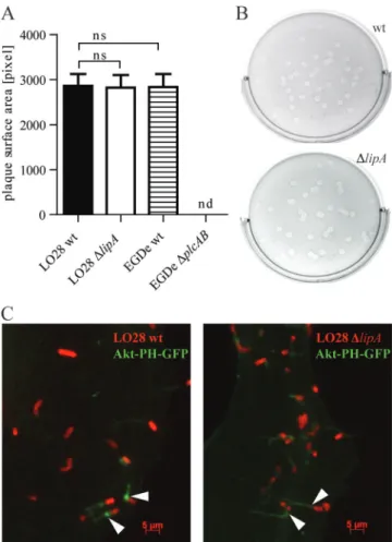
LipA is not required for cell-to-cell spread.
To test the
impact of LipA deletion on the ability of Listeria to spread
from cell to cell, a plaque assay was performed. Semiconfluent
layers of L2 fibroblast cells were infected at a low MOI and
overlaid with a mixture of tissue culture medium, agarose, and
gentamicin. After an incubation time of 3 days, plaques were
visualized by addition of another overlay containing neutral
red, a stain for live host cells. L. monocytogenes wt and
phos-pholipase
⌬plcAB bacteria were included as controls. ⌬plcAB
strains are not able to escape from secondary vacuoles and do
not form plaques. Comparison of the average surface areas of
plaques formed by wt and
⌬lipA bacteria showed no significant
difference, whereas the
⌬plcAB strain showed the expected
inability to form plaques (Fig. 4A and B).
During ActA-dependent movement within the cytoplasm,
Listeria bacteria accumulate PI(3,4,5)P
3around their surface
(51). The host Akt kinase contains a PH domain that binds
PI(3,4)P
2and PI(3,4,5)P
3, and a fusion protein of GFP linked
to the PH domain of Akt has been reported to concentrate on
moving Listeria and the actin-rich tails (51). We used this
construct to investigate Akt-PH recruitment to L.
monocyto-genes wt and
⌬lipA bacteria. Ptk-2 cells were transfected with
Akt-PH-GFP and infected with L. monocytogenes for 6 h.
Bac-teria were stained with a primary anti-LisBac-teria antibody and a
red secondary Alexa Fluor 594-conjugated antibody.
Recruit-ment of the Akt-PH-GFP fusion protein to one pole of L.
monocytogenes as well as to polarized tails, presumably
repre-senting the actin-rich tails, was detected upon microscopic
examination of cytoplasmic L. monocytogenes wt and
⌬lipA
strains (Fig. 4C).
FIG. 3. Effect of L. monocytogenes LipA on intracellular growth. Growth of L. monocytogenes LO28 or EGDe was assessed by plating serial
dilutions of cellular lysates at the indicated time points. Numbers of CFU are displayed as log CFU per 1
⫻ 10
6cells. BMM (A), CMT-93 (B),
and L2 (C) cells were infected with L. monocytogenes LO28 wt and
⌬lipA strains at MOIs of 10, 50, and 20, respectively. (D) J774 cells were infected
with L. monocytogenes EGDe wt and
⌬lipA strains at an MOI of 50.
FIG. 4. Effect of L. monocytogenes LipA on movement and
cell-to-cell spread. (A) Detection of L. monocytogenes cell-to-cell-to-cell-to-cell spread by a
plaque assay. L2 cells were infected with L. monocytogenes LO28 wt
and
⌬lipA strains at an MOI of 0.5. Three days after infection, relative
plaque surface area was calculated for 50 plaques per bacterial strain,
and the mean for two independent experiments was calculated. ns, not
significant; nd, not detected. (B) Representative pictures of plaques
formed by L. monocytogenes LO28 wt (upper panel) and
⌬lipA (lower
panel) strains. (C) Localization of Akt-PH-GFP (green) in Ptk-2 cells
infected with L. monocytogenes LO28 wt and
⌬lipA strains. Cells were
transfected with Akt-PH-GFP and infected with Listeria for 6 h.
Bac-teria were visualized using an antibody to LisBac-teria and a secondary
Alexa Fluor 594-conjugated antibody (red).
on September 1, 2017 by INRA - old
http://iai.asm.org/
LipA is highly expressed in blood.
To get insight into the
contribution of LipA to infection, we examined lipA
transcrip-tion after growth of Listeria in vitro and in vivo, using our L.
monocytogenes-specific tiling arrays and gene expression arrays
(54). The level of the lipA transcript in wild-type L.
monocy-togenes grown to exponential phase in BHI broth at 37°C, the
reference condition, was compared to the level of lipA
tran-scribed in bacteria grown to stationary phase in BHI broth at
37°C following growth in BHI broth at 37°C and 6% oxygen,
growth in BHI broth at 30°C, growth in the intestinal lumen
after oral inoculation of axenic mice, and growth in human
blood for 60 min. Strikingly, lipA transcription was induced
13.4-fold when bacteria were grown in human blood (Fig. 5).
The expression levels of lipA were similar to those under the
reference condition for all the other growth conditions and
were independent of PrfA, the main activator of Listeria
viru-lence genes. Likewise, lipA expression was not influenced by
the alternative sigma factor SigB.
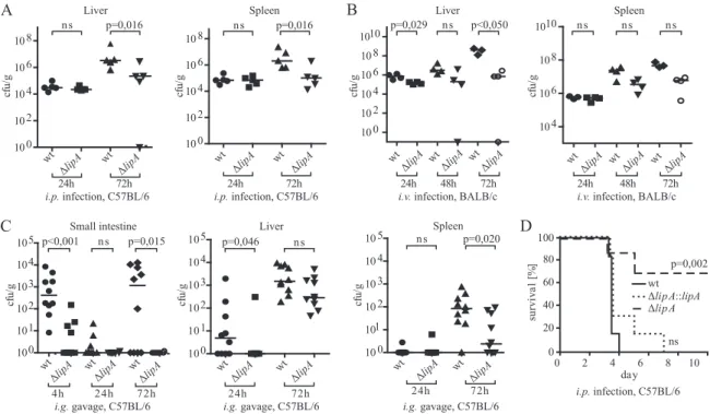
LipA is required for effective host colonization and Listeria
virulence in vivo.
To examine the contribution of LipA to
Listeria virulence in mice, the spread of
⌬lipA bacteria to
internal organs was measured. To exclude the possibility that
the consequences of lipA deletion were restricted to a
partic-ular genetic background of either L. monocytogenes or its
mu-rine host, C57BL/6 and BALB/c mice were infected with both
the EGD
⌬lipA and LO28 ⌬lipA strains and their respective
parental strains. Intraperitoneal (i.p.), intravenous (i.v.), and
intragastric (i.g.) routes were chosen for infection and
subse-quent measurement of spread to internal organs. In addition,
LO28 wt, LO28
⌬lipA, and LO28 ⌬lipA::lipA bacteria were
used to determine the survival of infected C57BL/6 mice.
Mice infected with 2
⫻ 10
6L. monocytogenes LO28
⌬lipA
bacteria exhibited a significant reduction of bacterial loads in
both the liver and spleen at day 3 postinfection compared to
the wt strain (Fig. 6A). Likewise, BALB/c mice injected i.v.
with 8
⫻ 10
3viable units of the EGDe or isogenic
⌬lipA mutant
strain showed reduced organ loads of mutant bacteria. The
differences were most pronounced in both organs at 72 h
postinfection, although statistical significance was not reached
in the spleen (Fig. 6B). Importantly, multiplication of the
mu-tant bacteria was controlled at 72 h postinfection in both the
liver and spleen, while the number of wild-type bacteria
con-tinued to increase. To assess whether the decrease in virulence
of
⌬lipA strains observed upon systemic infection also occurred
upon infection via the gastrointestinal tract, C57BL/6 mice
were infected with L. monocytogenes LO28 wt and
⌬lipA strains
via intragastric gavage (Fig. 6C). wt bacteria were taken up by
or tightly associated with intestinal tissue 4 h after infection,
whereas significantly fewer bacteria were found after infection
with the L. monocytogenes LO28
⌬lipA strain. This is unlikely
to result from a decreased resistance to acid conditions in the
gastrointestinal tract because the acid resistance of L.
mono-cytogenes was unchanged upon deletion of the lipA gene (see
Fig. S2 in the supplemental material; data not shown).
Bacte-rial counts were below the detection limit after 24 h and
in-creased approximately to the values found at 4 h 3 days after
infection. Again, numbers of
⌬lipA bacteria were significantly
lower. Likewise, a reduction in
⌬lipA counts 3 days after
in-fection was observed in the spleen and liver. Whereas the
difference was pronounced in the spleen, it did not reach
sta-tistical significance in the liver. The reduction of the pathogen
burden was in line with an increased survival of mice (Fig. 6D).
After i.p. infection of C57BL/6 mice with 1
⫻ 10
6L.
monocy-togenes LO28 wt bacteria, all animals died by day 4, whereas
70% of the population injected with the
⌬lipA strain survived.
To ascertain that the reduction of virulence was indeed due to
the lack of LipA, the
⌬lipA strain was complemented with an
integrative plasmid encoding the lipA gene to generate the L.
monocytogenes LO28
⌬lipA::lipA strain. The presence of this
plasmid restored PTP activity in Listeria extracts (see Fig. S6 in
the supplemental material) and lethal infection of mice upon
intraperitoneal infection (Fig. 6D).
Serum cytokine levels are reduced after infection with
⌬lipA
bacteria.
To test whether proinflammatory cytokine
produc-tion reflected bacterial loads, serum levels of IFN-
␥, TNF-␣,
and IL-6 were measured by ELISA. IFN-
␥ was robustly
pro-duced after i.p. infection of C57BL/6 mice with wt L.
monocy-togenes LO28 on days 1 and 3 after infection. A significant
reduction was detected after infection with
⌬lipA bacteria on
the third day of infection. Production of TNF-
␣ was detected
at low levels 3 days after infection with wt bacteria but with
quite a high variability between mice. After infection with the
⌬lipA strain, TNF-␣ levels were only marginally elevated
com-pared to those in uninfected mice and were significantly lower
than those in mice infected with wt bacteria. Likewise, serum
IL-6 levels were highly reduced during infection with
⌬lipA
bacteria compared to those during infection with wt bacteria
(Fig. 7). During infection with L. monocytogenes, leukocyte
populations in blood or lymphoid organs were subject to
changes in number and composition (see Fig. S7 in the
sup-plemental material). The extents to which these changes
oc-curred were similar for wt and
⌬lipA bacteria.
DISCUSSION
L. monocytogenes is a well-established model organism in
infection biology. Extensive studies on the infectious cycle of
Listeria have provided profound knowledge of the virulence
factors that bacteria require for successfully using host cells as
a replication niche. Posttranslational modification of host
pro-teins and pathways by bacterial enzymes is increasingly
recog-nized as an important facet of the host-pathogen relationship
FIG. 5. Expression of L. monocytogenes lipA in human blood.
Tran-scriptional tiling maps of L. monocytogenes EGDe grown in BHI to
exponential phase at 37°C (black dots) or in human blood at 37°C for
60 min (red dots) show normalized hybridization intensities (y axis, in
arbitrary units) and genomic coordinates at the lipA locus (x axis, in
bp). Each dot represents the average intensity signal for one probe
from three independent biological replicates. Annotated ORFs are
indicated as black boxes along the x axis.
VOL. 79, 2011
LipA PHOSPHATASE INVOLVED IN LISTERIA VIRULENCE
2495
on September 1, 2017 by INRA - old
http://iai.asm.org/
(48, 49). An enhancement of virulence through secretion of
phosphatases and their interference with host metabolism had
originally been associated with the pathogenicity of enteric
Gram-negative bacteria and the use of type III secretion
sys-tems (10, 25, 39). Whereas S/T dephosphorylation and the
ensuing adaptation to the host environment have previously
been found to contribute to L. monocytogenes virulence (2, 3),
we now provide the first report showing that tyrosine
dephos-phorylation of hitherto-unidentified host proteins similarly
contributes to the virulence of this bacterium. We characterize
lmo1800 as a gene encoding the phosphatase LipA, a member
of the classical tyrosine phosphatase family. Among
phospho-FIG. 6. Effect of L. monocytogenes LipA on virulence in mice. (A) Groups of 4 to 5 C57BL/6 wt mice were infected intraperitoneally with 2
⫻
10
6L. monocytogenes LO28 wt and
⌬lipA bacteria. At days 1 and 3, the spleen and liver were homogenized and the bacterial titer was determined.
Medians were plotted, and statistical significance was calculated using the Mann-Whitney U test. (B) L. monocytogenes EGDe wt and
⌬lipA strains
(8
⫻ 10
3CFU) were inoculated intravenously in groups of 4 BALB/c mice. Bacterial growth was followed in the spleen and liver at 24, 48, and
72 h. Medians were plotted, and statistical significance was calculated using the Mann-Whitney U test. (C) Groups of 10 C57BL/6 wt mice were
infected via intragastric gavage of 1
⫻ 10
9L. monocytogenes LO28 wt and
⌬lipA bacteria. At the indicated time points, numbers of CFU were
determined in the small intestine, liver, and spleen by plating serial dilutions on Oxford agar plates. Medians were plotted, and statistical
significance was calculated using the Mann-Whitney U test. (D) Survival assay. Groups of 6 to 7 C57BL/6 wt mice were infected intraperitoneally
with 1
⫻ 10
6L. monocytogenes LO28 wt,
⌬lipA, or ⌬lipA::lipA bacteria, and survival was monitored for 10 days. Data are depicted as Kaplan-Meier
plots. The survival of mice infected with the L. monocytogenes LO28
⌬lipA strain was strongly increased compared to that of mice infected with
the wt strain (P
⫽ 0.002). This difference was no longer observed when the mice were infected with the complemented strains (not significant for
the wt versus the
⌬lipA::lipA strain and P ⫽ 0.009 for the ⌬lipA strain versus the ⌬lipA::lipA strain). Statistical significance was calculated using the
Gehan-Breslow-Wilcoxon test.
FIG. 7. Effect of L. monocytogenes LipA on serum cytokine levels. Levels of IFN-
␥, TNF-␣, and IL-6 in sera of C57BL/6 wt mice after
intraperitoneal infection with L. monocytogenes LO28 wt and
⌬lipA strains was measured by ELISA. Cytokine levels in individual mice are
depicted, and means with standard deviation are shown. Statistical significance was calculated using the Student t test. ni, noninfected.
on September 1, 2017 by INRA - old
http://iai.asm.org/
amino acid substrates, recombinant LipA specifically
dephos-phorylated P-Tyr. Typically, tyrosine-specific phosphatases
dis-play a bell-shaped pH rate profile with a low pH optimum (30).
In line with this, LipA had its highest activity at pH 6.5. Besides
P-Tyr, LipA dephosphorylated the phosphoinositides PI(3)P,
PI(5)P, and PI(3,5)P
2. This dual activity as both
phospho-amino acid and phospholipid phosphatase is in line with the
high similarity between the predicted LipA structure and that
of phosphatase MPtpB, a virulence factor required for
intra-cellular growth of Mycobacterium tuberculosis (6, 8, 35). MPtpB
dephosphorylates P-Tyr, P-Ser, and P-Thr and, additionally, a
broad range of phosphoinositide substrates (8). MPtpB
con-tains a flexible lid formed by two helices that protects the active
site from oxidative inactivation. This is mediated by the
con-formational dynamics of the lid and might represent an
adap-tation to oxidative challenges in vivo (22). The predicted
three-dimensional (3D) model of LipA suggests that it also contains
this flexible loop structure. Important for the contribution of
LipA to L. monocytogenes virulence, our data show the
func-tionality of the predicted signal peptide for secretion or
mem-brane insertion. Using a LipA-GFP fusion construct, we found
that LipA-GFP was present in the supernatants of Listeria
cultured in broth, indicating that LipA is secreted. A recent
study suggested that LipA was among the proteins lipidated by
the prolipoprotein diacylglyceryl transferase (Lgt), leading to
its anchorage to the bacterial surface (7). Although the 30
N-terminal amino acids of LipA contain a potential lipobox for
acylation (L
19-G-G-C-G
23), the membrane association of LipA
could be only transient before its release into the extracellular
environment.
The important contribution of LipA to the virulence of L.
monocytogenes was revealed by a comprehensive study in mice.
LipA deficiency decreased pathogen load and inflammatory
cytokine production, irrespective of the genetic background of
the infected mice (C57BL/6 or BALB/c), the Listeria strain
(LO28 or EGDe), or the route of infection (i.v., i.p., or i.g.).
This strongly suggests that LipA targets host cell components
rather than bacterial components.
⌬lipA bacteria grew equally
well in broth and host cells, and they were equally resistant to
adverse conditions such as low pH, osmotic stress, and an
oxidizing environment. Importantly, the L. monocytogenes
ge-nome does not contain proven tyrosine kinases (26).
There-fore, it appears likely that the effect of LipA on virulence
results from dephosphorylation of host cell proteins or lipids.
Our data further suggest that LipA targets do not affect the L.
monocytogenes life cycle in either the phagocytes or the
nonphagocytic colonic epithelial cells investigated in this study.
Macrophages are relevant as both replication niches and
ef-fector cells, depending on activation status, which in turn
re-quires signals by pattern recognition receptors and IFN-
␥-mediated tyrosine phosphorylation of Stat transcription factors
(19). The intestinal epithelium represents the initial contact
between L. monocytogenes and its mammalian host. A plethora
of signals, including both tyrosine phosphorylation and
phos-pholipid-dependent events, derives from InlA and InlB
li-gands, pattern recognition and cytokine receptors, and the
invasion process per se to accompany bacterial uptake (15, 18,
40). Surprisingly, we could not detect an impact of LipA on
these pathways. Assuming that lipid phosphatase activity is
relevant for virulence, our future experiments to identify
events and pathways affected by LipA will have to take into
account that the enzyme shows pronounced preference for
some phosphoinositides over others. Strikingly, a phosphate at
C-4 strongly reduces dephosphorylation. Since many lipid
sig-naling pathways require PI(3,4)P
2or PI(3,4,5)P
3(12), our
search will be directed toward events such as endosome or
phagosome maturation that require PI(3)P (14, 28), PI(5)P
monophosphates (41, 42), or PI(3,5)P
2diphosphates (45, 57).
The decrease in the number of intestinal
⌬lipA bacteria
already 4 h after intragastric inoculation points toward a role
for LipA during the immediate innate phase of the anti-Listeria
immune response. Speculating about the possible function of
LipA, we hypothesize that it targets a pathway in a cell type not
analyzed so far or a cooperation of different cell types that may
not be readily amenable to studies ex vivo. Alternatively, LipA
might interfere with a biological process undetected using the
assays performed in our study. The reduction in serum
cyto-kines observed in infected mice would fit both explanations.
The identical compositions of blood and splenic leukocyte
pop-ulations rather argue that reduced serum levels are a
conse-quence of reduced bacterial numbers and not due to a change
in the quality of the immune response. Important effector cells
of early anti-Listeria immunity are neutrophils (55), a cell type
notoriously difficult to study due to its short life span in culture.
As LipA is highly upregulated in human blood, our future aims
must be directed at investigating the effect of LipA deletion
during infections of blood monocytes and neutrophils.
Taken together, we characterized a novel virulence
determi-nant of L. monocytogenes, namely, the secreted phosphatase
LipA. We demonstrated that LipA is essential for promoting
Listeria infections in vivo irrespective of the route of infection.
The importance of this newly described factor is highlighted by
the drastic defect in early host colonization after intragastric
delivery of
⌬lipA bacteria. Further investigation of LipA
func-tion and the identificafunc-tion of physiological LipA substrates will
be interesting future tasks for a better understanding of
Liste-ria interactions with its mammalian host.
ACKNOWLEDGMENTS
Work in the laboratory of T.D. was supported by the University of
Vienna through the research focus “Symbiosis Research and
Molecu-lar Principles of Recognition” and the Austrian Science Fund (FWF)
through grant P 20522-B05. Work in the laboratory of P.C. was
sup-ported by the Institut Pasteur (GPH9), Inserm, INRA, ANR
(ANR-06-PATHO-011-01), and ERC (Advanced Grant 233348).
P.C. is an International Research Scholar of the Howard Hughes
Medical Institute.
ADDENDUM IN PROOF
While our paper was under review, Beresford and colleagues
(N. J. Beresford, C. Saville, H. J. Bennett, I. S. Roberts, and L.
Tabernero, BMC Genomics 11:457–469, 2010) reported lipid
phosphatase activity of the protein encoded by lmo1800, in
agreement with the data reported in our study.
REFERENCES
1. Abachin, E., et al. 2002. Formation ofD-alanyl-lipoteichoic acid is required for adhesion and virulence of Listeria monocytogenes. Mol. Microbiol. 43: 1–14.
2. Archambaud, C., E. Gouin, J. Pizarro-Cerda, P. Cossart, and O. Dussurget. 2005. Translation elongation factor EF-Tu is a target for Stp, a serine-threonine phosphatase involved in virulence of Listeria monocytogenes. Mol. Microbiol. 56:383–396.
VOL. 79, 2011
LipA PHOSPHATASE INVOLVED IN LISTERIA VIRULENCE
2497
on September 1, 2017 by INRA - old
http://iai.asm.org/
3. Archambaud, C., M. A. Nahori, J. Pizarro-Cerda, P. Cossart, and O.
Dus-surget.2006. Control of Listeria superoxide dismutase by phosphorylation. J. Biol. Chem. 281:31812–31822.
4. Arnaud, M., A. Chastanet, and M. Debarbouille. 2004. New vector for efficient allelic replacement in naturally nontransformable, low-GC-content, gram-positive bacteria. Appl. Environ. Microbiol. 70:6887–6891.
5. Baccarini, M., F. Bistoni, and M. L. Lohmann Matthes. 1985. In vitro natural cell-mediated cytotoxicity against Candida albicans: macrophage precursors as effector cells. J. Immunol. 134:2658–2665.
6. Bach, H., K. G. Papavinasasundaram, D. Wong, Z. Hmama, and Y. Av-Gay. 2008. Mycobacterium tuberculosis virulence is mediated by PtpA dephos-phorylation of human vacuolar protein sorting 33B. Cell Host Microbe
3:316–322.
7. Baumgartner, M., et al. 2007. Inactivation of Lgt allows systematic charac-terization of lipoproteins from Listeria monocytogenes. J. Bacteriol. 189: 313–324.
8. Beresford, N., et al. 2007. MptpB, a virulence factor from Mycobacterium tuberculosis, exhibits triple-specificity phosphatase activity. Biochem. J. 406: 13–18.
9. Beresford, N. J., et al. 2009. Inhibition of MptpB phosphatase from Myco-bacterium tuberculosis impairs mycobacterial survival in macrophages. J. Antimicrob. Chemother. 63:928–936.
10. Bliska, J. B., and S. Falkow. 1993. The role of host tyrosine phosphorylation in bacterial pathogenesis. Trends Genet. 9:85–89.
11. Cabanes, D., O. Dussurget, P. Dehoux, and P. Cossart. 2004. Auto, a surface associated autolysin of Listeria monocytogenes required for entry into eu-karyotic cells and virulence. Mol. Microbiol. 51:1601–1614.
12. Cantley, L. C. 2002. The phosphoinositide 3-kinase pathway. Science 296: 1655–1657.
13. Chakraborty, T., et al. 1992. Coordinate regulation of virulence genes in Listeria monocytogenes requires the product of the prfA gene. J. Bacteriol.
174:568–574.
14. Coronas, S., et al. 2007. PtdIns5P: a little phosphoinositide with big func-tions? Biochem. Soc. Symp. 74:117–128.
15. Cossart, P., and H. Bierne. 2001. The use of host cell machinery in the pathogenesis of Listeria monocytogenes. Curr. Opin. Immunol. 13:96–103. 16. Cozzone, A. J., C. Grangeasse, P. Doublet, and B. Duclos. 2004. Protein
phosphorylation on tyrosine in bacteria. Arch. Microbiol. 181:171–181. 17. Darji, A., W. Mohamed, E. Domann, and T. Chakraborty. 2003. Induction of
immune responses by attenuated isogenic mutant strains of Listeria mono-cytogenes. Vaccine 21(Suppl. 2):S102–S109.
18. Decker, T., M. Muller, and S. Stockinger. 2005. The yin and yang of type I interferon activity in bacterial infection. Nat. Rev. Immunol. 5:675–687. 19. Decker, T., S. Stockinger, M. Karaghiosoff, M. Muller, and P. Kovarik. 2002.
IFNs and STATs in innate immunity to microorganisms. J. Clin. Invest.
109:1271–1277.
20. Domann, E., et al. 1993. Detection of a prfA-independent promoter respon-sible for listeriolysin gene expression in mutant Listeria monocytogenes strains lacking the PrfA regulator. Infect. Immun. 61:3073–3075. 21. Dussurget, O., J. Pizarro-Cerda, and P. Cossart. 2004. Molecular
determi-nants of Listeria monocytogenes virulence. Annu. Rev. Microbiol. 58:587– 610.
22. Flynn, E. M., J. A. Hanson, T. Alber, and H. Yang. 2010. Dynamic active-site protection by the M. tuberculosis protein tyrosine phosphatase PtpB lid domain. J. Am. Chem. Soc. 132:4772–4780.
23. Fujimoto, S., and Y. Ike. 2001. pAM401-based shuttle vectors that enable overexpression of promoterless genes and one-step purification of tag fusion proteins directly from Enterococcus faecalis. Appl. Environ. Microbiol. 67: 1262–1267.
24. Galan, J. E. 2001. Salmonella interactions with host cells: type III secretion at work. Annu. Rev. Cell Dev. Biol. 17:53–86.
25. Galan, J. E., and A. Collmer. 1999. Type III secretion machines: bacterial devices for protein delivery into host cells. Science 284:1322–1328. 26. Glaser, P., et al. 2001. Comparative genomics of Listeria species. Science
294:849–852.
27. Grangeasse, C., A. J. Cozzone, J. Deutscher, and I. Mijakovic. 2007. Tyrosine phosphorylation: an emerging regulatory device of bacterial physiology. Trends Biochem. Sci. 32:86–94.
28. Hilbi, H. 2006. Modulation of phosphoinositide metabolism by pathogenic bacteria. Cell. Microbiol. 8:1697–1706.
29. Kocks, C., et al. 1992. L. monocytogenes-induced actin assembly requires the actA gene product, a surface protein. Cell 68:521–531.
30. Kolmodin, K., and J. Aqvist. 2001. The catalytic mechanism of protein tyrosine phosphatases revisited. FEBS Lett. 498:208–213.
31. Kovarik, P., D. Stoiber, M. Novy, and T. Decker. 1998. Stat1 combines signals derived from IFN-gamma and LPS receptors during macrophage activation. EMBO J. 17:3660–3668.
32. Lalic-Multhaler, M., J. Bohne, and W. Goebel. 2001. In vitro transcription of PrfA-dependent and -independent genes of Listeria monocytogenes. Mol. Microbiol. 42:111–120.
33. Lingnau, A., et al. 1995. Expression of the Listeria monocytogenes EGD inlA and inlB genes, whose products mediate bacterial entry into tissue culture cell lines, by PrfA-dependent and -independent mechanisms. Infect. Immun.
63:3896–3903.
34. Luo, Q., M. Rauch, A. K. Marr, S. Muller-Altrock, and W. Goebel. 2004. In vitro transcription of the Listeria monocytogenes virulence genes inlC and mpl reveals overlapping PrfA-dependent and -independent promoters that are differentially activated by GTP. Mol. Microbiol. 52:39–52.
35. Madhurantakam, C., et al. 2005. Crystal structure of low-molecular-weight protein tyrosine phosphatase from Mycobacterium tuberculosis at 1.9-Å resolution. J. Bacteriol. 187:2175–2181.
36. Marquis, H. 2006. Tissue culture cell assays used to analyze Listeria mono-cytogenes. Curr. Protoc. Microbiol. 9:9B.4.
37. Mead, P. S., et al. 1999. Food-related illness and death in the United States. Emerg. Infect. Dis. 5:607–625.
38. Monk, I. R., C. G. Gahan, and C. Hill. 2008. Tools for functional postgen-omic analysis of Listeria monocytogenes. Appl. Environ. Microbiol. 74:3921– 3934.
39. Nhieu, G. T., J. Enninga, P. Sansonetti, and G. Grompone. 2005. Tyrosine kinase signaling and type III effectors orchestrating Shigella invasion. Curr. Opin. Microbiol. 8:16–20.
40. O’Riordan, M., and D. A. Portnoy. 2002. The host cytosol: front-line or home front? Trends Microbiol. 10:361–364.
41. Pendaries, C., et al. 2006. PtdIns5P activates the host cell PI3-kinase/Akt pathway during Shigella flexneri infection. EMBO J. 25:1024–1034. 42. Pendaries, C., et al. 2005. Emerging roles of phosphatidylinositol
monophos-phates in cellular signaling and trafficking. Adv. Enzyme Regul. 45:201–214. 43. Phan-Thanh, L., and T. Gormon. 1997. A chemically defined minimal me-dium for the optimal culture of Listeria. Int. J. Food Microbiol. 35:91–95. 44. Philpott, D. J., S. E. Girardin, and P. J. Sansonetti. 2001. Innate immune
responses of epithelial cells following infection with bacterial pathogens. Curr. Opin. Immunol. 13:410–416.
45. Pizarro-Cerda, J., and P. Cossart. 2004. Subversion of phosphoinositide metabolism by intracellular bacterial pathogens. Nat. Cell Biol. 6:1026–1033. 46. Portnoy, D. A., V. Auerbuch, and I. J. Glomski. 2002. The cell biology of Listeria monocytogenes infection: the intersection of bacterial pathogenesis and cell-mediated immunity. J. Cell Biol. 158:409–414.
47. Reutterer, B., et al. 2008. Type I IFN are host modulators of strain-specific Listeria monocytogenes virulence. Cell. Microbiol. 10:1116–1129. 48. Ribet, D., and P. Cossart. 2010. Pathogen-mediated posttranslational
mod-ifications: a re-emerging field. Cell 143:694–702.
49. Ribet, D., and P. Cossart. 2010. Post-translational modifications in host cells during bacterial infection. FEBS Lett. 584:2748–2758.
50. Scortti, M., H. J. Monzo, L. Lacharme-Lora, D. A. Lewis, and J. A.
Vazquez-Boland.2007. The PrfA virulence regulon. Microbes Infect. 9:1196–1207. 51. Sidhu, G., et al. 2005. Phosphoinositide 3-kinase is required for intracellular
Listeria monocytogenes actin-based motility and filopod formation. J. Biol. Chem. 280:11379–11386.
52. Stockinger, S., et al. 2009. Characterization of the interferon-producing cell in mice infected with Listeria monocytogenes. PLoS Pathog. 5:e1000355. 53. Tabernero, L., A. R. Aricescu, E. Y. Jones, and S. E. Szedlacsek. 2008.
Protein tyrosine phosphatases: structure-function relationships. FEBS J. 275: 867–882.
54. Toledo-Arana, A., et al. 2009. The Listeria transcriptional landscape from saprophytism to virulence. Nature 459:950–956.
55. Unanue, E. R. 1997. Inter-relationship among macrophages, natural killer cells and neutrophils in early stages of Listeria resistance. Curr. Opin. Im-munol. 9:35–43.
56. Vazquez-Boland, J. A., et al. 2001. Listeria pathogenesis and molecular virulence determinants. Clin. Microbiol. Rev. 14:584–640.
57. Weber, S. S., C. Ragaz, and H. Hilbi. 2009. Pathogen trafficking pathways and host phosphoinositide metabolism. Mol. Microbiol. 71:1341–1352.