HAL Id: inserm-00188498
https://www.hal.inserm.fr/inserm-00188498
Submitted on 19 Nov 2007
HAL is a multi-disciplinary open access
archive for the deposit and dissemination of sci-entific research documents, whether they are pub-lished or not. The documents may come from teaching and research institutions in France or abroad, or from public or private research centers.
L’archive ouverte pluridisciplinaire HAL, est destinée au dépôt et à la diffusion de documents scientifiques de niveau recherche, publiés ou non, émanant des établissements d’enseignement et de recherche français ou étrangers, des laboratoires publics ou privés.
Improving the dynamics of responses to amplitude
modulated stimuli by modeling inhibitory interneurons
in cochlear nucleus.
Pierre Dugué, Régine Le Bouquin Jeannès, Gérard Faucon
To cite this version:
Pierre Dugué, Régine Le Bouquin Jeannès, Gérard Faucon. Improving the dynamics of responses to amplitude modulated stimuli by modeling inhibitory interneurons in cochlear nucleus.. Confer-ence proceedings : .. Annual International ConferConfer-ence of the IEEE Engineering in Medicine and Biology Society. IEEE Engineering in Medicine and Biology Society. Annual Conference, Institute of Electrical and Electronics Engineers (IEEE), 2007, 1, pp.1286-9. �10.1109/IEMBS.2007.4352532�. �inserm-00188498�
Abstract— Amplitude modulation is an important feature of communication sounds. A phenomenological model of the auditory pathway that reproduces amplitude modulation coding from the outer ear to the inferior colliculus is presented. It is based on Hewitt and Meddis’ work. To improve the temporal coding for high level stimuli, high spontaneous rate and low spontaneous rate auditory nerve fibers innervate chopper cells of the cochlear nucleus. Wideband inhibitory interneurons which limit high spontaneous rate fibers connected to chopper units are added in this nucleus. The realistic structure we propose gives results closer to physiological data in terms of synchronization.
I. INTRODUCTION
OMPLEX sounds and especially communication sounds
such as speech are characterized by amplitude modulation, also referred to as temporal envelope. Psychoacoustic studies (e.g. [18]) indicate that envelope information plays an important role in many perceptual phenomena including speech identification and auditory streaming. Physiological studies show that temporal coding of amplitude modulation takes place from the inner ear to the auditory cortex (for review, see [5]). In this paper, a model of amplitude modulation coding from the middle ear to the inferior colliculus (IC) is presented. At this level, modulation frequencies are extracted from the stimulus as some neurons respond to specific modulation frequencies.
The point neuron model of the auditory pathway is based on Hewitt and Meddis’ work [4]. This is a four-stage model including: the cochlea multichannel filterbank decomposition, the auditory nerve response via inner hair cells that act as rectifiers and compressors, the cochlear nucleus decomposition in terms of modulation frequency, and the coincidence detection in the inferior colliculus. This model reproduces physiological data except for high level stimuli where neuron synchronization decreases. So, we propose in this paper an update of Hewitt and Meddis’ structure to improve synchronization and find results closer to physiological data.
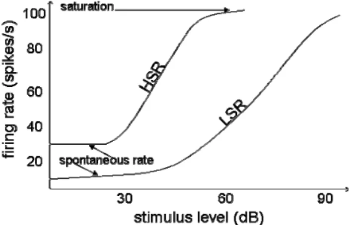
The main evolution is the use of high spontaneous rate (HSR) and low spontaneous rate (LSR) fibers to innervate ventral cochlear nucleus (VCN) cells. HSR fibers, which are used in [4], respond to low level stimuli and saturate for
Manuscript received April 2, 2007.
P. Dugué, R. Le Bouquin Jeannès, and G. Faucon are with INSERM, U 642, Rennes, F-35000 France ; and with Université de Rennes 1, LTSI, F-35000, France.
e-mail: (pierre.dugue, regine.le-bouquin-jeannes, gerard.faucon)@univ-rennes1.fr
high level ones. LSR fibers begin to fire for medium level stimuli and saturate for very high ones. Figure 1 illustrates HSR and LSR behaviors. The chopper cell receives HSR and LSR fibers afferent activity. Those fibers are tuned to the same center frequency (CF). This is not enough to expand chopper unit dynamics because it saturates. That is why an onset inhibitory interneuron is added. It receives afferent from HSR auditory nerve fibers with a wide range of CFs which makes it a wideband inhibitory interneuron (WBII). When the HRS fibers, afferent of the chopper unit, saturate, they are inhibited by the WBII, then LSR fibers relay temporal information. Figure 2 shows the principle of cells connection in the VCN.
Fig. 1. Raise of the firing rate of two auditory nerve fibers with the stimulus level. These auditory nerve fibers are characterized by their spontaneous firing rate. There are low and high spontaneous firing rate fibers (LSR and HSR fibers respectively). HSR is sensitive to low stimulus level and saturates for medium level stimulus. LSR is sensitive to medium level stimulus and saturates for high level stimulus.
Physiological elements support this scheme. Firstly, Liberman [7] showed that each inner hair cell is connected with the three types of fibers (HSR, medium spontaneous rate and LSR). Secondly, Ferragamo [1] noticed that, in the VCN, stellate T cells are inhibited by stellate D ones. Stellate T cells correspond to multipolar cells in the VCN [12]. Chopper responses are associated with this cell [16]. Stellate D cells may correspond to giant multipolar cells and therefore have an onset response. Concerning afferent connections to those cells, Ryugio [17] found that LSR fibers give rise to greater collaterals in VCN than HSR fibers. Moreover, LSR fibers have a greater number of terminals in VCN [13]. Geometrical considerations lead us to suppose that LSR fibers innervate giant multipolar units.
Improving the Dynamics of Responses to Amplitude Modulated
Stimuli by Modeling Inhibitory Interneurons in Cochlear Nucleus
Pierre Dugué, Régine Le Bouquin Jeannès, and Gérard Faucon
C
This material is presented to ensure timely dissemination of scholarly and technical work. Copyright and all rights therein are retained by authors or by other copyright holders.
All persons copying this information are expected to adhere to the terms and constraints invoked by each author's copyright. In most cases, these works may not be reposted without the explicit permission of the copyright holder.
HAL author manuscript inserm-00188498, version 1
HAL author manuscript
Fig. 2. Explicative scheme of cochlear nucleus neurons: innervations and connections. The chopper cell receives HSR and LSR auditory nerve afferents having the same CF. The onset inhibitory interneuron is connected with LSR fibers from a wide range of CFs. This interneuron inhibits HSR chopper cell excitatory afferent.
II. PROCEDURE FOR PAPER SUBMISSION
The model we propose is composed of five stages: the outer and middle ear, the basilar membrane, the inner hair cells, the auditory nerve fibers, the cochlear nucleus and the inferior colliculus. The outer and middle ear filtering consists in four second order Butterworth filters in cascade. This is a bandpass function whose maximum is reached at 1.5 kHz.
The sound is then decomposed by the basilar membrane of the cochlea. Here the ‘dual resonance non linear’ model is used ([10], [8]). This model is interesting in its behavior because it reproduces intensity influence on basilar membrane filters selectivity and center frequencies.
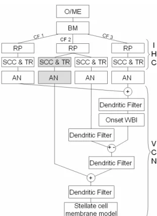
Each point of the modeled basilar membrane stimulates an inner hair cell. These cells compress and adapt the signal to the auditory nerve. One inner hair cell is modeled by a three-stage model: receptor potential, synaptic calcium channel and transmitter release [19]. The first part transforms basilar membrane motion in hair cell membrane potential (receptor potential). The second part turns this potential into transmitter release probability which is used in the third part to determine the amount of neurotransmitters in the synaptic cleft. This corresponds to a probability for the connected fiber to fire. In this model, this probability is realized by a geometrical law parameterized by the number of fibers. The IHC-AN complex allows us to model two kinds of auditory nerve fibers: HSR ones and LSR ones. Those fibers are obtained by changing synaptic calcium channel and the transmitter release parameters. These two kinds of fiber innervate cells of the VCN. The WBII receives LSR fibers outputs with different CFs (from one octave lower and two higher than the CF of the chopper cell these fibers inhibit). Chopper cell receives two excitatory afferents from LSR and HSR fibers of same CF. These afferent fibers determine the chopper cell CF. Figure 3 gives a detailed schematic organization of the model for a single couple of WBI-chopper VCN cells. Results presented here are obtained with afive-channel basilar membrane (corresponding to five CFs; the WBII is connected with four auditory nerves).
Fig. 3. Detailed scheme of the first stages of the model. After outer/middle ear (O/ME) filtering, the basilar membrane (BM) filterbank decomposes the signal in three nonlinearly filtered versions (characterized by their center frequency: CF1<CF2<CF3). They
correspond to the motion of three points on the BM. The motion of each point is transformed in receptor potential (RP) of the corresponding inner hair cell (IHC). It influences synaptic calcium channels (SCC) which controls transmitter release (TR). The amount of neurotransmitters is converted in discharge in the auditory nerve (AN). All HSR auditory nerve fibers excite the onset WBI cell. Then three filters are used to model dendritic connections of the chopper cell (see text). The soma is a McGregor model.
Fig. 4. Global scheme of the auditory pathway model from the outer ear to the inferior colliculus. First the stimulus is filtered by the outer/middle ear (O/ME), then the basilar membrane (BM) decomposes the sound in a tonotopic manner. Each center frequency (CF) of the BM filters activates an inner hair cell (IHC). Two kinds of IHC are used to model high spontaneous rate (HSR) auditory nerve fibers (white background) and low spontaneous rate (LSR) ones (grey background). Several fibers are connected to each IHC in order to connect 10 VCN groups of cells identically. Then the stellate cells outputs of those groups converge to the inferior colliculus (IC) coincidence detector unit.
Now the model of figure 3 has to be extended at the VCN level to produce inferior colliculus inputs. All IHC afferents are realized using a geometrical law and provide inputs to ten couples of WBI-chopper neurons. Outputs of these chopper cells converge in the inferior colliculus where they are averaged and filtered to excite coincidence detector soma. This is illustrated in figure 4.
III. METHODS A. Stimulus definition
Stimuli are defined to be as close as possible to experimental ones [15]. They consist in sinusoidal modulated tones. The carrier frequency corresponds to the CF of the channel with HSR and LSR fibers. The modulation depth is 50%. All stimuli last 0.6 s for VCN tests and 0.3 s for IC tests. They are shaped by rising and falling 25 ms cosine ramps.
B. Neuronal response measurement
Simulated responses are spike trains. To compare simulated data with physiological measurements, responses are evaluated in terms of average firing rate and envelope synchronization. Synchronization is evaluated using the modulation gain based on Goldberg and Brown index [2]. The relation between firing rate and modulation frequency is the rate modulation transfer function (r-MTF) and the relation between synchronization and modulation frequency is the temporal modulation transfer functions (t-MTF).
IV. RESULTS A. Cochlear nucleus responses
101 102 103 -20 -10 0 10 M o d ula tio n G a in ( d B ) (A) 101 102 103 -20 -10 0 10 Modulation Frequency (Hz) M o d ul a tio n G a in ( d B ) (B) 10 dB 30 dB 50 dB 10 dB 30 dB 50 dB
Fig. 5. Response of a cochlear nucleus chopper unit. t-MTF of the model (A) without and (B) with the WBII. For low level stimulus, the two models are similar with a good synchronization for a wide band of modulation frequencies. The VCN cell becomes more and more selective as the stimulus level increases. For medium level stimulus, the model that includes WBII still has a good synchronization at the neuron best modulation frequency (100Hz here).
Figure 5 illustrates the difference in the modulation gain obtained with and without the WBII in the VCN. At low
stimuli levels (10 dB and 30 dB), the synchronization of the two models is similar. At medium level (50 dB), the WBII improves VCN chopper response modulation gain of about 4 dB. Higher levels are not represented in figure 5 because physiological data to be reproduced correspond to 10, 30 and 50 dB stimuli levels. However, at high levels the new structure turns out to be more synchronized than the original one.
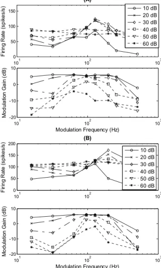
B. Cochlear nucleus responses
Figure 6 illustrates the impact of modeling WBII in the cochlear nucleus in terms of firing rate and synchronization. r-MTF curves are similar for both structures. They exhibit a peak at a specific modulation frequency at low intensities, and become flat at high intensities. At low intensity levels, t-MTF of the two models show a band-pass profile. As the intensity increases, it becomes more and more selective. At high level, the model without WBII has a noisy t-MTF contrary to the complete model whose t-MTF is still synchronized at the modulation frequency for which the neuron reaches its maximum firing rate (best modulation frequency). 101 102 103 0 50 100 150 (A) F iri n g R ate (s p ik e s/ s) 10 dB 20 dB 30 dB 40 dB 50 dB 60 dB 101 102 103 -20 -10 0 10 Modulation Frequency (Hz) M o d ula tio n G a in ( d B ) 101 102 103 0 50 100 150 200 (B) F iri n g R ate (s p ik e s/ s) 10 dB 20 dB 30 dB 40 dB 50 dB 60 dB 101 102 103 -20 -10 0 10 Modulation Frequency (Hz) Mo d ul a tio n Ga in ( d B )
Fig. 6. Response of an inferior colliculus coincidence detector unit. (A) r-MTF and t-r-MTF curves are obtained without WBII in the cochlear nucleus. (B) r-MTF and t-MTF are obtained with the model including WBII. According to physiological measurements [15], high level stimuli are synchronized at the best modulation frequency (see text) of the neuron with WBII included in the VCN.
V. DISCUSSION
The model response without WBII structure is closer to physiological measurements [15] than Hewitt and Meddis first results. This is due to the new basilar membrane model [10] and corresponding inner hair cells. The addition of WBII improves high level stimuli response. This is explained as follows. For low level stimuli, HSR fibers of the right CF drive this unit. At medium stimuli level, when HSR fibers saturate, LSR fibers convey synchrony information. Of course WBI inhibition is active, but it just acts as a limiter. For high level stimuli carrier, when both HSR and LSR fibers saturate, inhibition gives chopper cell synchronization by the wideband afferent. This is an example of how inhibition could be an information vector and not just an information modulator.
These results may be improved considering the study by Guérin [3]. It shows that relaxing constraint on chopper cell time constant makes the model more selective.
VI. CONCLUSION
This study confirms the natural relation that exists between temporal coding and dynamics. Moreover this work is a complement to other studies about temporal processing in the early stage of the auditory pathway.
Each stage of the physiological model (BM, IHC, AN) was already validated in different aspects, reproducing post stimulus time histograms, t-MTF, r-MTF, and forward masking measurements. The structure we proposed here improves results produced by Meddis [4] for high level stimulus in terms of response synchronization. Given Pressnitzer’s work [14], one can suppose that this model will probably be able to reproduce comodulation masking results. Moreover, this structure generalizes Nelson and Carney’s “same frequency inhibition and excitation” [11]. This lets us hope to reproduce other types of inferior colliculus responses like those measured by Krishna and Semple [6] when varying WBII and chopper cell time constant.
VII. MODEL SETTING
All notations used in this section correspond to those used in corresponding articles.
TABLEI
BASILAR MEMBRANE SETTINGS [10]
CF (Hz) 826 1297 1980 2970 4400
TABLEII
IHCRECEPTOR POTENTIAL SETTINGS [19] M GCA max (nS) [Ca2+]thr (*10-11)
HSR 10 8 4.48
LSR 8 2.75 4
TABLEIII
POINT NEURON SETTINGS [4],[9] τM ms τGk ms C B Th0 mV Ek mV Er mV VCN chopper 5 10 0.3 0.1 10 -16 0 VCN onset 5 10 0.3 0.1 14 -10 0 IC 1 0.1 0 0.1 14 -10 0 Dendritic filtering time constant is 0.1 ms. AP is 50 mV
REFERENCES
[1] M.J. Ferragamo, N.L. Golding, and D. Oertel, “Synaptic inputs to stellate cells in the ventral cochlear nucleus,” J. Neurophysiol. vol. 79, 1998, pp. 51-63.
[2] J.M. Goldberg and P.B. Brown, “Responses of binaural neurons of dog superior olivary complex to dichotic tonal stimulation: some physiological mechanisms of sound localization,” J. Neurophysiol, vol. 32, 1969, pp. 940-958.
[3] A. Guérin, R. Le Bouquin Jeannès, J. Bès, G. Faucon and C. Lorenzi, “Evaluation of two computational models of amplitude modulation coding in the inferior colliculus,” Hear. Res., 2006, vol. 211, pp. 54-62.
[4] M.J. Hewitt, and R. Meddis, “A computer model of amplitude modulation sensitivity of single units in the inferior colliculus,” J.
Acoust. Soc. Am. vol. 95, 1994, pp. 2145-2159.
[5] P.X. Joris, C.E. Schreiner and A. Rees, Neural representation of amplitude-modulated sounds, Physiol Rev, vol. 84, 2004, pp. 541-577. [6] B.S. Krishna and M.N. Semple “Auditory temporal processing:
response to sinusoidally amplitude modulated tone in the inferior colliculus,” J.Neurophysiol., vol. 22, 2000, pp. 255-273.
[7] M.C. Liberman, “Central projection of auditory-nerve fibers of differing spontaneous rate. I. Anteroventral cochlear nucleus,” J.
Comp. Neurol., vol. 313, 1991, pp. 240-258.
[8] E.A. Lopez-Poveda, “An approximate transfer function for the dual-resonance nonlinear filter model of auditory frequency selectivity,” J.
Acoust. Soc. Am., vol. 114, 2003, pp. 2112-2117.
[9] R.J. MacGregor, “Neural and Brain Modeling,” Academic, San Diego, CA, 1987.
[10] R. Meddis, L.P. O’Mard, and E.A. Lopez-Poveda, “A computational algorithm for computing nonlinear auditory frequency selectivity,” J.
Acous. Soc. Am., vol. 109, 2001, pp.2852-2861.
[11] P.C. Nelson, and L.H. Carney, “A phenomenological model of peripheral and central neural responses to amplitude modulated tones,” J. Acoust. Soc. Am., vol. 116, 2004, pp. 2173-2186.
[12] D. Oertel, S.H. Wu, M.W. Garb, and C. Dizack, “Morphology and physiology of cells in slice preparations of the posteroventral cochlear nucleus of mice,” J. Comp. Neurol., vol. 295, 1990, pp. 136-154. [13] D. Oertel, R.R. Fay, and A.N. Popper, Integrative function in the
mammalian auditory pathway, Springer-Verlag, New York, 2002.
[14] D. Pressnitzer, R. Meddis, and I.M. Winter, “Physiological correlates of comodulation masking release in the mammalian ventral cochlear nucleus,” J. Neurosci., vol. 21, 2001, pp. 6377-6386.
[15] A. Rees, and A.R. Palmer, “Neuronal response to amplitude modulated and pure tone stimuli in the guinea pig inferior colliculus, and their modification with broadband noise,” J. Acoust. Soc. Am., vol. 85, 1989, pp. 1978-1994.
[16] W.S. Rhode, P.H. Smith, and D. Oertel, “Physiologicalresponse properties of cells labeled intracellulary with horseradish peroxidase in the ventral cochlear nucleus,” J. Comp. Neurol., vol. 213, 1983, pp. 426-447.
[17] D.K. Ryugo, and T.N. Parks, “Primary innervation of the avian and mammalian cochlear nucleus,” Brain Res. Bulletin, vol. 60, 2003, pp. 435-456.
[18] Z.M. Smith, B. Delgutte, A.J. Oxenham, “Chimaeric sounds reveal dichotomies in auditory perception,” Nature, vol. 416, 2002, pp. 87– 90.
[19] C.J. Sumner, E.A. Lopez-Poveda, L.P. O’Mard, and R. Meddis, “A revised model of the inner-hair cell and auditory nerve complex,” J.
Acoust. Soc. Am., vol. 111, 2002, pp. 2178-2188.