HAL Id: hal-01266776
https://hal.archives-ouvertes.fr/hal-01266776
Submitted on 3 Feb 2016HAL is a multi-disciplinary open access archive for the deposit and dissemination of sci-entific research documents, whether they are pub-lished or not. The documents may come from teaching and research institutions in France or abroad, or from public or private research centers.
L’archive ouverte pluridisciplinaire HAL, est destinée au dépôt et à la diffusion de documents scientifiques de niveau recherche, publiés ou non, émanant des établissements d’enseignement et de recherche français ou étrangers, des laboratoires publics ou privés.
Excited States
Dimitra Markovitsi
To cite this version:
Dimitra Markovitsi. UV-induced DNA Damage: The Role of Electronic Excited States . Photochem-istry and Photobiology, Wiley, 2016, 92 (1), pp.45-51. �10.1111/php.12533�. �hal-01266776�
INVITED REVIEW
UV-induced DNA Damage: The Role of Electronic Excited States
†Dimitra Markovitsi
CNRS, IRAMIS, LIDYL, Laboratoire Francis Perrin, URA 2453, F-91191 Gif-sur-Yvette, France
*Corresponding author e-mail: Dimitra.markovitsi@cea.fr (Dimitra Markovitsi)
†
Part of the data in this paper were presented during the 16th International Congress on Photobiologyheld in Cordoba, Argentina, in September (8th - 12th), 2014.
ABSTRACT
The knowledge of the fundamental processes induced by the direct absorption of UV radiation by DNA allows extrapolating conclusions drawn from in vitro studies to the in-vivoDNA photo-reactivity. In this respect, the characterization of the DNA electronic excited states plays a key role. For a long time, the mechanisms of DNA lesion formation were discussed in terms of generic “singlet” and “triplet” excited state reactivity. However, since the beginning of the 21st century, both experimental and theoretical studies revealed the existence of “collective” excited states, i.e. excited states delocalized over at least two bases. Two limiting cases are distinguished: Frenkel excitons (delocalized * states) and charge transfer states in which positive and negative charges are located on different bases. The importance of collective excited states in photon absorption (in particular in the UVA spectral domain), the redistribution of the excitationenergy within DNA, and the formation of dimeric pyrimidine photoproducts is discussed. The dependence of the behavior of the collective excited states on conformational motions of the nucleic acids is highlighted.
INTRODUCTION
The effects resulting from the DNA damage induced by UV radiation (carcinogenic mutations, cell lethality, etc.) mobilize important efforts of the photobiology community.Considerable contributions in the characterization of chemical structures altering the genetic codehavebeen made by the chemists. To date, a large number ofcompounds, corresponding to both primary and secondary photoproducts,formed via subsequent dark reactions,have been identified, and highly sensitive analytical methods have been developed for their quantification(1-3). The mechanistic schemes proposed to explain the formation of these photoproducts often consider as precursors generic “singlet” or “triplet” excited states of individual bases.But, in the light of recent advances in the field of spectroscopy, the use of such generic terms may lead to erroneous conclusions.
Since the turn of the century, the electronic excited states of nucleic acids per sehave been the subject of an increasing number of experimental and theoretical studies.On one hand, the improvement of time-resolved techniqueshas allowedscientists to study their fateover a large time domain, starting from the femtosecond scale; this is achieved by detectingeither nucleic acids’ absorptionin the UV/visible or IR spectral domains or their fluorescence emission(4-11). On the other hand, with the development of advanced computational methods, it is now possible to include in the calculation of the excited states factors that are crucial for biological systems, such as water molecules, metal cations or even conformational motions of the phosphodeoxyribose backbone(12-18). The general picture emerging from all these studies is that electronic interactions operating among the basesdue to their close proximitywithin DNA strands play a key role in the properties of the excited states. A straightforward consequence is that excited states may be delocalized over two or more bases (collective states or excitons) and that their behavior depends strongly on the relative position of the bases and their sequence.
In the present short overview, which corresponds to a keynote lecture presented during the International Congress of Photobiology (Cordoba, Argentina, 2014), Iwill focus on singlet excited states that are formed following absorption of UV photons directly by DNA.Iwill introducetwo limiting cases of collective excited states: Frenkel excitons and charge transfer (CT) states, as well as combinations of them. Iwill discuss their reactivity, which may intervene either immediately after photon absorption or following redistribution of the excitation energy. The objective is not to present a thorough review but ratherto highlight recent advances in the study of complex processes.
COLLECTIVE EXCITED STATES: STATIC PICTURE
Before approaching the collective excited states of DNA bases,Irecall that there are various types of excited states localized on single bases: *, n*, n*, which may exist in both singlet and triplet multiplicity. The electronic transitions leading from the ground state to
* states are responsible for the characteristic absorption around 260 nm of the bases, nucleosides and nucleotides. In contrast, n* or n* states cannot be populated directly by photon absorption.
In DNA duplexes, G-quadruplexes and, to a lesser extent, single strands, the electronic coupling among * transitions gives rise to Frenkel excitons(19). These delocalized statesare linear combinations of the excited states of individual bases.DNA can then be compared to a molecular aggregate ofNmonomers, which is characterized by N Frenkel excitons(19),
described by the equation:
N n n n k C k 1
, , where kdenotes a Frenkel exciton and Ψan
excited state of a single monomer. For a given Frenkel excitonk, (Ck,n)
2
represents the contribution of each chromophore n to this state and definesits topography (Figure 1).Such topographies have been calculated for Frenkel excitons associated with the absorption spectra
of model double- and four-stranded structures(20-22).Each Frenkel excitonalso has its own energy and oscillator strength (related to the molar absorption coefficient), ranging from zero to values higher than those of thelocalized * states.Further, the properties of the Frenkel excitons of a given duplex strongly depend on the conformation of the double helix because the dipolar coupling between * transitions is sensitive to the relative orientation of the bases(20). Accordingly, the UV spectrum recorded for a solution of thisduplex, whichexists in multiple conformations, is in fact the sum of a very largenumber of spectra, each one corresponding to a specific helix conformation.
<Figure 1>
When the stacking distance between bases becomes very small, their orbitalsstartto overlap. In other words, the electronic density of one base starts to penetrate that of another, thereby generating an “orbital overlap” coupling (23). This orbital overlap gives rise to thesecond family of collective excited states, the charge transfer states,whosecharacteristic is that fractions of positive and negative charges are located on different bases. Due to such charge separation, CT states are more sensitive than Frenkel excitons to environmental factorssuch as the presence of water moleculesor ionic species in the vicinity. The oscillator strength associated with CT states is extremely low. But even ifthey do not contribute directly to the DNA absorption band around 260 nm, they do affect its molar absorption coefficient. In fact, the existence of CT states is related to the well-known hypochromism of DNA duplexes (24, 25).Although this property (via the melting curves) is widely exploited for the study of duplex stability, it is rarely perceived as an important difference between the excited statesof single and double strands.
Frenkel excitons and CT states are two limiting cases of collective excited states. However, intermediate schemes,e.g. collective excited states with partial * and partial CT character, are encountered quite often. The fingerprint of such mixed collective states may
appear as a tail onthe red side of the main absorption band. Weak absorption above 300 nm was already observed in 1981 for natural DNA of various origins and was correlated with the GC content(26). Recent spectroscopic studies on model systems have shownthat, incontrast to the behavior of isolated bases, adenine and thyminesingle strands are capable of absorbing UVA photons and that this capacity is enhanced by base-pairing(27). These observations prove that UVA absorption arises from a collective behavior of the bases. Theoretical calculations attributed these delocalized states to mixed */CT states involving the bases of one strand (18).
REACTIVITY OF COLLECTIVE EXCITED STATES
The reactivity of collective excited states in connection with pyrimidine dimeric photoproductshas been examined in quantum chemistry studies(28-31). These studies have shown that Frenkel excitons delocalized over two stacked thymines lead to the formation of cyclobutane dimers (CPDs); the reaction proceeds without an energy barrier(29). This picture is strongly supported by a series of experimental observations on thymine single strands. Time-resolved experiments with UVC excitation and infra-red detection revealed that CPD formationis an ultrafast reaction, occurringwithin1 ps(32, 33). Furthermore, continuous irradiations and subsequent analysis of photoproducts by mass spectrometry, combined with liquid chromatography, have shown that the CPD quantum yield is constant (0.05) throughout the main absorption band(29).In contrast, the quantum yield related to triplet formation (intersystem crossing) strongly decreases upon increasing the excitation wavelength, showing that 3* statescontribute to the dimerization reactionby less than 10%(29). This was confirmed by a transient infra-red absorption study of thymine single strands which detected the signature of3* states but did not observe their reaction toward CPDs; it was found instead that theencounter of 3* states with non-excitedthyminescreates transient species
(biradicals) which simply decay to the ground state(34).
Charge transfer states between two pyrimidines may evolve toward a reaction intermediate (oxetane),eventually leading to (6-4) adducts(29, 30). The existence of a reaction intermediate was demonstrated spectroscopically in the case of thymine single strands:it was found that the typical absorption of the (6-4) adducts around 320 nm appears only within 4 ms(35), a time scale on which all excited states have decayed. Such an observation proves that the reaction proceeds via a transient species which does not absorb between 290 and 800 nm, a feature compatiblewith the electronic structure of oxetane. According to quantum chemistry calculations, oxetane formation from CT states involves a reaction barrier(29) so that, if the UV photons do not provide the excess energy required to overcome it, the reaction cannot take place. Such a picture is corroborated by the observed important decrease of the quantum yield determined for (6-4) adducts with increasing irradiation wavelength(29).
Reaction paths similar to those described for the formation of thymine dimeric photoproducts, via collective states,have also been found for thymine-cytosine CPDs and (6-4) adducts, even when cytosine is methylated in the 5 position(30).In this case, a CT state corresponding to an electron transfer from the 5’ to the 3’ base (T+C-) results in the formation of an azetidine-like species, which is the analog of the oxetane for thymine-cytosine (6-4)adducts.
The above mentioned theoretical studies showed the reactivity of Frenkel excitons and CT states. However, the extent in which these reactions will effectively take place in a given DNA multimer depends on conformational factors. For example, since the lifetime of * states in thymine single strands is very short, only pairs that have appropriate mutual orientation in the ground state geometry will react(36). Similarly, steric factors may limit the formation of (6-4) adducts in duplexes. In addition to such “static” structural factors, “dynamic” effects, intervening during the lifetime of singlet excited states, may also prove
crucial for the DNA photoreactivity.
REDISTRIBUTION OF THE EXCITATION ENERGY
Time-resolved measurementshave shown that the fluorescence decays of DNA multimers, including natural DNA, are complex(37), spanning from fs to ns(38-40). All over this range, a redistribution of the excitation energy takes place, in the sense that various singlet excited statesare populated successively. We have seen previously that each collective excited state has its own topology (Figure 1). Consequently,interconversion among various collective states implies changes in the contribution of the basesto them, resulting to an energy transfer process. These changes are associated with conformational motionswhich may affect seriously the electronic coupling,even when they have very weak amplitude. In addition to conformational motions of nucleic acids taking place in the ground state, specific structural changes are triggered by the absorption of UV photons. Accordingly, we are faced a continuous spectrum of excited states. Therefore, the term “electronic excitation” is often used instead of “excited state”,commonly associated with well-defined properties.
Prior to discussing the fate of electronic excitations in DNA multimers, let’sexamine briefly what happens in the case of theirmonomeric building blockswhen they are “isolated”in aqueous solution,i.e.not interactingwith each other. Under such conditions, the lifetimes of the singlet * states are shorter than 1 ps and can be determined only by femtosecond spectroscopy(4, 6). The main reason for the ultrafast decay of * states is not their conversion to other singlet (n*, n*) or triplet excited states because the quantum yieldsfor these processesare too low(41-43). The ultrafast decay is due to the existence of “conical intersections”, allowing a very rapid return to the ground state. This processis accompanied by conformational changes, such as out-of-plane motions of substituentsat the 5 position of pyrimidines or the 2 position of purines(44).
When going from mononucleotides to polynucleotides, the deactivation mechanism of
* states just described remains valid but is now in competition with otherphotophysical processes.These are associated not only with internal movements of the base structure but also with changes in the relative distance and orientation of different bases.As a result, we may have interconversion both among various excited states of the same type (Frenkel excitons or CT states) and between * and CT states, including a series of mixed states with various degrees of CT character.
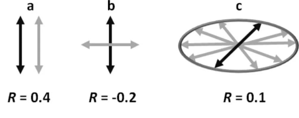
A suitable way to follow interconversion among singlet excited states of polynucleotides is to study their fluorescence anisotropy, which is connected to the polarization of the electronic transitions (Figure 2). A decrease in anisotropy occurring at times shorter than thoseneeded for rotational diffusion of the molecules shows that the emitted photons stem from different excited states than those populated initially by photon absorption.This short time decrease was indeed observed for the various model duplexes and natural DNA, as well as for G-quadruplexes:their fluorescence anisotropy diminishes already on the fs time-scale revealing that energy transfer takes place among the bases(45, 46, 40).
<Figure 2>
The ultrafast decrease in the fluorescence anisotropy observed at 330 nm, around the maximum of the * emission band,showsthat energy transfer involves Frenkel excitons(38, 40).Their internal conversion, also called “intraband scattering”, is a much faster process than Förster transfer, according to which excitation hops from one base to the other. In the case of calf thymus DNA, in addition to ultrafast energy transfer, a slower one was detected. Within 800 ps its fluorescence anisotropy decays to 0.1 and, subsequently, remains constant for several ns(46). This0.1 value corresponds to the in-plane depolarization (Figure 2) of the fluorescence (47). It arises from a statistical average of hops involving * transitions, all of them polarized within the aromatic plane of the bases, approximately perpendicular to the axis
of the helix.
Conformational motions tend to localize Frenkel excitons to single bases, where * states follow a deactivation mechanism similar to that determined for monomeric nucleic acids. Alternatively, the conformational motions may favor localization of the excitation on stacked dimers,the excitation acquiring a CT character with concomitant loweringof its energy. The fingerprint of this process is also encountered in the fluorescence anisotropy(48-50). Upon increasing emission wavelength, the fluorescence anisotropy of some DNA systems becomes lower and lower. For example, the anisotropy value obtained for the duplex d(AT)10·d(AT)10at 3 ps is 0.22 at 330 nm but dwindles down to -0.12 at 420 nm(51). The latterwavelength corresponds to the peak of the well-known exciplex fluorescence(52). The negative value originates from the CT component of the excited states, which, in contrast to
* transitions, is polarized out of the aromatic plane of the bases.
Low but positive fluorescence anisotropy (0.02) was also found for the peculiar emission band dominating the fluorescence spectrum of alternating guanine-cytosine duplexes, peaking at shorter wavelengths than * fluorescence(53, 54). This emission band, decaying on the ns time-scale,is quenched upon transformation of the B-form to the Z-form(55). Quantum chemical calculations have identified an exciton state responsible for this high energy fluorescence. It results from mixing of Frenkel excitons withCT states. This collective state, named HELM (High-energy Emitting Long-lived Mixed) state, is delocalized over at least four bases across both strands(55). The stacking distance d in the HELM state (3.18 Å) is shorter than in the ground state(3.68 Å) and the guanine-to-cytosine charge transfer amounts to 10-20%, depending on the type of calculation(Figure 3).
<Figure 3>
According to transient absorption studies using UVC excitation, the majority of CT states populated during the excited state relaxation of * states disappear, on average,
within a few hundred ps;their lifetimeis shorter in double strands than in single strands(56-58).The opposite lifetime trend was observed by fluorescence spectroscopy following UVA excitation: base-pairing slows down thedecays: the average lifetime determined for theduplex (dA)20·(dT)20 is 1.3 ns versus 0.6 ns for each one of the parent single strands(27). The UVA-induced fluorescence stems from excited states with mixed*/CT character(18), which are favored by the better base stacking in double-stranded structures.
The relaxation of * states toward CT states may give rise to excimer/exciplex fluorescence, characterized by a broad band peaking around 420 nm(52). In early studies, such a peak was also detected for natural DNA(59, 60). But recent experiments on ultrapure calf thymus DNA have shown that its fluorescence spectrum exhibits a single fluorescence band coinciding with that of its monomeric constituents(39). Despite this similarity, the fluorescence of this natural DNA decays on the ns time-scale, three decades of time later than the fluorescenceof mononucleotides(44). The strange “monomer-like” * delayed fluorescence observed for calf thymus DNA has been explained by interconversion between
and CT states(46). According to the proposed mechanism, the initially formed Frenkel excitons are trapped by CT states.Then, after charge separation and charge recombination, the excitation reaches a * state. Another possible route leading to the long-lived DNA fluorescence is the transformation of HELM states to* states localized on single bases,promoted by conformational motions(55).
The interplay between Frenkel excitons and CT states was modeled by quantum chemical calculations for a series of adenine stacks(61, 62).In general, if these two types of excited states are close in energy, it is possible that vibrational motions provide the necessary driving force for their interconversion.Such a conversion of mixed */CT states populated by absorption of UVA photons to Frenkel excitons, which give rise to CPDs, is certainly involved in the UVA-induced thymine dimerization. The existence of an additional step
between photon absorption and chemical reaction explains why the CPD quantum yield in thymine single strandsis much lower for UVA excitation compared to UVB/UVC excitation, populating directly * states (29). But base-pairing increases the quantum yield of the UVA-induced CPDs by an order of magnitude.In parallel, the UVA-UVA-induced andthe UVC-UVA-induced fluorescence spectra shift closer toeach other (Figure 4), indicating closer energy levels between mixed and reactive*states,in line with more efficient conversion.
<Figure 4>
Finally, dimerization reactions may be in competition with evolution of the excitation from Frenkel excitons to CT states, governed by, among others, the redox potentials of theflanking bases. Such a dependence was indeed demonstratedfor the formation of thymine CPDs(63). In this case, part of the excitations, instead of leading to the photoproducts, evolve toward low-lying CT states which eventually decay to the ground state (31).
CONCLUSION
In this brief overview I have tried to give a flavor of recent advances on understanding of the DNA singlet electronic excited states and to discuss their relevance in the UV-induced reactivity leading to damage of the genetic code. Below I highlight the most important points.
The close proximity of the bases gives rise to collective excited states delocalized over at least two bases. The properties of these states depend strongly on the conformation of the nucleic acids and the redox potentials of the bases. As a result, for a given DNA multimer, we have a continuous spectrum of excited states (electronic excitations) with various degrees of delocalization and various degrees of charge transfer character.The multitude of excited states is both static, depending on the precise configuration of the system at the moment of photon absorption, and dynamic, changing as a function of the motions that occur during the lifetime of the excitation.
The existence of excited states with CT character renders possible the absorption of UVA photons by DNA multimers.
Frenkel excitons (delocalized * states) lead to the formation of pyrimidine cyclobutane dimers while charge transfer states lead to reactive intermediates that give rise to (6-4) adducts.
During the lifetime of the excitation, interconversion among the various singlet excited states takes place. Thus, reactive excited states, characterized by very short lifetimes, may be populated either directly by the photon absorption or at later times by such interconversion.
Excitations begin decaying on the fs time-scale buta portion of them survives for several ns. The longest lifetimes have been reported for low-energy (UVA-induced) and high-energy (UVB- or UVC-inducedHigh high-energyEmittingLong-lived Mixed/HELM states) excited states. Small populations of long-lived excited states may play a key role in lesion formation because the probability that nucleic acids adopt conformations appropriate to photoreactions increases with time.
When the amplitude of conformational motions decreases the collective character of the electronic excitations is enhanced.
<Figure 5>
The above conclusions have been reached by numerous studies on model systems which are valuable for rationalizing the fate of electronic excitations. The model systems have allowedthe base sequence effects to be taken into account and the experimental results to be explained in the light of theoretical calculations. But the important point is that observations on natural DNA are very much in line with the overall picture provided by these studies. In particular with the fact that the cooperativity among the bases in respect with electronic excitations is boosted when the amplitude of conformational motions decreases. This
encourages us to speculate that the collective character of the electronic excitationsis particularly enhancedinside cells when DNA is condensed.
Acknowledgements- The French Agency for Research(projects ANR-10-BLAN-0809-01
REFERENCES
1. Douki, T. (2013) The variety of UV-induced pyrimidine dimeric photoproducts in DNA as
shown by chromatographic quantification methods. Photochem. Photobiol. Sci. 12, 1286-1302.
2. Cadet, J., T. Douki and J.-L. Ravanat (2015) Oxidatively Generated Damage to Cellular DNA by UVB and UVA Radiation. Photochem. Photobiol. 91, 140-155.
3. Cadet, J., A. Grand and T. Douki (2015) Solar UV radiation-induced DNA Bipyrimidine photoproducts: formation and mechanistic insights. Top. Curr. Chem. 356, 249-275. 4. Pecourt, J.-M. L., J. Peon and B. Kohler (2001) DNA excited-state dynamics: ultrafast
internal conversion and vibrational cooling in a series of nucleosides. J. Am. Chem. Soc. 123, 10370-10378.
5. Peon, J. and A. H. Zewail (2001) DNA/RNA nucleotides and nucleosides: direct
measurement of excited-state lifetimes by femtosecond fluorescence up-conversion. Chem. Phys. Lett. 348, 255-262.
6. Onidas, D., D. Markovitsi, S. Marguet, A. Sharonov and T. Gustavsson (2002)
Fluorescence properties of DNA nucleosides and nucleotides: a refined steady-state and femtosecond investigation. J. Phys. Chem. B 106, 11367-11374.
7. Markovitsi, D., A. Sharonov, D. Onidas and T. Gustavsson (2003) Effect of molecular organisation in DNA oligomers studied by femtosecond fluorescence spectroscopy. ChemPhysChem 3, 303-305.
8. Pancur, T., N. K. Schwalb, F. Renth and F. Temps (2005) Femtosecond fluorescence up-conversion spectroscopy of adenine and adenosine: experimental evidence for the * state? Chem. Phys. 313, 199-212.
9. Kuimova, M. K., J. Dyer, M. W. George, D. C. Grills, J. M. Kelly, P. Matousek, A. W. Parker, X. Z. Sun, M. Towrie and A. M. Whelan (2005) Monitoring the effect of ultrafast deactivation of the electronic excited states of DNA bases and polynucleotides following 267 nm laser excitation using picosecond time-resolved infrared spectroscopy. Chem. Comm., 1182-1184.
10. Kwok, W.-M., C. Ma and D. L. Phillips (2006) Femtosecond time- and wavelength-resolved fluorescence and absorption study of the excited states of adenosine and an adenine oligomer. J. Am. Chem. Soc. 128, 11894-11905
11. Chen, J., Y. Zhang and B. Kohler (2015) Excited states in DNA strands investigated by ultrafast laser spectroscopy. Top. Curr. Chem. 356, 39-87.
12. Improta, R. and V. Barone (2011) Interplay between “neutral” and “charge-transfer” excimers rules the excited state decay in adenine-rich polynucleotides. Angew. Chem. Int. Ed. 50, 12016-12019.
13. Olaso-Gonzalez, G., M. Merchan and L. Serrano-Andres (2009) The Role of Adenine Excimers in the Photophysics of Oligonucleotides. J. Am. Chem. Soc. 131, 4368-4377. 14. Improta, R. and V. Barone (2015) Excited states behavior of nucleobases in solution:
insights from computational studies. Top. Curr. Chem. 355, 329-357.
15. Voityuk, A. A. (2013) Effects of dynamic disorder on exciton delocalization and photoinduced charge separation in DNA. Photochem. Photobiol. Sci. 12, 1303-1309. 16. Plasser, F., A. Aquino, H. Lischka and D. Nachtigallová (2015) Electronic Excitation
Processes in Single-Strand and Double-Strand DNA: A Computational Approach. Top. Curr. Chem. 356, 1–38.
17. Lu, Y., Z. Lan and W. Thiel (2015) Computational modeling of photoexcitation in DNA single and double strands. Top. Curr. Chem. 356, 89-122.
18. Spata, V. A. and S. Matsika (2014) Role of Excitonic Coupling and Charge-Transfer States in the Absorption and CD Spectra of Adenine-Based Oligonucleotides Investigated through QM/MM Simulations. J. Phys. Chem. A 118, 12021-12030.
19. Kasha, M., H. R. Rawls and M. A. El-Bayoumi (1965) The exciton model in molecular spectroscopy. Pure & Appl. Chem. 11, 371-392.
20. Bouvier, B., J. P. Dognon, R. Lavery, D. Markovitsi, P. Millié, D. Onidas and K. Zakrzewska (2003) Influence of conformational dynamics on the exciton states of DNA oligomers. J. Phys. Chem. B 107, 13512-13522.
21. Emanuele, E., K. Zakrzewska, D. Markovitsi, R. Lavery and P. Millie (2005) Exciton states of dynamic DNA double helices: alternating dCdG sequences. J. Phys. Chem. B 109, 16109-16118.
22. Changenet-Barret, P., E. Emanuele, T. Gustavsson, R. Improta, A. B. Kotlyar, D. Markovitsi, I. Vaya, K. Zakrzewska and D. Zikich (2010) Optical properties of guanine nanowires: experimental and theoretical study. J. Phys. Chem. C 114, 14339–14346. 23. Blancafort, L. and A. A. Voityuk (2014) Exciton delocalization, charge transfer, and
electronic coupling for singlet excitation energy transfer between stacked nucleobases in DNA: An MS-CASPT2 study. J. Chem. Phys. 140.
24. Starikov, E. B. (2004) Importance of charge transfer excitations in DNA electron spectrum: a ZINDO semiempirical quantum-chemical study. Modern Phys. Lett. B 18, 825-831.
25. Varsano, D., R. Di Felice, M. A. L. Marques and A. Rubio (2006) A TDDFT study of the excited states of DNA bases and their assemblies. J. Phys. Chem. B 110, 7129-7138. 26. Sutherland, J. C. and K. P. Griffin (1981) Absorption spectrum of DNA for wavelengths
27. Banyasz, A., I. Vayá, P. Changenet-Barret, T. Gustavsson, T. Douki and D. Markovitsi (2011) Base-pairing enhances fluorescence and favors cyclobutane dimer formation induced upon absorption of UVA radiation by DNA. J. Am. Chem. Soc. 133, 5163-5165. 28. Roessle, S., J. Friedrichs and I. Frank (2010) The Formation of DNA Photodamage: The
Role of Exciton Localization. ChemPhysChem 11, 2011-2015.
29. Banyasz, A., T. Douki, R. Improta, T. Gustavsson, D. Onidas, I. Vayá, M. Perron and D. Markovitsi (2012) Electronic Excited States Responsible for Dimer Formation upon UV Absorption Directly by Thymine Strands: Joint Experimental and Theoretical Study. J. Am. Chem. Soc. 134, 14834–14845.
30. Esposito, L., A. Banyasz, T. Douki, M. Perron, D. Markovitsi and R. Improta (2014) Effect of C5-Methylation of Cytosine on the Photoreactivity of DNA: A Joint
Experimental and Computational Study of TCG Trinucleotides. J. Am. Chem. Soc. 136, 10838-10841.
31. Lee, W. and S. Matsika (2015) QM/MM studies reveal pathways leading to the quenching of the formation of thymine dimer photoproduct by flanking bases. Phys. Chem. Chem. Phys. 17, 9927-9935.
32. Schreier, W. J., T. B. Schrader, F. O. Koller, P. Gilch, C. Crespo-Hernàdes, V. N.
Swaminathan, T. Carell, W. Zinth and B. Kohler (2007) Thymine dimerization in DNA is an ultrafast photoreaction. Science 315, 625-629.
33. Schreier, W. J., J. Kubon, N. Regner, K. Haiser, T. E. Schrader, W. Zinth, P. Clivio and P. Gilch (2009) Thymine dimerization in DNA model systems: cyclobutane photolesion Is predominantly formed via the singlet channel. J. Am. Chem. Soc. 131, 5038-5039. 34. Pilles, B. M., D. B. Bucher, L. Z. Liu, P. Clivio, P. Gilch, W. Zinth and W. J. Schreier
(2014) Mechanism of the Decay of Thymine Triplets in DNA Single Strands. J. Phys. Chem. Lett. 5, 1616-1622.
35. Marguet, S. and D. Markovitsi (2005) Time-resolved study of thymine dimer formation. J. Am. Chem. Soc. 127, 5780-5781.
36. McCullagh, M., F. Lewis, D. Markovitsi, T. Douki and G. C. Schatz (2010)
Conformational control of TT dimerization in DNA conjugates. A molecular dynamics study. J. Phys. Chem. B 114, 5215–5221.
37. Markovitsi, D., F. Talbot, T. Gustavsson, D. Onidas, E. Lazzarotto and S. Marguet (2006) Complexity of excited state dynamics in DNA. Nature 441, E7.
38. Markovitsi, D., T. Gustavsson and I. Vayá (2010) Fluorescence of DNA Duplexes: From Model Helices to Natural DNA. J. Phys. Chem. Lett. 1, 3271–3276.
39. Vayá, I., T. Gustavsson, F. A. Miannay, T. Douki and D. Markovitsi (2010) Fluorescence of natural DNA: from the femtosecond to the nanosecond time-scales. J. Am. Chem. Soc. 132, 11834-11835.
40. Changenet-Barret, P., Y. Hua and D. Markovitsi (2015) Electronic excitations in guanine quadruplexes. Top. Curr. Chem. 356, 183–202.
41. Hare, P. M., C. Crespo-Hernández and B. Kohler (2007) Internal conversion to electronic ground state occurs via two distinct pathways for pyrimidine bases in aqueous solution. Proc. Natl. Acad. Sci. 104, 435-440.
42. Kwok, W. M., C. Ma and D. L. Phillips (2008) A doorway state leads to photostability or triplet photodamage in thymine DNA. J. Am. Chem. Soc. 130, 5131-5139.
43. Cheng, C. C.-W., C. Ma, C. T.-L. Chan, K. Y.-F. Ho and W.-M. Kwok (2013) The solvent effect and identification of a weakly emissive state in nonradiative dynamics of guanine nucleosides and nucleotides - a combined femtosecond broadband time-resolved
fluorescence and transient absorption study. Photochem. Photobiol. Sci. 12, 1351-1365. 44. Gustavsson, T., R. Improta and D. Markovitsi (2010) DNA/RNA: Building Blocks of Life
45. Markovitsi, D., T. Gustavsson and A. Banyasz (2010) Absorption of UV radiation by DNA: Spatial and temporal features. Mutat. Res. - Rev. Mutat. Res. 704, 21-28. 46. Vayá, I., T. Gustavsson, T. Douki, Y. Berlin and D. Markovitsi (2012) Electronic
Excitation Energy Transfer between Nucleobases of Natural DNA. J. Am. Chem. Soc. 134, 11366−11368.
47. Albrecht, A. C. (1961) Polarizations and assignments of transitions - method of photoselection. J. Mol. Spectroscopy 6, 84-108.
48. Changenet-Barret, P., Y. Hua, T. Gustavsson and D. Markovitsi (2015) Electronic excitations in G-quadruplexes formed by the human telomeric sequence: a time-resolved fluorescence study. Photochem. Photobiol., 759–765.
49. Banyasz, A., T. Gustavsson, D. Onidas, P. Changenet-Barret, D. Markovitsi and R.
Importa (2013) Multi-Pathway Excited State Relaxation of Adenine Oligomers in Aqueous Solution: A Joint Theoretical and Experimental Study. Chem. Eur. J. 19, 3762-3774 50. Hua, Y., P. Changenet-Barret, R. Improta, I. Vayá, T. Gustavsson, A. B. Kotlyar, D.
Zikich, P. Šket, J. Plavec and D. Markovitsi (2012) Cation Effect on the Electronic Excited States of Guanine Nanostructures Studied by Time-Resolved Fluorescence Spectroscopy. J. Phys. Chem. C 116, 14682–14689.
51. Onidas, D., T. Gustavsson, E. Lazzarotto and D. Markovitsi (2007) Fluorescence of the DNA double helices (dAdT)n.(dAdT)n studied by femtosecond spectroscopy. Phys. Chem. Chem. Phys. 9, 5143-5148.
52. Ge, G. and S. Georghiou (1991) Excited-state properties of the alternating polynucleotide poly(dA-dT)poly (dA-dT). Photochem. Photobiol. 54, 301-305.
53. Vayá, I., F. A. Miannay, T. Gustavsson and D. Markovitsi (2010) High energy long-lived excited states in DNA double strands. ChemPhysChem 11, 987-989.
54. Brazard, J., A. Thazhathveetil, I. Vayá, F. Lewis, T. Gustavsson and D. Markovitsi (2013) Electronic excited states of guanine-cytosine hairpins and duplexes studied by fluorescence spectroscopy Photochem. Photobiol. Sci. 12, 1453 - 1459.
55. Huix-Rotllant, M., J. Brazard, R. Improta, I. Burghardt and D. Markovitsi (2015) Stabilization of mixed Frenkel-charge transfer excitons extended across both strands of guanine-cytosine DNA duplexes J. Phys. Chem. Lett. 6, 2247-2251.
56. Chen, J., A. K. Thazhathveetil, F. D. Lewis and B. Kohler (2014) Ultrafast Excited-State Dynamics in Hexaethyleneglycol-Linked DNA Homoduplexes Made of A.T Base Pairs. J. Am. Chem. Soc. 135, 10290-10293.
57. Zhang, Y., K. de La Harpe, A. A. Beckstead, R. Improta and B. Kohler (2015) UV-Induced Proton Transfer between DNA Strands. J. Am. Chem. Soc. 137, 7059-7062. 58. Schreier, W. J., P. Gilch and W. Zinth (2015) Early Events of DNA Photodamage. Ann.
Rev. Phys. Chem. 66, 497-519.
59. Anders, A. (1981) DNA fluorescence at room temperature excited by means of a dye laser. Chem. Phys. Lett. 81, 270-272.
60. Ballini, J. P., P. Vigny and M. Daniels (1983) Synchrotron excitation of DNA
fluorescence: decay time evidence for excimer emission at room temperature. Biophys. Chem. 18, 61-65.
61. Improta, R. (2008) The excited states of pi-stacked 9-methyladenine oligomers: a TD-DFT study in aqueous solution. PhysChemChemPhys 10, 2656-2664.
62. Santoro, F., R. Improta, F. Avila, M. Segado and A. Lami (2013) The interplay between neutral exciton and charge transfer states in single-strand polyadenine: a quantum
dynamical investigation. Photochemical & Photobiological Sciences 12, 1527-1543. 63. Pan, Z., M. Hariharan, J. D. Arkin, A. S. Jalilov, M. McCullagh, G. C. Schatz and F. D.
Efficiency of Thymine Photodimerization (vol 133, pg 20793, 2011). J. Am. Chem. Soc. 134, 3611-3611.
Figure 1Topography of four Frenkel excitons (k) of a hypothetical molecular aggregate
composed of four monomers (n). Vertical bars represent the squares of coefficients
Ck,nwhich indicate the contribution of each chromophore n to the Frenkel exciton k.
Internal conversion among these states modifies the contribution of each monomer to the excitation and corresponds to energy transfer.
Figure 2 The fluorescence anisotropy r provides information about the angle formed
between the transition dipoles related to photon absorption (black) and photon emission (grey). a) Parallel dipoles: r = 0.4; b) orthogonal dipoles: r = -0.2; c) dipoles distributed randomly within a plane (in-plane depolarization): r = 0.1.
Figure 3 Schematic representation of two Watson-Crick pairs (yellow: cytosine; cyan:
guanine) involved in aHigh-energy Emitting Long-lived Mixed (HELM) state, identified for alternating guanine GC duplexes. d corresponds to the stacking distance and +/ -denote the partial charge transfer (55).
Figure 4 Fluorescence spectra of the single strand (dT)20 (a) and duplex (dA)20·(dT)20 (b)
obtained with UVC (dashes) and UVA (dots) excitation (27). E denotes the energy difference between the emission maxima.
Figure 5 UV photons may be absorbed collectively by several bases. Conformational
motions induce redistribution of the excitation energy.
Graphical abstract Electronic interactions among DNA bases give rise to excited states
delocalized over two or more bases. As a result, the excited state properties depend strongly on conformational motions.