HAL Id: hal-01918048
https://hal.archives-ouvertes.fr/hal-01918048
Submitted on 26 May 2020
HAL is a multi-disciplinary open access archive for the deposit and dissemination of sci-entific research documents, whether they are pub-lished or not. The documents may come from teaching and research institutions in France or abroad, or from public or private research centers.
L’archive ouverte pluridisciplinaire HAL, est destinée au dépôt et à la diffusion de documents scientifiques de niveau recherche, publiés ou non, émanant des établissements d’enseignement et de recherche français ou étrangers, des laboratoires publics ou privés.
genus Marafivirus
Lev G. Nemchinov, Sarah François, Philippe Roumagnac, Marie Helene
Ogliastro, Rosemarie W. Hammond, Dimitre S. Mollov, Denis Filloux
To cite this version:
Lev G. Nemchinov, Sarah François, Philippe Roumagnac, Marie Helene Ogliastro, Rosemarie W. Hammond, et al.. Characterization of alfalfa virus F, a new member of the genus Marafivirus. PLoS ONE, Public Library of Science, 2018, 13 (9), �10.1371/journal.pone.0203477�. �hal-01918048�
Characterization of alfalfa virus F, a new
member of the genus Marafivirus
Lev G. Nemchinov1
*, Sarah Franc¸ois2, Phillipe Roumagnac3,4, Mylène Ogliastro2, Rosemarie W. Hammond1, Dimitre S. Mollov5, Denis Filloux3,4
1 USDA-ARS, Molecular Plant Pathology Laboratory, Beltsville MD, United States of America, 2 INRA, UMR DGIMI, Montpellier, France, 3 CIRAD, UMR BGPI, Montpellier, France, 4 BGPI, CIRAD, INRA, Montpellier SupAgro, Univ Montpellier, Montpellier, 5 USDA-ARS, National Germplasm Recourses Laboratory, Beltsville MD, United States of America
*lev.nemchinov@ars.usda.gov
Abstract
Viral infections of alfalfa are widespread in major cultivation areas and their impact on alfalfa production may be underestimated. A new viral species, provisionally named alfalfa virus F (AVF), was identified using a virion-associated nucleic acid (VANA) metagenomics-based approach in alfalfa (Medicago sativa L.) samples collected in Southern France. The nucleo-tide sequence of the viral genome was determined by de-novo assembly of VANA reads and by 5’/3’ RACE with viral RNA extracted from enriched viral particles or with total RNA, respectively. The virus shares the greatest degree of overall sequence identity (~78%) with
Medicago sativa marafivirus 1 (MsMV1) recently deduced from alfalfa transcriptomic data.
The tentative nucleotide sequence of the AVF coat protein shares ~83% identity with the corresponding region of MsMV1. A sequence search of the predicted single large ORF encoding a polyprotein of 235kDa in the Pfam database resulted in identification of five domains, characteristic of the genus Marafivirus, family Tymoviridae. The AVF genome also contains a conserved “marafibox”, a 16-nt consensus sequence present in all known marafi-viruses. Phylogenetic analysis of the complete nucleotide sequences of AVF and other viruses of the family Tymoviridae grouped AVF in the same cluster with MsMV1. In addition to 5’ and 3’ terminal extensions, the identity of the virus was confirmed by RT-PCRs with primers derived from VANA-contigs, transmission electron microscopy with virus-infected tissues and transient expression of the viral coat protein gene using a heterologous virus-based vector. Based on the criteria demarcating species in the genus Marafivirus that include overall sequence identity less than 80% and coat protein identity less than 90%, we propose that AVF represents a distinct viral species in the genus Marafivirus, family
Tymoviridae.
Introduction
Alfalfa (Medicago sativa L.) is the most extensively cultivated forage legume in the world and
the fourth most widely grown crop in the U.S. Alfalfa productivity has often been limited by
a1111111111 a1111111111 a1111111111 a1111111111 a1111111111 OPEN ACCESS
Citation: Nemchinov LG, Franc¸ois S, Roumagnac
P, Ogliastro M, Hammond RW, Mollov DS, et al. (2018) Characterization of alfalfa virus F, a new member of the genus Marafivirus. PLoS ONE 13 (9): e0203477.https://doi.org/10.1371/journal. pone.0203477
Editor: Ulrich Melcher, Oklahoma State University,
UNITED STATES
Received: April 17, 2018 Accepted: August 21, 2018 Published: September 4, 2018
Copyright: This is an open access article, free of all
copyright, and may be freely reproduced, distributed, transmitted, modified, built upon, or otherwise used by anyone for any lawful purpose. The work is made available under theCreative Commons CC0public domain dedication.
Data Availability Statement: The genome
sequence of AVF has been deposited in NCBI GenBank on 12/24/2017 as accession MG676465.
Funding: This work was supported by the United
States Department of Agriculture, Agricultural Research Service and by CIRAD-INRA-SupAgro, UMR BGPI, 34398, Montpellier, France.
Competing interests: The authors have declared
various biotic and abiotic components in the ecosystem [1,2]. Minimizing these losses is a major area of concern in the alfalfa industry. Traditionally, viral infections of alfalfa are consid-ered diseases of limited importance even though they are widespread in major cultivation areas and their contribution to the severity of complex infections involving multi-pathogens is poorly known. More recently, emerging viral diseases of alfalfa have been described with the potential to cause serious yield losses. These include a rhabdovirus, diagnosed in alfalfa plants displaying multiple abnormalities [3]; a new enamovirus from Argentina, alfalfa enamovirus-1 (AEV-1), detected in alfalfa plants showing dwarfism symptoms [4]; an AEV isolate from Sudan, designated AEV-2 [5]; a new species of the familyAlphaflexiviridae discovered in alfalfa
samples exhibiting chlorosis and stunting [6]; and alfalfa leaf curl virus found in plants display-ing leaf curldisplay-ing symptoms [7].
Marafiviruses are positive strand RNA viruses transmitted by leafhoppers in a persistent manner. Their genome has a large open reading frame (ORF) of about 6.3–6.8 kb in size, encoding a precursor polypeptide composed of the replication-associated proteins and one of the two forms of the coat protein (CP) found in the virions [8,9]. The type species of the genus
Marafivirus is Maize rayado fino virus (MRFV) [10]. Until very recently, marafiviruses were not reported to infect alfalfa. In 2018, the nucleotide sequence of the proposed Medicagosativa
marafivirus 1 (MsMV1) was deduced from alfalfa transcriptomic data by Kim et al., [11]. These alfalfa transcriptomic data were previously deposited to NCBI and published by a differ-ent group of authors [12]. The reported sequence of MsMV1 appears to be missing the 5’ ter-minus and there is no evidence that terminal extensions were performed to confirm its sequence.
In this work, we report identification and the complete genomic sequence of a new alfalfa marafivirus, obtained byde-novo assembly of VANA reads and by 5’/3’ RACE with total RNA
extracted from infected plants. The identity of the virus reported in this study was also con-firmed by RT-PCRs with primers derived from VANA-contigs, transmission electron micros-copy observations, and transient expression of the viral coat protein gene using a heterologous plant virus-based vector. Based on the current species demarcation criteria, the virus, provi-sionally named alfalfa virus F (AVF), represents a distinct species in the genusMarafivirus,
familyTymoviridae.
Materials and methods
Plant samples
From 2010 to 2016, 33 alfalfa leaf samples were collected in Southern France to explore their virome using metagenomics approaches. The owners of the alfalfa fields where the plants were sampled gave permission to conduct the study on their sites. Samples were collected randomly, regardless of any potential symptoms. Two alfalfa plants, from which leaf samples were col-lected, were maintainedin vivo in insect proof growth chambers at Cirad, International
Cam-pus of Montpellier, France.
Virion-associated nucleic acids (VANA) metagenomics-based approach
Thirty-three alfalfa samples were processed using the VANA-based 454 pyrosequencing approach exactly as described in Franc¸ois et al. [13]. PCR amplicon libraries were sequenced on an Illumina MiSeq platform as 2x300 bp paired-end reads (Beckman Coulter Cogenics, Morrisville, NC, USA). Bioinformatics analyses were performed as described in Franc¸ois et al. [13]. Briefly, read cleanups and corrections were performed using the CutAdapt version 1.9 program (https://cutadapt.readthedocs.io/en/stable/) andde-novo assemblies were generatedTotal RNA extraction, RT-PCRs, cloning, 5’/3’ RACE, and
virion-associated nucleic acids (VANA) extraction
For 5’ and 3’ RACE systems and RT-PCR assays, total RNA was extracted from one alfalfa leaf sample collected in a commercial alfalfa field at Prades-le-Lez, Southern France (GPS location: 43˚42’16.44"N 3˚51’48.31"E) using TRIzol RNA isolation reagent as described by the manufac-turer (ThermoFisher Scientific, Waltham, MA). RT-PCRs were performed with total RNA employing the SuperScript RT-PCR system per the manufacturer’s directions (ThermoFisher Scientific). RT-PCR products were either sequenced directly or cloned into the pCRII-TOPO vector with dual promoter (ThermoFisher Scientific) for sequencing.
The 5’terminus of the virus was determined using the SMARTer1 RACE 5’/3’ Kit (Takara Bio USA, Inc., Madison. WI) with the genome-specific primer LN529 (Table 1). The 3’ termi-nal sequence was obtained with the 3’ RACE system (ThermoFisher Scientific) and genome-specific primers LN522 and LN523 (Table 1).
Phylogenetic analysis
A dataset consisting of the complete nucleotide sequences of alfalfa virus F and 12 ICTV-approved and tentative members of the genusMarafivirus was assembled. These sequences
were aligned and phylogenetic tree built with the CLC Genomics Workbench software (Qia-gen Inc) using neighbor-joining algorithm, Jukes-Cantor distances and 1000 bootstrap replicates.
Cloning into a potato virus X (PVX)-based vector, transcript preparation
and inoculation of plants
RT-PCR products amplified with primers LN568-LN569 were cloned into the pCR TOPO II vector (ThermoFisher Scientific), digested withEcoRV, gel-purified and sub-cloned into the EcoRV-linearized PVX-based vector pP2C2S [14], (pP2C2S obtained from D. Baulcombe, Sainsbury Laboratories, Norwich, England). pP2C2S plasmids were linearized withSpeI, and
capped transcripts were generated from cDNA clones using Ambion’s T7 mMessage Machine kit (ThermoFisher Scientific). The transcripts were mechanically inoculated onto fully expanded leaves ofNicotiana benthamiana.
Transmission electron microscopy
For transmission electron microscopy (TEM), viral particles from infected alfalfa tissues were partially purified using a protocol developed forPoinsettia mosaic virus [15]. For TEM obser-vation of virus-like particles generated via the PVX vector inN. benthamiana plants, samples
were processed as described in [6]. Virus captured on the TEM grids was stained with 1% phosphotungstate (PTA) solution. The grids were examined in a Hitachi H-7700 Electron
Table 1. Primers used for 5’RACE, 3’RACE and RT-PCR.
Primer name Primer position Primer sequence, 5’ to 3’
LN522 5517–5536 CGCCAACTTCGACTTCTTCT LN523 5776–5797 CTTGACTTCGCTCCTCTTGC LN529 902–924 GGATTCAGGAGGGACAACTAAAG LN568 5890–5919 gatatcATGGCCCTCTCTGCTATTGAA LN569 6584–6603 gatatcCTATTTGGAAGGGGTGGCGG https://doi.org/10.1371/journal.pone.0203477.t001
Microscope at the Electron and Confocal Microscope Unit, Beltsville Agricultural Research Center.
Results
Metagenomics-based discovery of a novel marafivirus from alfalfa plants
collected in Southern France
De novo assemblies of VANA reads revealed the presence of contigs sharing similarities with
marafiviruses in four alfalfa samples, including one plant sample from Prades-le-Lez (Mont-pellier region) and three plant samples from the Rhoˆne delta region. Interestingly, one of these three plants, originating from the Rhoˆne delta region, has been maintained at the Cirad labora-tory and is not exhibiting any visible symptoms that would differentiate it from healthy plants (S1 Fig).
Nucleotide sequence and genome organization
To confirm the identity of the sequenced sample asMedicago sativa species, all transcripts
gen-erated by the VANA metagenomics approach [13] were analyzed by BLASTn. Most of the eukaryotic transcripts with the length > 300 nucleotides (nt) and identity >90% aligned to the species in the subfamilyPapilionoideae, genus Medicago thus validating the host as Medicago sativa. Assembly of the raw data reads resulted in an incomplete viral genome lacking 5’ and
3’-end sequences of the mRNA, including the coat protein. After missing sequences were gen-erated using the Takara Smarter 5’/3’ RACE protocol and the ThermoFisher Scientific 3’RACE system, respectively, the full-length monopartite viral genome consisting of 6,818 nts was assembled and functionally annotated.
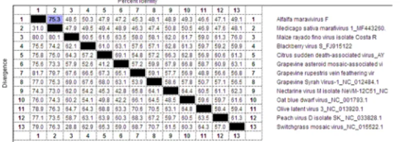
On the nucleotide level, AVF has 78% identity with the newly identified and proposed alfalfa marafivirus MsMV1 [11], (BLASTN query coverage 80%, E-value = 0.0, accession
MF443260.1). The next closest species from the genusMarafivirus, per PASC tool [16], is oat blue dwarf virus isolate OBDV2r (50.2% identities, accession GU396990), a member of the genusMarafivirus. Multiple nucleotide sequence alignment using the MegaAlign tool and
ClustalW algorithm of the DNAStar package (DNASTAR Inc., WI, USA) generated even lower identity between the complete genomes of AVF and MsMV1 (75.3%); identity with other viruses of the genus was at the 50% level or less (Fig 1). Therefore, based on the nucleo-tide sequence alignment of the compete AVF genome and criteria demarcating species in the genusMarafivirus that include overall sequence identity less than 80% [8], AVF represents a distinct viral species in the genusMarafivirus, family Tymoviridae.
Fig 1. Multiple nucleotide sequence alignment between complete genomes of AVF and other marafiviruses.
Alignment generated using DNASTAR software, MegAlign tool with ClustalW algorithm. Percent identity for each pair of sequences is shown on the upper right side and percent diversity (calculated by comparing sequence pairs in relation to the phylogeny reconstructed by MegAlign) is shown on the lower left side. Percent identity between AVF and MsMv1 (75.3%) highlighted in blue.
AVF encodes a single precursor polyprotein 2130 amino acids (aa) in length. In BLASTP query, the AVF polyprotein was 82% identical to the polyprotein of MsMV1 (99% coverage, E-value = 0.0, accession ATJ00054.1). The next closest species wasNectarine marafivirus M
(49% identities, 98% query cover, E-value = 0.0, accession YP_009222597.1). The Pfam data-base search and analysis of the AVF polyprotein (http://pfam.xfam.org/) resulted in five signif-icant Pfam-A matches corresponding to five viral domains characteristic for the genus
Marafivirus, family Tymoviridae: viral methyltransferase (PF01660; E-value 7.9e-55),
tymo-virus endopeptidase (PF05381; E-value 3.8e-19), viral (superfamily 1) RNA helicase (PF01443; E-value 5.3e-52), RNA dependent RNA polymerase (PF00978; E-value 5.9e-13) and tymovirus coat protein (PF00983; E-value 4.2e-20).
CP-encoding sequences in marafiviruses are in the same reading frame with the polyprotein ORF and are located at its 3’terminal end. Marafivirus particles were reported to contain so called major and minor CPs, of about 21kDa and 25kDa, respectively, that differ by an amino terminal extension in the minor CP and are found in the virus particles in molar ratios of approximately 3:1 (major CP21 vs minor CP25) [17,18]. However, it appears that unlike other marafiviruses, AVF as well as MsMV1, do not have a second initiation codon (Met) for the coding region of the major CP protein (CP21) and only encode methionine for the minor CP25 (5890–6603 nt; 1893–2130 aa). In this case, a possible strategy to produce two CPs could be a direct translation of the subgenomic RNA for the minor CP25 (rather than for CP21) and posttranslational cleavage of the larger precursor to produce the major protein CP21 (Fig 2A), [18]. A putative cleavage site to produce CP21 could be located downstream of the CP25 N-ter-minal end, speculatively between the amino acid residues Ala1934and Gly1935or Gly1935and
Ser1936(Fig 2B).
The nucleotide sequence of the AVF CP25 displays 80% identity with the MsMV1 polyprotein gene (query cover 100%, E-value = 4e-142; accession MF443260.1). On the amino acid level, AVF CP25 shares 83% identity with the polyprotein of the MsMV1 (query cover 100%, E-value = 2e-126, accession ATJ00054.1). Noteworthy, less than 90% aa sequence identity among coat protein sequences is another criterion demarcating species in the genusMarafivirus [8].
The AVF genome contains a conserved “marafibox”, a 16-nt consensus sequence of the subgenomic RNA (sgRNA) promoter that is present in all known marafiviruses, at positions 5734–5749 nt (5’ GAGGGTGAATTGCTTC 3’). The possible 5’ end of the subgenomic RNA encoding the viral CPs starts at the conserved adenine position 10 nt downstream of the core promoter sequence (A5759), [17,18].
Fig 2. (A) Tentative translation strategy for AVF coat proteins. The open reading frames are drawn as rectangles. The
black box indicates location of the “marafibox” subgenomic promoter; sgRNA, subgenomic RNA; CP21, major coat protein, molecular weight 21kDa; CP25, minor coat protein, molecular weight 25.2kDa; Triangle indicates putative cleavage site for viral or cellular proteases to produce CP21. (B) The amino acid sequence of the AVF CP region showing putative location of the methionine initiation codon for CP25 (1893 aa) and putative cleavage sites for CP21 between the amino acid residues Ala1934and Gly1935or Gly1935and Ser1936(arrows).
Interestingly, the marafivirus-related contigs, assembled from three alfalfa plants collected from the Rhoˆne delta region, shared 75–97% identities with the AVF genome (coverage, 1.e-138< E-value <2.e-9), indicating that these plants were most likely infected with the virus.
RT-PCR
A complete genome of the AVF obtained via VANA-based Illumina MiSeq and 5’/3’ RACE was used to design primers for RT-PCR amplification of viral sequences encoding the minor CP25 in order to confirm the virus identity and accuracy of the nucleotide sequence. Using primer pair LN568 (forward) and LN569 (complementary) (Table 1) in an RT-PCR assay with total RNA extracted from the infected plant sample, the respective gene was successfully ampli-fied (Fig 3), cloned into the pCRII-TOPO vector (Thermo Fisher Scientific) and sequenced. The nucleotide sequence of the amplified product matched the RNA-seq results thus validating the quality of the digital assembly and the presence of the predicted virus.
Transmission electron microscopy observations and transient expression of
AVF CP
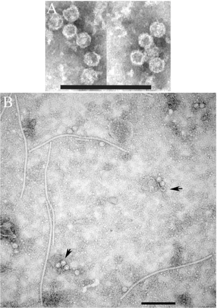
Routine sample preparation for negative staining [6] did not yield any results. Only when the virus was partially purified as described in [15], spherical virus particles ~ 30 nm in diameter, resembling T = 3 isometric virions of marafiviruses, were observed (Fig 4A).
To confirm functionality of the predicted CP coding region, a fragment of the viral genome encoding the tentative CP25 was transiently expressed inN. benthamiana plants via a PVX vector
[14]. To accomplish this, the RT-PCR product obtained with primers LN568-LN569 designed for amplification of the complete CP25, was sub-cloned into the PVX vector, transcribed and rub-inoculated onto leaves ofN. benthamiana, an experimental host species commonly used in plant
virology. Two weeks after inoculation, plants developed symptoms such as stunted growth, vein clearing and chlorotic mosaic (not shown). TEM of crude leaf extracts revealed characteristic PVX rods surrounded by isometric virus-like particles (VLPs) ~ 30 nm in diameter resembling typical marafivirus virions (Fig 4B). Therefore, the proposed sequence of CP25 is accurate and suf-ficient for assembly of AFV particles. Some virions appeared permeable and others stain-impermeable, suggesting that the latter may encapsidate CP mRNA.
Phylogenetic analysis
A phylogenetic tree was generated with the AVF genome and complete genomic sequences of several ICTV-approved and tentative members of the genusMarafivirus.(Fig 5). AVF grouped together with MsMV1 (78% identity vs AVF) and the cluster formed a sister-group with two other ICTV-proposed marafiviruses: grapevine rupestris vein feathering virus and grapevine Syrah virus-1. Both clusters branched out of the larger peach marafivirus D group, connected to the clusters of established species in the genusMarafivirus: type member Maize rayado fino virus, Oat blue dwarf virus, and Citrus sudden death-associated virus. Therefore, phylogenetic
analysis supported the tentative classification of AFV as a new member of the genus
Marafivirus.
Discussion
Here, we reported the discovery of a new alfalfa marafivirus originating from commercial alfalfa fields at Prades-le-Lez, Southern France. Our study indicates that AVF is also present in three alfalfa plants collected in three other areas of Southern France ~75 kms apart from Prades-le-Lez, suggesting that the distribution of AVF in Southern France is probably not
restricted to a single area. In this work, a complete genomic sequence of the virus was
obtained, identity of the virus was confirmed experimentally and methods suitable for specific
Fig 3. RT-PCR assay with primers LN568-LN569 (Table 1) to amplify a full-length AVF CP25. M, 1kb plus DNA
ladder (ThermoFisher Scientific). Lane 1, reaction performed with total RNA extracted from AVF-infected alfalfa sample. Lane 2, RT-PCR with total RNA extracted from healthy alfalfa material.
Fig 4. (A) Transmission electron microscopy of partially purified alfalfa virus F and (B) transient expression of AVF
virus-like particles inNicotiana benthamiana plants using a PVX-based vector. Scale bar represents 200 nm. Arrows
indicate AVF VLPs.
https://doi.org/10.1371/journal.pone.0203477.g004
Fig 5. Unrooted neighbor-joining phylogenetic tree built with the complete nucleotide sequences of alfalfa virus F and twelve ICTV-approved and tentative members of the genusMarafivirus. The percentage of replicate trees in which the associated taxa clustered together in the bootstrap test (1000 replicates) are shown next to the branches.
diagnostics of the virus were developed. In addition, a portion of the viral genome encoding a minor coat protein (CP25) was transiently expressed inN. benthamiana plants and
success-fully assembled into VLPs resembling natural virions, hence validating accuracy of the genome sequence and functional role of the projected CP domain. Since the transiently expressed genome fragment contains information to encode both AVF CP25 and smaller CP21, it is unclear whether the observed VLPs are composed of CP25 alone or of two proteins. If the lat-ter is true, CP25 capsids would have to be post-translationally cleaved with PVX- or host-encoded proteases to form CP21. This, however, requires further experimental confirmation. As previously suggested for MRFV, the ability of transiently expressed AMV CP25 to form VLPs may have potential value for applications in epitope presentation platforms [19].
The first proposed species of the genusMarafivirus in alfalfa, MsMV1, was described only
recently [11] and before that no members of this genus were identified in alfalfa. Although AVF is most closely related to MsMV1, it does represent a new species, based on the current demarcation criteria in the genusMarafivirus [8]. Notably, MsMV1 was not verified in plant samples as it was digitally deduced from alfalfa transcriptomic data published elsewhere and deposited to NCBI GenBank [12]. In addition, judging from the comparison with AVF, the reported sequence of MsMV1 appears to be missing the 5’ terminus and thus its genome length (6675 nt) is most likely inaccurate. Therefore, bioinformatic data alone, without experi-mental validation, might not be sufficient as the only criteria for characterization of new viral genomes. The final classification of AVF and MsMV1 as new marafivirus species is under con-sideration by the ICTVTymoviridae study group.
Primary research by Zhang et al. [12], whose transcriptomic data has been later adopted by Kim et. al. [11] for identification of MsMV1, originated from China and employed alfalfa standard varieties Maverick (FDC1) and CUF101 (FDC9) introduced to China from the United States.
Beyond identification and confirmation of AVF as a new viral species infecting alfalfa, the RT-PCR assay with virus-specific primers developed in this work may be of practical use for detection of the virus.
Notwithstanding, we conclude that AVF represents a unique virus species with a close resemblance to the members of the genusMarafivirus, family Tymoviridae and a more evident
relation to the unclassifiedMedicago sativa marafivirus 1. The genome sequence of AVF was
deposited in NCBI GenBank on 12/24/2017 as accession MG676465.
Supporting information
S1 Fig. AMV-infected plant, originating from the Rhoˆne delta region and maintained at the Cirad laboratory. The plant is not exhibiting any visible symptoms that would differenti-ate it from the healthy plants.
(TIF)
Acknowledgments
We are grateful to Joseph Mowery of the USDA-ARS Northeast Area Electron and Confocal Microscope Unit, Beltsville Agricultural Research Center, for his skillful assistance with TEM imaging and to Jonathan Shao and Maya Lee of the USDA-ARS Molecular Plant Pathology Laboratory for their assistance with phylogenetic analysis.
Author Contributions
Conceptualization: Lev G. Nemchinov, Sarah Franc¸ois, Phillipe Roumagnac, Dimitre S. Mol-lov, Denis Filloux.
Data curation: Lev G. Nemchinov, Phillipe Roumagnac, Dimitre S. Mollov, Denis Filloux. Formal analysis: Lev G. Nemchinov, Sarah Franc¸ois, Denis Filloux.
Funding acquisition: Lev G. Nemchinov, Phillipe Roumagnac, Dimitre S. Mollov, Denis Filloux.
Investigation: Lev G. Nemchinov, Sarah Franc¸ois, Phillipe Roumagnac, Mylène Ogliastro, Rosemarie W. Hammond, Dimitre S. Mollov, Denis Filloux.
Methodology: Lev G. Nemchinov, Sarah Franc¸ois, Phillipe Roumagnac, Mylène Ogliastro, Rosemarie W. Hammond, Denis Filloux.
Project administration: Lev G. Nemchinov, Denis Filloux.
Resources: Lev G. Nemchinov, Phillipe Roumagnac, Rosemarie W. Hammond, Dimitre S. Mollov, Denis Filloux.
Software: Lev G. Nemchinov, Sarah Franc¸ois, Denis Filloux.
Supervision: Lev G. Nemchinov, Phillipe Roumagnac, Mylène Ogliastro, Dimitre S. Mollov, Denis Filloux.
Validation: Lev G. Nemchinov, Phillipe Roumagnac, Dimitre S. Mollov, Denis Filloux. Visualization: Lev G. Nemchinov.
Writing – original draft: Lev G. Nemchinov.
Writing – review & editing: Lev G. Nemchinov, Phillipe Roumagnac, Rosemarie W. Ham-mond, Dimitre S. Mollov, Denis Filloux.
References
1. Li X, Brummer EC. Applied genetics and genomics in alfalfa breeding. Agronomy. 2012; 2:40–61. 2. Monteros MJ, Bouton JH. The future of alfalfa and forage crops In: Proceedings, Western Alfalfa &
For-age Conference, December 2–4, Reno, Nevada, 2009.
3. Bejerman N, Nome C, Giolitti F, Kitajima E, de Breuil S, Fernandez JP, et al. First report of a Rhabdovi-rus infecting alfalfa in Argentina. Plant Dis. 2011; 95:771.
4. Bejerman N, Giolitti F, Trucco V, de Breuil S, Dietzgen RG, Lenardon S. Complete genome sequence of a new enamovirus from Argentina infecting alfalfa plants showing dwarfism symptoms. Arch Virol. 2016; 161:2029–2032.https://doi.org/10.1007/s00705-016-2854-3PMID:27068164
5. Nemchinov LG, Grinstead SC, Mollov DS. First report and complete genome sequence of Alfalfa Enam-ovirus from Sudan. Genome Announc. 2017; 5(27). pii: e00531–17.https://doi.org/10.1128/genomeA. 00531-17PMID:28684567
6. Nemchinov LG, Grinstead SC, Mollov DS. Alfalfa virus S, a new species in the family Alphaflexiviridae. PLoS One. 2017; 12(5): e0178222.https://doi.org/10.1371/journal.pone.0178222PMID:28558046
7. Roumagnac P, Granier M, Bernardo P, Deshoux M, Ferdinand R, Galzi S, et al. Alfalfa Leaf Curl Virus: an aphid-transmitted geminivirus. J Virol. 2015; 89:9683–9688.https://doi.org/10.1128/JVI.00453-15
PMID:26109720
8. Dreher TW, Edwards MC, Haenni A-L, Hammond RW, Jupin I, Koenig R, et al. 2012. Family
Tymoviri-dae. In: King A.M.Q., Lefkowitz E., Adams M.J., Carstens E.B. (Eds.) Virus Taxonomy (9th Report of
the ICTV), Elsevier Academic Press, San Diego, 913–921.
9. Martelli GP, Sabanadzovic S, Abou-Ghanem Sabanadzovic N, Edwards MC, Dreher T. The family
Tymoviridae. Arch Virol. 2002; 147:1837–1846.https://doi.org/10.1007/s007050200045PMID:
12209322
10. Gamez R. Maize rayado fino virus. In: Descriptions of Plant Viruses. No. 220, Commonw. Mycol. Inst./ Assoc. Applied Biologists, Kew, Surrey, U.K, 1980.
11. Kim H, Park D, Hahn Y. Identification a novel RNA viruses in alfalfa (Medicago sativa): an
Alphapartiti-virus, DeltapartitiAlphapartiti-virus, and a Marafivirus. Gene. 2018; 638:7–12.https://doi.org/10.1016/j.gene.2017. 09.069PMID:28974471
12. Zhang S, Shi Y, Cheng N, Du H, Fan W, Wang C. De novo characterization of fall dormant and nondor-mant alfalfa (Medicago sativa L.) leaf transcriptome and identification of candidate genes related to fall dormancy. PLoS One. 2015; 10(3): e0122170.https://doi.org/10.1371/journal.pone.0122170PMID:
25799491
13. Francois S, Filloux D, Fernandez E, Ogliastro M, Roumagnac P. Viral Metagenomics Approaches for High-Resolution Screening of Multiplexed Arthropod and Plant Viral Communities. Methods in Molecu-lar Biology. 2018; 1746:77–95.https://doi.org/10.1007/978-1-4939-7683-6_7PMID:29492888
14. Chapman S, Kavanagh TA, Baulcombe DC. Potato virus X as a vector for gene expression in plants. Plant J. 1992; 2:549–557 PMID:1344890
15. Lesemann DE, Koenig R, Huth W, Brunt AA, Phillips S, Barton RJ. Poinsettia mosaic virus: A tymo-virus? Phytopath. Z. 1983; 107:250–262
16. Bao Y, Chetvernin V, Tatusova T. Improvements to pairwise sequence comparison (PASC): a genome-based web tool for virus classification. Arch Virol. 2014; 159:3293–3304.https://doi.org/10.1007/ s00705-014-2197-xPMID:25119676
17. Edwards MC, Weiland JJ. Coat protein expression strategy of oat blue dwarf virus. Virology, 2015; 450:290–296.
18. Hammond RW, Ramirez P. Molecular characterization of the genome of Maize rayado fino virus, the type member of the genus Marafivirus. Virology. 2001; 282:338–347.https://doi.org/10.1006/viro.2001. 0859PMID:11289816
19. Hammond RW, Hammond J. Maize rayado fino virus capsid proteins assemble into virus-like particles in Escherichia coli. Virus Res. 2010; 147:208–215.https://doi.org/10.1016/j.virusres.2009.11.002