HAL Id: inserm-00410111
https://www.hal.inserm.fr/inserm-00410111
Submitted on 8 Aug 2011
HAL is a multi-disciplinary open access
archive for the deposit and dissemination of
sci-entific research documents, whether they are
pub-lished or not. The documents may come from
teaching and research institutions in France or
abroad, or from public or private research centers.
L’archive ouverte pluridisciplinaire HAL, est
destinée au dépôt et à la diffusion de documents
scientifiques de niveau recherche, publiés ou non,
émanant des établissements d’enseignement et de
recherche français ou étrangers, des laboratoires
publics ou privés.
Evidence of in situ proliferation of adult adipose
tissue-derived progenitor cells: influence of fat mass
microenvironment and growth.
Marie Maumus, Coralie Sengenès, Pauline Decaunes, Alexia Zakaroff-Girard,
Virginie Bourlier, Max Lafontan, Jean Galitzky, Anne Bouloumié
To cite this version:
Marie Maumus, Coralie Sengenès, Pauline Decaunes, Alexia Zakaroff-Girard, Virginie Bourlier, et
al.. Evidence of in situ proliferation of adult adipose tissue-derived progenitor cells: influence of fat
mass microenvironment and growth.. Journal of Clinical Endocrinology and Metabolism, Endocrine
Society, 2008, 93 (10), pp.4098-106. �10.1210/jc.2008-0044�. �inserm-00410111�
Evidence of in Situ Proliferation of Adult Adipose
Tissue-Derived Progenitor Cells: Influence of Fat Mass
Microenvironment and Growth
Marie Maumus, Coralie Sengene`s, Pauline Decaunes, Alexia Zakaroff-Girard, Virginie Bourlier, Max Lafontan, Jean Galitzky, and Anne Bouloumie´
Institut National de la Sante´ et de la Recherche Me´dicale, Unit 858, Universite´ Toulouse III Paul Sabatier, Institut de Me´decine Mole´culaire de Rangueil, Institut Fe´de´ratif de Recherche 31, 31432 Toulouse, France
Context: Adipocyte formation in human adult adipose tissue (hAT) originates from resident
pro-genitor cell differentiation in the stroma vascular fraction of the AT. The processes involved in the self-renewal of this cell population remain to be defined.
Objective: The objective was to study in situ and in vitro hAT progenitor cell (defined as CD34⫹/ CD31⫺cells) proliferation.
Design and Participants: In situ progenitor cell proliferation was assessed by
immunohistochem-istry and flow cytometry analyses on hAT from lean to obese subjects using the proliferation marker Ki-67. The effects of adipokines, hypoxia, and conditioned media (CM) from adipocytes, capillary endothelial cells, and macrophages isolated by an immunoselection approach were studied on hAT progenitor cell growth. Cell death in hAT was assessed by the terminal deoxynucleotidyl trans-ferase-mediated dUTP-fluorescein end labeling method.
Results: Ki-67-positive staining was observed in AT progenitor cells. Fat mass enlargement in obese
patients was associated with an increased Ki-67⫹progenitor cell population together with a new fraction of small adipocytes and increased cell death. HIF-1␣ mRNA expression in freshly harvested progenitor cells was positively correlated with body mass index. Adipocyte- and capillary endo-thelial cell-CM, hypoxia, leptin, IL-6, lysophosphatidic acid, and vascular endoendo-thelial growth factor, all increased hAT progenitor cell proliferation in vitro. Macrophage-CM had an antiproliferative effect that was suppressed by an antioxidant.
Conclusions: The fraction of proliferative progenitor cells in adult hAT is modulated by the degree
of adiposity. Changes in the progenitor cell microenvironment involving adipokines, hypoxia, and oxidative stress might play a key role in the control of the self-renewal of the local pool of AT progenitor cells. (J Clin Endocrinol Metab 93: 4098 – 4106, 2008)
T
he excessive development of human adipose tissue (AT) results from both hypertrophy of preexisting adipocytes and hyperplasia due to the formation of new adipocytes (1, 2). Hypertrophy which consists of excessive triglyceride accumu-lation in mature adipocytes, is due to both positive energy balance and reduced lipolysis. It is the initial event occurring during the establishment of obesity (3). In severe forms of obesity, an apparent increase in number of adipocytes followsthe mature adipocyte hypertrophy (4, 5). Interestingly, Spal-ding et al. (6) recently showed that the number of mature fat cells is constant in adulthood in lean and obese individuals. Furthermore, the authors suggest that mature adipocytes ex-hibit a high turnover rate, indicating that fat cell number is tightly regulated during adulthood. Because adipocytes are terminally differentiated cells and, as such, are considered incapable of division (2, 7), the apparent increase in adipocyte
0021-972X/08/$15.00/0 Printed in U.S.A.
Copyright © 2008 by The Endocrine Society
doi: 10.1210/jc.2008-0044 Received January 7, 2008. Accepted July 28, 2008. First Published Online August 5, 2008
Abbreviations: AT, Adipose tissue; BMI, body mass index; BrdU, bromo-2⬘-deoxyuridine; CEC, capillary endothelial cell; CM, conditioned media; ECBM, endothelial cell basal me-dium; FCS, fetal calf serum; HIF-1␣, hypoxia-inducible factor-1␣; LPA, lysophosphatidic acid; NAC, N-acetyl cysteine; SVF, stroma-vascular fraction; TUNEL, terminal deoxynucleo-tidyl transferase-mediated dUTP nick-end labeling; VEGF-A, vascular endothelial growth factor A.
E n d o c r i n e R e s e a r c h
number is thought to originate from adipogenesis, the prolif-eration/differentiation of adipocyte progenitor cells (preadi-pocytes). Adipogenesis has been extensively studied in vitro on several cell models such as established murine preadipocyte cell lines and primary cultures of stroma-vascular fraction (SVF) cells in adipogenic culture conditions (8, 9). It was re-ported in mice that adipocytes may derive from circulating bone marrow cells (10), whereas a recent study found the opposite and suggests that bone-marrow-derived cells do not contribute in any way to AT development (11). Whatever the final issue of this debated question in rodents, clinical studies are requested to investigate adipogenesis in human AT. The exact nature of human preadipocytes still remains elusive. Because of the difficulty in studying cell turnover in humans, few data are available concerning human adipocyte precursor renewal within AT, although such a process is necessary to maintain a pool of preadipocytes to be recruited during AT enlargement. Our previous studies showed that human prea-dipocytes are present in the AT-derived SVF. Using a combi-nation of cell surface markers, CD34 (mucosialin) expressed on hematopoietic stem cells and capillary endothelial cells (CECs), CD31 (platelet endothelial cell adhesion molecule-1), expressed on leukocytes and endothelial cells and CD14 (li-popolysaccharide receptor), expressed on myeloid cells, the main cell types composing the AT-SVF were defined as AT CECs (CD34⫹/CD31⫹), AT macrophages (CD34⫺/ CD14⫹), and AT progenitor cells (CD34⫹/CD31⫺) (12). The CD34⫹/CD31⫺cell fraction was the only one, among the SVF-derived cells, to differentiate into adipocytes (13). Additionally, the CD34⫹/CD31⫺cells exhibited angiogenic potential (14) and were therefore defined as progenitor cells.
The present study was undertaken to evaluate the in situ proliferative status of the CD34⫹/CD31⫺progenitor cells in human sc AT and to determine whether self-renewal of a local pool of human progenitor cells could be modulated by adi-pocyte hypertrophy and/or hyperplasia. In vitro studies were performed on freshly isolated CD34⫹/CD31⫺cells to analyze the effects of local changes in the microenvironment in which the progenitor cells reside, notably the effects on the prolif-erative responsiveness of CD34⫹/CD31⫺cells of secreted fac-tors originating from adipocytes, AT-CECs, and AT-macro-phages as well as low oxygen tension.
Materials and Methods
Materials
Chemicals were from Sigma (Saint-Quentin Fallavier, France). Collagenase NB4 was from Serva (Coger, Paris, France). CD34⫹and CD14⫹ cell selection kits were from StemCell Technologies (Grenoble, France), and the CD31⫹cell selection kit was from Dynal (Invitrogen, Cergy-Pontoise, France). Culture media were from Pro-mocell (Heidelberg, Germany). Flow cytometry antibodies were from BD Biosciences (Le-Pont-de-Claix, France). Adiponectin, leptin, and vascular endothelial growth factor A (VEGF-A) were from Peprotech (Levallois-Perret, France) and IL-6 from R&D Systems (Lille, France). Lysophosphatidic acid (LPA) was a gift from Dr. Saulnier-Blache [Institut National de la Sante´ et de la Recherche Me´dicale (INSERM) Unit 858, France].
Patients
Human abdominal sc AT was obtained from a group of normal to class I obese women undergoing plastic surgery (dermolipectomy and/or liposuction) [n⫽ 61, body mass index (BMI) ranging from 20.8–34 kg/m2; mean age⫽ 42 ⫾ 1 yr] (15) and from a group of class II to class
III obese women (n⫽ 25, BMI ranging from 35–55 kg/m2; mean age⫽
40⫾ 3 yr) undergoing vertical banded Mason gastroplasty, with a stable weight for at least 3 months before surgery (supplemental data 1, pub-lished as supplemental data on The Endocrine Society’s Journals Online web site at http://jcem.endojournals.org). The AT samples were imme-diately processed after removal. The protocol has been conducted in accordance with the Declaration of Helsinki guidelines and was ap-proved by the Institutional Research Board of INSERM and the Tou-louse University Hospital.
Isolation of the cell types from human AT
Mature adipocytes and SVF were harvested as previously described (12, 16). The SVF cells were isolated using an immunoselection/depletion approach (14, 17). Freshly isolated CD34⫹/CD31⫺cells (progenitor cells), CD34⫹/CD31⫹cells (AT-CECs) and CD34⫺/CD14⫹cells (AT-macrophages) were cultured in appropriate media.
Isolation of human foreskin fibroblasts
Human foreskin fibroblasts were prepared according to a modified version of Rheinwald and Green protocol (18). The cells were cultured in DMEM/10% fetal calf serum (FCS) (passages 3–14 were used).
Adipocyte diameter determination
After isolation from dermolipectomies (normal to class I obese indi-viduals) or sc fat biopsies (class II and III obese patients) (n⫽ 51, BMI ranging from 20.8 –55 kg/m2), mature adipocytes were suspended in
endothelial cell basal medium (ECBM, containing no growth factors)/ 0.5% BSA (1/10, vol/vol), and 5l cell suspension was placed onto plastic slides. Three distinct calibrated fields were taken to measure man-ually adipocyte diameters with NIS software (Nikon, Champigny-sur-Marne, France). Fat cell-like shapes were labeled with the nucleus dye 4⬘,6-diamidino-2-phenylindole (DAPI). Among them, 94 ⫾ 1% were nucleated and identified as adipocytes (85⫾ 7 and 99 ⫾ 1%, for small and large size adipocytes, respectively).
Preparation of conditioned media (CM)
To prepare CM from the different cell fractions composing AT, cells were isolated from 21 abdominal liposuctions (BMI⫽ 30.2 ⫾ 0.9 kg/ m2). Adipocytes were plated in fibrin gels (12), and basal medium (i.e.
ECBM/0.1% BSA) was added. After 24 h, the adipocyte-CM was col-lected, centrifuged (20,000⫻ g for 3 min at room temperature), and stored at⫺20 C until further use. The AT-CECs were plated on fibronec-tin-coated plates (5g/cm2) in endothelial cell growth
medium-micro-vascular. At confluence, AT-CECs were rinsed, and basal medium was added. Twenty-four hours later, CM was collected, centrifuged, and stored at⫺20 C. AT-macrophages were plated in basal medium supple-mented or not with N-acetyl cysteine (NAC) for 24 h. The CM was collected, centrifuged and stored at⫺20C until further use.
Immunohistochemistry and cell death measurement
Immunohistochemistry analyses were performed on freshly har-vested human sc AT cut into small pieces and fixed in neutral buffered 4% (wt/vol) paraformaldehyde (24 h at room temperature). The micro-wave antigen retrieval was performed in citrate buffer (10 mmol/liter, pH 6) three times for 7 min each. The AT was permeabilized in PBS/0.1% Triton (20 min) followed by an overnight incubation in PBS/2% BSA with mouse monoclonal CD34 antibody (Santacruz, Le Perray-en-Yve-lines, France) (1/50), rabbit polyclonal Ki-67 antibody (Dako, Trappes, France) (1/50), or mouse polyclonal hypoxia-inducible factor-1␣
1␣) antibody (R&D Systems) (1/20). AT was then incubated for 1 h with the corresponding fluorescent-labeled second antibodies (goat anti-mouse or goat antirabbit coupled to AlexaFluor 488 or 546) (Invitrogen) (1/200). Cell death was evaluated by terminal deoxynucleotidyl trans-ferase-mediated dUTP nick-end-labeling (TUNEL) assays according to the manufacturer’s instructions (Roche, Meylan, France). Briefly, after fixation and permeabilization (PBS/0.5% Triton/0.1% sodium citrate for 20 min at room temperature) AT pieces were incubated in the dark with the TUNEL reaction mixture or with the control solution (for neg-ative controls) for 1 h at 37 C in a humidified atmosphere. Nuclei were
stained with Hoescht 33258 (Invitrogen). After washes, AT pieces were mounted between two slides, and positive TUNEL cells (green nuclei) were determined under fluorescent microscope analyses (Nikon) on three fields for each AT sample.
Cell proliferation assay
The CD34⫹/CD31⫺progenitor cells (or foreskin-derived fibroblasts) (n⫽ 14; BMI ⫽ 28.5 ⫾ 1.6 kg/m2) were plated at a density of 1.104
cells/cm2in ECBM/10% FCS. After 24 h, cells were rinsed and treated
with ECBM/0.1% BSA (control), 5% FCS (positive control), adipocyte-, CECs-, or macrophage-CM in normoxia or with ECBM/0.1% BSA in hypoxia chamber (1% O2;
Sanyo, Avon, France). Progenitor cells were also treated with adiponectin (1, 10, and 100 ng/ml), IL-6 (1, 10, and 100 ng/ml), leptin (1, 10, and 50 ng/ml), LPA (0.1, 1, and 10mol/l), or VEGF-A (0.1, 1 and 10 ng/ml). After 48 h, bromo-2 ⬘-de-oxyuridine (BrdU) was added to the medium (20 l at 100 mol/liter) for 6 h. The cell proliferation index was evaluated according to the manufac-turer’s instructions (Roche).
Flow cytometry analysis
AT-fixed SVF (CellFix; BD Bioscience; vol/ vol, 4 C, 30 min) were permeabilized (20 h at ⫺20C in 70% ethanol). At least 1 ⫻ 105cells in
PBS/0.5% BSA/2 mmol/liter EDTA were incu-bated with fluorescein isothiocyanate (FITC)-conjugated Ki-67 antibody, peridinin chlorophyll protein (PerCP)-conjugated CD34 antibody and phycoerythrin (PE)-conjugated CD14 antibody or the respective isotype controls. The labeled cells were analyzed by multiparameter flow cy-tometry using a FACSCalibur flow cytometer and the CellQuest Pro software (BD Bioscience).
RNA isolation and real-time PCR
Total RNA was extracted from CD34⫹/ CD31⫺cells or human foreskin fibroblasts using the RNeasy kit (QIAGEN, Courtaboeuf, France) and the RNA concentrations determined using a fluorometric assay (Ribogreen; Invitrogen). RNA (0.5g) was reverse-transcribed using the Super-script II (Invitrogen) (Random Hexamers and dNTPs were from Invitrogen). Reverse transcrip-tion was also carried out without the superscript enzyme on RNA samples. Primers for the adiponec-tin receptors (Adipo R1, Adipo R2), the lepadiponec-tin re-ceptor, the VEGF receptors (VEGF-R1, VEGF-R2, and VEGF-R3), the IL-6 receptor, and HIF-1␣ were provided by Applied Biosystems (Courtaboeuf, France) (assay on demand: hs00360422_m1, hs00226105_m1, hs00174497_m1, hs00176573_ m1, hs00176676_m1, hs00176607_m1, hs00169842_m1, and hs00153153_m1, respec-tively). The primers for the LPA receptor (LPA1R) were forward, 5 ⬘-TGGGCCATTTTCAACTT-GGT-3⬘, and reverse, 5⬘-TCTGGCGAACATAGC-CAAAGA-3⬘. The amplification reaction was car-ried out on 15 ng of cDNA samples in 96-well plates (Applied Biosystems) in a GeneAmp 7000 sequence detection system. The PCR mixture contained either 5 l TaqMan primers (5⫻ prediluted in water) or 900 nmol/liter for LPA1R and 10l 2⫻ TaqMan PCR Master Mix or 10l 2⫻ SybrGreen PCR Master Mix (Ap-plied Biosystems). All reactions were performed
FIG. 1. Evolution of adipocyte size and number with the growth of human sc AT. A, Adipocytes from human sc AT of lean/overweight (BMI⬍30 kg/m2; n⫽ 23; white bars) and obese (BMI ⱖ30 kg/m2; n⫽ 28; black bars) patients were isolated for counting and diameter determination. Results are expressed as frequency and presented in log10 scale. B and C, Correlations between the percentage of small (diameter ⬍60m, E and F) and large (diameter ⬎100 m, 䡺 and f) adipocytes according to the BMI in lean/overweight patients (B, white symbols; ***, P⬍ 0.01, Spearman r ⫽ ⫺0.55, n ⫽ 23; and ***, P ⬍ 0.001, Spearman r⫽ 0.62, n ⫽ 23, respectively) and in obese patients (C, black symbols; *, P ⬍ 0.05, Spearman r⫽ ⫺0.40, n ⫽ 28; and P ⫽ nonsignificant, Spearman r ⫽ ⫺0.23, n ⫽ 28, respectively).
under the same conditions: 50 C for 2 min, 95 C for 10 min, followed by 40 cycles of 95 C for 15 sec and 60 C for 1 min. The results were analyzed with the GeneAmp 7500 software, and all the values were normalized to 18S rRNA levels.
Statistical analysis
The statistical analysis was performed with GraphPad Software (San Diego, CA). Correlations were analyzed with a Spearman test. Values are given as mean⫾SEMof (n) separate experiments. The comparisons be-tween different groups were analyzed by one-way ANOVA followed by Dunnett post hoc test or with a Student’s t test for two groups. Differ-ences were considered significant when P⬍ 0.05.
Results
Changes in adipocyte size distribution with the growth of human sc AT
Fat cell size distribution was determined in AT from lean to overweight patients (20⬍ BMI ⬍ 30 kg/m2
; n⫽ 23) and was compared with the obese patients (BMIⱖ30 kg/m2
; n⫽ 28). As shown in Fig. 1A, the most representative population of adipo-cytes, with a mean diameter of 90m in lean/overweight AT, shifted to 100 –120m in obese AT. Moreover, the frequency of very small adipocytes (diameter 20 – 40 m) increased from 0.015 in lean/overweight AT to 0.045 in obese AT. To further analyze the influence of the degree of adiposity on adipocyte size, adipocytes were classified according to their diameter (i.e. large with a diameter more than 100m and small with a diameter less than 60m), and their percentage within the whole adipocyte population was determined. As shown in Fig. 1B, increased ad-iposity in lean to overweight patients was negatively correlated with the percentage of small adipocytes but positively with that of large adipocytes. The further increase in fat mass seen in obese patients was characterized by stabilization or a statistically non-significant decrease in the percentage of large adipocytes to-gether with an increase in the percentage of smaller adipocytes (Fig. 1C).
Changes in cell death with the growth of human sc AT
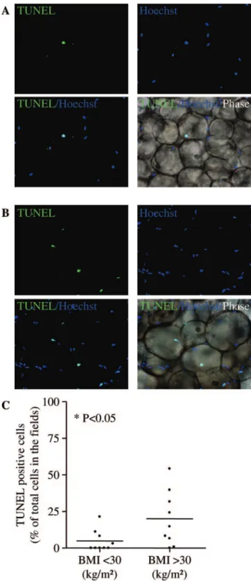
Cell death in human sc AT was assessed by TUNEL in lean/ overweight (n⫽ 9) and in obese patients (n ⫽ 9). As shown in Fig. 2A, occasional TUNEL-positive cells were observed in lean/over-weight AT (mean value of 5⫾ 2% of the total nuclei), whereas a clear increase was seen in obese AT (Fig. 2B) (mean value of 20⫾ 6% of total nuclei, P ⬍ 0.05; Fig. 2C).
Changes in the proliferation rate of the CD34ⴙ/CD31ⴚ progenitor cells with the growth of human sc AT
The expression of the Ki-67 protein that is expressed during all active phases of the cell cycle (G1, S, G2, and mitosis) but
absent from resting cells (G0) was studied in the sc AT of lean/
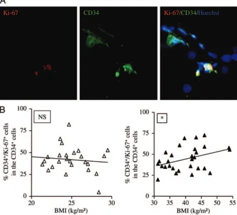
overweight and obese patients. As shown in a representative histological assay (Fig. 3A), immunohistochemistry revealed the presence of cells positive for CD34 in the stromal compartment surrounding mature adipocytes and defined as progenitor cells. Among the progenitor cells, Ki-67-positive cells were identified (Fig. 3A). To quantify the number of proliferative progenitor
cells, flow cytometry analyses were performed on the SVF from the sc AT of lean to obese patients using the peridinin chlorophyll protein (PerCP)-conjugated CD34 antibody together with the fluorescein isothiocyanate-conjugated Ki-67 antibody. The per-FIG. 2. Effect of human sc AT growth on cell death. A and B, Representative photomicrographs of TUNEL staining (green) and nuclei (blue) in fixed and permeabilized human sc AT of lean/overweight patients (BMI⬍30 kg/m2) (A) and obese patients (BMIⱖ30 kg/m2) (B) (original magnification,⫻200); C, mean values of TUNEL-positive cells expressed as percentage of the total cell number in nine lean/ overweight patients and nine obese patients determined on three independent microscopic fields for each adipose tissue sample. *, P⬍ 0.05, lean/overweight vs. obese.
centage of progenitor cells positive for Ki-67 remained constant with increasing BMI in the SVF of lean/overweight patients (Fig. 3B; n⫽ 26). Interestingly, positive Ki-67 progenitor cell per-centage was positively correlated with the BMI in obese patients (Fig. 3B; n⫽ 29; P ⬍ 0.05). As shown in Fig. 3B, large inter-obese individual differences exist; one cannot rule out that they might be the consequence of each patient’s weight history (weight cy-cling, pregnancy, and number of diet restriction periods). Even though the present work was not designed as a follow-up study, prebariatric data such as obesity duration and waist circumfer-ence were retrieved from 16 class II to class III obese patients of a group of 25 (supplemental data 1). Obesity duration was not correlated with the BMI or with the percentage of progenitor cells positive for Ki-67 (not shown). Finally, the age of the pa-tients included in the present work was not correlated to the percentage of progenitor cells positive for Ki-67 (not shown).
Local control of CD34ⴙ/CD31ⴚprogenitor cell proliferation by adipokines
Freshly harvested AT progenitor cells were treated for 48 h with CM originating from mature adipocytes, AT-CECs, and AT-macrophages. Their proliferation was determined by BrdU incorporation assays as well as by nuclei counting after Hoescht staining. As shown in Fig. 4A, CM originating from mature adipocytes and from AT-CECs increased the prolif-eration of the AT progenitor cells as shown by the enhanced
BrdU incorporation (Fig. 4A) and nuclei number (data not shown). The stimula-tory effects of the adipocyte- and CEC-derived CM on the progenitor cell pro-liferation was cell type specific because they did not affect the proliferation ac-tivity of human foreskin-derived fibro-blasts treated under the same conditions (Fig. 4B). Finally, the growth responsive-ness of the AT progenitor cells to kines known to be derived from adipo-cytes and CECs and whose production is known to be related to fat mass growth, such as leptin, adiponectin, LPA, IL-6, and VEGF-A, was studied. First, the ex-pression of their corresponding receptors in the AT progenitor cells was deter-mined by real-time PCR analysis and compared with the one of human fore-skin-derived fibroblasts. As described in Table 1, the AT progenitor cells specifi-cally expressed the mRNA transcripts en-coding for the receptor for leptin (LEPR), adiponectin (Adipo R1 and R2), LPA, IL-6, the VEGF-R2 for the VEGF-A re-ceptor as well as VEGF-R1 and -R3. Lep-tin triggered a concentration-dependent increase in BrdU incorporation (Fig. 4C) whereas adiponectin was without effect (data not shown). In parallel, VEGF-A, LPA, and IL-6 increased the proliferation of the progenitor cells (Fig. 4D).
AT macrophage-derived CM led to a reduction in the pro-liferation rate of the progenitor cells (Fig. 5). To investigate whether reactive oxygen species could be responsible for the antiproliferative effect of macrophage-CM, an antioxidant, NAC, was also added. As shown in Fig. 5, the presence of NAC abolished the antiproliferative effect of macrophage-CM.
Local control of progenitor cell proliferation by oxygen tension
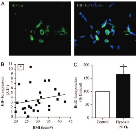
The potential presence of a hypoxic area within human sc AT was investigated by immunohistochemistry. As shown in Fig. 6A, the hypoxia-sensitive transcription factor HIF-1␣ could be identified in human sc AT. Moreover, the expression of HIF-1␣ was determined by real-time PCR analysis in the progenitor cells isolated from sc AT from patients with in-creasing BMI. As shown in Fig. 6B, the expression of HIF-1␣ in CD34⫹/CD31⫺progenitor cells was positively correlated with the BMI of the patients. Finally, the proliferation rate of the CD34⫹/CD31⫺progenitor cells was studied using BrdU incorporation assays under 20 and 1% O2in a hypoxia
cham-ber for 48 h. As shown in Fig. 6C, hypoxia increased the BrdU incorporation by the CD34⫹/CD31⫺progenitor cells (1.7⫾ 0.3-fold compared with normoxic conditions; n ⫽ 6;
P⬍ 0.05).
FIG. 3. Effect of human sc AT growth on the expression of Ki-67 in AT-progenitor cells. A, Representative photomicrographs of immunohistochemical analyses in fixed and permeabilized human sc AT stained with Ki-67-antibody (red), CD34-Ki-67-antibody (green), and Hoescht 33258 (original magnification,⫻200). B, Two-color flow cytometry analyses were performed on freshly harvested SVF using fluorescent-labeled antibodies directed against CD34 and Ki-67. The percentage of CD34⫹/Ki-67⫹cells within the CD34⫹cell population according to BMI is shown in lean/overweight patients (BMI⬍30 kg/m2
, ‚; P⫽ nonsignificant; Spearman r ⫽ 0.008; n ⫽ 26) and in obese patients (BMIⱖ30 kg/m2, Œ; *, P⬍ 0.05; Spearman r ⫽ 0.34; n ⫽ 29).
Discussion
The present study demonstrates that human AT progenitor cells characterized by the expression of the mucosialin CD34 and the
lack of platelet endothelial cell adhesion molecule-1 (CD31) exhibit in situ expres-sion of markers of proliferative activity, the proportion of which increases in obese pa-tients. Moreover, in vitro assays showed that the growth of AT progenitor cells is modulated by the AT microenvironment and more particularly by soluble factors originating from adjacent cells (adipocytes, AT-CECs, and AT-macrophages) and by hypoxia.
The enlargement of the human fat mass is associated with two major cell changes in adipocytes: size enlargement and increased number (3). In the present study covering a BMI range from 20 –55 kg/m2
, the progres-sive development of adipocyte hypertrophy was characterized by the concomitant in-crease in large adipocytes and reduction in small adipocytes in lean to overweight pa-tients. Such a phenomenon may be ex-plained by an increase in lipogenic activity due to positive energy balance leading to higher BMI. Further increases in the degree of adiposity in obese patients were associ-ated with the gradual replenishment of a pool of small adipocytes, whereas the frac-tion of large adipocytes remained constant or tended to decrease. This observation fits with previous reports showing that hyper-plasia follows hypertrophy in patients dur-ing fat mass increment (1, 3). Moreover, and in accordance with Spalding et al. (6), cell death in human AT was found. Altogether, the present results suggest that cell death occurring in AT might be, in addition to adipocyte hypertrophy and hyperplasia, a con-tributor to the remodeling events that take place in the growing fat mass. The appearance of small adipocytes in the AT of obese patients was more likely related to the formation of new
adipo-FIG. 5. Effects of macrophage-secreted factors on CD34⫹/CD31⫺progenitor cell proliferation. CD34⫹/CD31⫺progenitor cells were treated for 48 h with macrophage (M)-CM, in the presence or absence of the antioxidant NAC (0.5 mmol/liter). Proliferation was evaluated by BrdU incorporation assays. Results are expressed as the percentage of control (basal medium) and are presented as means⫾SEMfrom five to seven separate experiments. *, P⬍ 0.05.
TABLE 1. mRNA receptor expression of adipokines receptors
by freshly harvested CD34⫹/CD31⫺progenitor cells and human foreskin fibroblasts
Receptor
mRNA expression (AU) in CD34ⴙ/CD31ⴚ
progenitor cells
mRNA expression (AU) in human foreskin fibroblasts LEPR 1.17⫾ 0.34 NDa Adipo R1 0.84⫾ 0.24 0.05⫾ 0.04a Adipo R2 0.48⫾ 0.14 NDa LPA R 2.03⫾ 0.62 0.60⫾ 0.11a IL-6 R 1.15⫾ 0.42 ND VEGF-R1 0.07⫾ 0.04 ND VEGF-R2 0.42⫾ 0.31 ND VEGF-R3 0.05⫾ 0.03 ND
The mRNA expression of the receptors was determined in CD34⫹/CD31⫺ progenitor cells and human foreskin fibroblasts. Total RNAs were extracted from cells and reverse transcribed, and real-time RT-PCR analysis was performed using specific cDNA primers. The results obtained were normalized to the levels of 18S rRNA and expressed in arbitrary unit (AU). The presented data are mean⫾SEM for four to eight independent experiments. ND, Not detectable.
aP⬍ 0.05 progenitor cells vs. fibroblasts.
FIG. 4. Effects of adipokines on CD34⫹/CD31⫺progenitor cell proliferation. The effects of CM from adipocytes and CECs were evaluated on the proliferation of CD34⫹/CD31⫺progenitor cells (A) and compared with human foreskin-derived fibroblasts (B). Concentration response curves of leptin (C) and maximal concentration effects of LPA, IL-6, and VEGF-A (D) on the proliferation of the CD34⫹/CD31⫺ progenitor cells are presented. Proliferation was evaluated by BrdU incorporation assays. Results are expressed as the percentage of control (basal medium) and are presented as means⫾SEMfrom eight to 10 separate experiments. *, P⬍ 0.05; **, P ⬍ 0.01.
cytes rather than an increased lipolysis because the proportion of hypertrophic adipocytes was not statistically altered. The for-mation of new adipocytes is thought to occur via the differenti-ation of the preadipocytes. Although several lines of evidence show that human preadipocytes are present in the AT-SVF, the exact nature of this cell population needs to be clearly defined (19). Moreover, whether this cell population is resident within the fat mass and/or originates from the recruitment of bone marrow-derived circulating progenitor cells, as recently sug-gested in mice (10), remains to be determined, because conflict-ing results have been recently published (11). The study of Nish-imura et al. (20) in db/db obese mice has shown that newly formed adipocytes originated from AT stromal cells that had recently undergone cell division. Moreover, postconfluent mi-tosis and clonal expansion have been described as being required for the final expression of the adipogenic differentiation program in established preadipocyte cell lines (21). Finally, the study of Strawford et al. (22) demonstrated, using heavy water labeling, the presence of proliferating cells within the human sc AT. The present study demonstrates stromal cells that coexpress CD34 and the proliferation marker Ki-67 (23, 24). Because our previ-ous reports defined the stromal CD34⫹cells as endothelial and adipocyte progenitor cells (13, 14), this result indicates that hu-man AT contains proliferating progenitor cells located in the
stromal fraction. Moreover, although a basal level of proliferative cells in lean/over-weight patients with large interindividual differences was observed, adipocyte hyper-plasia in obese patients was associated with an increase in the proportion of progenitor cells expressing the proliferative marker. Even though a potential effect of weight cy-cling on the control of progenitor cell pro-liferation cannot be ruled out, our results are in agreement with the study of Neese et al. (25) showing, by the use of heavy water la-beling, an increased proliferation rate within the AT of obese vs. lean mice. In ad-dition, obesity duration correlated neither with the percentage of progenitor cells ex-pressing Ki-67 molecular marker of prolif-eration nor with the age of the class II to class III obese patients. Our results are in agree-ment with the study of Spalding et al. (6) because they propose, on one hand, that adi-pocyte number stays constant during adult-hood, the number of which is the balance between adipocyte death and generation, and, on the other hand, that adipocyte cell number in adult obese subjects is not caused by a prolonged expansion period in adult-hood. Whether proliferation could be con-sidered as an index of progenitor cell entry into adipogenesis and/or a mechanism to re-plenish the local pool of immature progen-itor cells of the expanding AT remains to be determined. The observation that the in-creased progenitor cell proliferation was associated with adipo-cyte hypertrophy suggests that hypertrophied adipoadipo-cytes could control the growth of AT-derived progenitor cells. Changes in the production of adipokines such as leptin, IL-6, and LPA, de-scribed as being increased with the adipocyte size (26, 27), may be key players in such a process. In addition, hypoxia, recently reported to occur in the AT of obese mice (28), might also be considered as a consequence of an increased adipocyte diameter (29, 30). To note, there is no direct evidence of hypoxia in human AT. Freshly harvested AT progenitor cells expressed receptors for leptin, IL-6, and LPA and thus could be targets for such factors released in their neighborhood. Moreover, immunohis-tochemistry showed stromal cells positive for HIF-1␣ within AT, the expression of which was positively correlated with BMI. HIF-1␣ expression has been described to be increased in the entire AT of obese vs. lean subjects (31). To note, in addition to its major role in low oxygen sensing, studies performed on var-ious preadipocyte models showed an increased expression of HIF-1␣ during adipogenesis and under normoxic culture con-ditions (32–34). Moreover, the pharmacological inhibition of HIF-1␣ activity led to the abrogation of adipocyte formation (34), strongly suggesting that HIF-1␣ could control adipogene-sis. Thus, it is tempting to speculate that the HIF-1␣ transcript level increase with BMI in the progenitor cells might be related FIG. 6. Control of progenitor cell proliferation by low oxygen tension. A, Representative photomicrographs
of immunohistochemical analyses of fixed and permeabilized human sc AT stained with HIF-1␣ antibody (green) and Hoescht 33258 (original magnification,⫻200); B, correlation between HIF-1␣ mRNA levels in freshly harvested progenitor cells according to BMI (P⬍ 0.05; Spearman r ⫽ 0.36; n ⫽ 27); C, CD34⫹/ CD31⫺progenitor cells were incubated for 48 h in basal medium in normoxia (20% O2) (control) or in hypoxia (1% O2). Proliferation was evaluated by BrdU incorporation assays. Results are expressed as a percentage of control (basal medium) and are presented as mean⫾SEMfrom six separate experiments. *,
to adipogenic events. CM originating from mature adipocytes, as well as leptin, IL-6, and LPA, stimulated the AT progenitor cell proliferation. Such an effect of leptin, IL-6, and LPA has already been described in established rodent preadipocyte cell lines or primary cultures of AT-derived SVF (35, 36). In addition, hy-poxic culture conditions and CM from AT-CECs as well as the proangiogenic and hypoxia-regulated VEGF enhanced the growth of progenitor cells, suggesting that in addition to the direct effects of adipokines and hypoxia, the proliferation of the AT progenitor cells might be modulated by indirect effects through endothelial-derived hypoxia-sensitive factors. Such an observation is in agreement with Hutley et al. showing that fac-tors secreted by human AT-derived endothelial cells promoted the human preadipocytes’ proliferation (37). AT progenitor cells are considered to be mesenchymal stem cells (38). Interestingly recent investigations suggest that oxygen tension affects the physiology of mesenchymal stem cells including growth and in
vitro development (39). Finally, the effects of
macrophage-de-rived secretions were also studied. Macrophage accumulation within AT is associated with fat mass enlargement (12). Histo-logical analyses have shown that macrophages localize to crown-like structures at sites of adipocyte death, the frequency of which is positively correlated with adipocyte size or inflammation (40, 41). In the same way, we found enhanced cell death in the AT from obese patients using the TUNEL method. Conflicting re-sults were recently published by Spalding et al. (6) reporting no adipocyte death rate increase in obese patients, which might be explained by the use of a different approach. It is anyway tempt-ing to speculate that adipocyte hypertrophy may be a triggertempt-ing signal for macrophage accumulation. In the present study, mac-rophage-CM reduced the proliferation rate of the AT progenitor cells. This effect was suppressed in the presence of the antioxi-dant NAC, suggesting that reactive oxygen species produced by AT macrophages are able to inhibit the AT progenitor growth. Such an antiproliferative effect mediated by oxidative stress has been reported in the 3T3-L1 preadipocyte cell line (42).
Taken together, the present results demonstrate that human AT progenitor cells in situ exhibit a proliferative potential that is modulated by fat mass enlargement. The microenvironment in which the AT progenitor cells reside plays a key role in the regulation of the progenitor growth via the balance between proliferative signals originating from paracrine soluble fac-tors released by the cellular partners of the AT-progenitor cells, adipocytes, endothelial cells, and oxygen tension. The antiproliferative reactive oxygen species derived from mac-rophages also contribute to the control of these progenitor cells. Adipocyte hypertrophy and/or death might also trigger the initial signals leading to increased AT-progenitor cell pro-liferation. Such an hypothesis is in agreement with the early observations of Hirsch and Batchelor (1) showing that new adipocyte formation occurs in AT once mature adipocytes have reached a critical size. Considering recent results in ro-dents demonstrating that blood-bone contribution to adipo-genesis does not happen with detectable frequency, the study of fat cell progenitor proliferation/differentiation determi-nants in vivo remains a challenge.
Acknowledgments
We acknowledge Dr. Chiotasso and Dr. Be´narous for their help in obtaining AT samples and Dr. Chavanas for the human foreskin fibroblasts.
Address all correspondence and requests for reprints to: Sengene`s Coralie, Equipe 1/INSERM U858, Hoˆpital Rangueil Bat L4, BP 84225, 31432 Toulouse Cedex 04, France. E-mail: coralie.sengenes@inserm.fr. This work was supported by INSERM/Midi-Pyre´ne´es Regional Authority, Afero-Roche grant.
Disclosure Statement: The authors have nothing to disclose.
References
1. Hirsch J, Batchelor B 1976 Adipose tissue cellularity in human obesity. Clin Endocrinol Metab 5:299 –311
2. Hausman DB, DiGirolamo M, Bartness TJ, Hausman GJ, Martin RJ 2001 The biology of white adipocyte proliferation. Obes Rev 2:239 –254
3. Bjorntorp P 1974 Size, number and function of adipose tissue cells in human obesity. Horm Metab Res Suppl 4:77– 83
4. Sjostrom L, Bjorntorp P 1974 Body composition and adipose cellularity in human obesity. Acta Med Scand 195:201–211
5. Hirsch J, Fried SK, Edens NK, Leibel RL 1989 The fat cell. Med Clin North Am 73:83–96
6. Spalding KL, Arner E, Westermark PO, Bernard S, Buchholz BA, Bergmann O, Blomqvist L, Hoffstedt J, Naslund E, Britton T, Concha H, Hassan M, Ryden M, Frisen J, Arner P 2008 Dynamics of fat cell turnover in humans. Nature 453:783–787
7. Butterwith SC 1997 Regulators of adipocyte precursor cells. Poult Sci 76:118 –123 8. Gregoire FM, Smas CM, Sul HS 1998 Understanding adipocyte
differentia-tion. Physiol Rev 78:783– 809
9. Rosen ED, Spiegelman BM 2000 Molecular regulation of adipogenesis. Annu Rev Cell Dev Biol 16:145–171
10. Crossno Jr JT, Majka SM, Grazia T, Gill RG, Klemm DJ 2006 Rosiglitazone promotes development of a novel adipocyte population from bone marrow-derived circulating progenitor cells. J Clin Invest 116:3220 –3228 11. Koh Y, Kang S, Lee H, Choi TS, Lee H, Cho C, Koh G 2007 Bone
marrow-derived circulating progenitor cells fail to transdifferentiate into adipocytes in adult adipose tissues in mice. J Clin Invest 117:3684 –3695
12. Curat CA, Miranville A, Sengenes C, Diehl M, Tonus C, Busse R, Bouloumie A 2004 From blood monocytes to adipose tissue-resident macrophages: in-duction of diapedesis by human mature adipocytes. Diabetes 53:1285–1292 13. Sengenes C, Lolmede K, Zakaroff-Girard A, Busse R, Bouloumie A 2005 Preadipocytes in the human subcutaneous adipose tissue display distinct fea-tures from the adult mesenchymal and hematopoietic stem cells. J Cell Physiol 205:114 –122
14. Miranville A, Heeschen C, Sengenes C, Curat CA, Busse R, Bouloumie A 2004 Improvement of postnatal neovascularization by human adipose tissue-de-rived stem cells. Circulation 110:349 –355
15. World Health Organization 2000 Obesity: preventing and managing the global epidemic. Report of WHO consultation. Tech Rep Ser 894. Geneva: World Health Organization; 1–253
16. Hauner H, Entenmann G 1991 Regional variation of adipose differentiation in cultured stromal-vascular cells from the abdominal and femoral adipose tissue of obese women. Int J Obes 15:121–126
17. Curat CA, Wegner V, Sengenes C, Miranville A, Tonus C, Busse R, Bouloumie A 2006 Macrophages in human visceral adipose tissue: increased accumula-tion in obesity and a source of resistin and visfatin. Diabetologia 49:744 –747 18. Rheinwald JG, Green H 1975 Serial cultivation of strains of human epidermal keratinocytes: the formation of keratinizing colonies from single cells. Cell 6:331–343
19. Gesta S, Tseng YH, Kahn CR 2007 Developmental origin of fat: tracking obesity to its source. Cell 131:242–256
20. Nishimura S, Manabe I, Nagasaki M, Hosoya Y, Yamashita H, Fujita H, Ohsugi M, Tobe K, Kadowaki T, Nagai R, Sugiura S 2007 Adipogenesis in obesity requires close interplay between differentiating adipocytes, stromal cells, and blood vessels. Diabetes 56:1517–1526
21. Tang QQ, Otto TC, Lane MD 2003 Mitotic clonal expansion: a synchronous process required for adipogenesis. Proc Natl Acad Sci USA 100:44 – 49 22. Strawford A, Antelo F, Christiansen M, Hellerstein MK 2004 Adipose tissue
triglyceride turnover, de novo lipogenesis, and cell proliferation in humans measured with2H
2O. Am J Physiol Endocrinol Metab 286:E577–E588 23. Endl E, Gerdes J 2000 The Ki-67 protein: fascinating forms and an unknown
function. Exp Cell Res 257:231–237
24. Scholzen T, Gerdes J 2000 The Ki-67 protein: from the known and the un-known. J Cell Physiol 182:311–322
25. Neese RA, Misell LM, Turner S, Chu A, Kim J, Cesar D, Hoh R, Antelo F, Strawford A, McCune JM, Christiansen M, Hellerstein MK 2002 Measure-ment in vivo of proliferation rates of slow turnover cells by2H
2O labeling of the deoxyribose moiety of DNA. Proc Natl Acad Sci USA 99:15345–15350 26. Ferry G, Tellier E, Try A, Gres S, Naime I, Simon MF, Rodriguez M, Boucher
J, Tack I, Gesta S, Chomarat P, Dieu M, Raes M, Galizzi JP, Valet P, Boutin JA, Saulnier-Blache JS 2003 Autotaxin is released from adipocytes, catalyzes lysophosphatidic acid synthesis, and activates preadipocyte proliferation. Up-regulated expression with adipocyte differentiation and obesity. J Biol Chem 278:18162–18169
27. Skurk T, Alberti-Huber C, Herder C, Hauner H 2007 Relationship between adipocyte size and adipokine expression and secretion. J Clin Endocrinol Metab 92:1023–1033
28. Hosogai N, Fukuhara A, Oshima K, Miyata Y, Tanaka S, Segawa K, Furukawa S, Tochino Y, Komuro R, Matsuda M, Shimomura I 2007 Adipose tissue hypoxia in obesity and its impact on adipocytokine dysregulation. Diabetes 56:901–911
29. Lolmede K, Durand de Saint Front V, Galitzky J, Lafontan M, Bouloumie A 2003 Effects of hypoxia on the expression of proangiogenic factors in differentiated 3T3–F442A adipocytes. Int J Obes Relat Metab Disord 27: 1187–1195
30. Trayhurn P, Wood IS 2004 Adipokines: inflammation and the pleiotropic role of white adipose tissue. Br J Nutr 92:347–355
31. Cancello R, Henegar C, Viguerie N, Taleb S, Poitou C, Rouault C, Coupaye M, Pelloux V, Hugol D, Bouillot JL, Bouloumie A, Barbatelli G, Cinti S, Svensson PA, Barsh GS, Zucker JD, Basdevant A, Langin D, Clement K 2005 Reduction of macrophage infiltration and chemoattractant gene expression changes in white adipose tissue of morbidly obese subjects after surgery-in-duced weight loss. Diabetes 54:2277–2286
32. Imagawa M, Tsuchiya T, Nishihara T 1999 Identification of inducible genes at the early stage of adipocyte differentiation of 3T3–L1 cells. Biochem Biophys Res Commun 254:299 –305
33. Irwin R, LaPres JJ, Kinser S, McCabe LR 2007 Prolyl-hydroxylase inhibition and HIF activation in osteoblasts promotes an adipocytic phenotype. J Cell Biochem 100:762–772
34. Floyd ZE, Kilroy G, Wu X, Gimble JM 2007 Effects of prolyl hydroxylase inhibitors on adipogenesis and hypoxia inducible factor 1␣ levels under nor-moxic conditions. J Cell Biochem 101:1545–1557
35. Valet P, Pages C, Jeanneton O, Daviaud D, Barbe P, Record M, Saulnier-Blache JS, Lafontan M 1998␣2-Adrenergic receptor-mediated release of ly-sophosphatidic acid by adipocytes. A paracrine signal for preadipocyte growth. J Clin Invest 101:1431–1438
36. Wagoner B, Hausman DB, Harris RB 2006 Direct and indirect effects of leptin on preadipocyte proliferation and differentiation. Am J Physiol Regul Integr Comp Physiol 290:R1557–R1564
37. Hutley LJ, Herington AC, Shurety W, Cheung C, Vesey DA, Cameron DP, Prins JB 2001 Human adipose tissue endothelial cells promote preadipocyte proliferation. Am J Physiol Endocrinol Metab 281:E1037–E1044 38. Zuk PA, Zhu M, Ashjian P, De Ugarte DA, Huang JI, Mizuno H, Alfonso ZC,
Fraser JK, Benhaim P, Hedrick MH 2002 Human adipose tissue is a source of multipotent stem cells. Mol Biol Cell 13:4279 – 4295
39. Grayson WL, Zhao F, Bunnell B, Ma T 2007 Hypoxia enhances proliferation and tissue formation of human mesenchymal stem cells. Biochem Biophys Res Commun 358:948 –953
40. Cinti S, Mitchell G, Barbatelli G, Murano I, Ceresi E, Faloia E, Wang S, Fortier M, Greenberg AS, Obin MS 2005 Adipocyte death defines macrophage lo-calization and function in adipose tissue of obese mice and humans. J Lipid Res 46:2347–2355
41. Strissel KJ, Stancheva Z, Miyoshi H, Perfield 2nd JW, DeFuria J, Jick Z, Greenberg AS, Obin MS 2007 Adipocyte death, adipose tissue remodeling, and obesity complications. Diabetes 56:2910 –2918
42. Carriere A, Fernandez Y, Rigoulet M, Penicaud L, Casteilla L 2003 Inhibition of preadipocyte proliferation by mitochondrial reactive oxygen species. FEBS Lett 550:163–167