Changes in the Firing Patterns in Neurons of the
Sensorimotor Striatum during Learning: What Changes
and Why.
By Terra D. Barnes
B.S. Biology
University of Illinois Urbana-Champaign (2001)
OF TECHNOLOGY
APR 01 2010
LIBRARIES
SUBMITTED TO THE DEPARTMENT OF BRAIN AND COGNITIVE SCIENCES IN PARTIAL FULFULLMENT OF THE REQUIRMENTS FOR THE DEGREE OF
DOCTOR OF PHILOSOPHY IN NEUROSCIENCE AT THE
MASSACHRUSETTS INSTITUTE OF TECHNOLOGY March 2010
C 2010 Massachusetts Institute of Technology. All rights reserved.
ARCHIVES
Signature of Author:
Deartment of Brain and Cognitive Sciences ---)?March 10, 2010 Certified by: Ai M.
Giraybiel,
Ph.D. Institute Professor Th-c Supervisor Accepted by: Earl K. Miller, Ph.D. Pico er Professor of Neuroscience Chairman, Committee for Graduate StudentsChanges in the Firing Patterns in Neurons of the
Sensorimotor Striatum during Learning: What Changes
and Why.
By
Terra D. Barnes
Submitted to the Department of Brain and Cognitive Sciences on February 1, 2010 in Partial Fulfillment of the Requirements for the Degree of Doctor of Philosophy in
Neuroscience
ABSTRACT
The basal ganglia, and specifically the sensorimotor (dorsolateral) striatum, have been implicated in stimulus-response learning. Here, I analyze the role the striatum plays in learning. We recorded from neurons of the sensorimotor striatum as rats learn, are over-trained, are extinguished, and are re-trained on a discriminative T-maze task. In this T-T-maze a gate was lowered immediately after an auditory click and the rats were allowed to proceed down the long arm of the maze. Mid-run, one of two tones was played. Rats had to choose to turn down either the left or right arm of the T-maze based on which tone was played. We discovered that population neural activity becomes restructured during learning and overtraining to emphasize the beginning and end of each trial. We also created a short-term memory version of the T-maze task by moving the location of the tone cue in order to determine if this affects the strength of the restructuring seen in the firing patterns of the striatum as learning progressed. Lastly, we examined the relationship the training induced pattern had to learning the tone-turn association and to other things that changed systematically throughout learning, such as speed.
Thesis Supervisor: Ann Graybiel, Ph.D. Title: Institute Professor
TABLE OF CONTENTS
Chapter I. Introduction... 4
Acknowledgem ents... 4
Introduction... 5
References... 12
Chapter 2. Activity of Striatal Neurons Reflects Dynamic Encoding and R ecoding of Procedural M em ories ... 15
Abstract ... 15
R e su lts ... 16
M ethods... 22
References... 24
Figures... 27
Supplem entary M ethods ... 31
Supplem entary Discussion... 41
Supplem ental References... 44
Supplem entary Figures ... 45
Chapter 3: Firing Patterns of Striatal Neurons Reflect Chunking of a Motor Program ... 56 Abstract... 56 Introduction... 57 R e su lts ... 5 8 Discussion... 67 M ethods... 70 References... 78 Figures...---. 79
Supplem ental Figures... 88
Chapter 4: The Relationship between the Firing Rate in the Dorsolateral Striatum and M otor M ovem ent ... 92
Abstract ... 92 Introduction... 93 R e su lts ... 9 5 Conclusion ... 105 References... 106 F ig u re s... 1 10
Chapter One
Introduction
Acknowledgements
Chapter One was has been previously published in Nature. 2005 Oct 20; 437 (7062): 1158-61. It was written by Dr. Ann Graybiel. Data was gathered by Dr. Dan Hu and Terra Barnes. Data was analyzed by Dr .Yasuo Kubota, Dr. Dezhe Jin, Dr. Ann Graybiel and Terra Barnes.
The data for Chapter Two was gathered by Dr. Jian Bin Mao. The data were analyzed by Terra Barnes, Dr. Ann Graybiel, Dr. Anna Dreyer and Dr. Emery Brown, Dr. Dan Hu, Dr. Dan Gibson and Dr. Yasuo Kubota. The data from Chapter Three was gathered by Dr. Dan Hu, Mark Howe, and Terra Barnes. The data were analyzed by Terra Barnes. I would like to thank all the people from the Graybiel laboratory especially Ann Graybiel, Katy Thorn, Dan Gibson, Hu Dan, Yasuo Kubota, Mark Howe, Ken Amemori, Hisham Atallah, Kyle Smith, Gila
Fakterman, Theresa Desrochers-Moore, Pat Harlen, Brandy Baker, and Henry Hall, the people on my committee; Matt Wilson, Chris Moore and Terrence Sejnowski. I would also like to thank Andrew Young, Robert Ajemian and Dezhe Jin.
Introduction
The basal ganglia have been implicated in a variety of neurological disorders including Parkinson's disease, Huntington's disease, Tourette's syndrome, schizophrenia, obsessive compulsive disorder, certain dsytonias, addiction, and others (Kish SJ 1988; Freeman, Cicchetti et al. 2000; Murer, Tseng et al. 2002; Saka and Graybiel 2003; Sato, Sumi-Ichinose et al. 2008). In a number of these disorders there are gross motor deficiencies. This, together with the fact that the basal ganglia are intimately connected to several motor areas, lead to the belief that the major role of the basal ganglia was that of motor control. However, there is a large body of data - both anatomical and physiological -- that implicates the basal ganglia, and more specifically the striatum, in learning as well.
Anatomically, the striatum serves as the primary input structure to the basal ganglia and receives input from virtually all of the cortex and much of the thalamus (Flaherty and Graybiel 1994). It also receives a dopaminergic input from the substantia nigra pars compacta, sometimes referred to as a "learning signal" (Schultz 1998). In some cases, these dopaminergic inputs synapse on the neck of spines receiving input from the cortex, acting as a gate for the glutamatergic signal for the cortex. Therefore, the reward information that the learning signal is thought to possess can potentially control the level of contribution from cortex to the input cells of the striatum (Reynolds, Hyland et al. 2001).
The structures and connections within the striatum also suggest a role in learning. Within the striatum, there are mu-opioid receptor rich sub-regions called striosomes that are highly connected to limbic parts of the brain. Striosomes are interspersed among the
mu-opioid receptor poor matrix, which is connected to sensory and motor areas. These sub-regions have strikingly different levels of neurotransmitters, as well as different inputs, and, therefore, may have different roles to play in learning and motor movements (Saka and Graybiel 2003; Canales 2005). At the same time, input from one motor or sensory cortical area sends divergent input to several different matrisomes -- yet another type of functional sub-region within the striatum. This input reconverges as it is sent to the globus pallidus (Flaherty and Graybiel 1994).
Another prominent feature of the cytoarchitecture of the striatum is that it has two distinct pathways; the direct and indirect pathway. The direct pathway originates in the striatal medium spiny neurons that have D1 type dopaminergic receptors. These neurons project directly to and have a net excitatory effect on the thalamic output nuclei (via the globus pallidus internal and the substantia nigra pars reticulata). By contrast, the indirect pathway originates in the striatal medium spiny neurons that have D2 type dopaminergic receptors. These neurons project to the globus pallidus external which then projects to the sub thalamic nuclei which in turns projects to the globus pallidus internal and the substantia nigra pars reticulate resulting in a net inhibitory effect on the thalamic output nuclei (Mink 1996).
Finally, there are data to suggest that there are several closed-loop circuits between the striatum-thalamus and cortex that allow for further specialty and ever more complex analysis (Kelly RM 2004 ). Often, the thalamic nuclei receiving projections from the basal ganglia send projections back into the same frontal cortical regions of origin resulting in multiple closed cortico-basal ganglia loops. There are many regionally distinct areas of the striatum. The connections of the sensorimotor (dorsolateral) striatum
is functionally and anatomically distinct from the associative (dorsomedial) striatum (Yin and Knowlton 2006; Corbit and Janak 2007). Indeed these regions of the striatum evolve into two distinct brain regions in the primate; the putamen and caudate nucleus respectively. With all of this information flowing into the striatum and its complex cytoarchitecture, the striatum is well situated to play a role in learning.
Physiologically, there is evidence from human pathology that the basal ganglia, and specifically the striatum, play a central role in certain types of learning. Parkinson's disease is categorized by neuronal cell death of dopaminergic neurons in the substantia nigra pars compacta which project to the striatum. Patients with Parkinson's disease show deficiencies in learning procedural and sequential tasks (Mink 1996; Graybiel 2000; Siegert 2006; Yin and Knowlton 2006; Grahn, Parkinson et al. 2009). For example, Parkinson's disease patients show deficiencies in learning the probabilistic classification task in which several cues are probabilistically associated with two different outcomes. Patients must choose one of two cues and learn through repeated experiences, which cue has a higher probability of reward. Because of the probabilistic associations, this task makes it difficult to memorize the relationship between the stimuli. Patients with medial temporal lesions can learn this task normally and are thought to use a type of procedural learning. Parkinson's disease patients are impaired in learning these associations (Knowlton, Mangels et al. 1996).
Lesion studies in animals also support the idea that the striatum plays a role in instrumental learning. Divac and Kornorski showed that lesions of the striatum (and other parts of the basal ganglia) do not impair skilled movements or sequences of complex behaviors per se, but rather impair the ability to perform these actions in
response to well-learned stimuli (Divac, Rosvold et al. 1967; Konorski 1967; Yin and Knowlton 2006). Packard and McGaugh showed that inactivation of the striatum lead to a deficiency in rats' ability to use a striatal-dependent response strategy to learn to retrieve chocolate from a consistent location on a plus maze (e.g. they always turn right). Well-trained animals prefer to use this striatal dependent response strategy (Packard and McGaugh 1996). Adams and Kesner et al. showed that lesions of the sensorimotor striatum impair performance on a discriminative task in which well-trained rats were required to find a reward in one of two places (either on top or underneath an apparatus) based on which tone was played (Adams, Kesner et al. 2001). A variety of other studies have also demonstrated that lesions of the striatum impair animals' abilities to learn instrumental tasks (McDonald and White 1994; Knowlton, Mangels et al. 1996; Adams, Kesner et al. 2001; White and McDonald 2002; Yin, Knowlton et al. 2004; Featherstone and McDonald 2005).
Electrophysiological studies have further elucidated the function the striatum plays in learning by identifying behavioral correlates of striatal neuron firing and tracking changes in firing throughout learning. Recording in the putamen, the primate equivalent of the sensorimotor striatum, Samejima et al. trained two primates to choose to turn a handle either left or right. The animals were rewarded with either a large or small amount of water. The association between turn direction and reward probability was changed during each of five blocks of training per day. They found that cells in the striatum did not just encode direction of movement. In fact, more than a third of cells encoded the value of one of the two actions (left turn or right turn) during a delay period before the
choice was made. Their results suggest action values represented in the striatum could guide action selection and learning in the basal ganglia (Samejima, Ueda et al. 2005).
Firing patterns in the striatum have been shown to change systematically as rats learn a variety of tasks (Jog, Kubota et al. 1999; Chang and Gold 2003; Kitabatake, Hikida et al. 2003; Wickens, Reynolds et al. 2003; Henry H. Yin 2004; Samejima, Ueda et al. 2005; Tang, Pawlak et al. 2007; Hori, Minamimoto et al. 2009; Tang, Root et al. 2009; Yin, Mulcare et al. 2009). Tang et al. trained rats to move their head in an upwards motion in order to receive a water reward. They found the majority of cells in the sensorimotor striatum decreased their firing rate as training progressed but that a smaller subset increased or maintained their firing rate. They conclude that early in training the striatum uses a large population of neurons to modulate the behavior, but with habit development, the striatum uses fewer neurons to modulate or maintain the habitual movement (Tang, Pawlak et al. 2007). In our T-maze task, there is also evidence that cells in the striatum systemically change their firing patterns during learning. Jog et al found that as learning progressed in a discriminative T-maze task the percentage of cells responding to the beginning and end of trials increased while the percentage of cells responding during the middle of trials decreased (Jog, Kubota et al. 1999).
Here, I attempt to further elucidate the role the striatum plays in learning with a series of related studies on a discriminative T-maze task. In our discriminative task, rats hear one of two tones. Each tone is associated with chocolate reward at one end arm of the maze. Through trial and error, rats must learn which way to turn in order to receive reward. As rats learn this task, the percentage of neurons responding to the beginning and of trials increases while the percentage of neurons responding to events in the middle
of the task decreases (Jog, Kubota et al. 1999). Note that this task has three characteristics that distinguish it from a simple stimulus response task. First, it takes rats several days, often weeks, to become proficient at this task, which is true for many types of procedural learning (Neal J. Cohen 1985). Second, when rats are extinguished (i.e. reward is withheld), they continue to perform for several days despite the lack of reward. This is true of habits once they are formed (Yin and Knowlton 2006). Third, there is no approach to the conditional stimulus (the tone cue) in this maze. The rats learn an arbitrary association between a tone (that originates from a single speaker mounted at the choice point of the T-maze) and an end arm of the maze. In contrast, many studies require the animal to approach the conditioned stimulus (e.g. approaching a light, speaker or smell) (Atallah, Lopez-Paniagua et al. 2007). The underlying circuitry for discriminative approach learning is not necessarily the same as that used to learn an arbitrary association.
In Chapter 2, we classified the changes in neuronal firing patterns in the striatum that occur as rats are trained on our standard T-maze task. We found that early in training cells fire throughout trials and that later in learning cells fire more at the beginning and end of trials (Barnes, Kubota et al. 2005). We suggest that there is a process analogous to an explore/exploit model of learning that occurs at a cellular level during learning in the sensorimotor striatum.
This accentuation at the beginning and end of trials is especially interesting because the tone that tells the animal which direction to turn to receive chocolate reward occurs in the middle of the trial, when striatal activity is low. We suggest that the striatum may be chunking the entire motor program marking the beginning and end of the motor portion
of the task. To test this chunking hypothesis, we performed an experiment detailed in Chapter 3, in which the associative tone was presented at the beginning of the task. This manipulation allowed the animals to access all of the information necessary for successful task performance before beginning their motor sequence, and we hypothesized that this ability to pre-plan may enable stronger chunking. Consistent with this idea, we found that the beginning and end annunciation is much stronger in rats trained on the early-tone version of the task as compared to those trained on the standard version (Barnes et .al, in prep).
We further investigated which aspect of learning is responsible for the training-induced changes found in the striatal firing patterns. In Chapter 4, we performed an in depth analysis of the relationship between speed and firing rate in the sensorimotor striatum. We also include a control experiment in which animals received the same exposure to the task, experienced the same tones as rats trained on the classic task, and learned to run in the maze at the same speed. However, reward delivery for these rats was yoked to the average performance of the animals in Chapter 2, thus both groups of rats received similar levels of reward throughout training. Importantly, unlike the rats trained on the standard task, the rats trained on the non-associative version were not required to learn the tone-turn associations to obtain reward. The non-associative animals do not show the same accentuation of the beginning and end of the task as training progressed, suggesting that learning the cue-response relationship is necessary for acquisition of patterned neural activity in the sensorimotor striatum.
References
Adams, S., R. P. Kesner, et al. (2001). "Role of the Medial and Lateral Caudate-Putamen in Mediating an Auditory Conditional Response Association." Neurobiology of Learning and Memory 76(1): 106-116.
Atallah, H. E., D. Lopez-Paniagua, et al. (2007). "Separate neural substrates for skill learning and performance in the ventral and dorsal striatum." Nat Neurosci 10(1): 126-131.
Canales, J. J. (2005). "Stimulant-induced adaptations in neostriatal matrix and striosome systems: Transiting from instrumental responding to habitual behavior in drug addiction." Neurobiology of Learning and Memory 83(2): 93-103.
Chang,
Q.
and P. E. Gold (2003). "Switching Memory Systems during Learning: Changes in Patterns of Brain Acetylcholine Release in the Hippocampus and Striatum in Rats." J. Neurosci. 23(7): 3001-3005.Divac, I., H. E. Rosvold, et al. (1967). "Behavioral effects of selective ablation of the caudate nucleus." Journal of comparative and physiological psychology. 63(2): 184-90.
Featherstone, R. E. and R. J. McDonald (2005). "Lesions of the dorsolateral striatum impair the acquisition of a simplified stimulus-response dependent conditional discrimination task." Neuroscience 136(2): 387-395.
Flaherty, A. W. and A. M. Graybiel (1994). "Input-output organization of the sensorimotor striatum in the squirrel monkey." J. Neurosci. 14(2): 599-610. Freeman, T. B., F. Cicchetti, et al. (2000). "Transplanted fetal striatum in Huntington's
disease: Phenotypic development and lack of pathology." Proceedings of the National Academy of Sciences of the United States of America 97(25):
13877-13882.
Grahn, J. A., J. A. Parkinson, et al. (2009). "The role of the basal ganglia in learning and memory: Neuropsychological studies." Behavioural Brain Research 199(1): 53-60.
Graybiel, A. M. (2000). "The basal ganglia." Current Biology 10(14): R509-R511. Henry H. Yin, B. J. K. B. W. B. (2004). "Lesions of dorsolateral striatum preserve outcome expectancy but disrupt habit formation in instrumental learning." European Journal of Neuroscience 19(1): 181-189.
Hori, Y., T. Minamimoto, et al. (2009). "Neuronal Encoding of Reward Value and Direction of Actions in the Primate Putamen." J Neurophysiol 102(6): 3530-3543. Jog, M., Y. Kubota, et al. (1999). "Building neural representations of habits." Science
286: 1745-1749.
Jog, M. S., Y. Kubota, et al. (1999). "Building Neural Representations of Habits." Science 286(5445): 1745-1749.
Kelly RM, S. P. (2004 ). "Macro-architecture of basal ganglia loops with the cerebral cortex: use of rabies virus to reveal multisynaptic circuits." Prog Brain Res 143: 449-59.
Kish SJ, S. K., Hornykiewicz 0. (1988). "Uneven pattern of dopamine loss in the striatum of patients with idiopathic Parkinson's disease. Pathophysiologic and clinical implications." N Engl J Med. 318(14): 876-80.
Kitabatake, Y., T. Hikida, et al. (2003). "Impairment of reward-related learning by
cholinergic cell ablation in the striatum." Proceedings of the National Academy of Sciences of the United States of America 100(13): 7965-7970.
Knowlton, B. J., J. A. Mangels, et al. (1996). "A Neostriatal Habit Learning System in Humans." Science 273(5280): 1399-1402.
Konorski, J. (1967). Integrative activty of the brain. Chicago, University of Chicago Press.
McDonald, R. J. and N. M. White (1994). "Parallel information processing in the water maze: Evidence for independent memory systems involving dorsal striatum and hippocampus." Behavioral and Neural Biology 61(3): 260-270.
Mink, J. W. (1996). "THE BASAL GANGLIA: FOCUSED SELECTION AND INHIBITION OF COMPETING MOTOR PROGRAMS." Progress in Neurobiology 50(4): 381-425.
Murer, M. G., K. Y. Tseng, et al. (2002). "Brain Oscillations, Medium Spiny Neurons, and Dopamine." Cellular and Molecular Neurobiology 22(5 -6): 611-632. Neal J. Cohen, H. E. B. S. D. S. C. (1985). "Different Memory Systems Underlying
Acquisition of Procedural and Declarative Knowledge<sup>a</sup>." Annals of the New York Academy of Sciences 444(Memory Dysfunctions: An Integration of Animal and Human Research From Preclinical and Clinical Perspectives):
54-71.
Packard, M. G. and J. L. McGaugh (1996). "Inactivation of Hippocampus or Caudate Nucleus with Lidocaine Differentially Affects Expression of Place and Response
Learning." Neurobiology of Learning and Memory 65(1): 65-72.
Reynolds, J. N. J., B. I. Hyland, et al. (2001). "A cellular mechanism of reward-related learning." Nature 413(6851): 67-70.
Saka, E. and A. M. Graybiel (2003). "Pathophysiology of Tourette's syndrome: striatal pathways revisited." Brain and Development 25(Supplement 1): S15-S19.
Samejima, K., Y. Ueda, et al. (2005). "Representation of Action-Specific Reward Values in the Striatum." Science 310(5752): 1337-1340.
Sato, K., C. Sumi-Ichinose, et al. (2008). "Differential involvement of striosome and matrix dopamine systems in a transgenic model of dopa-responsive dystonia." Proceedings of the National Academy of Sciences 105(34): 12551-12556. Schultz, W. (1998). "Predictive Reward Signal of Dopamine Neurons." J Neurophysiol
80(1): 1-27.
Siegert, R. J. T., Kathryn D.; Weatherall, Mark; Abernethy, David A. (2006). "Is implicit sequence learning impaired in Parkinson's disease? A meta-analysis."
Neuropsychology 20(4): 490-495.
Tang, C., A. P. Pawlak, et al. (2007). "Changes in activity of the striatum during
formation of a motor habit." European Journal of Neuroscience 25(4): 1212-1227. Tang, C. C., D. H. Root, et al. (2009). "Decreased Firing of Striatal Neurons Related to
Licking during Acquisition and Overtraining of a Licking Task." J. Neurosci. 29(44): 13952-13961.
White, N. M. and R. J. McDonald (2002). "Multiple Parallel Memory Systems in the Brain of the Rat." Neurobiology of Learning and Memory 77(2): 125-184. Wickens, J. R., J. N. J. Reynolds, et al. (2003). "Neural mechanisms of reward-related
motor learning." Current Opinion in Neurobiology 13(6): 685-690.
Yin, H. H. and B. J. Knowlton (2006). "The role of the basal ganglia in habit formation." Nat Rev Neurosci 7(6): 464-476.
Yin, H. H., B. J. Knowlton, et al. (2004). "Lesions of dorsolateral striatum preserve outcome expectancy but disrupt habit formation in instrumental learning." Eur. J. Neurosci. 19: 181-189.
Chapter Two
Activity of Striatal Neurons Reflects Dynamic
Encoding and Recoding of Procedural Memories
Terra Barnes,l* Yasuo Kubota,l* Dan Hu,1 Dezhe Z. Jin"2 and Ann M. Graybiell
Department of Brain and Cognitive Sciences and the McGovern Institute for Brain Research Massachusetts Institute of Technology
45 Carleton Street, E25-618
Cambridge, Massachusetts 02139 USA
2Department of Physics
Pennsylvania State University 0104 Davey Laboratory
University Park, Pennsylvania 16802 USA *These authors contributed equally to this work
This paper was published in Nature. 2005 Oct 20;437 (7062):1158-61.
Abstract
Learning to perform a behavioural procedure as a well-ingrained habit requires extensive repetition of the behavioural sequence, and learning not to perform such behaviours is notoriously difficult. Yet regaining a habit can occur quickly, with even one or a few exposures to cues previously triggering the behaviour". To identify neural mechanisms that might underlie such learning dynamics, we made long-term recordings from multiple neurons in the sensorimotor striatum, a basal ganglia structure implicated in habit formation - , as rats were successively trained
on a reward-based procedural task, given extinction training and then given reacquisition training. The spike activity of striatal output neurons, nodal points in cortico-basal ganglia circuits, changed dramatically across multiple dimensions during each of these phases of learning. First, new patterns of task-related ensemble firing successively formed, reversed and then re-emerged. Second, task-irrelevant firing was suppressed, then rebounded, and then was suppressed again. These changing spike activity patterns were highly correlated with changes in behavioural performance. We propose that these changes in task representation in cortico-basal ganglia circuits represent neural equivalents of the explore-exploit behaviour
9 characteristic of habit learning .
Results
The ability to establish habits, procedures and stereotyped behaviours brings great biological advantages to active organisms, and much evidence suggests that cortico-basal ganglia loops are critical for such learning4-8' 10' 11. If this view were correct, changes in the activity of basal ganglia neurons should accompany changes in behaviour not only as habits and procedures are initially acquired, but also as they are changed in response to altered behavioural contexts. To test for such restructuring of basal ganglia activity, we recorded chronically with multiple tetrodes for up to 63 sessions from the sensorimotor striatum of rats undergoing consecutive acquisition, over-training, extinction and reacquisition training on a conditional T-maze task (Fig. 1, Supplementary Fig. 1 and Table 1). The rats navigated the T-maze and turned right or left in response to auditory cues indicating whether chocolate reward was at the left or right choice-arm of the maze (Fig. 1c). This task requires trial-and-error learning, in which initial "exploration" of the
environment over successive trials leads, with successful learning, to "exploitation", in which correct choices are consistently made9. Performance accuracy increased during acquisition and was at or near asymptote during over-training (Fig. 1d). Accuracy then steadily deteriorated during extinction training, when reward was reduced (n = 4) or withheld entirely (n = 3), but recovered rapidly during retraining after extinction. Running times similarly fell, rose and fell (Fig. le and f).
As these behavioural changes occurred, the spiking of striatal neurons became redistributed across task-time (Fig. 2, Supplementary Figs. 2 and 3). We focused on the spike activity of neurons classified as striatal projection neurons, which directly participate in cortico-basal ganglia loop processing1 2 (Fig. la, Supplementary Fig. 1 and Supplementary Methods). At the start of acquisition training, the spike responses of the task-responsive projection neurons, as a group, occurred throughout the maze runs (Fig. 2a). By the time the learning criterion had been met, however, the strongest per-unit firing occurred near the start and end of the runs. This progressive concentration of spike activity continued during the over-training period, even though behavioural performance had reached near-asymptotic values. In addition, early activity advanced from the time of locomotion onset toward the waiting period after the warning cue, and late activity shifted from around goal-reaching to around the end of turning (Fig. 2a and c, Supplementary Figs. 2 and 3).
These acquired spiking patterns were largely reversed during the extinction period (Fig. 2a). Mid-trial firing increased, and the temporal shifts, particularly for the early activity, reversed. When the reacquisition period then was initiated by returning the reward at the end of each correct run, there was another sudden shift in the spike patterns,
producing reduced mid-trial firing and a temporal advance of start activity resembling that seen during initial acquisition (Fig. 2a, Supplementary Figs. 2-5).
To estimate the randomness of the population spike activity across the entire trial time, we calculated the entropy of the average per-neuron firing across learning stages (Supplementary Methods). The entropy fell sharply during acquisition, rebounded during extinction, and fell again during reacquisition (Fig. 2e), in the absence of significant changes in average per-trial firing rates (Supplementary Fig. 6). The changes in the spike patterns were highly correlated with the changes in behavioural accuracy (Fig. 2f and g).
Remarkably, we found equally striking lability in the spiking patterns of the striatal projection neurons that lacked detectable phasic peri-event activity during the task (Fig. 2b). Some of these non task-responsive neurons fired at low rates both in-task and out-of-task, and some fired more out-of-task than in-task (Fig. 2d). The in-task activity of these neurons dwindled during acquisition and then nearly ceased. Yet, on the first day of extinction, the average per-neuron firing of these neurons returned to pre-training levels and remained elevated. Then, when reacquisition training began, their activity declined sharply. These abrupt shifts were evident whether the activity of the neurons during the task was classified with respect to pre-trial baseline firing (Fig. 2b) or was classified relative to in-trial activity (Supplementary Fig. 2).
To determine whether the tuning of task-related responses changed during learning, we measured multiple parameters (e.g., height, width, peri-event peak timing) of the phasic spike responses detected by a slope threshold (Supplementary Methods). None of these was altered during learning. By contrast, we found large-scale changes in the
proportion of spikes per entire trial-run that occurred within phasic responses (Figs. 3a and b). This proportion tripled during acquisition, fell abruptly during extinction and abruptly rose again during reacquisition (Fig. 3b). The number of phasic responses also successively changed (Fig. 3c). Reinforcing these redistributions of spike activity, the proportions of task-responsive projection neurons responding to different task events also progressively emphasized7, de-emphasized and re-emphasized the beginning and end of the task (Fig. 3d, Supplementary Fig. 6). Notably, the sharpening of phasic responses during acquisition held not only for those occurring in the early and late parts of the task-runs in which overall spiking increased, but also for responses occurring in the middle parts of the runs in which spike activity decreased (Supplementary Fig. 6). This result suggests that even when fewer neurons responded, some "expert" responders with sharpened responses developed in the striatum as the task was acquired. This property, too, was subject to reversal and reappearance during subsequent extinction and reacquisition training.
Both the increase in concentration of spikes within phasic peaks during acquisition and the redistribution of spikes across run-times had the effect of reducing the spread of spiking across trial performance time as the rats learned the task. We looked for, but did not observe, significant changes in the variability of firing rates within peri-event or phasic-response windows across learning. However, we found major changes in the entropy (Fig. 2e) and in the variance (Supplementary Fig. 6) of spiking activity across the entire maze runs. Changes in spike distribution within the time frame of the entire procedural performance thus represented the key modulation of spike variability that we detected during learning.
Together, our findings demonstrate that per-trial spike distributions, response tuning and task selectivity were dynamically reconfigured as the procedural behaviour was acquired, extinguished and reacquired. Composite neural activity scores based on these factors were highly correlated with both behavioural accuracy and running times, especially during acquisition and extinction (Fig. 4, Supplementary Fig. 7 and Supplementary Methods). Restructuring of the day by day neural activity patterns in the "fast learners" (n = 5) but not in the "slow learners" (n = 2) early during acquisition (Supplementary Fig. 8) favoured a primary correspondence between the time evolution of the neural restructuring and associative learning. The acquired patterns were detectable in both correct and incorrect trials (Supplementary Fig. 9), however, so that the ensemble patterns were not tied to individual trial performance.
It has been proposed that the basal ganglia promote variability in behaviour during trial and error learning (exploration) and serve to evaluate behavioural changes to promote acquisition of optimal behaviour (exploitation)'0' 1,3 14. Our findings suggest that there may be a direct neural analogue to such explore-exploit behaviour in the firing patterns of projection neurons in the sensorimotor striatum. We demonstrate two fundamental changes in the spike activity of striatal projection neurons during procedural learning. First, there was a global modulation of the firing of projection neurons. Early in training, the spike activity of the task-responsive population was spread throughout task time, as though all task events were salient (neural exploration). Even neurons without detectable phasic task-responsive activity fired at low rates during the task. Then, with continued training, this widespread spiking of the task-responsive population diminished, and their spike activity became focused (neural exploitation). At the same time, the non
task-responsive population fell silent, further reducing the task-irrelevant firing of the total projection neuron population. These changes in firing thus altered the distribution of striatal output neuron firing across the actual time-frame of the behaviour to be learned (the entire task run time). The reversal of the acquired task-related patterning during extinction and its reappearance in reacquisition fits the idea of increased neural exploration in the new extinction context and then a return to neural exploitation in the reinstated original context during reacquisition 1517. The vivid modulation of the spiking of striatal projection neurons without detectable task-related activity also accords with this interpretation.
Second, in the exploitation phases of learning, ensemble firing at the start and end of the learned procedure strengthened, and sharply tuned responses of "expert neurons" appeared. These changes suggest that early in training many candidate neurons fired, but that, with training and presumably competitive selection18-2 0, the neurons with sharply tuned responses appeared, and, as a population, were tuned preferentially to respond near the start and end of the entire procedure performed. Our experiments leave open the question of where within the sensorimotor cortico-basal ganglia loop such changes were initiated. Because we recorded from striatal projection neurons, however, our findings demonstrate that such learning-related changes in neuronal firing occur as part of cortico-basal ganglia loop processing. The learning-related reduction in firing during the middle of task-time could indicate that striatal activity during this time was no longer needed for task execution, but could reflect the marking of behavioural boundaries in the process of chunking of the entire task performance5. These changing patterns could, in turn, reflect
reflect neural representations related to the ready release of the learned behaviours by the
appropriate context5.
Cortico-basal ganglia circuits likely act in determining, through reinforcement-based evaluation, which actions to enhance or diminish as learning proceeds4~6, 10, 11, 19, 20,
24.30 Viewed in the context of such selection functions, our findings suggest that dynamic
neural representations in the striatum could adjust the encoded salience of task events and behavioural responses as habits are formed, lost and regained.
Methods
The spike activity of neurons in the sensorimotor striatum was recorded chronically during behavioural training on a conditional reward-based T-maze task for 24-63 daily sessions from seven rats in which seven tetrode headstage assemblies had been implanted. Recordings began on the first day that the rats received training (about 40 trials per day) on the task, and were continued through successive acquisition training (stages 1-5), over-training (stages 6-15), extinction (stages 1-6) and reacquisition training (stages 1-6; Fig. lb, Supplementary Table 2 and Supplementary Methods). In this task, rats learned to run down the maze and to turn right or left as instructed by auditory cues in order to receive reward. Behavioural data were acquired by means of photobeams and a CCD camera. Neural data (32 kHz sampling) were collected by means of a Cheetah Data Acquisition System (Neuralynx Inc.). Well-isolated units accepted after cluster cutting were classified as striatal projection neurons or interneurons (Supplementary Fig. lIb-d). Behavioural and neural data were aligned by time stamps and
were analysed by in-house software. The properties of both task-responsive and non-task-responsive projection neurons were analysed. Task-related responses of putative projection neurons were identified with respect to activity during a pre-trial 500-ms baseline period (threshold: 2 s.d. above baseline mean) and used to define task-responsive and non-taskresponsive populations (Supplementary Methods). Unit data were analysed per neuron and per neuronal population across task events (Fig. 1c). To analyse population activity, normalized firing rates were averaged for each learning stage, and indices of spike firing patterns across learning stages were computed. The proportions of neurons with different task-related response types, the proportions of spikes that occurred within peri-event phasic responses per session, and trial-to-trial spike variability were also calculated, along with composite neural scores and measures of the entropy of neural firing. Changes in these measures were compared to changes in per cent correct performance and running times of the rats across stages of training.
Acknowledgements We thank H.F. Hall, P.A. Harlan and C. Thorn for their help. This work was funded
References
1. James, W. in The Principles of Psychology 104-127 (Dover, New York, 1890). 2. Dickinson, A. Actions and habits: the development of behavioural autonomy.
Phil. Trans. R. Soc. Lond. B 308, 67-78 (1985).
3. Pavlov, I. P. in Conditioned Reflexes: An Investigation of the Physiological Activity of the Cerebral Cortex (GV Anrep, Ed. & Trans.) (Oxford University Press: Humphrey Milford, London, 1927).
4. Packard, M. G. & Knowlton, B. J. Learning and memory functions of the basal ganglia. Annu Rev Neurosci 25, 563-93 (2002).
5. Graybiel, A. M. The basal ganglia and chunking of action repertoires. Neurobiol Learn Mem 70, 119-136 (1998).
6. Poldrack, R. A. et al. Interactive memory systems in the human brain. Nature 414, 546-50 (2001).
7. Jog, M., Kubota, Y., Connolly, C. I., Hillegaart, V. & Graybiel, A. M. Building neural representations of habits. Science 286, 1745-1749 (1999).
8. Yin, H. H., Knowlton, B. J. & Balleine, B. W. Lesions of dorsolateral striatum preserve outcome expectancy but disrupt habit formation in instrumental learning. Eur J Neurosci 19, 181-9 (2004).
9. Sutton, R. S. & Barto, A. G. Reinforcement Learning: An Introduction (MIT Press, Cambridge, MA, 1998).
10. Olveczky, B. P., Andalman, A. S. & Fee, M. S. Vocal experimentation in the juvenile songbird requires a basal ganglia circuit. PLoS Biol 3, e153 (2005).
11. Kao, M. H., Doupe, A. J. & Brainard, M. S. Contributions of an avian basal ganglia-forebrain circuit to real-time modulation of song. Nature 433, 638-43 (2005).
12. Wilson, C. J. in The Synaptic Organization of the Brain (ed. Shepherd, G. M.) 361-413 (Oxford University Press, New York, 2004).
13. Doya, K. & Sejnowski, T. J. in Advances in Neural Information Processing Systems. Vol. 7 (eds. Tesauro, G., Touretzky, D. S. & Leen, T. K.) 101-108 (MIT Press, Cambridge, MA, 1995).
14. Doya, K. & Sejnowski, T. in The New Cognitive Neurosciences (ed. Gazzaniga, M. S.) 469-482 (MIT Press, Cambridge, MA, 2000).
15. Bouton, M. E. Context and behavioral processes in extinction. Learn Mem 11, 485-94 (2004).
16. Routtenberg, A. & Kim, H.-J. in Cholinergic-monoaminergic Interactions in the Brain (ed. Butcher, L. L.) 305-331 (Academic Press, New York, 1978).
17. Myers, K. M. & Davis, M. Behavioral and neural analysis of extinction. Neuron 36, 567-84 (2002).
18. Gurney, K., Prescott, T. J. & Redgrave, P. A computational model of action selection in the basal ganglia. I. A new functional anatomy. Biol Cybern 84,
401-10 (2001).
19. Graybiel, A. M., Aosaki, T., Flaherty, A. W. & Kimura, M. The basal ganglia and adaptive motor control. Science 265, 1826-1831 (1994).
20. Djurfeldt, M., Ekeberg,
0.
& Graybiel, A. M. Cortex-basal ganglia interaction and attractor states. Neurocomputing 38-40, 573-579 (2001).21. O'Reilly, R. C. & Munakata, Y. Computational Explorations in Cognitive Neuroscience: Understanding the Mind by Stimulating the Brain (MIT Press,
Cambridge, MA, 2000).
22. Houk, J. C. & Wise, S. P. Distributed modular architectures linking basal ganglia, cerebellum, and cerebral cortex: their role in planning and controlling action.
Cereb Cortex 5, 95-110 (1995).
23. Doya, K. Metalearning and neuromodulation. Neural Netw 15, 495-506 (2002). 24. Montague, P. R., Hyman, S. E. & Cohen, J. D. Computational roles for dopamine
in behavioural control. Nature 431, 760-7 (2004).
25. Tanaka, S. C. et al. Prediction of immediate and future rewards differentially recruits cortico-basal ganglia loops. Nat Neurosci 7, 887-93 (2004).
26. Reynolds, J. N. J., Hyland, B. I. & Wickens, J. R. A cellular mechanism of reward-related learning. Nature 413, 67-70 (2001).
27. Mink, J. W. The basal ganglia: focused selection and inhibition of competing motor programs. Prog Neurobiol 50, 381-425 (1996).
28. McClure, S. M., Berns, G. S. & Montague, P. R. Temporal prediction errors in a passive learning task activate human striatum. Neuron 38, 339-46 (2003).
29. Barto, A. G. in Models of information processing in the basal ganglia (eds. Houk, J., Davis, J. & Beiser, D.) 215-232 (MIT Press, Cambridge, MA, 1995).
30.
Dickinson, A. & B., B. Motivational control of goal-directed action. Anim
b
Acq
1-5
Learning
OT
6-15
-5
72.5% correct
Click
4
d
100
t5
'080
860
::40
520
0N
e
'14
w12
'p1 0.E8
C6
.E4
E2
1 510151 61 6
Acq OT ExtRea
Learning stage
.1~
'. -1510151 61 6
Acq OT ExtRea
Learning stage
0
400 800
Trial number
Figure 1 T-maze task and behavioural learning. a, Simplified cortico-basal ganglia circuit, indicating recording of striatal projection neurons. Neocortex (N), striatum (S), thalamus (T), substantia nigra (SN). b, Training stages (acquisition
{}:
1-5, over-training [OT, grey]: 6-15, extinction [Ext, blue]: 1-6, reacquisition [Rea, red]: 1-6, described in Supplementary Methods). c, Run trajectories for one over-training session. d-e, Average percentages of correct responses (d) and average per-trial running times (e). Error bars represent standard errors of mean. f, Trial-by-trial running times for the tone onset to turn onset interval by a rat during successive training. Each dot represents performance in one trial. Figuresa
a
stages
Ext
1-6
Rea
1-6
W
O
a Task-responsive
Warning Gate Start
b Non task-responsive
Warnng Gat Start C AAcq
6 6
OTJ.g!8
OT:
fla
U N Ex Iii Ii I1-2 Rea Rea 6 200 msec
Seso:11 1 3 14 1 6 d Warning Loc. Tone Tur Turn Goal
Session 11 12 13 14 15 16t set
0 0
Session: 11 13 15 17 19 21
400
-1 0 +1-1 0+1-1 0 +1-1 0 +1-1 0+1-1 0 +1 -1 0+1-1 0+1-1 0+1-1 0+1-1 0+1-1 0+1
sec sec sec sec sec sec sec sec sec sec sec sec
e f 1.0 g 1.0 -0.0002- 9 0. C'aRe * .8 L-0.0004 - .2 0.5 .2 0.6-0 20 0 20.4 1.-0.0006 -0 0 o 0.0 - 0-0.2 -0.0008 -L CL 0.0 I V ~ ~~~~~~-0.5 ... .... ... 1.05 61 .. 6 1 510151 61 6 40 50 60 70 80 90 Acq OT Ext Rea Acq OT Ext Rea Percent correct behaviour
Learning stage Learning stage
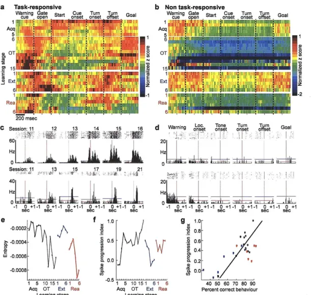
Figure 2 Plasticity in spike activity patterns of striatal projection neurons. a-b, Average activity of units classified as task-responsive (a) and non task-responsive (b) neurons plotted in 10-msec bins as z-scores normalized relative to that neuron's baseline activity, according to pseudo-colour scales shown at right, with one row per training session. Plots show ±200 msec time-windows around task events, abutted in the order of occurrence within a trial. c, Peri-event time histograms (PETHs, ±1 sec window) for units recorded on consecutive days at single sites (putative single units) illustrating strengthening and time-shift in responses around locomotion onset over 6 consecutive sessions (top) and sharpening of phasic responses at turn onset over 13 sessions (bottom). Horizontal lines indicate mean pre-trial baseline firing rates (red) and 2 SDs above the mean (blue, threshold for task-related activity). d, Typical PETHs for neurons lacking in-trial phasic peri-event activity ("non task-responsive" units). e, Entropy of per-trial spike activity of task-responsive units calculated for each training stage. f, Spike progression index (SPI) illustrating correlation of per-trial spike activity of task-responsive projection neurons at each training stage to the neural activity at the last stage of over-training. g, Significant correlation (R = 0.74, P < 0.0001) between SPI and progressive changes in percent correct behaviour during training. Acquisition and over-training (black), R = 0.82, P
0.0002; extinction (blue), R = 0.87, P = 0.02; reacquisition (red), R = 0.09, P = 0.87.
a
Warning
cue
oDen
Gate
200 msec
b
0.4
o0.450.3
0.
-
1 5 10151 61 6
Acq OT Ext Rea
Learning stage
Start
Cue
Turn
Turn
.
onset
.
onset . offset
Goal
I I
I
-oI3
ONz
-1
""1 5 10151 61 6 5
~~1
5 10151 61 6
Acq OT Ext Rea
-
Acq OT Ext Rea
Learning stage
Learning stage
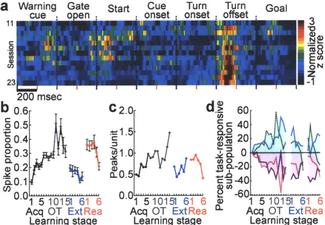
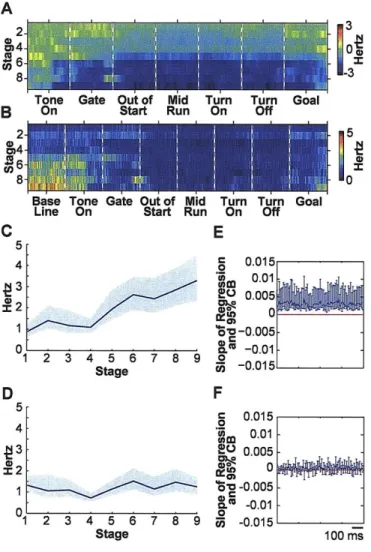
Figure 3 Multiple changes in projection neuron activity in the sensorimotor striatum during acquisition, extinction and reacquisition training. a, Unit activity at a single site recorded over 13 sessions. Each row represents a session. b, Proportions of spikes concentrated in phasic responses in each trial, averaged for each session. Error bars indicate standard errors of mean. c, Average numbers of phasic responses per unit. d, Percentages of task-responsive projection neurons with responses at warning cue (solid blue line), goal reaching (dotted green line), locomotion onset (solid purple line) or turning (dotted magenta line). Values plotted relative to first training stage.
a
8
Acq OT Ext Rea Learning stage 290- ' , 880-370- , 60-( 50-$40' ,, 30 -020 40 50 60 70 80 90 Percent correct C 290 'i. C 80 - i70-860 -250 -*40 , . E 30- . 0. 2 3 0 0 20 2 4 6 8 10 Running time
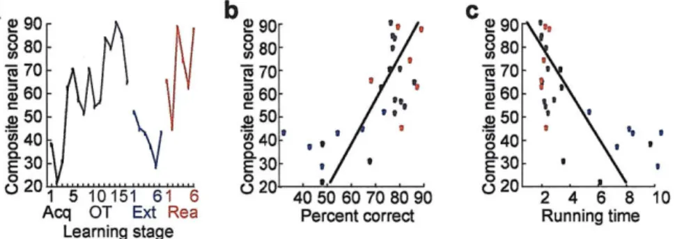
Figure 4 Striatal neural activity predictive of behavioural performance. a, Composite neural scores based on weighted neural measures at trial start (normalized per-neuron firing rates during the +200-msec interval around warning cue, proportions of warning cue-responsive neurons, and proportions of spikes within phasic warning cue responses). b, Significant correlation between the composite scores (shown in a) and actual behavioural accuracy for each training stage (colour-coded as in a, R = 0.69, P < 0.001). c, Plot as in b, showing significant correlation between the composite neural scores and actual running times for each training stage (R = -0.69, P < 0.001).
Supplementary Methods
Experimental protocol. The spike activity of neurons in the sensorimotor striatum was recorded chronically during behavioural training on a conditional reward-based T-maze task for 24 to 63 daily sessions from seven rats in which seven tetrode headstage
assemblies had been implanted. Recordings began on the first day that the rats received training (ca. 40 trials/day) on a conditional reward-based T-maze task and were continued through successive acquisition training (stages 1-5), over-training (stages 6-15),
extinction (stages 1-6) and reacquisition training (stages 1-6, Fig. 1 b, Supplementary Table 2). In this task, rats learned to run down the maze and to turn right or left as
instructed by auditory cues in order to receive reward. Behavioural data were acquired by means of photobeams and a CCD camera. Neural data (32 kHz sampling) were collected by means of a Cheetah Data Acquisition System (Neuralynx Inc.). Well-isolated units accepted after cluster cutting were classified as striatal projection neurons or interneurons (Supplementary Fig. lb-d). Behavioural and neural data were aligned by time stamps and were analyzed by in-house software. The properties of both responsive and non task-responsive projection neurons were analyzed. Task-related responses of putative
projection neurons were identified with respect to activity during a pre-trial 500-msec baseline period (threshold: 2 SDs above baseline mean) and used to define
task-responsive and non task-task-responsive populations. Unit data were analyzed per neuron and per neuronal population across task events (Fig. 1c). To analyze population activity, normalized firing rates were averaged for each learning stage, and indices of spike firing patterns across learning stages were computed, the proportions of neurons with different task-related response types, the proportions of spikes that occurred within peri-event
phasic responses per session, and trial-to-trial spike variability were also calculated, along with composite neural scores and measures of the entropy of neural firing, as described below. Changes in these measures were compared to changes in percent correct performance and running times of the rats across stages of training.
Surgical procedures. Headstages carrying tetrodes (200-250 KQ) in each of seven independently-moveable microdrives (six for recording and one for reference) were mounted on the skull above an opening overlying the dorsolateral caudoputamen (AP =
+0.5 mm, ML = 3.6 mm) in male Sprague-Dawley rats (250-350 g) anesthetized with ketamine (75-100 mg/kg) and xylazine (10-20 mg/kg). An anchor screw served as animal ground. All procedures were approved by the Committee on Animal Care of the
Massachusetts Institute of Technology.
Behavioural procedures. Each rat was first handled in the animal colony room (3-5 days) and then was habituated to the T-maze chamber for 3-5 days. About one week after surgery, acquisition training began (Fig. lb and c). In each trial, a warning cue (~70 dB click) was presented 250 msec before the opening of the start gate, while the rat was at the start location. When the gate opened, the rat was allowed to run down the maze. Half-way down the main alley, one of two tones (1 and 8 kHz pure tones, -80 dB) was
sounded to indicate which of the choice arm goals was baited with reward (chocolate sprinkles). The tones remained on until the rat reached one of the goals or the trial was terminated (Fig. Ic). Tone-goal arm assignments were randomized and counterbalanced
among rats. Approximately 40 trials separated by 1-3 minute inter-trial intervals were given each day.
Each rat was required to reach the correct goal in at least 72.5% of trials in a session to attain the acquisition criterion for significant correct performance (p < 0.01, chi-square tests) and then had to perform at or above this level in 10 out of 11
consecutive daily sessions to reach the over-training criterion. The numbers of initial acquisition training sessions ranged from 3 to 21, and the numbers of over-training
sessions varied from 10 to 38. The rats were then given extinction training, in which reward was reduced to 1-3 trials per session (n = 4) or withdrawn altogether (n = 3). Extinction training lasted 2-11 days. Immediately thereafter, reacquisition training on the
original task began and continued until the rat performed at the 72.5% correct criterion level or headstages failed (Supplementary Tables 1 and 2). Two to eleven reacquisition sessions were given. During all training phases, sessions were terminated if the rat
stopped performing the task before completing 40 trials. Each day, recordings were made for an average of 38.1 trials during acquisition, 33.3 trials during extinction and 38.4 trials during reacquisition.
Neuronal and behavioural data acquisition. Tetrodes were gradually lowered through the brain toward the striatum (3.5-5.0 mm) during the 1-week recovery period after
surgery. Once they reached the target, the position of each tetrode was adjusted until 3-5 distinguishable units appeared in the recordings. Task training then began. During
training, tetrodes were moved as little as possible, and then in small (e.g., <100 pm) steps to maintain high quality, multiple single-unit recordings. The average distance of tetrode
movement throughout the recording periods is shown for each rat in Supplementary Table 1. We recorded an average of 10.8 units per daily session. The absolute numbers of units recorded could not be accurately determined, given probable repeated recording from individual neurons on successive days. Data were thus compiled in terms of units per session and were then averaged.
In selected sessions, sensory responses of recorded units were tested before or after behavioural training by tactile stimulation of contra-lateral body areas (e.g., front and hind limb, neck, back and body) with a glass stir-bar and by manipulation of joints. This examination identified sensory responses of units recorded by a tetrode, but did not provide information about which unit was activated by the stimulation. Despite this limitation, the results did not suggest any clear relationships between sensory responsiveness and task-related activity of recorded units.
Unit activity (gain: 200-10000, filter: 600-6000 Hz) was recorded during all training sessions with a Cheetah Data Acquisition System (Neuralynx Inc.). Spikes
exceeding a preset voltage threshold were sampled at 32 kHz per channel and were stored with time stamps. The animal ground or a single tetrode channel served as reference. The movement of the rat was monitored continuously and recorded (sampling rate: 60 Hz) by a video tracker that received images from an overhead CCD camera. The times of
occurrence of behavioural and stimulus events were determined either online by the use of photobeams (Med Associates, Inc.) or offline by analyzing the tracker data.
At the end of training, rats were deeply anesthetized (Nembutal, 50-100 mg/kg), and lesions were made to mark the final recording sites (25 piA, 10 sec). Rats were then perfused with 4% paraformaldehyde in 0.1 M phosphate buffer, and 30 ptm thick
transverse frozen brain sections were stained for Nissl substance to identify recording tracks and lesion sites (Supplementary Fig. la).
Data Analysis.
1. Behavioural data. The performance of each rat in each training session was measured by the accuracy of responses (percent correct) and the time that elapsed as the rat ran the maze from gate opening to goal reaching (running time), averaged over all trials per session. Changes in these measures during training were analyzed by repeated measures analysis of variance (ANOVA). In order to combine data from different rats to detect learning-related changes in neural responses, we defined stages of learning according to the response accuracy in each training session as follows: stage 1 = first training session, stage 2 = second training session, stage 3 = first session with >60% correct responses, stage 4 = first session with >70% correct responses, and then subsequent stages as pairs of consecutive sessions with >72.5% correct performance. Some pairs of consecutive sessions were on consecutive days of training, but others were separated by gaps in which per-session performance fell below 72.5% correct (Supplementary Table 2). For
extinction and reacquisition sessions, stages were: stage 1 = first training session, stage 2 = second training session, and then stages 3-6 = pairs of consecutive sessions.
2. Spike sorting and unit classification. Unit activity recorded by each tetrode was first
sorted into single units by the use of AutoCut (DataWave Technologies) under manual control, and the quality of sorted units was tested by analyzing auto-correlograms and overlays of spike waveforms. Each unit was included for analysis if its total number of spikes exceeded a threshold of 100 spikes/session, and each accepted unit was classified,
as shown in Supplementary Fig Ib-d, as either a putative projection neuron, a putative fast-firing interneuron (FFN) or a putative tonically firing interneuron (TFN). Units classified as putative projection neurons made up 2091 of 3149 accepted units (66.4%). These were the focus of this study and, for convenience, are termed projection neurons in the text. Smaller numbers of units were classified as FFNs (n = 942, 29.9%) or TFNs (n =
116, 3.7%). The relatively small numbers of FFNs and TFNs precluded conclusive analysis of changes in their firing patterns across all task events and the 27 learning stages; but in the data available, we did not observe the large-scale, multiple changes in firing patterns that we found for the neurons classified as projection neurons.
3. Task-related activity of individual units. Peri-event time histograms (PETHs) were made for each unit for each time-stamped task event (warning cue, gate opening, locomotion onset, tone onset, turn onset and offset, and goal reaching). Task-related responses were defined as responses in which the spike counts in four or more
consecutive 20-msec bins, with at least one of those bins occurring in +200-msec peri-event time windows, had 2 or more spikes and exceeded the criterion level, which was set at two standard deviations (SDs) above the mean activity recorded during the pre-trial baseline 1900 to 1400 msec before warning cue. For units that did not fire during the baseline period, task-related responses were defined as epochs with four or more
consecutive bins with spike counts of at least 2. The proportions of units with such event-related phasic discharges ("task-responsive units") were calculated for each task event for each learning stage. The remaining units were designated as "non task-responsive units." The proportions of task-responsive units increased from 55-80% of all accepted units during acquisition to 80-100% late in over-training, then decreased to 45-75% during