HAL Id: hal-01219119
https://hal-amu.archives-ouvertes.fr/hal-01219119
Submitted on 22 Oct 2015
HAL is a multi-disciplinary open access
archive for the deposit and dissemination of
sci-entific research documents, whether they are
pub-lished or not. The documents may come from
teaching and research institutions in France or
abroad, or from public or private research centers.
L’archive ouverte pluridisciplinaire HAL, est
destinée au dépôt et à la diffusion de documents
scientifiques de niveau recherche, publiés ou non,
émanant des établissements d’enseignement et de
recherche français ou étrangers, des laboratoires
publics ou privés.
A robust and rapid xenograft model to assess efficacy of
chemotherapeutic agents for human acute myeloid
leukemia
E Saland, H Boutzen, R Castellano, L Pouyet, E Griessinger, C Larrue, F de
Toni, S Scotland, Mathieu David, G Danet-Desnoyers, et al.
To cite this version:
E Saland, H Boutzen, R Castellano, L Pouyet, E Griessinger, et al.. A robust and rapid xenograft
model to assess efficacy of chemotherapeutic agents for human acute myeloid leukemia. Blood Cancer
Journal, Nature Publishing Group, 2015, 5 (e297), �10.1038/bcj.2015.19�. �hal-01219119�
ORIGINAL ARTICLE
A robust and rapid xenograft model to assess ef
ficacy of
chemotherapeutic agents for human acute myeloid leukemia
E Saland1,2, H Boutzen1,2, R Castellano3,4,5,6, L Pouyet3,4,5,6, E Griessinger7,8, C Larrue1,2, F de Toni1,2, S Scotland1,2, M David1,2, G Danet-Desnoyers9, F Vergez1,2, Y Barreira10, Y Collette3,4,5,6, C Récher1,2,11,12and J-E Sarry1,2,12
Relevant preclinical mouse models are crucial to screen new therapeutic agents for acute myeloid leukemia (AML). Current in vivo models based on the use of patient samples are not easy to establish and manipulate in the laboratory. Our objective was to develop robust xenograft models of human AML using well-characterized cell lines as a more accessible and faster alternative to those incorporating the use of patient-derived AML cells. Five widely used AML cell lines representing various AML subtypes were transplanted and expanded into highly immunodeficient non-obese diabetic/LtSz-severe combined immunodeficiency IL2Rγcnull mice (for example, cell line-derived xenografts). We show here that bone marrow sublethal conditioning with busulfan or irradiation has equal efficiency for the xenotransplantation of AML cell lines. Although higher number of injected AML cells did not change tumor engraftment in bone marrow and spleen, it significantly reduced the overall survival in mice for all tested AML cell lines. On the basis of AML cell characteristics, these models also exhibited a broad range of overall mouse survival, engraftment, tissue infiltration and aggressiveness. Thus, we have established a robust, rapid and straightforward in vivo model based on engraftment behavior of AML cell lines, all vital prerequisites for testing new therapeutic agents in preclinical studies.
Blood Cancer Journal (2015)5, e297; doi:10.1038/bcj.2015.19; published online 20 March 2015
INTRODUCTION
Acute myeloid leukemia (AML) is the most common adult acute leukemia and is characterized by clonal expansion of immature myeloblasts, initiating from rare leukemic stem or progenitor cells. In Europe and the USA, the incidence and mortality rates of AML are about 5 to 8/100.000 and 4 to 6/100.000 per year, respectively.1 Despite a high rate of complete remission after treatment with genotoxic agents, the relapse rate remains very high and the prognosis very poor. Overall survival at 5 years is ~ 30–40% in patients younger than 60 years, and o20% in patients over 60 years. Front-line chemotherapy is highly effective in ablating leukemic cells, but distant relapses are observed in the majority of patients, characterized by a refractory phase during which no other treatment has shown any efficacy thus far. Relapses are caused by malignant cell regrowth initiated by chemoresistant leukemic clones.2,3
This unfavorable situation leads to a strong need for new therapeutic strategies, as well as relevant preclinical mouse models in which to test them. We have previously established a robust xenotransplantation model to study primary human AML biology and stem cell function based on the engraftment of primary AML samples into non-obese diabetic (NOD)/LtSz-severe combined immunodeficiency (SCID) IL2Rγcnull(NSG) mice.4,5These highly immunodeficient mice have the advantages of a longer life span and higher levels of engraftment of human AML cells compared with other immunocompromised mouse strains such as SCID, NOD-SCID.4,6,7However, this model is based on three-to-six
month-long experiments with a large excess of cells and requires access to a biobank of diverse primary AML patient samples. Moreover, extensive characterization of this xenograft model with more commonly used and easily accessible AML cell lines has not been reported to date in the same setting to compare models in vivo. Herein, we report experimental conditions (bone marrow preconditioning and injected cell number) for a faster and easier preclinical mouse model of AML based on the xenotransplantation of a panel of six adult and childhood human AML cell lines, representing different FAB types, genetic variants and chromoso-mal abnorchromoso-malities. In addition, we have also shown that our cell line-derived xenograft (CLDX as mirrored to patient-derived xenograft) models exhibited a broad range of overall mice survival, engraftment, tissue infiltration and aggressiveness based on their AML cell line characteristics.
MATERIALS AND METHODS Cell lines and culture conditions
Human AML cell lines U937, MV4-11, MOLM-14, HL-60 (DSMZ, Braunsch-weig, Germany) and KG1a (ATCC, Manassas, VA, USA) were maintained in
minimum essential medium-α medium supplemented with 10% fetal
bovine serum (Invitrogen, Carlsbad, CA, USA). All cell lines were grown
in the presence of 100 units per ml of penicillin and 100μg/ml of
streptomycin, and were incubated at 37 °C with 5% CO2. The cultured cells
were split every 2–3 days and maintained in an exponential growth phase.
All cell lines were annually re-ordered to DSMZ or ATCC stocks and
1
Cancer Research Center of Toulouse, INSERM, U1037, Toulouse, France;2
University of Toulouse, Toulouse, France;3
Cancer Research Center of Marseille, INSERM, U1068, Marseille, France;4Institut Paoli-Calmettes, Marseille, France;5Aix-Marseille Université, Marseille, France;6CNRS, UMR7258, Marseille, France;7Centre Méditerranéen de Médecine Moléculaire, INSERM, U1065, Nice, France;8
Université de Nice Sophia Antipolis, Nice, France; 9
Department of Medicine, University of Pennsylvania School of Medicine, Philadelphia, PA, USA;10
Service d'Expérimentation Animale, UMS006, 31059, Toulouse cedex, France and11
Département d’Hématologie, Centre Hospitalier Universitaire de Toulouse, Institut Universitaire du Cancer Toulouse Oncopole, Toulouse cedex, France. Correspondence: Dr J-E Sarry, Cancer Research Center of Toulouse, INSERM, U1037, 2 Avenue Hubert Curien CHU Purpan, Toulouse 31024, France.
E-mail: jean-emmanuel.sarry@inserm.fr
12
These authors contributed equally to this work. Received 19 January 2015; accepted 2 February 2015
periodically authenticated by morphologic inspection and mutational sequencing, and tested negative for Mycoplasma. The clinical and mutational features of our AML cell lines are described in Table 1.
Flow cytometric analysis
CD45-APC (BD, San Jose, CA, USA; 555485), CD33-PE (BD 555450), CD44-FITC (BD 555478), CD44-PE-Cy7 (BD 560533), CD45.1-PerCP-Cy5.5 (BD 560580), CD34-APC-Cy7 (Biolegend, Ozyme, Saint Quentin Yvelines, France; 343514), Lin1-FITC (BD 340546), CD123-PE (BD 340545),
CD45RA-Alexa Fluor 700 (BD 560673) and CD38-APC (BD 555362) fluorescent
antibodies were used to analyze leukemic cells before and after injection into animals to determine phenotypic analysis of engrafted cells and percentage of leukemic cell engraftment. Absolute cell counts were determined with CountBright absolute counting beads (Invitrogen) following the manufacturer's recommendations.
Xenotransplantation of human leukemic cells
Animals were used in accordance with a protocol reviewed and approved by the Institutional Animal Care and User Ethical Committee of the UMS006 and Région Midi-Pyrénées (Approval#13-U1037-JES08). NSG mice were produced at the Genotoul Anexplo platform at Toulouse (France) using breeders obtained from The Charles River Laboratory. Mice were
housed in sterile conditions using high-efficiency particulate arrestance
filtered micro-isolators and fed with irradiated food and acidified water.
Adult mice (6–8 weeks old) were sublethally irradiated with 250 cGy of
total body irradiation or treated with 20 mg/kg busulfan (Busilvex, Pierre Fabre, France) by intraperitoneal administration 24 h before injection of leukemic cells. Cultured AML cell lines were washed twice in phosphate-buffered saline (PBS) and cleared of aggregates and debris using a 0.2-mm
cellfilter, and suspended in PBS at a final concentration of 0.2–2 million
cells per 200μl of PBS per mouse for intravenous injection. Xenograft
tumors were generated by injecting AML cells (in 200μl of PBS) in the tail
vein of NSG mice. Daily monitoring of mice for symptoms of disease
(ruffled coat, hunched back, weakness and reduced motility) determined
the time of killing for injected animals with signs of distress.
Hematopoietic cells count
Peripheral blood was obtained with retro-orbital bleeding. Fifty microliter of blood were collected in heparin-coated collection tube for analyzed with hematology counter (ABX Micros 60, Horiba, Montpellier, France).
Assessment of leukemic engraftment
NSG mice were humanely killed in accordance with the Institutional Animal Care and User Ethical Committee of the UMS006 and Région Midi-Pyrénées Protocols. Bone marrow (mixed from tibias and femurs) and spleen were dissected, crushed in PBS and made into single cell
suspensions for analysis by flow cytometry (FACS Calibur, FACS Canto,
FACS LSR II–BD Biosciences, San Jose, CA, USA).
Statistical analysis
Mann–Whitney test was used to calculate final P-values. Significance is
represented as followed: *Po0.05, **Po0.01 and ***Po0.005.
RESULTS AND DISCUSSION
The objective of this study was to develop robust xenograft models by establishing the specific experimental conditions (cell dose, dissemination tropism, growth kinetics, symptoms, lethality and stability of cell surface markers) required for the evaluation of therapeutic agents in routine and easy-to-use setting. The advantages of using well-established AML cell lines as compared with primary patient samples are the unlimited access to a large amount of human AML cells and a faster engraftment in immunodeficient mice. We analyzed in vivo the five most commonly used AML cell lines with a range of molecular abnormalities, clinical, biological and immunophenotypical char-acteristics (Table 1). MOLM-14 and MV4-11 cells are a widely studied model for FLT3-ITD AML (30% of AML patients associated with worst prognosis). U937 cells are a model for monocytic
Table 1. Cli nical, mu tational and biological featu res of AML cell lines used in thi s in vivo study Name Gender FAB Karyotype M odel for FLT3 NPM1 IDH1 R132 IDH2 R140 IDH2 R172 DNMT3A CEBPa Kit NRas KRas WT1 p53 c-Myc PTEN ITD TKD K G 1a M M 0 Relapse C omplex FGFR1OP2-FGFR1 w t w t w t w t w t w t w t w t w t + wt Mutated Overexpressed Mutated HL-60 F M2 Dx C omplex wt wt wt wt wt wt wt wt wt + w t + Null; deleted Ampli fi ed wt MV4-11 a M M 5 D x C omplex MLL-AF4 w/FL T3-ITD ITD wt wt wt wt wt wt wt wt wt wt Overexpressed w t MOLM14 M M 5 Relapse C omplex MLL-AF4 w/FL T3-ITD ITD wt wt wt wt wt wt wt wt wt wt + w t Overexpressed w t U937 M M 5 Refractor y t(10;11)(p13;q14) CALM-AF10 wt wt wt wt wt wt wt wt wt wt wt + Null; deleted Overexpressed Null Abbreviations: F, female; M, male; wt, w ild type. aChildhood .
CLDX models for AML E Saland et al
2
development and translocation t(10;11)(p13;q14) leading to PICALM-MLLZ10/AF10 fusion gene found in 7% of AML patients with good prognosis factor. HL-60 cells are a model for human myeloid promyelocytic cell differentiation and proliferation, and KG1a is a model for immature myeloid progenitor phenotype and FGFR1 kinase fusion genes (Table 1). These cells were transplanted intravenously in adult NSG mice. Importantly, AML cell lines kill mice as does AML in patients, whereas primary AML cells in NSG mice rarely kill the animals.
Busulfan or irradiation conditioning step does not change the xenotransplantation efficacy of AML cells
Xenotransplantation of primary human AML or normal CD34+cells is greatly enhanced if recipients receive a chemical conditioning regimen such as busulfan (a myeloablative alkylating agent, 25–35 mg/kg) or total body irradiation (up to 4 Gy).8–11However, it
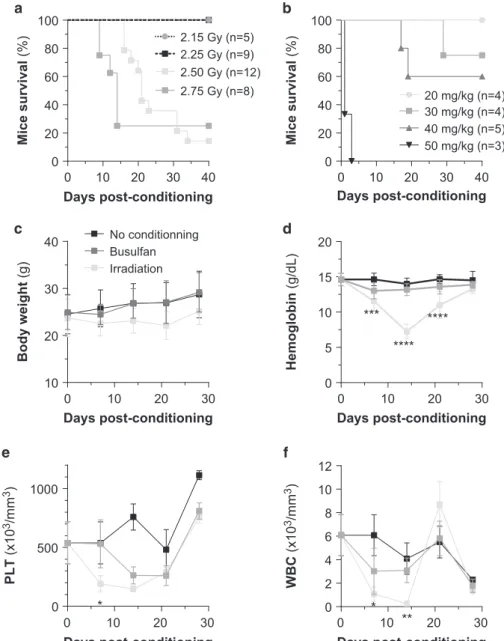
is not clear whether conditioning improves the engraftment of human AML cell lines especially in the most recently developed NSG mouse model. Wefirst performed a maximally tolerated dose study to define the best conditioning procedures (Figures 1a and b) and then tested the effect of the determined sublethal conditions (20 mg/kg busulfan vs 2.25 Gy irradiation) on NSG mice. Body weight, hemoglobin, platelet number and white blood cell number were monitored over 4 weeks following the conditioning procedure. Irradiation severely impacts mice body weight and blood counts, whereas busulfan injection has a milder effect on these parameters (Figures 1c and f). In both the procedures, normal mouse hematopoiesis is recovered 3 weeks post conditioning.
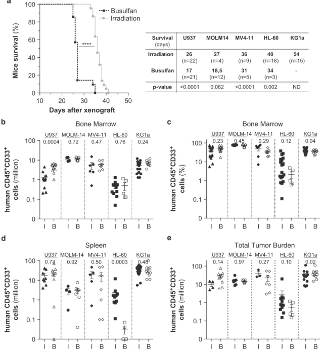
We observed a significant reduction in overall survival of the xenografted mice in the busulfan conditioning setting as compared with irradiation setting (Figure 2a) but observed no
Mice survival (%) Mice survival (%) Days post-conditioning 0 10 20 30 40 0 20 40 60 80 100 2.50 Gy (n=12) 2.75 Gy (n=8) 2.25 Gy (n=9) 2.15 Gy (n=5) Days post-conditioning 0 10 20 30 40 0 20 40 60 80 100 20 mg/kg (n=4) 30 mg/kg (n=4) 40 mg/kg (n=5) 50 mg/kg (n=3) 0 10 20 30 10 20 30 40 Days post-conditioning Body weight (g) Irradiation Busulfan No conditionning 0 10 20 30 0 5 10 15 20 Days post-conditioning Hemoglobin (g/dL) *** **** **** 0 10 20 30 0 500 1000 Days post-conditioning PLT (x10 3/mm 3) * 0 10 20 30 0 2 4 6 8 10 12 Days post-conditioning WBC (x10 3/mm 3) * **
Figure 1. Impact of various bone marrow conditioning treatments on the overall and hematological toxicity and the engraftment efficacy of
AML cell lines in highly immunodeficient NSG mice. Kaplan–Meyer curve of the mice overall survival during the maximal tolerated dose
study (a, with irradiation dose escalade; b, with busulfan dose escalade). Adult NSG mice non-conditioned (n= 5) or irradiated at 2.25 Gy
(n= 5) or treated with 20 mg/kg busulfan (n = 5) were weighed (c) and their blood was collected each week post conditioning for assays of
hemoglobin (d), platelets (e) and white blood cells (f).
significant difference in leukemic engraftment level in bone marrow and spleen or total cell tumor burden after transplanta-tion of four out thefive AML (U937, KG1a, MOLM-14 and MV4-11) cell lines tested (Figures 1b and e), as previously observed in NOD-SCID for human cell engraftment.11
AML cell lines exhibit a broad range of engraftment, tissue infiltration and aggressiveness
A recurrent problem in leukemic adoptive transfer is the issue of the cell dose injected, to avoid either engraftment failure or delay or, on the contrary, a tumor burden outgrowth leading to a premature animal death precluding any investigation. Thus, we asked whether the cell number injected into NSG mice would
affect the overall mice survival. For 0.2 million injected AML cells, recipient mice survival was dependent upon the AML cell line and ranged from 28 to 68 days (U937 oMOLM-14oMV4-11oHL-60oKG1a; Figure 3a). As expected, increasing the injected cell number (up to 2M) decreased the overall mice survival for all AML
cell lines tested (Figure 3a). For both doses, U937 cells appeared to be the most aggressive AML cell line, paralyzing and killing the mice within 4 weeks, whereas mice engrafted with KG1a cells had the longest overall survival with a median of 54 and 68 days for 2Mversus 0.2Mcells injected, respectively (Figure 3a).
The variability in mice survival between cell lines was not associated with differences in leukemic cells infiltration in the bone marrow (Figures 3b and c), spleen (Figure 3d) or the total
Days after xenograft
Mice survival (%) 10 20 30 40 50 0 20 40 60 80 100 **** Busulfan Irradiation p-value Survival (days)
U937 MOLM14 MV4-11 HL-60 KG1a
Irradia on 26 (n=22) 27 (n=4) 36 (n=9) 40 (n=18) 54 (n=15) Busulfan 17 (n=21) 18,5 (n=12) 31 (n=5) 34 (n=3) -<0.0001 0.062 <0.0001 0.002 ND human CD45 +CD33 + cells (million) human CD45 +CD33 + cells (million) human CD45 +CD33 + cells (million) 0.1 1 10 100 0
U937 MOLM-14 MV4-11 HL-60 KG1a 0.0004 0.72 0.47 0.76 0.24
Bone Marrow Bone Marrow
0.1 1 10 100 0 0.23 0.45 0.29 0.12 0.04 U937 MOLM-14 MV4-11 HL-60 KG1a
0.1 1 10 100 0 0.73 0.92 0.50 0.0003 0.48 U937 MOLM-14 MV4-11 HL-60 KG1a
I 0.1 1 10 100 0 0.14 0.97 0.27 0.10 0.02 U937 MOLM-14 MV4-11 HL-60 KG1a
Spleen Total Tumor Burden
human CD45 +CD33 + cells (%) B I B I B I B I B I B I B I B I B I B I B I B I B I B I B I B I B I B I B I B
Figure 2. Impact of the conditioning methods on the xenotransplantation of different AML cell lines in highly immunodeficient
NSG mice. Adult NSG mice were injected with 2 × 10⁶ of different AML cells (HL-60, MOLM-14, MV4-11, U937 and KG1a) 24 h post
conditioning (I, irradiation at 2.25 Gy; B, 20 mg/kg busulfan). Mice survival is analyzed with Kaplan–Meyer curve (a) and the engraftment
level is assessed byflow cytometry in bone marrow (million, b; percent, c), spleen (million, d) and in bone marrow+spleen (total cell tumor
burden, e).
CLDX models for AML E Saland et al
4
tumor burden (Figure 3e), regardless of the injected leukemic cell dose. Accordingly, decreasing injected cell number increases the time to engraft without changing the engraftment capacity at the dissection time and has the disadvantage of extending the in vivo efficacy assay. Furthermore, we noted a variable total cell tumor burden in hematopoietic tissues (bone marrow and spleen)
depending on the AML cell line type (2M for HL-60; 9M, U937;
12Mfor MOLM-14; 13M, KG1a; and 30MMV4-11; Figure 3e). The
engraftment level of HL-60 cells was lowest compared with other cell lines. MV4-11 cells have the highest expansion level in hematopoietic tissues of xenografted mice. Although most of the AML cell lines invade the spleen and exhibit a spleenomegaly,
Survival
(days)
U937 MOLM-14 MV4-11 HL-60 KG1a
0.2M 29 (n=8) 34 (n=18) 48 (n=15) 55 (n=12) 68 (n=4) 2M 27 (n=22) 27 (n=4) 36 (n=9) 40 (n=30) 54 (n=15) p-value 0.0086 0.0002 0.0001 <0.0001 0.0008 20 30 40 50 60 70 0 20 40 60 80 100 0.2M 2M
Days after xenograft
Mice survival (%)
****
0.1 1 10 100Cell dose (Million)
0
0.46 0.33 0.35 0.84 0.19 U937 MOLM-14 MV4-11 HL-60 KG1a
0.1 1 10 100
Cell dose (Million)
0.90 0.92 >0.99 0.01 0.38 U937 MOLM-14 MV4-11 HL-60 KG1a
0.1 1 10 100
Cell dose (Million)
0
0.17 0.52 0.55 0.07 0.72 U937 MOLM-14 MV4-11 HL-60 KG1a
0.2 2 0.2 2 0.2 2 0.2 2 0.2 2
0.1 1 10 100
Cell dose (Million)
0
0.04 >0.99 0.85 0.40 0.52 U937 MOLM-14 MV4-11 HL-60 KG1a
No engrafted HL-60 MOLM-14MV4-11 U937 KG1a 0 1 2 *** ** *** *** Spleen size (cm) HL-60 MOLM-14 MV4-11 U937 KG1a 0 20 40 60 80 100 BM SP Tissue distribution
(% of total tumor BM+SP burden)
Bone Marrow Spleen
Bone Marrow Total Tumor Burden
0.2 2 0.2 2 0.2 2 0.2 2 0.2 2 human CD45 +CD33 + cells (million) human CD45 +CD33 + cells (million) human CD45 +CD33 + cells (million) human CD45 +CD33 + cells (million) 0.2 2 0.2 2 0.2 2 0.2 2 0.2 2 0.2 2 0.2 2 0.2 2 0.2 2 0.2 2
Figure 3. Impact of the injected cell dose on the xenotransplantation of different AML cell lines in highly immunodeficient NSG mice. NSG mice were injected with two doses (0.2 or 2 Millions) of different AML cells (HL-60, MOLM-14, MV4-11, U937 and KG1a). Mice survival was analyzed after xenotranplantation (a). The engraftment level was evaluated in the bone marrow (million, b; percent, c) and spleen (million, d), as well as the total tumor cell burden in hematopoietic mice organs (million, e). Tumor invasion in hematopoietic organs was shown through
the spleen size (f) and the tissue distribution in hematopoietic organs (g). BM, bone marrow; SP, spleen. **Po0.01 and ***Po0.005.
HL-60 and MOLM-14 cells are preferentially located in the bone marrow (Figures 3f and g), indicating differential tissue tropism for this panel of AML cell lines.
These results show a diversity of engraftment capacities and aggressiveness in these AML cell lines, as well as a diversity in tissue tropism, consistent with what we observed with AML patient cells transplanted in the same immunodeficient mice strain.4Engraftment levels, tissue distribution and overall survival are not associated with any clinical features and cytogenetic abnormalities. Interestingly, we also observed that AML cell lines
bearing differentiation markers such as U937, MOLM-14 and MV4-11 appeared to have a more aggressive in vivo behavior than immature like AML cell lines such as HL-60 or KG1a.
The overall immunophenotype of AML cells is conserved in vivo We next addressed whether the immunophenotype of AML cell lines was conserved in vivo. The expression level of various myeloid cell surface markers such as CD45, CD33, CD44, CD34, CD38, CD45RA and CD123 was analyzed for four AML cell lines HL-60 MV4-11 Before 31% 44% 6% 19% 63% 3% 34% 0% 3% 28% 0% 69% 96% 3% 1% 0% After 60% 40% 64% 12% 1% 21% 0% 0% Before 1% 99% 21% 1% 1% 77% 0% 0% Gated on CD45+CD44+CD33+ Gated on CD45+ U937 After Before 0% 20% 0% 80% 54% 7% 37% 2% 0% 3% 0% 97% 6% 3% 78% 13% KG1a After Before 3% 16% 1% 80% 6% 3% 78% 13% 0% 49% 0% 51% 0% 1% 0% 99% CD45 SSC CD44 CD33 CD48 CD34 CD123 CD45RA After
Figure 4. Analysis of the expression of major myeloid cell surface markers in AML cell lines before and after xenotransplantation. The immunophenotype of HL-60, MV4-11, U937 and KG1a cells was analyzed on BD LSRII Fortessa Flow Cytometer using human CD45, CD44,
CD33, CD34, CD38, CD45RA and CD123 before and after xenotransplantation in our NSG mice model. All AML cell lines are SSClow, CD45,
CD44 and CD33 positive in vitro and in vivo.
CLDX models for AML E Saland et al
6
before and after xenotransplantation (Figure 4). Most of those markers appeared globally unchanged during engraftment, however, we found that the transplantation of HL-60 cells in mice also led to the appearance of a second population CD44dim (Figure 4, second column). Moreover, the frequency of CD34+ CD38+was significantly increased in vivo to the detriment of the CD34+CD38- population (Figure 4, third column). With the sole exception of the CD38 marker, the NSG-based model of AML maintains cell phenotype more consistently than in the NOD-SCID model.12
Here we provide necessary technical details and conditions that other laboratories may use to quickly and routinely set up in vivo AML experiments. A xenografts based on native or engineered cell lines are not novel, this is thefirst study that compares the five most well-characterized AML cell lines in the same experimental settings and mouse strain. The establishment of this preclinical AML model is of special relevance and significance to drug-sensitivity studies as in vitro cell culture-based screens do not accurately reflect in vivo effects and responses. In conclusion, we have found that the xenotransplantation models of five well-characterized AML cell lines (for example, KG1a, HL-60, MOLM-14, MV4-11 and U937) exhibited a broad range of mice survival, engraftment, tissue infiltration and aggressiveness. By increasing or decreasing the number of injected leukemic cells, we can modulate mice survival without changing the tissue distribution of leukemic cells and their engraftment in NSG mice. This work has also shown that AML cell lines kill mice in a manner similar to that as patients in clinics but different from the xenograft of primary AML patient specimens in NSG mice. Moreover, in contrast to patient-derived xenograft models, these CLDX models are a powerful, rapid and straightforward in vivo assay available to all leukemia research laboratories to perform preclinical studies to assess the in vivo efficacy of conventional or targeted therapies in AML.
CONFLICT OF INTEREST
The authors declare no conflict of interest.
ACKNOWLEDGEMENTS
We thank all members of mice core facilities (UMS006, INSERM, Toulouse) for their support and technical assistance and Dr. Mary Selak for helpful discussion and reading of the manuscript. We thank Valérie Duplan-Eche, Delphine Lestrade and Fatima L’Faqihi-Olive for technical assistance at the flow cytometry core facility of INSERM UMR1043. FdT is a fellowship from the Fondation de France. This work was
supported by grants from INSERM, Association Laurette Fugain (J-ES), Fondation ARC (SFI20121205478; J-ES), Région Midi-Pyrénée (J-ES), the association G.A.E.L (CR) and Fondation InNaBioSanté (CR and J-ES).
REFERENCES
1 Burnett A, Wetzler M, Löwenberg B. Therapeutic advances in acute myeloid leukemia. J Clin Oncol 2011;29: 487–494.
2 Lübbert M, Müller-Tidow C, Hofmann WK, Koeffler HP. Advances in the treatment of acute myeloid leukemia: from chromosomal aberrations to biologically targeted therapy. J Cell Biochem 2008;104: 2059–2070.
3 Estey EH. Acute myeloid leukemia: 2012 update on diagnosis, risk stratification, and management. Am J Hemat 2012;87: 89–99.
4 Sanchez PV, Perry RL, Sarry JE, Perl AE, Murphy K, Swider CR et al. A robust xenotransplantation model for acute myeloid leukemia. Leukemia 2009; 23: 2109–2117.
5 Sarry JE, Murphy K, Perry R, Sanchez PV, Secreto A, Keefer C et al. Human acute myelogenous leukemia stem cells are rare and heterogeneous when assayed in NOD/SCID/IL2Rγc-deficient mice. J Clin Invest 2011; 121: 384–395.
6 Ailles LE, Gerhard B, Kawagoe H, Hogge DE. Growth characteristics of acute mye-logenous leukemia progenitors that initiate malignant hematopoiesis in nonobese diabetic/severe combined immunodeficient mice. Blood 1999; 94: 1761–1772. 7 Lapidot T, Fajerman Y, Kollet O. Immune-deficient SCID and NOD/SCID mice
models as functional assays for studying normal and malignant human hema-topoiesis. J Mol Med 1997;75: 664–673.
8 Mauch P, Down JD, Warhol M, Hellman S. Recipient preparation for bone marrow transplantation. I. Efficacy of total-body irradiation and busulfan. Transplantation 1988;46: 205–210.
9 Down JD, Ploemacher RE. Transient and permanent engraftment potential of murine hematopoietic stem cell subsets: differential effects of host conditioning with gamma radiation and cytotoxic drugs. Exp Hematol 1993;21: 913–921. 10 Ishikawa F, Livingston AG, Wingard JR, Si Nishikawa, Ogawa M. An assay for
long-term engrafting human hematopoietic cells based on newborn NOD/SCID/beta2-microglobulin(null) mice. Exp Hematol 2002;30: 488–494.
11 Robert-Richard E, Ged C, Ortet J, Santarelli X, Lamrissi-Garcia I, de Verneuil H et al. Human cell engraftment after busulfan or irradiation conditioning of NOD/ SCID mice. Haematologica 2006;91: 1384.
12 Henschler R, Göttig S, Junghahn I, Bug G, Seifried E, Müller AM et al. Transplantation of human acute myeloid leukemia (AML) cells in immuno-deficient mice reveals altered cell surface phenotypes and expression of human endothelial markers. Leuk Res 2005;29: 1191–1199.
This work is licensed under a Creative Commons Attribution 4.0 International License. The images or other third party material in this article are included in the article’s Creative Commons license, unless indicated otherwise in the credit line; if the material is not included under the Creative Commons license, users will need to obtain permission from the license holder to reproduce the material. To view a copy of this license, visit http://creativecommons.org/licenses/ by/4.0/


