1389
Special Issue: Brain Aging
The Neural Basis of Age-Related Changes in Motor
Imagery of Gait: An fMRI Study
Gilles Allali,
1,2Marian van der Meulen,
1–4Olivier Beauchet,
5Sebastian W. Rieger,
6,7Patrik Vuilleumier,
1–4and Frédéric Assal
1,21Department of Neurology, Geneva University Hospitals and 2Faculty of Medicine, University of Geneva, Switzerland.
3Laboratory for Neurology and Imaging of Cognition, Department of Neurosciences, University Medical Center and 4Department of Clinical Neurosciences, University Hospital Geneva, University of Geneva, Switzerland.
5Department of Neuroscience, Division of Geriatric Medicine, UPRES EA 4638, UNAM, Angers University Hospital, France. 6Brain & Behaviour Laboratory, Swiss Center for Affective Sciences and
7Geneva Neuroscience Center, University of Geneva, Switzerland.
Address correspondence to Gilles Allali, MD, PhD, Department of Neurology, Geneva University Hospitals, University of Geneva, 4 rue Gabrielle-Perret-Gentil, 1211 Geneva, Switzerland. Email: gilles.allali@hcuge.ch
Background. Aging is often associated with modifications of gait. Recent studies have revealed a strong relationship
between gait and executive functions in healthy and pathological aging. We hypothesized that modification of gait due to aging may be related to changes in frontal lobe function.
Methods. Fourteen younger (27.0 ± 3.6 years) and 14 older healthy adults (66.0 ± 3.5 years) performed a motor
imagery task of gait as well as a matched visual imagery task. Task difficulty was modulated to investigate differential activation for precise control of gait. Task performance was assessed by recording motor imagery latencies, eye move-ments, and electromyography during functional magnetic resonance imaging scanning.
Results. Our results showed that both healthy older and young adults recruited a network of brain regions comprising
the bilateral supplementary motor cortex and primary motor cortex, right prefrontal cortex, and cerebellum, during motor imagery of gait. We observed an age-related increase in brain activity in the right supplementary motor area (BA6), the right orbitofrontal cortex (BA11), and the left dorsolateral frontal cortex (BA10). Activity in the left hippocampus was significantly modulated by task difficulty in the elderly participants. Executive functioning correlated with magnitude of increases in right primary motor cortex (BA4) during the motor imagery task.
Conclusions. Besides demonstrating a general overlap in brain regions recruited in young and older participants,
this study shows age-related changes in cerebral activation during mental imagery of gait. Our results underscore the importance of executive function (dorsolateral frontal cortex) and spatial navigation or memory function (hippocampus) in gait control in elderly individuals.
Key Words: Mental imagery—Aging—Gait—Neuroimaging.
Received July 16, 2013; Accepted November 16, 2013 Decision Editor: James Goodwin, PhD
G
AIT disorders are very common in the elderly individu-als; it is estimated that only 20% of very old individuals walk normally (1–3). Specific age-related changes in spatial and temporal gait parameters have been associated with the risk of falling (4). For example, increased stride time varia-bility predicts incidental falls in older adults. Aging can also result in increased step width variability or stride length vari-ability, highlighting a disturbance in automatic and rhythmic motor control (5). Several neurophysiological factors may play a role in the modulation of gait in aging, from regional muscular atrophy (6) through to the degeneration of particu-lar neurotransmitter systems, like the cholinergic system (7)or dopaminergic system (8). The presence of a gait disor-der can be a marker of the future development of disease, like dementia, pathologies disturbing the frontal lobe or the connections between subcortical regions and frontal cortices (9). The frontal lobes are closely related to executive func-tioning, but also appear crucial for the control of gait (10). Gait disturbances may thus be indicative of subtle preclini-cal changes in higher order cognitive functioning.
In recent years, this relationship between executive function-ing and gait control has received considerable attention (10). Impaired executive functioning has been proposed to under-lie gait changes in healthy aging (11), but also in Alzheimer’s November
disease (12), vascular dementia (13), frontotemporal demen-tia (14), and Parkinson’s disease (15). The role of executive functioning in gait control becomes apparent especially in demanding situations such as dual tasking (ie, walking while performing a cognitive task) or walking on uneven ground (10).
The use of functional neuroimaging techniques has greatly contributed to the understanding of cerebral control of gait in humans. Near-infrared spectroscopy (16), tran-scranial magnetic stimulation (17), nuclear neuroimaging techniques (18), and more recently electroencephalography (19) allow for direct measures of cerebral activity during actual gait performance. Functional magnetic resonance imaging (fMRI) and positron emission tomography have also been employed to study neural processes associated with gait control, through the use of motor imagery (MI) paradigms. This approach exploits the documented overlap in cerebral regions recruited during mental simulation of a movement and during actual execution of the same move-ment (20,21). Together, these studies have identified a net-work associated with MI of gait, comprising bilateral SMA, superior parietal lobules, hippocampus, parahippocampal gyri, pedunculopontine nuclei, and cerebellum (22,23).
The effects of aging on the neural substrate of gait con-trol merits more systematic investigations given the high fre-quency of gait disturbances in the elderly individuals and its significance for future disease. The modification of quantita-tive gait parameters and the implication of cogniquantita-tive functions in gait control in the elderly individuals suggest a specific pat-tern of brain activation during MI of gait in comparison with young adults. Our aim was therefore to compare brain func-tion in a group of young participants and a group of healthy older adults using fMRI and a validated gait imagery task.
Methods Participants
Fourteen healthy young participants (4 men/10 women, mean age: 27.0 ± 3.6 years, education: 16.2 ± 2.8 years) and 14 healthy old participants (4 men/10 women, mean age: 66.0 ± 3.5 years, education: 14.6 ± 2.5 years) took part in this study. They were recruited from the community through word of mouth and public advertising. The study was approved by the local ethics committee. All participants gave writ-ten informed consent. All participants were right handed, as verified with the Edinburgh Handedness Inventory (EHI, Oldfield, 1971). Young participants obtained a mean score of 85.3 ± 17.6 and elderly participants of 87.1 ± 14.4 (p = nonsignificant). All participants had normal or corrected-to- normal vision and no neurological or orthopedic distur-bances. Cognitive functioning of all elderly participants was assessed using a detailed neuropsychological test bat-tery (Table 1) and the young participants were screened for any neurological or medical conditions interfering with brain functioning. All the older participants presented nor-mal performances in the neuropsychological assessment.
Motor Performance and Chronometry Ability
To assess individual participant’s motor performance and chronometry ability (CA), we administered two men-tal chronometry tests, both evaluating performed and imagined times of an action. The first was a version of the Timed “Up and Go” (TUG) test developed by Beauchet and coworkers (24). The initiation, acceleration, decel-eration, preparation of turning, and turning component of the TUG are also involved in the “difficult” condition of our fMRI task. The second was the Ten Meter test. Participants completed two trials on both the real and imagined version of both tasks. On the imagined TUG, the experimenter gave the cue to start imagining, and the participant was instructed to pronounce the word “stop” when they had completed the mental task. The Ten Meter test was similar in design to the TUG, but assessed CA while walking in a straight line, similar to the fMRI task (which also involved walking straight for 10 m). All participants performed two trials of the Ten Meter and the TUG and the mean of these two trials were used as outcome.
Individual CA scores were calculated on the basis of the delta time scores on the TUG and Ten Meter test. Delta times were calculated according to the following formula: delta = [treal − timag]/[(treal + timag)/2] (24). CA score was
Table 1. Demographic Data and Neuropsychological Test Scores of the 14 Healthy Older Participants
EC Mean ± SD Sociodemographic Female/male 10/4 Age 66.0 ± 3.5 Education 14.6 ± 2.5 Global measures MMSE 29.1 ± 0.9 Mattis 142.9 ± 1.2 Memory Buschke—48 items 26.9 ± 3.9 Doors Test A 11.2 ± 1.1 Doors Test B 8.6 ± 1.7
Digit span forwards 5.9 ± 0.7
Attention/executive
Stroop Test 112.3 ± 16.3
Trail-Making Test A 34.6 ± 5.9
Trail-Making Test B 76.1 ± 20.7
Digit span backwards 4.2 ± 0.7
Language
Boston Naming A* 31.3 ± 1.9
Boston Naming C* 20.0 ± 0.0
Verbal fluency (category) 33.9 ± 7.4
Verbal fluency (letter) 25.0 ± 6.4
Depression/anxiety
HAD (depression) 1.4 ± 1.2
HAD (anxiety) 4.4 ± 2.7
Notes: EC = elderly controls; HAD = Hospital Depression and Anxiety scale.
*Version A of the Boston Naming test is for the age group <70 years and version C for >70 years.
taken as the positive mean of the two delta times for each participant. The lower the CA score, the smaller the dif-ference between real and imagined performance times, and therefore the higher CA.
fMRI Procedure
Materials.—Participants were scanned during a single
fMRI session lasting about 30 minutes. All functional and structural MRI data were acquired on a 3T whole body MRI system (Trio TIM, Siemens, Germany) with a 12-channel head coil at the Brain & Behaviour Laboratory (BBL) of the University of Geneva. Visual stimuli were presented on a projection screen inside the scanner using E-prime (E-prime 1.0, Psychology Software Tools Inc., Pittsburgh, PA). Gaze direction and pupil diameter were logged with an MRI compatible long range eye tracker (EyeTrac 6, Applied Science Laboratories).
Scanning protocol.—Whole-brain functional images were
collected using a susceptibility-weighted EPI sequence (TR/TE = 2,100/30 ms; flip angle = 80°; PAT factor = 2; FOV = 205 mm; matrix size = 64 × 64 pixels). Thirty-six transversal slices were acquired sequentially with a 3.2 mm thickness and an interslice gap of 0.64 mm, yielding a voxel size of 3.2 mm isotropic. High-resolution whole-brain ana-tomical scans were acquired with a T1-weighted, 3D sequence (MPRAGE; TR/TI/TE = 1,900/900/2.27 ms; flip angle = 9°; voxel dimensions = 1 mm isotropic; 256 × 256 × 192 voxels).
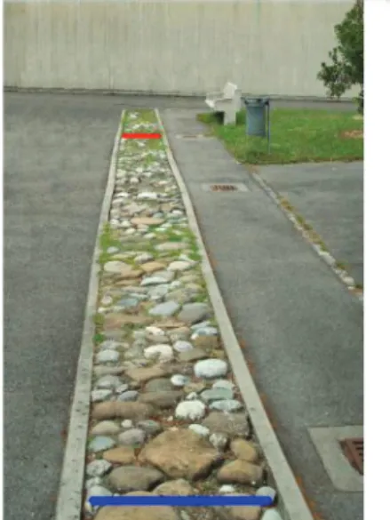
fMRI activation task.—Stimuli for the fMRI task
con-sisted of 12 photos, 6 each of 2 different outdoor walk-ways, located in the garden of the rehabilitation unit of the Geneva University Hospital, each taken from a slightly dif-ferent perspective, side or angle (Figure 1) that have been described previously (25). Both walkways were about 1 m wide. One walkway had a smooth tarred surface (providing stimuli for the “easy” condition), whereas the other had a surface consisting of cobble stones of between 4 and 30 cm in diameter (“difficult” condition). Participants performed three different tasks on the two kinds of walkways: MI, visual imagery (VI), and a perceptual control (C) condition.
In the MI task, participants were shown the photos of the different walkways with two horizontal lines drawn across them: a grey line at the bottom to indicate the start of the trajectory and a black line at a distance of 10 m (as meas-ured on the actual path). Participants were asked to start imagine walking from the grey line to the black line as soon as the picture appeared. They had to press a button when they imagined that they had reached the red line on the walkway. Focusing on the kinesthetic component of MI, they were instructed to imagine walking as vividly as pos-sible, as if feeling their limbs moving (but without actually moving them), and from a first-person perspective.
In the VI task, participants were presented with the same photos as in the MI task, but this time a black disk (25 cm in diameter) was shown on each photo at the start of the trajec-tory (on the grey line). Participants were asked to imagine this disk moving from the blue to the red line at a constant speed, similar to walking speed. They pressed a button when they had imagined the disk to arrive at the red line.
In the control task, participants were presented with the same pictures as in the MI task, but in this case, the lines on the walkways were either both grey or both black. Participants were instructed to simply inspect each photo-graph for a fixed period of time (6 seconds in half of the trials and 10 seconds in the other half). After offset of the picture, participants were cued to press either one button if the lines on the walkway were black or another in case they were blue.
Procedure.—The three different tasks were presented in
a block-wise fashion, with two blocks per task, and there-fore six blocks in total, that have been described previously (25). All participants received detailed instructions and underwent a training session on all three conditions of the task before the scanning session, until they were confident that they were able to perform the task at the best of their capacities.
Behavioral Data Analysis
For each participant, the mean imagery time for both MI and VI was calculated by averaging the trials for each condi-tion. Imagery time was taken as the time between the onset of the picture and the button press. We used an eye-tracker monitor to check online that all participants kept their eyes open and adhered to the actual task instructions (ie, moved their eyes along the walkways). Moreover, following the procedure by Bakker and coworkers (22), we also verified task adherence by investigating the effect of path surface (ie, task difficulty) for the MI and the VI tasks. As the black disk is not impeded by path surface, task difficulty should have a larger effect on MI than on VI. Therefore, a 2 × 2 repeated-measures analysis of variance (ANOVA) of imagery times was carried out, with task (MI vs VI) and difficulty (easy vs difficult) as within-participant variables.
EMG Recording/Analysis
During the fMRI session, muscle activity of the right leg was recorded to control for overt muscle movements. Muscle activity (EMG) was recorded with a modular data acquisition system (MP150, BIOPAC Systems Inc.), with a sampling rate of 10,000 Hz. A pair of carbon-wired MRI compatible electrodes were placed 5 cm apart along the muscle bellies of the right tibialis anterior. The reference electrode was placed on the center of the patella. Details of EMG analysis have been previously described (25).
functional Data Analysis
first-level analysis.—Functional images were preprocessed
and analyzed using standard methods implemented in SPM5 (Wellcome Department of Imaging Neuroscience, London), which have been described previously (25). The experimen-tal conditions were Motor Imagery—Easy (MIE) Motor Imagery—Difficult (MID) Visual Imagery—Easy (VIE) Visual Imagery—Difficult (VID) and Control (C) (Figure 1).
Second-level analysis.—Contrast images obtained in the
individual analyses were entered into one flexible factorial design, which included group and condition as main factors,
as well as their interaction. The main contrasts of interest were MI > C, probing for activation associated with MI, and MID > MIE, investigating the effect of path surface (ie, task difficulty). MI > VI was also included to investigate the motor specificity of the effects. Imagination times were added as a linear parametric nuisance covariate to make sure that any difference between conditions or groups was not due simply to a difference in time taken to perform the task. CA scores were also added as a second covariate to control for differences between groups in CA. Statistical analyses were performed on a voxel-wise basis across the whole brain, all thresholded at p < .001 (uncorrected) with a mini-mum cluster size of 20 voxels, unless otherwise specified.
Anatomical labels of activated clusters were determined using the xjView toolbox (http://www.alivelearn.net/xjview) and the Anatomy Toolbox version 1.5.
Regression.—To explore whether there were any areas in
which activity during gait imagery correlated with execu-tive functioning and according to the previous observa-tions in matter of association between cognitive function and gait (25), we added the Stroop interference score as a linear regressor to the MI > C contrast of our task using the multiple regression option in SPM5. The Stroop interfer-ence score refers to the differinterfer-ence in response times related to the suppression of word reading in favor of color nam-ing, which assesses inhibitory control. Walking requires the quick online processing of a flow of information, to adapt motor actions to the environmental conditions and inhibit-ing the nonrelevant information, which is crucial for gait.
Results
Behavioral Results
Motor performance and CA.—Mean performed (treal)
and imagined times (timag) of the two mental chronometry tests, as well as CA scores, are presented in Table 2. From the table, it can be seen that elderly participants took longer to perform both tasks (significant only for the TUG), indi-cating a slight reduction in walking speed.
Both groups always imagined doing the tasks faster than they executed them in reality. CA scores differed signifi-cantly between groups (f(1,27) = 7.21, p < .012), indicating that CA was slightly worse in the elderly participants.
fMRI gait imagery task.—Mean imagery latencies during
the different conditions of our fMRI task are summarized
in Table 3. Responses in the control condition (deciding
on the color of the lines) were 100% correct for all par-ticipants. A repeated-measure ANOVA with task (MI and VI) and condition (easy and difficult) as within-participant factors revealed that for the young participants, there was a main effect of condition (f(1,13) = 40.64, p < .001) and an interaction between task and condition (f(1,13) = 9.40, p < .01), but no main effect of task (f(1,13) = 1.11, p = nonsig-nificant). This indicates that they took longer to perform the difficult than the easy condition for both MI and VI and that
the difference between easy and difficult was significantly greater for the MI than for the VI task. For the old partici-pants, there was only a main effect of condition (f(1,13) = 79.63, p < .001), but no main effect of task (f(1,13) = 0.23,
p = nonsignificant) and no interaction (f(1,13) = 0.38, p = nonsignificant). Therefore, elderly participants showed an equal difference in imagery times between the easy and dif-ficult condition for both the MI and VI tasks. When the two participant groups were collapsed, we found a main effect of condition (f(1,27) = 106.95, p < .001) and an interaction between tasks and condition (f(1,27) = 5.59, p < .05).
EMG Results
The mean normalized muscle activity values for MI and VI are presented in Table 4. A repeated-measures ANOVA with task as within-participants factor revealed no difference in EMG values between MI and VI for the young partici-pants (f(1,13) = 1.47, p = nonsignificant) nor for the elderly participants (f(1,13) = 0.87, p = nonsignificant). There were also no differences between groups for MI (f(1,27) = 0.002,
p = nonsignificant) nor for VI (f(1,27) = 1.34, p = nonsig-nificant). This indicates that young and old participants pre-sented the same muscle activity during MI and VI and that this muscle activity was similar in both groups.
Imaging Results
Motor imagery effect (MI > C).—For the main contrast
MI > C, we found widespread activation in both groups in a network of areas including bilateral supplementary motor area (BA6) extending to midcingulate cortex, supe-rior medial prefrontal cortex, lateral infesupe-rior BA4, right inferior prefrontal cortex (incl. BA44/45/47) extending to the insula and right postcentral gyrus, and the cerebellum. When we compared groups directly for this contrast ((MI > C_elderly) > (MI > C_young)), we found stronger activa-tion in elderly participants in the left middle frontal gyrus (BA10), right SMA, and right superior orbitofrontal cortex (BA11). The parameter estimates from these three clusters showed a group difference only for the MI task and not for the VI task, indicating that only imagery for gait (and not imagery for visual movement) in our task is modulated by age (Figure 2 and Supplementary Table 1). There were no clusters that were activated stronger in the young than in the
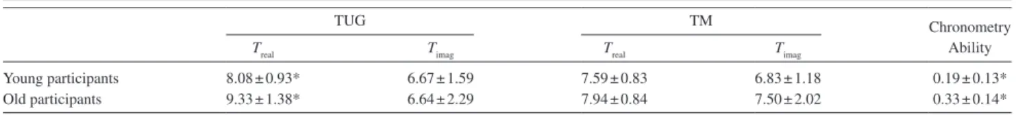
Table 2. Mean ± SD treal, timag, on the TUG and TM, and Chronometry Ability Scores
TUG TM Chronometry
Ability
treal timag treal timag
Young participants 8.08 ± 0.93* 6.67 ± 1.59 7.59 ± 0.83 6.83 ± 1.18 0.19 ± 0.13*
Old participants 9.33 ± 1.38* 6.64 ± 2.29 7.94 ± 0.84 7.50 ± 2.02 0.33 ± 0.14*
Notes: timag = imagined performance time; treal = real performance time; TM = Ten Meter; TUG = Timed Up and Go test.
elderly participants. When we compared groups for the con-trast MI > VI, using the same threshold as for the concon-trast MI > C, we did not find any stronger activation in elderly participants.
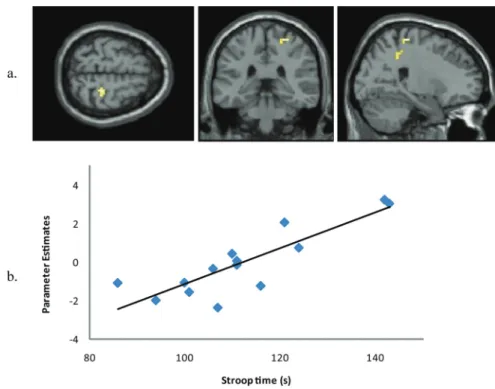
Correlation of activation with Stroop task.—For the
elderly participants, we investigated whether there were any regions that showed a correlation between activation during imagery of gait and behavioral performance on the Stroop test (interference condition). We found a single clus-ter in the right primary motor cortex (BA4), where activity was tightly and positively correlated with performance on the Stroop (Figure 3 and Supplementary Table 2), indicat-ing that the better executive functionindicat-ing, the stronger the recruitment of the right primary motor area during MI.
task difficulty (MID > MIE).—To investigate
differen-tial activation for precise control of gait, we looked at the effect of path surface during imagine walking (ie, task diffi-culty). In the elderly group, we found differential activation for difficult paths (MID > MIE) in bilateral hippocampus, left primary motor cortex (BA4), right insula, left middle and inferior temporal gyrus, and bilateral occipital cortex. For the same contrast, the young participants activated right putamen, bilateral superior temporal gyrus, and bilat-eral occipital cortex. When we compared the two groups directly, we found stronger activation in the elderly partici-pants in the left hippocampus (Figure 4 and Supplementary Table 3). There were no areas with stronger activation in the young compared with the elderly participants.
Discussion
In this study, we explored the effects of aging on the neural substrate of gait control, using a MI paradigm. We have identified a network of activation in young participants during MI of gait (MI > C), comprising bilateral primary motor cortex and SMA, right prefrontal cortex and cerebel-lum, which is highly consistent with previous neuroimaging
studies on gait imagery (22,26). Elderly participants recruited a network that was similar to that recruited by young participants, but with some critical differences, dis-cussed later.
As predicted, the elderly participants exhibited signifi-cantly greater activation than the young participants during MI in two prefrontal regions (left BA10 and right BA11) and the right SMA. The given instruction focusing on the kinesthetic modality of MI could contribute to these results. Indeed, Guillot and coworkers (27) showed that the kines-thetic modality of MI was associated with more activity in motor-associated brain regions. Remarkably, the elderly participants also showed a correlation between activation during imagery of gait and performance on the Stroop test in the right primary motor cortex (BA4). Additionally, they showed stronger activation in some subcortical structures, in particular the substantia nigra and putamen. In the only previous study assessing the effect of aging on the human supraspinal locomotor and postural control by fMRI, although the authors did not assess the cognitive function of their elderly participants, a prominent activation was also observed in the SMA (26). The correlation between activa-tion during imagery of gait and performance on the Stroop test in the right primary motor cortex that we found rein-forces the notion of a strong link between executive func-tion and gait.
The frontal lobes are highly susceptible to age-associated changes, including disruption of frontostriatal circuits by diffuse white matter changes, gray matter atrophy (28), and reduction of dopamine activity in the frontal cortex (29). Interestingly, with regard to the greater activation in the orbitofrontal cortex in the elderly participants, previous studies indicate that elderly patients with freezing of gait present a specific hypometabolism in these orbitofrontal regions (30). These metabolic and morphological changes may contribute to deficits in executive functioning as com-monly seen in the elderly participants (28,29).
Another main result of this study was a greater activation in the left hippocampus in elderly participants relative to young participants, specifically for the MID > MIE contrast that probed activations associated with the higher order, more precise control of gait. The hippocampus and adja-cent regions are well known to be involved in age-related changes in memory and spatial navigation but have also been implicated in gait. In a positron emission tomography study on gait, Malouin and coworkers (23) have shown the involvement of a significant recruitment of the left parahip-pocampal region during walking. The specific role of the hippocampus/medial temporal lobe in gait may therefore be related to topographical memory (31) and mental naviga-tion (32). Animal models also suggest that the hippocampal formation provides voluntary motor systems with continu-ally updated feedback on environmental conditions (33). Previous studies on anesthetized rats revealed a strong inter-action between the hippocampus and the pedoculopontine
Table 4. Mean ± SD EMG Values for the Motor Imagery and Visual Imagery Normalized for the Control Condition
MI VI
Young participants 1.05 ± 0.30 0.99 ± 0.28
Old participants 1.05 ± 0.22 1.10 ± 0.16
Notes: MI = motor imagery; VI = visual imagery.
Table 3. Mean ± SD Imagery Times in Seconds
Task MI VI
Condition Easy Difficult Easy Difficult
Young participants 8.7 ± 2.4 10.5 ± 3.2 8.8 ± 3.1 9.6 ± 3.4 Old participants 9.0 ± 3.0 10.9 ± 3.8 8.9 ± 3.7 10.6 ± 3.7
nucleus mediated by cholinergic input (34). One speculative hypothesis is that the cholinergic pathway reduction due to aging (7) may have contributed to this increased activation in the hippocampus in the elderly participants.
These effects of aging on brain activation during cogni-tive tasks could be explained by the compensation hypoth-esis, which holds that aging is associated with a decline in brain function and performance, inducing hyperactivation of specific brain regions as a compensatory mechanism (35). The dedifferentiation hypothesis, according to which older adults tend to present a nonselective recruitment of brain regions relative to young adults (36), could be another explanation. The greater activation in the right SMA (BA6), the right orbitofrontal cortex (BA11), the left dorsolateral frontal cortex (BA10), and the left hippocampus observed in
old adults reflects a difficulty in recruiting specialized neu-ral mechanisms (37). Perhaps associated with this inability of selective task-associated cerebral recruitment, the elderly participants may have used different strategies to perform the task. Our behavioral data indicate that the young partici-pants showed a greater modulation of imagination time by task difficulty during the MI task than during the VI task. In contrast, the elderly participants showed an equal modula-tion by type of path for both tasks. Performance on our task could be regarded as worse in elderly participants than in young participants, given that they fail to show an interac-tion between task and difficulty. Moreover, like previously observed (38), the motor CA of our elderly participants was worse than that of the young participants, as evidenced by a larger discrepancy between performed and imagined times
Figure 3. Area that shows a significant positive correlation between activity during motor imagery (a) and performance on the Stroop test (b).
on the two mental chronometry tests. Finally, the increased left hippocampal activation seen for the MID > MIE con-trast in the elderly participants relative to young partici-pants suggests that the navigational aspect of walking on cobble stones may have been more challenging to them than to young participants. Therefore, the additional cortical and subcortical areas recruited in older adults may constitute a compensatory mechanism in an attempt to achieve normal performance levels on the MI task, in the light of reduced imagery performance and ability.
As a study limitation, it should be noted that most of the activations were obtained with a standard strict threshold of
p < .001 at the voxel level, with a cluster threshold of k > 20. This combination of threshold and cluster criteria selects reliable effects when changes in BOLD (Blood-oxygen-level depedent) have relatively weak amplitudes and imprecise onsets as for mental imagery or other purely internal (non-sensory) mental events (39). Importantly, all activations reported were selective and concerned areas well known to be involved in MI of gait. Furthermore, we found a signifi-cant correlation in the right primary motor cortex, prefrontal cortex, and an independent cognitive measure (Stroop test). So this suggests that our effects were reliable and task related.
Conclusion
We have demonstrated that aging results in an increased activation during imagery of gait in the prefrontal cortex (specifically, in the left MFG [BA10] and right superior OFC [BA11]) and the right SMA, with additional increases in left hippocampal activation during more precise gait control. In addition, executive functioning of our elderly participants correlated with activity during MI of gait in the right pri-mary motor cortex. These findings emphasize the age-related modification of brain functioning and the important role of executive function in the control of gait in healthy human participants. Future studies should investigate how these neu-ral circuits are modified in pathologies affecting gait control, such as primary progressive gait apraxia, and further help to guide and assess neurorehabilitation techniques.
Supplementary Material
Supplementary material can be found at: http://biomedgerontology. oxfordjournals.org/
Funding
G.A. was supported by « la Bourse d’encouragement de la Société Suisse de Neurologie », M.v.d.M. and F.A. by a grant from the Société Académique de Genève (NAC 08-025), and P.V. by a grant from the Société Académique de Genève (Fonds Forename).
Acknowledgments
We thank Cecile Lederrey for her contribution to the neuropsychologi-cal assessments. We are grateful to the participants for their cooperation. Conflict of Interest
The authors report no conflicts of interest.
References
1. Bloem BR, Haan J, Lagaay AM, van Beek W, Wintzen AR, Roos RA. Investigation of gait in elderly subjects over 88 years of age. J Geriatr
Psychiatry Neurol. 1992;5:78–84.
2. Snijders AH, van de Warrenburg BP, Giladi N, Bloem BR. Neurological gait disorders in elderly people: clinical approach and classification.
Lancet Neurol. 2007;6:63–74.
3. Oh-Park M, Holtzer R, Xue X, Verghese J. Conventional and robust quan-titative gait norms in community-dwelling older adults. J Am Geriatr
Soc. 2010;58:1512–1518. doi:10.1111/j.1532-5415.2010.02962 4. Hausdorff JM, Rios DA, Edelberg HK. Gait variability and fall risk in
community-living older adults: a 1-year prospective study. Arch Phys
Med Rehabil. 2001;82:1050–1056.
5. Beauchet O, Kressig RW, Najafi B, Aminian K, Dubost V, Mourey F. Age-related decline of gait control under a dual-task condition. J Am
Geriatr Soc. 2003;51:1187–1188.
6. Kennedy KM, Raz N. Age, sex and regional brain volumes predict perceptual-motor skill acquisition. Cortex. 2005;41:560–569. 7. Bartus RT, Dean RL 3rd, Beer B, Lippa AS. The cholinergic
hypoth-esis of geriatric memory dysfunction. Science. 1982;217:408–414. 8. Kaasinen V, Rinne JO. Functional imaging studies of dopamine
sys-tem and cognition in normal aging and Parkinson’s disease. Neurosci
Biobehav Rev. 2002;26:785–793.
9. Verghese J, Lipton RB, Hall CB, Kuslansky G, Katz MJ, Buschke H. Abnormality of gait as a predictor of non-Alzheimer’s dementia. N
Engl J Med. 2002;347:1761–1768.
10. Yogev-Seligmann G, Hausdorff JM, Giladi N. The role of executive function and attention in gait. Mov Disord. 2008;23:329–342; quiz 472. 11. Hausdorff JM, Schweiger A, Herman T, Yogev-Seligmann
G, Giladi N. Dual-task decrements in gait: contributing fac-tors among healthy older adults. J Gerontol A Biol Sci Med Sci. 2008;63:1335–1343.
12. Allali G, Kressig RW, Assal F, Herrmann FR, Dubost V, Beauchet O. Changes in gait while backward counting in demented older adults with frontal lobe dysfunction. Gait Posture. 2007;26:572–576. 13. Allali G, Assal F, Kressig RW, Dubost V, Herrmann FR, Beauchet
O. Impact of impaired executive function on gait stability. Dement
Geriatr Cogn Disord. 2008;26:364–369. doi:10.1159/000162358 14. Allali G, Dubois B, Assal F, et al. Frontotemporal dementia: pathology
of gait? Mov Disord. 2010;25:723–729. doi:10.1002/mds.22927 15. Amboni M, Cozzolino A, Longo K, Picillo M, Barone P. Freezing of
gait and executive functions in patients with Parkinson’s disease. Mov
Disord. 2008;23:395–400.
16. Holtzer R, Mahoney JR, Izzetoglu M, Izzetoglu K, Onaral B, Verghese J. fNIRS study of walking and walking while talking in young and old individuals. J Gerontol A Biol Sci Med Sci. 2011;66:879–887. doi:10.1093/gerona/glr068
17. Schubert M, Curt A, Jensen L, Dietz V. Corticospinal input in human gait: modulation of magnetically evoked motor responses. Exp Brain
Res. 1997;115:234–246.
18. Fukuyama H, Ouchi Y, Matsuzaki S, et al. Brain functional activ-ity during gait in normal subjects: a SPECT study. Neurosci Lett. 1997;228:183–186.
19. Gwin JT, Gramann K, Makeig S, Ferris DP. Removal of movement artifact from high-density EEG recorded during walking and running.
J Neurophysiol. 2010;103:3526–3534. doi:10.1152/jn.00105.2010 20. Porro CA, Francescato MP, Cettolo V, et al. Primary motor and
sensory cortex activation during motor performance and motor imagery: a functional magnetic resonance imaging study. J Neurosci. 1996;16:7688–7698.
21. Roth M, Decety J, Raybaudi M, et al. Possible involvement of primary motor cortex in mentally simulated movement: a functional magnetic resonance imaging study. Neuroreport. 1996;7:1280–1284.
22. Bakker M, De Lange FP, Helmich RC, Scheeringa R, Bloem BR, Toni I. Cerebral correlates of motor imagery of normal and precision gait.
23. Malouin F, Richards CL, Jackson PL, Dumas F, Doyon J. Brain activa-tions during motor imagery of locomotor-related tasks: a PET study.
hum Brain Mapp. 2003;19:47–62.
24. Beauchet O, Annweiler C, Assal F, et al. Imagined Timed Up & Go test: a new tool to assess higher-level gait and balance disorders in older adults? J Neurol Sci. 2010;294:102–106. doi:10.1016/j.jns.2010.03.021 25. Van der Meulen M, Allali G, Rieger SW, Assal F, Vuilleumier P. The
influence of individual motor imagery ability on cerebral recruitment during gait imagery. hum Brain Mapp. 2012; doi:10.1002/hbm.22192. 26. Zwergal A, Linn J, Xiong G, Brandt T, Strupp M, Jahn K. Aging of
human supraspinal locomotor and postural control in fMRI. Neurobiol
Aging. 2012;33:1073–1084.
27. Guillot A, Collet C, Nguyen VA, Malouin F, Richards C, Doyon J. Brain activity during visual versus kinesthetic imagery: an fMRI study. hum Brain Mapp. 2009;30:2157–2172.
28. Raz N, Gunning FM, Head D, et al. Selective aging of the human cerebral cortex observed in vivo: differential vulnerability of the pre-frontal gray matter. Cereb Cortex. 1997;7:268–282.
29. Volkow ND, Wang GJ, Fowler JS, et al. Effects of methylphenidate on regional brain glucose metabolism in humans: relationship to dopa-mine D2 receptors. Am J Psychiatry. 1997;154:50–55.
30. Bartels AL, Leenders KL. Brain imaging in patients with freezing of gait. Mov Disord. 2008;23(suppl 2):S461–S467.
31. Maguire EA, Burke T, Phillips J, Staunton H. Topographical diso-rientation following unilateral temporal lobe lesions in humans.
Neuropsychologia. 1996;34:993–1001.
32. Berthoz A. Parietal and hippocampal contribution to topokinetic and topographic memory. Philos trans R Soc Lond B Biol Sci. 1997;352:1437–1448.
33. Bland BH, Oddie SD. Theta band oscillation and synchrony in the hippocampal formation and associated structures: the case for its role in sensorimotor integration. Behav Brain Res. 2001;127:119–136.
34. Vertes RP, Colom LV, Fortin WJ, Bland BH. Brainstem sites for the carbachol elicitation of the hippocampal theta rhythm in the rat. Exp
Brain Res. 1993;96:419–429.
35. Rypma B, D’Esposito M. Isolating the neural mechanisms of age-related changes in human working memory. Nat Neurosci. 2000;3:509–515. 36. Seidler RD, Bernard JA, Burutolu TB, et al. Motor control and aging:
links to age-related brain structural, functional, and biochemical effects. Neurosci Biobehav Rev. 2010;34:721–733.
37. Logan JM, Sanders AL, Snyder AZ, Morris JC, Buckner RL. Under-recruitment and nonselective recruitment: dis-sociable neural mechanisms associated with aging. Neuron. 2002;33:827–840.
38. Esposito G, Kirkby BS, Van Horn JD, Ellmore TM, Berman KF. Context-dependent, neural system-specific neurophysiological con-comitants of ageing: mapping PET correlates during cognitive activa-tion. Brain. 1999;122(Pt 5):963–979.
39. Lieberman MD, Cunningham WA. Type I and type II error concerns in fMRI research: re-balancing the scale. Soc Cogn Affect Neurosci. 2009;4:423–428.