QJMed, 1994; 87:385-401
Original papers
Hereditary complement factor I deficiency
T.J. VYSE, P.J. SPATH
1, K.A. DAVIES, B.J. MORLEY, P. PHILIPPE
2,
P. A T H A N A S S I O U , C M . GILES and M.J. WALPORT
From the Rheumatology Unit, RPMS, Hammersmith Hospital, London, UK,
^ZLB, Central Laboratory, Blood Transfusion Service, SRC, Berne, Switzerland,
and
2Regional Hospital Delemont, Detemont, Switzerland
Received 17 March 1994; Accepted 13 April 1994
QJM
Summary
We describe four cases (from three families) of hereditary factor I deficiency, bringing the total number of cases now reported to 23. In one family there are two affected siblings: one has suffered recurrent pyogenic infections; the other is asympto-matic. In the second family, the patient had recur-rent pyogenic infections and a self-limiting vasculitic illness; in the third family, the patient suffered recur-rent pyogenic and neisserial infections. All four patients had markedly reduced concentrations of C3 in the serum (family 1 propositus: 28%; family 1 asymptomatic sibling: 15%; family 2: 3 1 % ; and
family 3: 31 % normal human serum) which was in the form of C3b. Low lgG2 levels may occur in
primary C3 deficiency, and a reduction in lgG2
concentration to 1.14 g/l (normal: 1.30-5.90 g/l) was found in the patient from family 2. Using radio-ligand binding assays, we demonstrated increased binding of C3b to erythrocytes in a patient with factor I deficiency. This C3b could not be cleaved by autologous serum but could be cleaved by normal serum or purified factor I. We review and compare the published cases of C3, factor H and factor I deficiency.
Introduction
Complement factor I (Fl) is a serine esterase that acts to control the amplification loop of the alternative pathway of the complement cascade. Hereditary deficiency of Fl is a rare autosomal recessive condi-tion. In the absence of Fl, the amplification loop of the alternative pathway is activated in an uncon-trolled fashion, so that there is consumptive loss of complement C3.1 - t Secondary depletion of C3 is also caused by genetic deficiency of complement component factor H (FH), which is a cofactor for Fl. The study of these hereditary deficiencies is important because of the evidence that they provide in relation to the roles of the complement system in vivo.
Furthermore, it was the investigation of the physi-ology of the complement system in the first case of Fl deficiency1"* that generated much of the early evidence for the existence of the amplification loop of the alternative pathway.5
The clinical effects of C3, Fl and FH deficiency are similar: a propensity to suffer recurrent pyogenic infections and an increased incidence of glomerulo-nephritis and SLE-like illness. The clinical con-sequences of C3 deficiency reflect the role played by the complement system in the opsonization of pathogens and the clearance of immune complexes.6 Although there are subtle differences in the effects
Address correspondence to Professor M.J. Walport, Rheumatology Unit, RPMS, Hammersmith Hospital, Du Cane Road, London W12 ONN
386
T.J. Vyseetal.of these three conditions on the complement system,
the predominant influence on the clinical outcome
in all three states is that of C3 deficiency.
To date, 19 patients with Fl deficiency have been
reported.
7"
19We report an additional four patients
from three families. In one family, there were two
homozygously affected siblings; one of whom
suf-fered recurrent pyogenic infections, the other being
healthy. In the other two families there were single,
homozygous, symptomatic individuals. We review
the clinical features of Fl deficiency, and then
compare them with the clinical consequences of FH
and C3 deficiencies. The clinical features of all the
reported cases of these hereditary deficiencies are
tabulated (see Table 1) and their pathogenesis and
treatment are briefly discussed.
Patients
Family 1
A 14-year-old boy presented with a history of
recur-rent pyogenic infections that began at the age of 18
months. At this time he developed a septic arthritis
of the left shoulder from which Staph. epidermidis
was cultured. At the age of 5 years he suffered
orbital cellulitis, and over the next 5 years he had
recurrent episodes of sinusitis. When 8 years old, an
abscess on the side of his neck was drained. At the
age of 13 he had a single episode of meningococcal
meningitis. He has subsequently had a number of
minor respiratory tract infections. Physical
examina-tion was unremarkable. There was no family history
of immunodeficiency or autoimmune disease, and
no consanguinity, although both parents originate
from western Scotland. He has one sibling, a sister
who is 5 years older, who has neither suffered from
any major episodes of infection with pyogenic
organ-isms, nor had recurrent sinusitis or otitis media.
Investigation of both siblings revealed a normal
full blood count and basic biochemistry profile. The
immunoglobulin concentrations of both siblings were
within the normal ranges: the affected male's values
were: IgG 10.5 g/l (normal: 7.2-16.2); IgA 1.3 g/1
(0.8-3.9); IgM 2.3 g/l (0.5-3.5); his sister's values
were: IgG 9.5 g/l (7.2-16.2); IgA 1.1 g/l (0.8-3.9);
IgM 1.5 g/l (0.5-3.5). The IgG subtype concentrations
gave no evidence of selective isotype deficiency: the
affected male's concentrations were: IgG, 9.05 g/l
(3.2-10.2); lgG
22.97 g/l (1.2-6.6); lgG
31.25 g/l
(0.2-1.9); lgG
40.20 g/l (0.10-1.3); the IgG subtype
levels of the asymptomatic sister were as follows:
IgG, 10.05 g/l (3.2-10.2); IgG, 2.73 g/l (1.2-6.6);
lgG
31.55 g/l (0.2-1.9); lgG
40.20 g/l (0.10-1.3).
Antinuclear antibodies (ANA) (measured by indirect
immunofluorescence on Hep-2 cells) and rheumatoid
factor (measured by Latex agglutination) were not
detected. His complement system was investigated
together with that of all four family members (see
Table 2). The results demonstrate the diagnosis of Fl
deficiency in the patient and in his asymptomatic
elder sister: Fl was undetectable in both siblings, as
was factor B (FB); there was no detectable alternate
pathway activity (APH50); and both FH and
proper-din levels were reduced. Both parents had
approxi-mately half the normal concentration of circulating Fl.
The patient is being treated with prophylactic
phenoxymethylpenicillin, 250 mg bd. He was
vac-cinated with Pneumovax and mounted a significant
lgG
2response: pre-immunization 1:16;
post-immunization 1 :110; control 1 :160 (measured by
Dr Kumaratne, Immunology Dept, Dudley Road
Hospital, Birmingham). He is now aged 19 years
and has had no further significant infective episodes.
His sister, who is 24 years old, also remains well.
Family 2
The affected male in this pedigree has suffered from
recurrent otitis media since infancy. He had one
episode of idiopathic keratitis when aged 5 years.
Because of recurrent sinusitis, surgery to the nasal
septum was performed at age 14 and it was noted
that he was allergic to both penicillin and
sulphon-amides. When he was 16 years old, he suffered four
bouts of left-sided bronchopneumonia together with
sinusitis. Str. pneumoniae and Staph. aureus were
cultured from the sputum. The following year a
bronchogram was performed, no lesion was
demon-strated, but an episode of bronchopneumonia was
precipitated. When aged 20, a sixth episode of
bronchopneumonia occurred. Five years later, he
had a self-limiting illness characterized by hepatitis,
pneumonitis, myositis, possible meningitis, and
pur-pura, with histological evidence of a
microangi-opathic vasculitis on skin biopsy. During the next
ten years, he had five additional episodes of
broncho-pneumonia.
At the age of 36 he was fully investigated, and
the diagnosis of Fl deficiency was established. He
had a normal full blood count and biochemistry. His
ANA and rheumatoid factor were negative. The
concentrations of immunoglobulin isotypes were
within the normal ranges, however, analysis of IgG
subtypes: IgG, 11.49 g/l (3.55-11.25); lgG
21.14 g/l
(1.30-5.90); lgG
30.44 g/l (0.15-1.05); lgG
40.54 g/l
(0.10-1.10), indicated that the lgG
2concentration
was diminished. Analysis of the complement system
confirmed the diagnosis of Fl deficiency (see Table 3).
Additional family members were also studied (see
Figure 1) indicating the inheritance of the trait
throughout the family. This family is Swiss/German
in origin, there is no known consanguinity. The
Table 1a Hereditary Factor I deficiency: twenty-three patients from nineteen families (separated by lines)
Patient Age Sex Nationality Consanguinity Age of (years) Race onset
Infections Other complications
Klinefelter's syndrome XXY
Urticaria
Notes
Became C3 Coombs negative for 14 days and had
undetectable C3b and 17 days after infusion of normal plasma C3 and C5 levels raised for 12 days but FB levels maintained for only 3 days after infusion of purified Fl Reference (1) (2) (3) (4)
1 25 M USA 1 year Haemorrhagic measles
Recurrent otitis media Recurrent sinusitis Inguinal abscess
Auricular abscess: C. diphtheriae Septicaemia x 2 : Str. pyogenes N. meningitidis Pneumonia: H. influenzae 2 11 F English no Caucasoid 4 months Meningitis x 4: Str. pneumoniae x 1 N. meningitidis x 2 Otitis media (7) o
I
I"
8-1'
5
? F USA ? 4 ? Meningitis x 3 H. influenzae b N. meningitidis 3 months Pneumonia x 1 (8)5 3 M Germany 6 months Gastroenteritis
Recurrent otitis media Septicaemia
UTI x 3
IgM rheumatoid Abnormal FH mobility factor positive (by immunoelectrophoresis)
probably due to its binding to C3b ANA negative Infusion of plasma (FFP):
C5 and FH elevated above pre-infusion levels for up to 1 month FB increased for 6 h only
No complications after 22 infusions of FFP
(9)
28 M Canada
Caucasian
No Infancy Recurrent otitis, bronchitis, and mastoiditis
Pneumonia x 4 Serum sickness-like Pleural empyema: Str. pneumoniae illness following the Bronchiectasis administration of Meningitis penicillin
5 of 10 siblings died in infancy
3 from sepsis, ?FI status
IgM rheumatoid factor (1 :1280) ANA negative
(10)
Table l a Patient 7 (continued) Age Sex (years) 19 F Nationality Race Denmark Caucasoid Consanguinity No Age of onset 19 years Infections Otitis media Meningitis: N. meningitidis CpB Herpes zoster
Other complications Notes Reference
(11) (12)
8 15/12 F USA 3 weeks Septicaemia: Str. pneumoniae
Pneumonia: Str. pneumoniae Otitis media
?Osteomyelitis
low IgA (0.36 g/1) Infusion of Fl:
C3 and FB increased for 4 days FH and CH50 increased for 14 days Fl remained undetectable (13) 00 00 9 9 ? France ?
? Recurrent bronchitis, otitis media and mastoiditis Reduced erythrocyte CR1 Normal range 300-1320 (14) 10 ? ? France ? 11 ? France
9 years Arthritis: septic ?agent
11 years Arthritis: N. meningitidis
Reduced erythrocyte CR1 Normal range 300-1320 patients: 10-115; 11-182
Defective CRT- and CR3-dependent phagocytosis (14) 12 13 14 15 37 37 27 ? M F M M ? Caucasoid Caucasoid Tunisian No No No
Childhood Recurrent otitis, sinusitis Pneumonia x 8 Meningitis x 5:
N. meningitidis (GpB x 1, W135
x l )
Septicaemia with DIC x 2
37 years Fatal systemic vasculitis: cutaneous leucocytoclastic lesions
Haematuria and proteinuria Deep venous thrombosis Perivenous encephalomyelitis (diagnosed at post-mortem)
Childhood Recurrent otitis media and cutaneous abscesses Vasculitic illness followed the administration of penicillin for pharyngitis Cryoglobulinaemia ANA not reported
Asymptomatic
Treatment with FFP: (15) C3 increase 16 days
Native FB increase 4 days
Fall in C4 and rise in C4d duration - 2 days
Fl half-life estimated 29 h
Anaphylaxis with 8th and 9th FFP infusions
Very low, but detectable levels of (15) Fl, C3d and C4d also detectable
(?from blood transfusion)
(16)
20 France Yes ?Caucasian (second
cousins)
17 months Recurrent pharyngitis Meningitis x 3 : N. meningitidis (GpC x 1) N. meningitidis (untypable x 1) Pneumonia x 1 (Str. pneumoniae) ?FI status
Older brother died age 12 of meningitis (17) 17 12 F France Turkish Parents from same village 4 Vi years Pneumonia x 2
Meningitis: H. influenzae type b
Two siblings both well (17)
18 M USA ?
Caucasian
Recurrent sinusitis and pulmonary Urticaria infections IgA < 6 mg/dl (35-209) some Fi detected 2.6% (46-159) C3 27 mg/dl (83-177), FB <12 mg/dl (17-42) (18)
19 28 F Greek ? 24 years Aseptic meningitis x 11 Sulphonamide allergy Weakly positive ANA 1 :40 to
1 :80 No anti-dsDNA antibodies (19)
I
1
?5 20 21 22 15 18 M Scottish No Caucasoid18 months Septic arthritis: 5. epidermidis Orbital cellulitis, bronchitis Recurrent otitis media and sinusitis Cutaneous abscess Meningitis: N. meningitidis ANA negative Asymptomatic
I
3'
43
36 M Swiss No Childhood Recurrent otitis media, sinusitis
bronchopneumonia x 1 0
[Str. pneumoniae H. influenzae)
Sulphonamide/ penicillin
Allergy one episode of a multisystem inflammatory illness ?vasculitic ANA negative 23 M Spanish Parents from nearby villages
3 weeks Cutaneous sepsis (Staph. aureus) Meningitis x 4: N. meningitidis x 3
Str. pneumoniae x 1
Conjunctivitis
Otitis media, enteritis, septicaemia Herpes zoster
low lgG2 1.14 g/l (1.30-5.90)
ANA negative
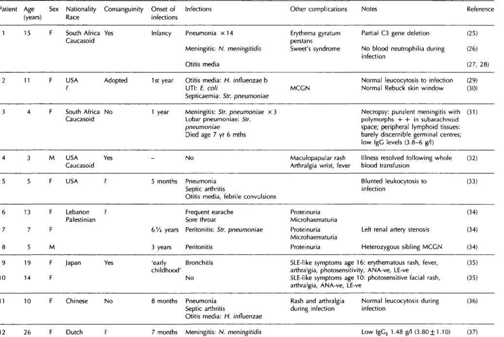
Table 1b Reported human C3 deficiency: twenty-two patients from fifteen families (separated by lines)
o
Patient Age Sex Nationality Consanguinity Onset of Infections (years) Race infections
Other complications Notes Reference
1 15 F South Africa Yes Caucasoid Infancy P n e u m o n i a x 1 4 Meningitis: N. meningitidis Otitis media Erythema gyratum perstans Sweet's syndrome MCGN
Partial C3 gene deletion No blood neutrophilia during infection
Normal leucocytosis to infection Normal Rebuck skin window
(25) (26) (27, 28) (29) (30)
11 F USA Adopted 1st year Otitis media: H. influenzae bO t t s eda
UTI: E. coli Septicaemia: Str. pneumoniae South Africa No Caucasoid 6 7 8 9 10 13 7 5 19 14 F F M F F Lebanon Palestinian Japan
1 year Meningitis: Str. pneumoniae x 3 Lobar pneumoniae: Str.
pneumoniae
Died age 7 yr 6 mths
Necropsy: purulent meningitis with polymorphs + + in subarachnoid
space; peripheral lymphoid tissues: barely discernible germinal centres; low IgC levels (3.8-6 g/l)
(31) 4 5 3 5 M F USA Caucasoid USA Yes ? -5 months No Pneumonia Septic arthritis
Otitis media, febrile convulsions
Maculopapular Arthralgia wrist,
rash fever
Illness resolved following whole blood transfusion Blunted leukocytosis to infection (32) (33) Frequent earache Sore throat
bVi years Peritonitis: Str. pneumoniae
3 years Peritonitis Proteinuria Microhaematuria Proteinuria Microhaematuria Proteinuria
Left renal artery stenosis Heterozygous sibling MCCN
(34) (34) (34)
I
Yes 'early Bronchitis
childhood'
No
SLE-like symptoms age 16: erythematous rash, fever, (35) arthralgia, photosensitivity, ANA-ve, LE-ve
SLE-like symptoms age 10: photosensitive facial rash, (35) arthralgia, ANA-ve, LE-ve
11 10 F Chinese No 8 months Pneumonia
Septic arthritis
Otitis media: H. influenzae
Rash and arthralgia during infection
Normal leucocytosis during (36) infection
13 14 19 16 Meningitis: Str. pneumoniae 'sepsis': S. aureus
years Meningitis: Str. pneumoniae Otitis media 8 months Osteomyelitis Otitis media Transient maculopapular rash during infectious episodes Transient maculopapular rash during infectious episodes MCGN type I Low lgC2 0.11 g/1 (3.80±1.10) Low lgC2 0.44 g/l (3.80±1.10) Administration of FFP of no benefit (64)
15 M Laos No 5 months Lobar pneumonia
Meningitis: Str. pneumoniae x 2
MCGN Acute administration of FFP not
associated with renal deterioration
(38) (39) 16 12 Kuwait Dysfunctional C3 molecules 1 47 F Swedish 2 42 F No Nil Microhematuria Nephrotic syndrome Renal failure MCGN Type I
Recurrent otitis media
Meningitis N. meningitidis Gp Y None SLE
Weak ANA, IgG and IgM at dermo-epidermal junction
+ ve ANA, anti-dsDNA antibody, anti-centromere antibody (40) 17 18 19 20 21 22 6 10 23 14 19 7 M M M F M F Brazil England Japan N. Zealand/ Israel Yes Yes Yes ? 3 months 4 Childhood Meningitis: N. meningitidis Bronchopneumonia x 4 Otitis media, osteomyelitis
Otitis media URTI: Str. pyogenes Meningitis Pneumonia Meningitis: N. meningitidis x 3 x 2 Transient erythema multiforme at time of infection IgA nephropathy Lupus-like illness
Normal leucocytosis to infection
First cousin of patient 19
Asthma, rhinitis, otherwise healthy (41) (41) (42) (42) (43) (50)
8
I
r
i
8
I
Table 1c Hereditary Factor H deficiency: twelve patients from six families
Patient Age Sex Nationality Consanguinity Age of Infections (years) Race onset
Other features Notes Reference
1 8/12 M Indian
M
Yes 8 months Otitis media: H. influenzae (first cousins)
Haemolytic-uraemic syndrome (HUS) Asymptomatic
Very low factor H detected (6% (44) NHS)
Renal biopsy characteristic of HUS
5 M Algerian Yes 14 months Recurrent otitis and bronchitis (first cousins) followed by haematuria
Glomerulonephritis
MCGN-like
6/12 M 4 months Otitis media and bronchitis
Septicaemia: E. coli UTI: Proteus sp. Persistent haematuria
Glomerulonephritis
MCGN-like
FH 12% NHS in both cases renal biopsy appearance similar in both: intramembranous dense deposits detected atypical
immunofluorescence (IF): anti-C3 staining in mesangium and
capillary walls, not in the basement membrane H antigen and C5b-9 neoantigens in the areas of C3 staining (45)
I
2-EU Spain No N. meningitidis N. meningitidisMCGN
MCGN
MCGN
No factor H antigen detectable in all sibs
Reduced C5 ( < 1 0 % NHS) and C9 (10% NHS)
No details of renal biopsy given All ANA negative
(46)
11
Italy Yes None
11 years
Asthma Haematuria SLE with nephritis Anti-dsDNA antibodies
FH undetectable in all 3 sibs Heterozygous C2 deficiency Heterozygous C2 deficiency Renal biopsy: diffuse proliferative nephritis 6 of 25 glomeruli with crescents C3 ( 3 + ) and IgG (1 + ) in mesangium by IF Responded to prednisolone (2 mg/kg/day)
(47)
11 15 Danish No 10 years Meningitis x 2:
N. meningitidis Cp B
No detectable FH (48) Depressed C5 (7% NHS) and C7 ( < 0 . 5 % NHS) ?C7 deficiency too
12 49 Netherlands Meningitis: N. meningitidis Gp X SLE/lupus-like illness No details of autoantibodies (49)
Partial factor H deficiency
1 17 M USA ? Polish 11 years Henoch-Schonlein purpura Thrombocytopenia and splenomegaly
Three other asymptomatic family (51) members
(52)
F USA ? Anglo-Irish
63 years ?Recurrent UTIs
38 M 33 26 M 30 years 36 years 26 years Hypertension IgA nephropathy leading to end-stage renal failure IgA nephropathy (mild) Proteinuria (52)
Diagnosis on renal biopsy
Renal biopsy performed (sister of patient 2)
Brother of patient 2
1
I"
I
F USA No 18 months Recurrent respiratory tract
infections
Haemolytic-uraemic syndrome x 3
Died age 2 yrs
Null allele at C4A and C4B Abnormal C3 variant with reduced total C3
(53)
394
T.J. Vyseetal.Table 2 Complement profile of family 1
Mother Father Propositus Sister Mother Father Propositus Sister Fl 41 74 -C2 84 96 77 90
All results expressed as -, undetectable. FH 96 155 46 42 C3 84 99 28 15 FB 71 71 -C4 71 147 60 105 P 51 52 38 29 C5 137 91 99 48 APH50 50 45 -CH50 92 102
-% normal human serum.
Table 3 Complement profiles of the propositi from family
2 and family 3 Fl FH FB P C4BP APH50 FD100 Propositus 2 - 53 17 34 107 - 82 Propositus 3 - 42 9 38 72 - 68 C2 C3 C4 C5 C7 CH50 Propositus 2 ND 31 159 53 30 47 Propositus 3 ND 31 124 34 36 40
-, undetectable; ND, not determined.
All results expressed as % normal human serum.
patient is now 43 years old and is well apart from occasional febrile illnesses which respond rapidly to the administration of antibiotics.
Family 3
A 6-year-old presented with a history of recurrent pyogenic infections which began at the age of 3 weeks with a cutaneous abscess and omphalitis due to Staph. aureus. At 3 months, he developed a second abscess, and at 6 months he started having persistent folliculitis. When 4 months old, he became septicaemic with Str. pneumoniae, and at 9 months he had a further pyrexial illness. At 14, 15 and 16 months of age he had an abscess on the right cheek, enteritis, and purulent conjunctivitis, respectively. When 19 months old he suffered a pyrexial illness with erythematous rash that responded to penicillin. Four months later, he had two episodes of meningitis in rapid succession; the first due to N. meningitidis and the second due to Str. pneumoniae. The follow-ing month he had otitis media and then septicaemia with Str. pneumoniae. Between the ages of 3 and 6 years the patient suffered from otitis media, bron-chitis, enteritis, purulent pharyngitis, and aphthous stomatitis. When 6 years old, he had an episode of meningococcal septicaemia after which complement studies were performed (see Table 2) and the dia-gnosis of Fl deficiency was made. The immunoglob-ulin concentrations were: IgG 8.24 g/1 (6.86-14.9); IgA 1.5 g/l (0.45-3.09); IgM 0.74 g/l (0.41-3.00) and IgG subtypes were: IgG, 8.07 g/l (3.55-11.25); lgG2 2.67 g/l (1.30-5.90); lgG3 0.26 g/l (0.15-1.05); lgG4 0.26 g/I (0.10-1.10). The concentration of Fl in other family members is shown in Figure 2. Although the propositus and his parents now live in Switzerland, the family was originally from northern Spain, and the parents of the Fl-deficient boy were from neigh-bouring villages. There is no history of consanguinity, however.
The patient was vaccinated with Pneumovax and
II
I l l 107( ) 110 57
IV
Figure 1. The pedigree shows the inheritance of factor I deficiency in the family of Swiss/German origin (family 2). The
propositus is marked with an arrow. The number to the left of the individuals is the factor I concentration in the serum of that individual expressed as a percentage of the value of normal human serum: normal range 75%—135%.
Hereditary complement factor I deficiency
395
NT
NT
NT
3
NT
4 r^iNTQ
II
III
Figure 2. The pedigree shows the inheritance of factor I deficiency in the family of Spanish origin (family 3). The propositus
is marked with an arrow. The number to the left of the individuals is the factor I concentration in the serum of that individual expressed as a percentage of the value of normal human serum: normal range 75%-135%. NT signifies an individual whose Fl concentration was not tested.
maintained on prophylactic penicillin for 3 years. During this period he had intermittent bouts of bronchitis and sinusitis, and when 8 years old, varicella-zoster. He is now 15, and having withdrawn prophylactic penicillin of his own volition for 3 years, has remained relatively well with occasional pyrexial illnesses but no septicaemic episodes.
Methods
Complement assays
Antigenic and functional assays
Serum concentrations of the complement proteins were assayed by radial immunodiffusion using poly-clonal antisera (sheep anti-C2, anti-C3, anti-C4, anti-C5, anti-FI, anti-FH, and anti-properdin: The Binding Site, Birmingham). Functional complement assays were performed as follows:20 classical path-way activity (CH50) was assessed using a haemolytic assay with sheep red blood cells sensitized with rabbit antibody (Tissue Culture Services, Buckingham), in a 1.5% CFD/agarose gel (Oxoid, Basingstoke); alternative pathway activity (APH50) was measured by haemolytic assay with guinea pig erythrocytes (Tissue Culture Services, Buckingham) in a 1.5% agarose gel in the presence of 7 j i M MgCI2 and I O U M ethyleneglycol tetraacetic acid (EGTA). The FD100 is an assessment of factor D (FD) activity. It was measured in the same way as APH50, except that the guinea pig erythrocytes were sus-pended in FD-depleted (by affinity chromatography) serum. The investigation of the state of circulating C3 in the Fl-deficient members of family 1 was carried out by crossed immunoelectrophoresis according to the method of Laurell, as described in reference 20.
Assays for cell-bound C3 and CR1
The binding of C3b and its degradation products on the red cell surface was analysed by radioligand binding assay as previously described.21 Three anti-C3 monoclonal antibodies were used (clones 3, 4 and 9, kindly provided by Prof. P. Lachmann, Cambridge) which recognize C3d, C3c and C3g, respectively.22 Erythrocyte CR1 numbers were also measured by radioligand binding assay using the monoclonal antibody El 1 (kindly provided by Dr Nancy Hogg, ICRF, London). The binding of this antibody to the receptor is not affected by the ligation of CR1 with C3b.23
The purified Fl and FH reagents, used in the ligand binding studies, were prepared by affinity chromato-graphy using specific monoclonal antibodies coupled to cyanogen-bromide-activated Sepharose (Pharmacia): anti-FI: MRC OX21; anti-FH: MRC OX23 (Serotec, Oxford).24
Results
Complement estimation
The complement profiles of the four homozygously affected patients from the three families are given in Tables 2 and 3. In none of them was Fl detected in the circulation. The complement profiles of these patients were similar. They had very low or undetect-able levels of factor B (FB), and no detectundetect-able alternative pathway activity (APH50). The C3 con-centrations were also very low. Crossed immuno-electrophoresis of serum from the two affected sib-lings from family 1, indicated that 90% of the C3 was in the form of C3b (data not shown). The reduced concentrations of FH and properdin in all the propositi to approximately one half of their
396
TJ. Vyse etal.normal value have been documented in all nine
cases of Fl deficiency in which these components
have been assessed.
4'
9'
10'
13151719In one report,
15normal FH and properdin levels were probably the
result of a prior infusion of blood. Reduced levels of
the terminal pathway components C5 and C7 were
found in the affected individuals from families 2 and
3 and the sister of the propositus from family 1 (C5
only). A reduction in C5 has been reported in seven
previous cases,
5'
910'
13'
15'
17and a reduction in C7 in
four cases.
5'
10'
17In none of the reports in which
these two complement components were measured
were normal values recorded.
The results of the radioligand binding studies
performed on family 1 (see Table 4) indicate that the
amount of C3b present on the erythrocyte surface
was increased ten-fold in both homozygotes, as
demonstrated by the binding of the two monoclonal
antibodies to the C3d and C3c portions of C3b. No
increase in binding was observed in the
heterozyg-ously-affected parents. The antibody specific for C3g,
clone 9, recognises a neoantigen on C3 revealed by
the action of Fl. No significant binding was found
with this antibody to erythrocytes from normal or
affected individuals.
Following the incubation of erythrocytes from the
Fl-deficient patient with autologous serum, there was
no change in the pattern of binding observed with
the three different anti-C3 monoclonal antibodies
(see Figure 3). The incubation of the patient's
erythro-cytes with either normal serum, purified Fl, or a
mixture of Fl and FH, resulted in a similar outcome
as regards monoclonal antibody binding: an increase
in clone 9 binding (indicating conversion of C3b to
iC3b and C3dg by Fl); no apparent change in the
binding of clone 3 (which binds to epitopes expressed
in both C3b and in its breakdown products that
remain surface-bound); but a fall in clone 4 binding
(this antibody binds C3c which is cleaved from iC3b
by Fl and released into the fluid phase).
Review of the cases of hereditary Fl, FH and
C3 deficiencies
All the reported cases of hereditary Fl, FH and C3
deficiencies are outlined in Table 1. There are 22
M o A b : H c l o n e 9 W clone 3 • clone 4
300
en c c !o "co CO.a
200
100
1 1
'Fl-def. normal
serum serum
Fl FH + Fl
Figure 3. The treatment of erythrocytes from the propositus
of family 1 with either normal serum or purified factor I ( + / — factor H) resulted in a 2-3 fold increase in the binding of the monoclonal clone 9 (which binds to neoepitopes in C3g). The concentration of factor I used was 10 Hg/ml and the concentration of factor H used was 50 Hg/ml. There was no change in clone 3 binding (which binds to C3b and its degradation products), and a fall in clone 4 binding (which binds to the C3c portion of C3).
patients with homozygous C3 deficiency,
25"
4323
with Fl deficiency,
1"
4'
7-
19and 12 who lack FH.
44^
19Of these, all but one case of homozygous C3
deficiency is symptomatic;
43two cases of Fl
16(family 1, this report) and FH deficiency
44'
47are
asymptomatic. In addition, we tabulate one family
in which complete C3 deficiency has resulted from
the inheritance of one null allele and one
dysfunc-tional molecule;
50six instances of partial FH
defi-ciency are also summarized.
51"
53Discussion
The most consistent clinical feature of these three
inherited complement deficiencies is susceptibility
to infection. This is predominantly due to pyogenic
organisms and Neisseria meningitidis. Infection with
N. meningitidis was documented in 11/22 cases of
Fl deficiency, in 4/12 cases of FH deficiency, and in
Table 4 Assessment of C3 and CR1 numbers on erythrocytes (family 1)
C3 fragment.. C3d C3c C3g CR1 Monoclonal antibody... (clone 3) (clone 4) (clone 9) (E11)
Mother
Father
Proband Sister
Normal
"The number of erythrocyte CR1 is subject to a genetic influence, and hence an absolute normal range has limited meaning.
41
74
475
516
45
59
52
573
621
93
15
24
29
27
31
908
645
311
339
300-1300*Hereditary complement factor I deficiency 397
4/22 cases of C3 deficiency. The efficacy of comple-ment as an opsonin is indicated by the recurrent pyogenic infections sustained by C3-deficient indi-viduals. In both Fl and C3 deficiency, defective opsonization and killing of microbes has been dem-onstrated with phagocytes from affected individuals using in vitro assays.4'14'37'39 Moreover, the defective opsonization and bactericidal activity of leucocytes were improved, in vitro, if either infusions of FFP were given,39 or the missing complement component was replaced, either Fl4 or C3.37
Isolated hereditary deficiency of the terminal path-way components increases susceptibility to neisserial infection, implying that the membrane attack com-plex is important in the eradication of these organ-isms.54 The increased incidence of neisserial infections in C3 deficiency is presumably due to the reduced efficiency of the terminal pathway. Low C5 and C7 levels were found in the propositi from families 2 and 3 in this paper, confirming observa-tions from other reports of Fl deficiency.5'9'10'13'15'17 A reduction in the concentration of C5 has been reported in nine of the ten symptomatic cases of FH deficiency,45"46 and was normal in only one case.44 In this latter case there was a low level of circulating FH detected, and no instance of neisserial infection was recorded in the propositus. C5 is consumed in FH and Fl deficiency because of the C5 convertase activity (C3bBbC3b) derived from the alternative pathway C3 convertase (C3bBbP) which is present in excess. An additional factor in the predisposition of these patients to infection may be that opsoniz-ation of the pathogens by antibody and C3 to form immune complexes facilitates their delivery to the splenic macrophage system.55 Therefore, C3-, FH-and Fl-deficient individuals may be functionally hyposplenic. The infections typically commence in early childhood: the median age of the first pyogenic infection in Fl deficiency was 17 months; in FH deficiency it was 14 months; and in primary C3 deficiency it was 8 months. Evidence from the three symptomatic cases presented in this paper suggests that the frequency of infection declines with age. A possible explanation of this phenomenon is that as the immunological memory of the adaptive immune system expands with increasing age, so the role of the innate immune system becomes less important.
It is apparent from the data in Table 1 that there is a marked contrast in the incidence of renal disease in Fl deficiency compared to the incidence in C3 and FH deficiencies. Clomerulonephritis has not been described in any of the reported cases of Fl deficiency, including the four examples reported in this paper. In C3 and FH deficiencies, there is frequent evidence of some renal involvement. FH deficiency seems to have the strongest association with renal disease. From a total of 12 patients, seven
had definite nephritis: five of these had mesangio-capillary glomerulonephritis (MCGN),45'46 one had the haemolytic-uraemic syndrome,44 and another had crescentic nephritis47 (this patient aJso +iad heterozygous C2 deficiency). In C3 deficiency, glom-erulonephritis was recorded in 8/20 patients and in four of these was of MCGN-type.30-37"39'40 In two of these cases,37'40 it was specified to be type 1 MCGN, of which the characteristic feature is subendothelial deposits. The exact mechanism by which nephritis develops in these two C3 deficiency states is unclear. That C3 and FH deficiency patients suffer an MCGN-like nephritis is interesting because of the association of this disease with the C3 depletion found in conjunction with a C3 nephritic factor. The C3 nephritic factor is usually associated with type II (dense-deposit) MCGN. The association between C3 deficiency and MCGN-like nephritis is further strengthened by the observation that dogs with hereditary C3 deficiency develop a nephritis whose histological pattern is that of MCGN.5 7
The absence of Fl produces marked abnormalities of the complement system (see Table 5 for a compar-ison of the effects of C3, FH and Fl deficiency), and yet has not been associated with glomerulonephritis. Thus there is no straightforward relationship between the derangement of the complement system as meas-ured in vitro and the predisposition to disease. In Fl deficiency, only three cases of 'immune complex' illnesses have been described; one being the multisy-stem inflammatory disorder that occurred in the propositus from family 2. Another patient succumbed to a fatal vasculitic syndrome,15 and the third had a serum sickness-like syndrome following the adminis-tration of penicillin.10 The association of immune complex disease with recurrent infection raises the possibility that the two are related. Recurrent infec-tions, by stimulating an acute-phase response, may theoretically exacerbate an immune-mediated inflammatory process; alternatively, microbial patho-gens may act as the antigenic source for immune complex disease. In one case of FH deficiency,45 in which there was MCGN-like glomerulonephritis, recurrent episodes of otitis media and bronchitis were followed by episodic haematuria. In six patients with hereditary C3 deficiency, transient maculopapu-lar skin rashes have developed during infective episodes.2532-36-37'41 In one study37 circulating immune complexes (identified by C1q binding assay) were present at the time when a rash developed in two patients. Skin biopsy revealed local deposition of IgG, IgM, C1q, but not C3, by immunofluores-cence. It is of note that in the three cases of Fl deficiency in which 'immune complex' type illness developed (the case from family 2, and those reported in references 10 and 15) the illness followed the administration of antibiotic. The complement
398
T.J. Vyseetal.
system is also an important effector mechanism in
the inflammatory response. It is therefore possible
that if some residual C3 activity remained the
con-sequences of immune complex deposition/formation
would be aggravated. In one instance of FH
defi-ciency,
45in which there was some FH antigen
detected (12% normal), two siblings both developed
MCGN-like glomerulonephritis. Renal biopsies were
performed and subjected to immunofluorescence.
The results indicated that C3 was deposited in the
mesangium and capillary walls, and that FH and
C5b-9 neoantigens were present in a similar
distribu-tion to C3. The localizadistribu-tion of C5b-9 neoantigens
suggests that there was formation of the C5
con-vertase and that complement was playing an active
part in the inflammatory process.
There may be an increased incidence of nephritis
in cases of partial (heterozygous) complement
defi-ciency. Two families are summarized in Table 1 in
which partial FH deficiency occurs with IgA
nephro-pathy.
51-
52There is one instance of IgA nephropathy
in a patient with homozygous C3 deficiency.
42No
disease has been associated with heterozygous Fl
deficiency. In the three families reported, there were
no unusual clinical problems in any of the
heterozyg-ous Fl-deficient individuals. In addition, in one family
in which partial Fl deficiency occurred in
conjunc-tion with C1 -inhibitor deficiency,
57the clinical
mani-festations of the C1-inhibitor deficiency were not
altered by the coexistence of heterozygous Fl
defi-ciency.
A reduced concentration of lgG
2was found in the
propositus from family 3. Low lgG
2concentrations
were found in one family to be associated with
homozygous C3 deficiency
37together with a reduced
antibody response to pneumococcal capsular
poly-saccharide (which is lgG
2-dependent).
MAntibody
isotypes have not been studied in FH deficiency,
and in family 1, one of the cases of Fl deficiency
reported here, normal concentrations of IgG, and
lgG
2were measured together with a normal lgG
2response to pneumococcal capsular polysaccharide.
In one systematic investigation of IgG subclasses in
complement deficiencies,
59lower mean levels of
lgG
2were found in primary C3 deficiency compared
to normals, and a marked reduction in lgG
4was a
consistent finding in classical pathway and
C3-deficiency states. Using bacteriophage <f>X'\74 as
a test antigen, two C3-deficient patients produced
normal titres of IgM following primary and secondary
immunization, but failed to make an isotype switch
to IgG when the antigen was used at low dose.
60In
C3, Fl, and FH deficiencies, normal antibody
responses to antigens such as tetanus toxoid,
dip-theria, and pertussis vaccine have been recorded.
37'
48In one study
61which included one FH-deficient
pedigree in Italy, high titres of antibody against
meningococcal polysaccharides A and C were
observed, presumably as a consequence of natural
infection. However, only a modest response was
then generated by immunization with the
meningoc-occal vaccine, Menpovax A + C.
The mechanism by which five patients (one
with-out C3, and two withwith-out FH and Fl) are asymptomatic
is not known. The explanation presumably lies in
the fact that within both the innate and adaptive
immune systems there is a great deal of redundancy.
Thus genetic variation at many loci may influence
the penetrance as well as the expressivity of these
monogenic disorders. The asymptomatic cases of Fl
deficiency were detected because their siblings were
clinically unwell. Because Fl deficiency may be
asymptomatic, it is possible that a substantial
propor-tion of cases are undetected. In one report
15two
cases were identified in two non-consanguinous
families from the Danish island of Funen. The authors
estimate the minimum frequency for the deficient
gene to be 0.002 using the Funen island data.
However, in two large studies, one in 41 083 Swiss
Army recruits,
62and a second in 145 640 blood
donors from Osaka, Japan,
63no instances of C3
deficiency were identified.
The mainstay of treatment at the present time for
these inherited conditions is immunization against
pathogens to which affected individuals are
particu-larly susceptible, Str. pneumoniae, H. influenzae b,
N. meningitidis (vaccination with polysaccharide
antigens types A and C is currently available),
together with the administration of prophylactic
anti-biotics.
An additional potential therapeutic option is
replacement of the deficient complement
compon-ent. Replacement treatment using either fresh-frozen
plasma (FFP) or purified protein has been used in
C3
37-39,64
a n d F|deficiencies.
1'
91315In both
circum-stances, this form of treatment is limited by the high
rate of turnover of the deficient protein. Moreover,
there are two potential drawbacks: firstly, the
replace-ment of a genetically-absent protein may stimulate
an alloimmune response against it; secondly, the
reconstitution of the complement system in the acute
phase may exacerbate the underlying illness. FFP
has been administered in two cases of C3 deficiency
and MCGN with no evidence of improvement or
deterioration in renal function.
14"
39'
64In one of these
cases a renal biopsy was performed before and after
two months of infusion therapy without evidence of
change in renal function, but there was some
histo-logical improvement, with a reduction of staining for
IgG and C4.
64The nephritis subsequently showed a
definite response to corticosteroid therapy. FFP must
still be used cautiously because of the observation
that in C3-deficient dogs, the nephritis was worsened
by replacement of C3.
56Hereditary complement factor I deficiency 399
The administration of FFP in Fl deficiency has resulted in a rapid increase in FB and C3 levels. There was a transient rise in C3d and C4d concentra-tions, accompanied by a loss of C3 from the patient's red ceHs together with a slight fall in C4 concentra-tion.1'9'13'15 No deterioration in clinical condition has been reported in response to FFP administration, although in one case anaphylaxis occurred with the eighth and ninth FFP infusions which were then halted.15 After an FFP infusion, the decline in the serum concentration of Fl is paralleled by that of FB. It has been observed in three instances4'9'15 that despite the fall in Fl and FB levels there is a prolonged effect of Fl replacement on the C3 concen-tration. This starts to decline only after 14 days, at which time there is no antigenically detectable Fl, and FB has returned to its baseline level. The mechanism of this discrepancy is unclear. C2 defi-ciency has been successfully managed with regular FFP replacement therapy, and the clinical improve-ment, in arthralgia and skin rash for instance, has been observed to last 4 - 8 weeks, considerably longer than the half-life of C2.65
In summary, the four cases of hereditary Fl defi-ciency described in this paper reflect the range of clinical manifestations that can occur in this comple-ment deficiency. The spectrum of illness was from one individual who was completely asymptomatic, to another patient who had recurrent pyogenic infections starting in infancy and continuing until he was diagnosed at the age of 36 years, together with a single episode of a multisystem vasculitic illness. The mechanism by which this variation in disease expression is generated is not known, but it can not be accounted for by any differential affects of Fl deficiency on the complement system that can be measured in the laboratory. The clinical con-sequences of primary C3 deficiency and FH defi-ciency are similar to those of Fl defidefi-ciency. There are some differences, however, notably the predis-position towards renal disease in C3 and FH defi-ciency that has not been found in Fl defidefi-ciency.
Acknowledgements
The authors wish to thank Liselotte Meyer-Haenni and Roland Zehnder for technical help in the comple-ment analysis of the two Swiss pedigrees. Much of the clinical data from these two pedigrees was assembled by S. Jakob in preparation for his M D thesis which was submitted to the Medical Faculty of the University of Bern. For secretarial help, we acknowledge Ms. Crete Voegeli. We also wish to thank the physicians who originally referred the factor-l-deficient individuals reported in this paper for investigation. Family 1 was referred by Dr David
Webster, Clinical Research Centre, Harrow; family 2 was referred by Dr B. Ott, Tiefenauspital, Bern; and family 3 was referred by Dr P. Imbach, Inselspital, Bern. Dr Kevin Davies is a Senior Clinical Research Fellow funded by the Arthritis and Rheumatism Council (ARC), Dr Tim Vyse is a Junior Clinical Research Fellow also funded by the ARC.
References
1. Alper CA, Abramson N, Johnston JB, Jandl JH, Rosen FS. Increased susceptibility to infection associated with abnormalities of complement-mediated functions and of the third component of complement (C3). New Eng J Med 1970; 282:349-52.
2. Abramson N, Alper CA, Lachmann PJ, Rosen FS, Jandl JH. Deficiency of C3 inactivator in man. J Immunol 1971; 107:19-27.
3. Alper CA, Rosen FS, Lachmann PJ. Inactivator of the third component of complement as an inhibitor in the properdin pathway. Proc Natl AcadSci USA 1972; 69:2910-13. 4. Ziegler JB, Alper CA, Rosen RS, Lachmann PJ, Sherington L.
Restoration by purified C3b inactivator of complement-mediated function in vivo in a patient with C3b inactivator deficiency. J Clin Invest 1975; 55:668-72.
5. Lachmann PJ, Nicol P. Reaction mechanism of the alternative pathway of complement fixation. Lancet 1973; 1:465-7.
6. Lambris JD. The multifunctional role of C3, the third component of complement. Immunol Today 1988; 9:387-93.
7. Thompson RA, Lachmann PJ. A second case of human C3b inhibitor (KAF) deficiency. Clin Exp Immunol 1977; 27:23-9.
8. Eng RHK, Seligman SJ, Arnaout MA, Alper CA. Variable expression of homozygous C3b inactivator deficiency. Clin
Res 1978; 26:394 (Abstract).
9. Wahn V, Rother U, Rauterberg EW, Day NK, Laurell AB. C3b inactivator deficiency: association with an alpha-migrating Factor H. J Clin Immunol 1981; 1:228-33. 10. Solal-Celigny P, Laviolette M, HebertJ, Atkins PC, SiriosM,
Brun C, Lehner-Netsch C, DelSge JM. O b inactivator deficiency with immune complex manifestations. Clin Exp
Immunol 1982; 47:197-205.
11. Teisner B, Brandslund I, Folkersen J, Rasmussen JM, Poulsen LO, Svehag S-E. Factor I deficiency and C3 nephritic factor: immunochemical findings and association with Neissena
meningitidis infection in two patients. ScandJ Immunol
1984; 20:291-7.
12. Rasmussen JM, Teisner B, Brandslund I, Svehag S-E. A family with complement factor I deficiency. Scsnd
J Immunol 1986; 23:711-15.
13. Barrett DJ, Boyle MDP. Restoration of complement function
in vivoby plasma infusion in factor I (C3b inactivator)
deficiency. J Pediatr 1984; 104:76-81.
14. Porteu F, Fischer A, Descamps-Latscha B, Halbwachs-Mecarelli L. Defective complement receptors (CR1 and CR3) on erythrocytes and leucocytes of factor I (C3b inactivator) deficient patients. Clin Exp Immunol 1986; 66:463-71.
15. Rasmussen JM, Teisner B, Jepsen HH, Svehag S-E, Knudsen F, Kirstein H, Buhl M. Three cases of factor I
400 T.J. Vyseetal.
deficiency: The effect of treatment with plasma. Clin Exp
Immunol 1988; 74:131-6.
16. Maillet F, Weiss L, Chibani), Kazatchkine MD. Deficit en facteur I, une proteine regulatrice du complement. Presse
Med 1990; 19:762. |Fr.|
17. Floret D, Stamm D, Ponard D. Increased susceptibility to infection in children with congenital deficiency of Factor I.
Pediatr Infect Dis J 1991; 10:615-18.
18. Tottori DH, Hilman B, Daul CB. Concomitant factor I and IgA deficiencies. Ann Allergy 1992; 68:115. (abstract). 19. Bonnin AJ, Zeitz HJ, Cewurz A. Complement factor I
deficiency with recurrent aseptic meningitis. Arch Intern A<fed1993; 153:1380-3.
20. Harrison RA, Lachmann PJ. Complement technology. In: Weir DM, Herzenberg LA, Blackzell C, eds. Handbook of
Experimental Immunology (4th edn). Oxford, Blackwell
Scientific Publications, 1986.
21. Walport MJ, Ross GD, Mackworth-Young C, Watson JV, Hogg N, Lachmann PJ. Family studies of erythrocyte complement receptor type 1 levels: reduced levels in patients with SLE are acquired not inherited. Clin Exp
Immunol 1985; 59:547-54.
22. Lachmann P), Oldroyd RC, Milstein C, Wright BW. Three rat monoclonal antibodies to human C3. Immunology 1980; 41:503-15.
23. Hogg N, Ross CD, Jones DB, Slusarenko M, Walport MJ, Lachmann PJ. Identification of an anti-monocyte monoclonal antibody that is specific for membrane complement receptor type one (CR1). EurJ Immunol 1984; 14:236-43.
24. Sim RB, Day AJ, Moffatt BE, Fontaine M. Complement factor I and cofactors in control of complement system convertase enzymes. Methods Enzymol 1993; 223:14-35. 25. Alper CA, Colten HR, Rosen FS, Rabson AR, MacNab CM,
Gear JSS. Homozygous deficiency of O in a patient with repeated infections. Lancet 1972; 2:1179-81.
26 Alper CA, Colten HR, Gear JSS, Rabson AR, Rosen FS. Homozygous human C3 deficiency. The role of C3 in antibody production, Cis-induced vasopermeability, and cobra venom-induced passive hemolysis. ) Clin Invest 1976; 57:222-9.
27. Weiss RM, Schulz EJ. Complement deficiency in Sweet's syndrome [letter). Br J Dermatol 1989; 121:413-15. 28. Botto M, Fong KY, So AK, Morley BJ, Barlow R, Routier R,
Walport MJ. Homozygous hereditary C3 deficiency due to a partial gene deletion. Proc Natl Acad Sci USA 1992; 89:4957-61.
29. Ballow M, Shira JE, Harden L, Yang Soo Young, Day NK. Complete absence of the third component of complement in man. J Clin Invest 1973; 56:703-10.
30. Berger M, Balow JE, Wilson CB, Frank MM. Circulating immune complexes and glomerulonephritis in a patient with congenital absence of the third component of complement.
N Engl J Med 1983; 308:1009-13.
31. Grace HJ, Brereton-Stiles GG, Vos GH, Schonland M. A family with partial and total deficiency of complement C3. 5
AfrMedJ 1976; 50:139-40.
32. Osofsky SG, Thompson BH, Lint TF, Gewurz H. Hereditary deficiency of the third component of complement in a child with fever, skin rash, and arthralgias: response to transfusion of whole blood. J Pediatr 1977; 90:180-6.
33. Davis III AE, Davis IV JS, Rabson AR, Osofsky SG, Colten HR, Rosen FS, Alper CA. Homozygous C3 deficiency:
detection of C3 by radioimmunoassay. Clin Immunol
Immunopathoh977; 8:543-50.
34. Pussell BA, Bourke E, Nayef M, Morris S, Peters DK. Complement deficiency and nephritis. Lancet 1980; 1:675-7.
35. Sano Y, Nishimukai H, Kitamura H, Nagaki K, Inai S, Hamasaki Y, Maruyama I, Igata A. Hereditary deficiency of the third component of complement in two sisters with systemic lupus erythematosus-like symptoms. Arthritis
Rheum 1981; 241255-60.
36. Hsieh KH, Lin CY, Lee TC. Complete absence of the third component of complement in a patient with repeated infections. Clin Immunol Immunopathol 1981; 20:305-12. 37. Roord JJ, Daha M, Kuis W, Verburgh HA, Verhoef), Zegers BJM, Stoop JW. Inherited deficiency of the third component of complement associated with recurrent pyogenic infections, circulating immune complexes, and vasculitis in a Dutch family. Pediatrics 1983; 71:81-7.
38. Borzy MS, Houghton D. Mixed pattern immune complex deposit glomerulonephritis in a child with inherited deficiency of the third component of complement. Am
J Kidney Dis 1985; 5:54-9.
39. Borzy MS, Gewurz A, Wolff L, Houghton D, Lovrien E. Inherited C3 deficiency with recurrent infections and glomerulonephritis. Am J Dis Child 1988; 142:79-83. 40. Cozma G, Aburumeih S, Malik-Cozma MC, Johny KV. CAPD in a patient with a complete absence of O . Clin
Nephrol 1987; 27:269 (Abstract).
41. Grumach AS, Vilela MM, Gonzalez CH, Starobinas N, Pereira AB, Dias-da-Silva W, Carneiro-Sampaio MMS. Inherited C3 deficiency of the complement system. Braz
] Med Biol Res 1988; 21:247-57.
42. Imai K, Nakajima K, Eguchi K, Miyazaki M, Endoh H, Tomino Y, Nomoto Y, Sakai H, Hyodo Y. Homozygous C3 deficiency associated with IgA nephropathy. Nephron 1991; 59:148-52.
43. Peleg D, Harit-Bustan H, Katz Y, Peller S, Schlesinger M, Schonfeld S. Inherited C3 deficiency and meningococcal disease in a teenager. Pediatr Infect Dis J 1992; 11:401 - 4 . 44. Thompson RA, Winterborn MH. Hypocomplementaemia
due to a genetic deficiency of betai H globulin. Clin Exp
Immunol 1981; 46:110-19.
45. Levy M, Halbachs-Mecarelli L, Gubler M-C, Kohout G, Bensenouci A, Niaudet P, Hauptmann G, Lesavre P. H deficiency in two brothers with atypical intramembranous deposit disease. Kidney Int 1986; 30:949-56.
46. Lopez-Larrea C, Diegez MA, Enguix A, Dominguez O, Marin B, Gomez E. A family deficiency of complement factor H. Biochem Soc Trans 1987; 15:648-9.
47. Brai M, Misiano G, Maringhini S, Cutaja I, Hauptmann G. Combined homozygous factor H and heterozygous C2 deficiency in an Italian family. J Clin Immunol 1988; 8:50-6.
48. Nielsen HE, Christensen KC, Koch C, Thomsen BS, Heegaard NH, Tranum Jensen J. Hereditary, complete deficiency of complement factor H associated with recurrent meningococcal disease. ScandJ Immunol 1989; 30:711-18.
49. Fijen CA, Kuijper EJ, Hannema AJ, Sjoholm AG, van Putten JP. Complement deficiencies in patients over ten years old with meningococcal disease due to uncommon serogroups.
Lancet 1989; 2:585-8.
50. Nilsson UR, Nilsson B, Storm KE, Sjolin-Forsberg G, Hallgren R. Hereditary dysfunction of the third component
Hereditary complement factor I deficiency 401
of complement associated with an SLE-like syndrome and meningococcal meningitis. Arthritis Rheum 1992; 35:580-5.
51. McClean RH, Weinstem A, Chapitis J, Lowenstein M, Rothfield NF. Familial partial deficiency of the third component of complement (C3) and the
hypocomplementaemic vasculitis syndrome. Ami Med 1980; 68:549-58.
52. Wyatt RJ, Julian BA, Weinstein A, Rothfield NF, McClean RH. Partial H (betai H) deficiency with glomerulonephritis in two families. I Clin Immunol 1982; 2:110-17. 53. Roodhooft AM, McClean RH, Elst E, Van Acker
KJ. Recurrent haemolytic uraemic syndrome and acquired hypomorphic variant of the third complement component
Pediatr Nephrol 1990; 4:597-9.
54. Ross SC, Densen P. Complement deficiency states and infection: epidemiology, pathogenesis and consequences of Neisserial and other infections in an immune deficiency.
Medicine (Baltimore) 1984; 63:243-73.
55. Davies KA, Erlendsson K, Beynon HLC, Peters AM, Steinsson K, Valdimarsson H, Walport MJ. Splenic uptake of immune complexes in man is complement-dependent.
) Immunol 1993; 151:3866-73.
56. Cork CL, Morris JM, Olson JL, Krakowka S, Swift AJ, Winkelstein JA. Membranoproliferative glomerulonephritis in dogs with genetically-determined deficiency of the third component of complement. Chn Immunol Immunopathol 1991;60:455-70S.
57. Spath PJ, Misiano G, Goetz G, Wurthrich B, Hauptmann G, Butler R. Heterozygous condition of factor I, C4A or C4B in a kindred with hereditary angioedema (HAE). Complement
1985,2:73.
58. Hazlewood MA, Kumaratne DS, Webster ADB, Goodall M, Bird P, Daha M. An association between homozygous C3 deficiency and low levels of anti-pneumococcal polysaccharide. Clin Exp Immunol 1992; 87:404-9. 59. Bird P, Lachmann PJ. The regulation of IgG subclass
production in man: low serum lgG4 in inherited deficiencies of the classical pathway of C3 activation, fur
J Immunol 1988; 18:1217-22.
60. Ochs HD, Wedgwood RJ, Heller SR, Beatty PG. Complement, membrane glycoproteins, and complement receptors: their role in regulation of the immune response.
Clin Immunol Immunopathol 1986; 40:94-104.
61. Biselli R, Casapollo I, fJAmelio R, Salvato S, Matricardi PM, Brai M. Antibody response to meningococcal
polysaccharides A and C with complement defects. Scand
J Immunol 1993; 37:644-50.
62. HSssig Von A, Borel JF, Ammann P, Thoni M, Butler R. Essentialle hypokomplementSmie. Pathol Microbiol. 1964; 27:542-7.
63. Fukumori Y, Yoshimura K, Ohnoki S, Yamaguchi H, Akagaki Y, Inai S. A high incidence of C9 deficiency among healthy blood donors in Osaka, Japan. Int Immunol 1988; 1:85-9.
64. Roord JJ, van Dienn van Steenvoorde RAAM, Schuurmann HJ, Rijkers GT, Zegers BJM, Gmelig-Meyling FHJG, Stoop JW. Membranoproliferative glomerulonephritis in a patient with congenital deficiency of the third component of complement: effect of treatment with plasma. Am J Kidney
Dis 1989; 13:413-17.
65. Steinsson K, Erlendsson K, Valdimarsson H. Successful plasma infusion treatment of a patient with C2 deficiency and systemic lupus erythematosus: clinical experience over forty-five months. Arthritis Rheum 1989; 32:906-13.