Journal of Analytical Toxicology, Vol. 29, November/December 2005
Detection of Exogenous GHB in Blood by Gas
Chromatography-Combustion-Isotope Ratio Mass
Spectrometry: Implications in Postmortem Toxicology
Christophe Saudan 1,*, Marc Augsburger 2, Pascal Kintz 3, Martial Saugy 1, and Patrice Mangin 1,2
~ Laboratoire Suisse d'Analyse du Dopage, Institut Universitaire de M6decine L6gafe, Rue du Bugnon 21, t005 Lausanne, Switzerland; 2Laboratoire de Toxicologie et de Chimie Forensiques, Institut Universitaire de Mddecine Ldgale,
Rue du Bugnon 21, 1005 Lausanne, Switzerland; and 31nstitut de Mddecine Ldgale, Universitd Louis Pasteur (ULP), 11 rue Humann, 67000 Strasbourg, France
Abstract
]
Because GHB (y-hydroxybutyrate) is present in both blood and urine of the general population, toxicologists must be able to discriminate between endogenous levels and a concentration resulting from exposure. In this paper, we propose a procedure for the detection of exogenous GHB in blood by gas
chromatography-combustion-isotope ratio mass spectrometry (GC-C-IRMS). Following liquid-liquid and solid-phase extractions, GHB is derivatized to GHB di-TMS before analysis by GC-C-IRMS. Significant differences in the carbon isotopic ratio (A613C-values > 13.5%o) were found between endogenous and synthetic GHB. Indeed, for postmortem blood samples with different GHB concentrations (range: 13.8-86.3 rag/L), we have obtained GHB 813C-values ranging from -20.6 to -24.7%o, whereas ~13C.values for the GHB from police seizure were in the range -38.2 to -50.2%0. In contrast to the use of cut-off concentrations for positive postmortem blood GHB concentrations, this method should provide an unambiguous indication of the drug origin.
Introduction
y-Hydroxybutyrate (GHB) is a substance naturally present within mammalian species. Neurotransmitter or neuromodu- lator properties are generally associated with this substance (1). GHB has been used medically for induction of anesthesia (2), treatment of narcolepsy (3), and treatment of alcohol and opiate withdrawal (4,5). Besides its therapeutic effects, this drug has become popular with people such as body builders for stimu- lating the release of growth hormone (6) and ravers or club at- tendees for its intoxicating effects such as euphoria, reduced inhibitions, sedation, and muscle relaxation (7). Moreover, the purported enhancement of sexuality, coupled with a possible
* Author to whom correspondence should be addressed. Laboratoire Suisse d'Analyse du Dopage, Institut Unlversitaire de M~decine L~gale, Rue du Bugnon 21, 1005 Lausanne, Switzerland. E-mail: [email protected].
abrupt coma-inducing effect, ease of administration in spiked drinks, and potential amnesia has resulted in the use of GHB as an assault-related drug. GHB is also attractive to rapists as it is readily available on the Internet, on the street, and in dance clubs or fitness centers.
In both clinical and forensic investigations, determining whether any GHB detected is due to endogenous production or exogenous ingestion is usually required. There have been in- creasing data suggesting that antemortem endogenous GHB concentrations are less than 10 and 5 mg/L in urine and blood, respectively (8,9). The presence of GHB in postmortem fluid has also been evaluated, although the available data have been sub- ject to controversy (10,11). Very recently, interpretative cut-offs based on GHB concentrations measured in cases unrelated to GHB ingestion have been proposed for both postmortem blood and urine (12). Indeed, it has been estimated that at blood con- centrations less than 30 mg/L and at urine concentrations less than 20 mg/L, it may be possible that any GHB detected is en- dogenous. However, it has also been shown that a positive post- mortem blood GHB concentration cannot support drug exposure alone and that it is essential to document the case with other specimens, including peripheral blood and vitreous humor (13).
To have an alternative to the use of GHB interpretative con- centration cut-offs, we propose here a method using contin- uous-flow gas chromatography-combustion-isotopic ratio mass spectrometry (GC-C-IRMS) technique to discriminate between endogenous and exogenous GHB in blood. Carbon isotope ratios (13C/12C) of synthetic GHB is expected to depend on both the origin of the precursors and on the synthetic route used, whereas the carbon isotopic composition of endogenous GHB will depend strongly on the food diet and will be influ- enced by additional effects of human biological processing. In this study, we report the carbon isotope ratios of GHB in post- mortem blood samples with endogenous GHB production and in a blood sample from a subject exposed to GHB. The carbon isotope ratios of GHB in these blood samples are also corn-
pared with those of GHB samples from police seizures and manufacturers. To date, there is no report of application of the isotope ratio technology in the detection of exogenous GHB in biological media.
Experimental
Chemicals
Methanol (CH3OH), dimethyformamide (DMF), hexane, am- monium hydroxide (NH4OH), and potassium hydroxide of an- alytical grade purity were obtained from Fluka (Buchs, Switzerland). NaH2PO4 of analytical grade purity was from Merck (Darmstadt, Germany). The 0.1M phosphate buffer (pH 6.0 • 0.1) was prepared from 0.1M NaH2PO 4 and 5M sodium hy- droxide. GHB standards (1 mg/mL in methanol) were pur- chased from Cerilliant (Austin, TX), Lipomed (Arlesheim, Switzerland) and Cambridge Isotope Laboratories (CIL) (Andover, MA). 7-Butyrolactone (GBL) with a purity > 99% was obtained from Aldrich (Steinheim, Germany). Bis(trimethyl- silyl)-trifluoroacetamide (BSTFA) with 1% trimethylchlorosi- lane (TMCS) was purchased from Supelco (Bellefonte, PA). Deionized water was prepared using a Millipore purification system. The extraction columns were CLEAN SCREEN | CSGHB203 from United Chemical Technologies, Inc. (Bristol, PA). The reference carbon dioxide gas was purchased from Carbagaz (Domdidier, Switzerland). The mixture of three alkanes, C15 (n-pentadecane), C20 (n-eicosane), and C25 (n-pen- tacosane) with 613C-values of-30.22,-33.06, and -28.21%o, respectively, was supplied by Chiron AS (Trondheim, Norway).
GHB seizure samples and blood specimens
GHB seizure samples in powder form were obtained from dif- ferent police departments in France. Blood specimens with the presence of endogenous GHB (from 13.8 to 86.3 IJg/mL) were obtained from the autopsy room. For all these postmortem blood specimens collected in the autopsy room, there was no suspicion of GHB consumption before death. The blood sample of a subject exposed to GHB (215 mg/L) was collected at the emergency unit of the local hospital. A positive control with a GHB concentration of 50 t~g/mL was obtained from the blood of a healthy subject spiked with a GHB standard (1 mg/mL in methanol) purchased from Cerilliant. Each blood sample was preserved in EDTA tubes and stored at +4~ until GC-MS mea- surements. Afterwards, the tubes were stored at -20~ until GC-C-IRMS analysis.
Sample preparation
The concentration of GHB is determined previously by a rapid standard procedure (14). Knowing the concentration of GHB, a minimum blood volume corresponding to 4 mg GHB is treated with 1 mL acetone in a 15-mL glass tube. After a me- chanical agitation for 5 min and centrifugation at 2500 rpm for 5 min, the supernatant is collected and evaporated to dryness at 50~ under a nitrogen stream (Turbo Vap LV evaporator, Zy- mark, Hopkinton, MA). Subsequently, the residue is redissolved in I mL of 0.1M phosphate buffer (pH 6.0 • 0.1), and the mix-
ture is vortex mixed for about 10 s. Then, the solution is applied onto a CLEAN SCREEN GHB extraction column, which is pre- viously conditioned with successive addition of 3 mL methanol, 3 mL deionized water, and 1.0 mL 0.1M phosphate buffer (pH 6.0 • 0.1). The sample is loaded onto the extraction column, and the aspirate is collected in a 10-mL glass tube. The original sample tube is washed with I mL of CH3OH/NH4OH (99:1), and the wash solution is decanted into the column. The column is aspirated into the 15-mL glass tube that contained the first aspirate. The resulting solution is evaporated to dryness at 50~ under a nitrogen stream. Dimethylformamide (400 IJL) is added to the tube containing the residue followed by I mL of hexane saturated with dimethylformamide. Then, the tube is mixed by inversion for 5 min and centrifuged at 2500 rpm for 5 min. The top hexane layer is discarded, and the lower DMF layer is transferred to a clean 15-mL glass tube. The DMF layer is evaporated to dryness at 50~ under a nitrogen stream and
derivatized with 50 IJL of BSTFA + 1% TMCS. The extract is vortex mixed and heated for 5 rain at 60~ Finally, the solution is transferred to an auto-sampler vial for GC-MS and contin- uous flow GC-C-IRMS analysis.
GC-MS analysis
The GC-MS analysis was performed on a Hewlett-Packard 5890 series II Plus chromatograph (HP Analytical Division, Waldbronn, Germany) equipped with an HP 7673 autosampler and coupled with an HP 5971 mass selective detector. GC sep- aration was achieved on an HP cross-linked 50% phenyl- methylsiloxane fused silica capillary column (30 m x 0.25-ram i.d., 0.15-lJm film thickness) from J&W Scientific (Folsom, CA). For the analysis of GHB di-trimethylsilyl (TMS), the oven temperature was increased from 70~ (1 rain) to 100~ at 10~ then to 295~ at 30~ and maintained at the final temperature for 1 rain. Injections of 1-1JL samples were made at 280~ in the splitless mode. Et mass spectra were recorded by continuous scanning in the mass range 50-450 at an ionization potential of 70 eV.
GC-C-IRMS analysis
The carbon isotope measurements were performed on a Delta Plus IRMS system (Thermo Finnigan MAT, Bremen, Ger- many) coupled to an Agilent 6890A GC (HP Analytical Division, Waldbronn, Germany) via a Finnigan GC Combustion III in- terface (Thermo Finnigan MAT) and a CTC Analytics CombiPal autosampler (CTC Analytics AG, Zwingen, Switzerland). Chro- matographic separations were achieved on a HP cross-linked 50% phenylmethylsiloxane fused silica capillary column (30 m x 0.25-mm i.d., 0.15-]Jm film thickness) from J&W Scientific (Folsom, CA). The injector temperature was set to 280~ The combustion and reduction oven temperatures were set to 940~ and 600~ respectively. Reference carbon dioxide gas pulses (20 s durations) were introduced at different times during the course of the chromatographic separation. For the calibration of the reference gas with the alkane mixture of known 613C - values, the oven temperature was increased from 80~ (1 rain) to 270~ (7.0 rain) at 15~ For the analysis of GHB di- TMS, the oven temperature was increased from 70~ (1 rain) to 100~ at 10~ then to 295~ at 30~ and main-
Journal of Analytical Toxicology, Vol. 29, November/December 2005
tained at the final temperature for 1 min. The volume of injec- tion was 2 IJL, and the extracts were injected in the splitless mode.
The symbol a is the standard notation for expressing carbon isotope ratios. It is defined as parts per thousand deviation of isotopic compositions from that of Pee Dee Belemnite and is calculated according to
(~13C/%o =
(13C/12C)sample
- (13C/12C)standard X 1000 Eq. 1(13C/12C)standard
If there is no isotopic fractionation during the GHB silylation reaction, then the ~13C-value of the derivatized GHB di-TMS is the carbon-weighted average of the isotope signatures of GHB and TMS:
a13CGHB
di-TMS
= ~al3CGHB + ~ t~13CoTMS
Eq. 2 According to a published procedure (15), the 513C-value of the added silyl carbon atoms (~paCoTM s) was -47.9 + 0.30%0 for the reagent batch used throughout the study. Therefore, the [i13C-value of GHB can be calculated from the 513C-value of the GHB di-TMS measured by GC-C-IRMS.Results and Discussion
~13C values of GHB drug seizures and synthetic GHB
A synthetic GHB standard from Cerilliant (1 mg/mL in methanol) was analyzed by GC-MS to determine the retention time and purity of the drug. The GC-MS analysis of GHB re- quired the formation of a TMS derivative to avoid peak tailing and loss of sample. This derivatization leads to the addition of six carbon atoms to a molecule possessing four carbon atoms (Figure 1). Therefore, using Eq. 2, the 813C-value of GHB may be determined from the ii13C-value of GHB di-TMS measured by continuous flow GC-C-IRMS. In contrast to GHB, the TMS derivative of GHB (minjected = 400 ng) was found to give a stable signal by GC-C-IRMS and very reproducible carbon isotopic values (Table I). Moreover, the IRMS response for GHB di-TMS was linear from 60 to 900 ng. GC-C-IRMS analyses of four GHB standards at a concentration of 1 mg/mL gave ~p3C-value of GIIB di-TMS ranging from -44.0 to -48.3%0 (Table I). After correction of the carbon isotopic ratio according to Eq. 1, we calculate GHB 513C-value ranging from -38.2 to -48.2%0. Sim- ilarly, the carbon isotopic values of five GHB samples (solid form) from drug seizures dissolved in methanol at a concen- tration of I mg/mL are ranging from -38.2 to-50.2%o (Table I). It is noteworthy that the uncertainty for the GHB 813C-value is relatively high because the derivatization process adds more
O CH3 O
OH J V V " O _ _ S i - - C H 3
C:H3
~H3
GHB GHB di-TMS
Figure 1. Chemical structures of/-hydroxybutyrate and T-hydroxybutyrate di-TMS.
carbons than that of the molecule to be analyzed. According to a procedure given in the literature (16), we would obtain a theoretical uncertainty of 1.09%o on the GHB i513C-value with a standard deviation of 0.50%0 for TMS and GHB di-TMS 813C - values. Despite the disadvantages caused by derivatization with two TMS groups, addition of six carbons to GHB will signifi- cantly increase the sensitivity of IRMS measurements.
Isotope measurements of GHB in blood samples and accuracy of the method
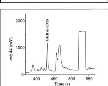
Routine procedures for the quantification of GHB in blood in- volve GC coupled to MS with two different approaches: GHB can be detected after cyclization to GBL in strong acid medium (17) or directly after liquid-liquid extraction (14,18). The liquid-liquid extraction of GHB resulted in no analyzable blood samples by GC-C-IRMS. Satisfying results were obtained by coupling liquid-liquid extraction with a solid-phase method developed for isolation of GHB in urine and blood (19,20). Figure 2 shows a typical example of a GC-C-IRMS chro-
Table I. 813C-Value of GHB di-TMS and GHB for Four Synthetic Standards and Five Seizure Samples (n = 3)*
GHB di.TMS GHB 813C-value/% o SD 813C-value/%~ Standard (Cerilliant) -44.02 0.09 -38.2 + 0.5 Standard (CIL) -44.05 0.08 -38.3 + 0.5 Standard (Lipomed) -48.27 0.10 -48.2 __. 0.5 Standard * (Aldrich) -45.05 0.tl -40.8 + 0.5 Seizure sample 1 -45.09 0.10 -40.9 + 0.5 Seizure sample 2 -48.81 0.10 -50.2 _+ 0.5 Seizure sample 3 -44.13 0.08 -38.5 -+ 0.5 Seizure sample 4 -46.92 0.05 -45.5 _+ 0.5 Seizure sample 5 -44.00 0.09 -38.2 _+ 0.5
* Uncertainty in 613C-values of GHB has been calculated according to a procedure given in the literature (16).
* Synthesized from y-butyrolactone (GBL) according to a published procedure (26).
i , u " 1000 0 400 4,50 500 550 Time (s)
Figure 2. GC-C-IRMS chromatogram (m/z 44) of derivatized GHB after extraction of a blood sample (100 pL) containing GHB at a concentra- tion of 50 mg/L. The square-topped peak represents a pulse of CO2 ref- erence gas.
matogram of GHB extracted from blood. The chromatogram shows symmetrical peaks for the compounds of interest and no tailing. It should be noted that the poorly resolved signals be- tween 455 and 480 s were identified by GC-MS to be urea di- TMS and phosphoric acid tri-TMS. Finally, it is also noteworthy that the presence of GHB in the extracts was verified by GC-MS prior to any GC-C-IRMS analysis.
Carbon isotopic measurements of GHB di-TMS are performed in the linear range of the instrument, and also include a posi- tive quality control. The purpose of the quality control is to verify the reproducibility of the extraction procedure and the GC-C-IRMS measurements. The within-assay precision was determined by extracting six aliquots of the positive quality control and injecting each once within the same day. For the de- termination of the between-assay precision, six aliquots of the positive quality control were extracted over a period of two months, and injected once each. For the within-assay precision, we obtained a mean GHB di-TMS 813C-value of -43.68%o and SD = + 0.22%0 with the values ranging from--43.32 to -43.99%o. The statistics of the between-assay precision were --43.73%0 and 0.43%0 for the mean GHB di-TMS 613C-value and SD, re- spectively, with a13C-values ranging from -42.99 to --44.12%o. These results indicate that the SD obtained for between-assay precision is slightly higher compared to the within-assay pre- cision. Moreover, the mean GHB di-TMS 813C-values of within- assay and between-assay precisions are similar to the GHB di-TMS 81gC-value of the standard from Cerilliant (Table I). These findings indicate a good reproducibility of the method and no significant isotopic fractionation during the extraction and analysis processes.
Discrimination between endogenous and exogenous GHB
To illicitly manufacture GHB, the industrial solvent GBL can be made alkaline with lye and heated; GHB powder is then pro- duced by adding acetone and by drying the mixture (21). GBL is produced in Japan based on the hydrogenation of maleic an- hydride, whereas in the United States and Europe, a major portion of GBL is currently being produced via the dehydro- genation of 1,4-butanediol, which is manufactured by the clas- sical Reppe process based on acetylene and formaldehyde (22). The isotopic signature of GHB will depend on the manufac- turing process and on the carbon feed stocks of the starting ma- terials. Based on the GHB 813C-value of five seizure samples and
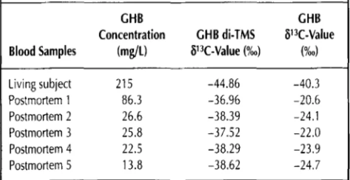
Table II. ~13C-Values of GHB di-TMS and GHB for Five Postmortem Blood Samples and One Blood Sample of a Living Subject Exposed to GHB
GHB GHB
Concentration GHB di-TMS ~13C-Value Blood Samples (mg/t) [~13C-Value (%0) (%0) Living subject 215 -44.86 -40.3 Postmortem 1 86.3 -36,96 -20.6 Postmortem 2 26.6 -38.39 -24.1 Postmortem 3 25.8 -37.52 -22.0 Postmortem 4 22.5 -38.29 -23.9 Postmortem 5 13.8 -38.62 -24.7
four standard (Table I), we may distinguish four isotopic signa- tures. If a manufacturing by the classical Reppe process is hy- pothesized, it is likely that the analyzed GHB samples have arisen from different carbon feed stocks with different isotopic signatures. It should be noted that GC-MS analyses of the seizure samples revealed no traces of GHB precursors (e.g., 1,4-butanediol, maleic anhydride, GBL) and hence did not pro- vide information about the route of synthesis.
Endogenous concentrations of living people may be influ- enced by factors such as biochemical, drug-induced, and dietary effects (23). Endogenous production of GHB results in the brain from the metabolism of GABA with glutamic acid as a pre- cursor, whereas 1,4-butanediol, an endogenous product from fatty acids, may be a source of GHB in peripheral tissues (24). The most likely source of endogenous postmortem GHB pro- duction is putrescine (1,4-butanediamine), which will be sub- sequently converted to GABA by the action of several enzymes (21). Hence, it may be hypothesized that the 13C and 12C content in the natural endogenous form of GHB will depend on the iso- topic carbon composition of the food diet and is influenced by additional effects of human biological processing. For the five postmortem blood samples with different GHB concentrations (range: 13.8-86.3 mg/L), we have obtained GHB di-TMS 813C - values ranging from -36.96 to -38.62%0 (Table II). After cor- rection of the carbon isotopic ratio following Eq. 1, we calculated GHB 813C-values ranging from -20.6 to -24.7%o.
Although more data are needed to have a more descriptive statistic, these findings significantly show differences in the 13C and 12C content of the natural endogenous with the syn- thetic form of GHB. The mean GHB di-TMS 8UC-value of the analyzed postmortem bloods is -37.96%o (n = 5, SD = 0.69%0 and differs by more than 6%o from the synthetic GHB sample having the highest 13C content (Table I). A difference of 6.90%0 is obtained between the mean GHB di-TMS 81:~C-value of the an- alyzed postmortem blood samples and the GHB di-TMS 81.~C - value of the living subject with a GHB concentration of 215 mg/L. Although the GHB concentration is far above the adopted cut-off for antemortem endogenous GHB concentrations in blood, the carbon isotopic measurement clearly reveals the synthetic origin of GHB. When a correction following Eq. 2 is applied, differences in the 813C-values between endogenous and exogenous GHB become more pronounced (A813C-values > 13.5%o).
Conclusions
This method allows the discrimination between synthetic and endogenous GHB in blood samples with large variations of the carbon isotopic ratio. Nevertheless, further studies are still needed to adjust the positive identification threshold. For in- stance, it would be possible that the origin of GHB is both syn- thetic and endogenous in a blood sample with a total GHB concentration of 5 rng/L. To allow a discrimination in this case, a carbon isotopic study of GHB in a reference population of healthy subjects would be needed to determine a threshold value defined by the mean 813C-value - 3SD. Then, the sample
Journal of Analytical Toxicology, Vol. 29, November/December 2005 could be declared positive with exogenous GHB if the carbon isotope value is lower than the threshold value. This proce- dure is applied in the detection of exogenous testosterone in urine by means of GC-C-IRMS (25).
Although this method was successful for the analyses of post- mortem blood samples with GHB concentrations ranging from 13.8 to 86.3 rag/L, further developments in the extraction pro- cess should be undertaken for postmortem blood samples with GHB concentrations lower than 10 mg/L. Indeed, this method requires the use of about 2.5 mL of blood for a sample with a GHB concentration of 2 mg/L. In addition, other developments should be made to analyze GHB in other matrices with the aim of determining if the carbon isotopic ratio of this sub- stance is maintained or if an isotopic discrimination occurs because of the mode of GHB production and absorption into the body.
References
1. P. Vayer, P. Mandel, and M. Maitre. Gamma-hydroxybutyrate, a possible neurotransmitter. Life Sci. 41' 1547-1557 (1987). 2. S. Kleinschmidt, C. Schellhase, and F. Mertzluff. Continuous se-
dation during spinal anaesthesia: gamma-hydroxybutyrate vs. propofol. Eur. J. Anaesthesiol. 16:23-30 (1999).
3. M. Mamelak, M.B. Scharf, and M. Woods. Treatment of narcolepsy with gamma-hydroxybutyrate. A review of clinical and sleep lab- oratory findings. Sleep 9' 285-289 (1986).
4. F. Polodrugo and G. Addolorato. The role of gamma-hydroxybu- tyric acid in the treatment of alcoholism: from animals to clinical studies. Alcohol Alcohol. 34:15-24 (1999).
5. L. Gallimberti, M. Cibin, P. Pagnin, R. Sabbion, P.P. Pani, R. Pirastu, S.D. Ferrara, and G.L. Gessa. Gamma hydroxybutyric acid for the treatment of opiate withdrawal syndrome. Neuropharmacology 9:
77-81 (1993).
6. J. Takahara, S. Yunoki, W. Yakushiji, J. Yamauchi, Y. Yanane, and T. Ofuji. Gamma Stimulatory effects of gamma-hydroxybutyric acid on growth hormone and prolactin release in humans. J. Clin. Endocrinol. Metab. 44:1014-1017 (1977).
7. P.C.A. Kam and F.F.Y. Yoong. Gamma-hyxroxybutyric acid: an emerging recreational drug. Anaesthesia 53:1195-1198 (1998). 8. M.A. LeBeau, R.H. Christenson, B. Levine, W.D. Darwin, and
M.A. Huestis. Intra- and interindividual variations in urinary con- centrations of endogenous gamma-hydroxybutyrate. J. Anal. Tox- icol. 26:340-346 (2002).
9. A.A. Elian. Determination of endogenous gamma-hydroxybutyric acid (GHB) levels in antemortem urine and blood. Forensic Sci. Int.
128:120-122 (2002).
10. S. Elliott. The presence of gamma-hydroxybutyric acid (GHB)in postmortem biological fluids. J. Anal. Toxicol. 25:152 (2001). 11. L.J. Marinetti, D.S. Isenschmid, B.R. Hepler, and R.L. Commissaris.
Response to the presence of gamma-hydroxybutyric acid (GHB) in
postmortem biological fluids. J. Anal Toxicol. 25:356-357 (2001). 12. S.P. Elliott. Further evidence for the presence of GHB in post- mortem biological fluid: implications for the interpretation of find- ings. J. Anal. Toxicol. 28:20-26 (2004).
13. P. Kintz, M. Villain, V. Cirimele, and B. Ludes. GHB in postmortem toxicology: Discrimination between endogenous production from exposure using multiple specimens. Forensic ScL Int. 143:177-181 (2004).
14. M. Villain, V. Cirimele, B. Ludes, and P. Kintz. Ultra-rapid proce- dure to test for/-hydroxybutyric acid in blood and urine by gas chromatography-mass spectrometry. J. Chromatogr. B 792:83-87 (2003).
15. D.M. Jones, J.F. Carter, G. Eglinton, E.J. Jumeau, and C.S. Fen- wick. Determination of ~13 C values of sedimentary straight chain and cyclic alcohols by gas chromatography/isotope ratio mass spectrometry. BioL Mass. Spec. 20:641-646 (1991 ).
16. G. Rieley. Derivatization of organic compounds prior to gas chro- matography-combustion-isotope ratio mass spectrometric analysis: identification of isotope fractionation processes. Analyst 119:
915-919 (1994).
17. Y. Fukui, E. Matsusima, K. Muramoto, N. Nagai, K. Ohama, and K. Yamashita. Validation of a simple gas chromatographic-mass spectrometric method for the determination of gamma-butyrolac- tone in human plasma. J. Chromatogr. B 785:73-80 (2003). 18. A.A. Elian. GC-MS determination of gamma-hydroxybutyric acid
(GHB) in blood. Forensic Sci. Int. 122:43-47 (2001).
19. R.R. McCusker, H. Paget-Wilkes, C.W. Chronister, B.A. Gold- berger, and M.A. EISohly. Analysis of gamma-hydroxybutyrate (GHB) in urine by gas chromatography-mass spectrometry. J. Anal.
Toxicol. 23:301-305 (1999).
20. K.S. Kalasinsky, M.M. Dixon, G.A. Schmunk, and S.J. Kish. Blood, brain, and hair GHB concentrations following fatal ingestion.
J. Forensic. Sci. 46:728-730 (1999).
21. F.J. Couper and L.J. Marinetti./-Hydroxybutyrate (GHB)~effects on human performance and behavior. Forensic. Sci. Rev. 14:101-121 (2002).
22. Y. Hara, H. Kusaka, H. Inagaki, K. Takahashi, and K. Wada. A novel production of y-butyrolactone catalyzed by ruthenium complexes.
J. Catal. 194:188-197 (2000).
23. S.P. Elliott. Gamma hydroxybutyric acid (GHB) concentrations in humans and factors affecting endogenous production. Forensic ScL Int. 133:9-16 (2003).
24. S.D. Ferrara, G. Frison, U Tedeschi, and M. LeBeau. Gamma- hydroxybutyrate (GHB) and related products. In Drug-Facilitated Sexual Assault, a Forensic Handbook, M.A. LeBeau and A. Moza- yani, Eds. Academic Press, San Diego, CA, 2001, pp 107-126. 25. R. Aguilera, T.E. Chapman, B. Starcevic, C.K. Hatton, and
D.H. Catlin. Performance characteristics of a carbon isotope ratio method for detecting doping with testosterone based on urine diols: controls and athletes with elevated testosterone/epitestos- terone ratios. Clin. Chem. 47:292-300 (2001).
26. C.S. Marvel and E.R. Birkhimer. The preparation of the sodium salts of omega-hydroxybutyric, -valeric and -caproic acids. J. Am. Chem. Soc. 51:260-262 (1929).
Manuscript received July 16, 2004; revision received January 17, 2005.