TCR–MHC class II interaction is required for
peripheral expansion of CD4 cells in a T
cell-deficient host
Ulrich Beutner
1and
H. Robson MacDonald
Ludwig Institute for Cancer Research, Lausanne Branch, University of Lausanne, Ch. des Boveresses 155, 1066 Epalinges, Switzerland
1Present address: Chirurgische Universita¨tsklinik und -Poliklinik, Josef-Schneider-Strasse 2, 97080
Wu¨rzburg, Germany
Keywords: CD4 cell reconstitution, knockout mice, peripheral expansion, TCR–MHC interaction
Abstract
It is well established that T cell-deficient nude and SCID mice can be reconstituted by i.v. injection of small numbers of purified peripheral CD4FT cells; however, the requirements for expansion of the transferred T cells in such systems are not clear. We show here that blood and lymphoid organs of MHC class II-deficient mice (which selectively lack mature CD4FT cells) cannot be reconstituted by transfer of purified splenic CD4FT cells, whereas TCRα-deficient mice (which lack both CD4Fand CD8Fmature T cells) are readily reconstituted. The failure of CD4FT cell
reconstitution in MHC class II-deficient mice was not due to the presence of CD8FT cells, since similar results were obtained in TCRα–MHC class II double-deficient mice. Consistent with most previous studies CD4FT cells in reconstituted TCRα-deficient mice had a diverse TCR Vβ repertoire and were predominantly of an activated/memory (CD44high) phenotype. Collectively our
data demonstrate that the expansion of peripheral CD4FT cells in a T cell-deficient host is dependent upon interactions of the TCR with MHC class II.
Introduction
T cells found in the periphery of humans or mice have an enormous potential of expansion and self renewal. If the thymus, the source of newly developing T cells, is removed after puberty in man or only.2 days after birth in mice, the size of the peripheral T cell pool stays constant, shows diversity and can respond to environmental antigens (reviewed in 1). Very little is known about how this peripheral T cell homeostasis is maintained in the absence of T cell emigration from the thymus. The classical experiments to analyze the repopulation potential of peripheral T cells involved reconstitution of T cell-deficient nude or lymphocyte-deficient SCID mice with total splenocytes or sorted CD41T cells (reviewed in 2,3). These experiments showed that small numbers of CD41T cells (102to 103sorted cells) were already
sufficient to repopulate the periphery. Nevertheless, the CD41 T cell repertoire (judged by the expression of various Vβ genes) was quite diverse and similar to the distribution in the donor mice. Thus, the expansion of transferred peripheral CD41T cells is polyclonal and presumably some regulatory mechanism is responsible for maintaining a broadly distrib-uted T cell repertoire.
Correspondence to: H. R. MacDonald
Transmitting editor: T. Hu¨nig Received 28 August 1997, accepted 12 November 1997
One obvious candidate for such a regulatory system would be the MHC molecules of the recipient. According to such a scenario peripheral CD41T cells with a certain minimal TCR affinity for self MHC class II molecules would be allowed to survive and/or proliferate following transfer, whereas T cells with affinities below this threshold would die. In essence, peripheral T cells would undergo a process similar to the MHC-mediated positive selection mechanism operating in the thymus.
To test this hypothesis we used the classical approach of reconstituting immunodeficient mice with sorted CD41splenic T cells. However, instead of nude or SCID recipients, we used the better defined TCRα- and MHC class II-deficient mice derived by gene targeted mutation. As expected T cell-deficient TCRα–/– mice could be easily reconstituted with
peripheral CD41T cells. However MHC class II-deficient mice (which lack CD41 T cells in the periphery) could not be reconstituted. To exclude a regulatory role of CD81 T cells present in the MHC class II-deficient mice we generated TCRα-deficient mice which do not express MHC class II. No
306 MHC class II-dependent expansion of peripheral CD41T cells
significant CD41 T cell reconstitution was observed in these ‘double-deficient’ mice, indicating a critical role for TCR–MHC class II interactions in this model system.
Methods
Mice
MHC class II-deficient (MHC II–/–) mice were obtained from RCC-BRL (Fu¨llinsdorf, Switzerland). These mice were gener-ated on the genetic background of C57BL/6 mice which do not express MHC class II E molecules. The MHC class II I-Aa chain gene was inactivated by gene targeted mutation leading to a total loss of MHC class II expression in these mice (4). TCRα-deficient (TCRα–/–) mice (backcrossed 12
generations to C57BL/6) (5) and B6.PL-Thy-1a/Cy (Thy-1.11) mice were obtained from the Jackson Laboratories (Bar Harbor, ME). MHC II–/– female mice were crossed with a TCRα–/– mouse and the offspring was inbred to generate
MHC II–/– TCRα–/– double-deficient mice. Double-deficient
mice were identified either by PCR or by FACS analysis of peripheral blood lymphocytes (PBL) using anti-MHC class II (M5/114; Boehringer Mannheim, Mannheim, Germany) and anti-CD4 (H129.19; Boehringer) antibodies. All mice were kept under specific pathogen-free conditions.
Cell transfer
One or two spleens from male B6.PL (Thy-1.1) mice were homogenized through a steel mesh and red blood cells were removed by hypoosmotic shock. Cells were incubated in 0.5–1.0 ml cold PBS containing 5% FCS and anti-CD4– phycoerythrin (PE) (Boehringer) for 30 min. Cells were washed twice and diluted to 23106cells/ml, before CD41T cells were
sorted using a FACStar Plus (Becton Dickinson, San Jose, CA). Purity of the sorted cells was.98%. Sorted cells were washed in PBS and resuspended to 107cells/ml. Typically 0.5–1.03106cells were injected in the tail vein of male mice.
Blood analysis
Every week after the transfer 3–4 drops of blood were collected from the tail vein into an Eppendorf tube containing 3µl of 0.5 M EDTA. Then 50µl of blood was transferred into a fresh tube containing Thy-1.1–FITC (HO-22.1.1) (6) and anti-CD4–PE in a total volume of 3–5µl PBS. Blood was incubated for 30 min at room temperature with the antibodies before it was transferred into 0.5 ml FACS lysing solution (Becton Dickinson). After 2–6 min incubation the cells were pelleted and washed in 1 ml PBS with 5% FCS. Cells were resuspended in 200µl PBS/FCS and analyzed using a FACScan (Becton Dickinson). For each sample 40,000 events in a FSC/SSC lymphocyte gate were acquired. Data were analyzed using the program WinMDI (7).
Spleen and lymph node (LN) analysis
Spleen and LN (mesenteric LN or a pool of two inguinal and two submaxillary LN) were homogenized through a steel mesh. Red blood cells were removed from the splenocytes by osmotic shock. Viable cells were counted using a hemocy-tometer and ~106cells were used for each sample of antibody labeling. For the determination of reconstitution cells were
incubated with anti-Thy-1.1–FITC (HO-22.1.1) and anti-CD4– PE for 30 min on ice, washed and analyzed on a FACScan. For the analysis of Vβ repertoire and CD44 expression, splenocytes were incubated with biotin conjugates of the following antibodies: anti-Vβ5 (MR9-4), anti-Vβ6 (44-22.1), anti-Vβ8 (F23.1), anti-Vβ10 (B21.5) and anti-CD44 (IM7.8.1). Samples were then washed and incubated with anti-Thy-1.1– FITC, streptavidin–PE (Caltag, San Francisco, CA) and anti-CD4–Red613 (Gibco/BRL, Basel, Switzerland). If not other-wise indicated, antibodies were prepared and conjugated at the Ludwig Institute. Three-color analysis was performed on a FACScan (Becton Dickinson) and data were analyzed using WinMDI (7).
Results
T cell-deficient but not MHC class II-deficient mice can be reconstituted with CD4 T cells
Many previous reports have described the reconstitution of nude or SCID mice with peripheral T cells or total splenocytes (8–11). These immunodeficient mice were derived from spon-taneously occurring mutations leading to the loss of peripheral T cells in the case of the nude mice, and the lack of B and T cells in the SCID mouse. However, both nude and SCID mice are considered ‘leaky’ as small numbers of lymphocytes can still be generated, a fact which potentially complicates reconstitution experiments (12). Therefore, we chose T cell-deficient (TCRα–/–) mice generated by a gene-targeted
muta-tion of the TCRα constant region (5) for our reconstitution experiments. No αβ TCR can be expressed in these mice leading to the absence of conventional CD4 and CD8 T cells, whileγδT cells and B cells are still present.
TCRα–/– mice were reconstituted by i.v. injection of 0.5– 1.03106FACS sorted CD41 splenocytes from B6.PL-Thy-1a
mice. The TCRα–/– mice as well as all other recipient mice used in the following experiments express the Thy-1.2 allele on T cells. The donor CD4 T cells, however, express the Thy-1.1 allele, allowing easy detection of donor-derived cells.
Already 1 week after injection reconstitution of CD41T cells in the blood could be observed, reaching a plateau at ~4–5 weeks (Figs 1 and 2). The percentage of CD41 T cells in the blood of the reconstituted mice never reached values observed in normal C57BL/6 mice; however, similar observa-tions have been made in several previous reports using nude or SCID mice as recipients (8–11). Similar to these earlier reports we also found reconstitution of other lymphatic organs (Fig. 3). Although also reported earlier it is worth mentioning that reconstitution of ‘peripheral’ (inguinal, submaxillary) LN was much less efficient than of mesenteric LN. (Fig. 3). Thus, TCRα–/–mice can efficiently be reconstituted with peripheral
CD4 T cells and will serve as a positive control for the following experiments.
MHC class II-deficient (MHC II–/–) mice have very few CD41 T cells due to a selective block in positive selection (4,13,14). The small number of CD41 cells that can still be found in these mice are selected on MHC class I or CD1 and express a lower level of CD4 than conventional CD41T cells (Fig. 1) (15). Thus the CD41 T cell compartment in these mice is nearly empty and one should be able to reconstitute MHC
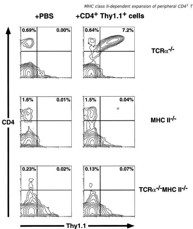
Fig. 1.CD41T cell reconstitution of TCRα–/–mice is MHC class II dependent. Shown are FACS contour plots of PBL (labeled with anti-Thy-1.1–FITC and anti-CD4–PE) from mice 4 weeks after injection of Thy-1.11CD41T cells (right panels) or PBS (left panels). The type of recipient is indicated on the right-hand side. Threshold for the contour plots was 0%.
II–/–mice with CD41T cells if this expansion is independent
of MHC class II expression.
However, only trace numbers of CD41 Thy-1.11 donor-derived T cells (0.04%) could be found in the blood of reconstituted MHC II–/– mice in the first few weeks after
reconstitution (Fig. 1). These numbers were slightly, but con-sistently, above the background staining values of PBS injected littermates (0.01%). At later time points (.6 weeks) donor-derived T cells were virtually undetectable. In the spleen and LN the number of donor-derived CD41T cells was also barely over the background values found in littermates injected with PBS (Fig. 3). Furthermore, donor-derived CD41
T cells could not be detected in the thymus, bone marrow, gut epithelium, lamina propria or liver of MHC II–/–mice (data not shown).
CD41 T cells in reconstituted TCRα–/–mice have a random TCR Vβrepertoire and are CD44hi
Previous studies of CD4 T cell reconstitution of nude and SCID mice have shown that reconstitution does not significantly bias the TCR Vβ repertoire (16,17). As shown in Fig. 4, donor-derived splenic CD4 T cells in reconstituted TCRα–/– mice
likewise exhibited an apparently random Vβ repertoire, although variations in Vβexpression between individual mice
308 MHC class II-dependent expansion of peripheral CD41T cells
Fig. 2. Kinetics of CD41 T cell reconstitution. The percentage of donor-derived CD41T cells was determined by flow cytometry in the blood of immunodeficient mice at various times after reconstitution (see Fig. 1). In all cases background values (usually ,0.03%) of PBS injected littermates were subtracted. Data points represent the averages of nine TCRα–/–, seven TCRα–/–MHC II–/–and five MHC II–/– mice (except for the last two data points of the TCRα–/–mice, which are the averages of three mice) from two independent experiments. Error bars indicate SD.
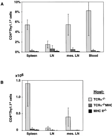
Fig. 3.CD41T cell reconstitution of peripheral lymphoid organs. The percentage of donor-derived T cells in peripheral lymphatic organs was determined by flow cytometry 3–5 weeks after CD41 T cell reconstitution. All values are corrected for the background values found in PBS injected control animals. Histograms represent the averages of six TCRα–/–, five TCRα–/–MHC II–/–and three MHC II–/– mice; error bars indicate SD. LN represents a pool of two inguinal and two submaxillary LN, mes. LN represents the mesenteric LN. (A) The percentage of donor-derived CD41T cells among lymphocytes (determined by a FSC/SSC gate). (B) The absolute number of donor-derived T cells in these organs.
Fig. 4.TCR Vβrepertoire and CD44 phenotype of donor-derived T cells in reconstituted TCRα–/–mice. (A) TCR V
βusage was analyzed among CD41splenocytes in control mice and among CD41Thy-1.11 splenocytes of CD4 T cell reconstituted TCRα–/–mice. Histograms represent the average percentage of cells positive for a given TCR Vβamong CD41 T cells; error bars indicate SD. The control group consisted of one C57BL/6, one B6.PL and three (MHC II–/–3 TCRα–/–)F
1 mice. (B) The percentage of CD44hiCD41T cells was determined as in (A).
(assessed by the magnitude of the standard derivation) were somewhat greater than in normal controls. These data argue against any major selective expansion of the transferred CD4 T cells driven by environmental antigens or pathogens, which might be expected to significantly alter the TCR repertoire.
CD44 is a phenotypic marker that is up-regulated on activated and/or memory T cells (18). Reconstitution of nude or SCID mice with low numbers of CD4 T cells leads to accumulation of cells with an activated/memory phenotype in lymphoid organs (reviewed in 19). Figure 4 shows that, in contrast to the original donor cell inoculum, most splenic CD4 T cells present in reconstituted TCRα–/–mice have a CD44hi phenotype, consistent with the fact that considerable CD4 T cell expansion has occurred.
TCRα and MHC class II double-deficient mice cannot be reconstituted with CD4 T cells
The failure to reconstitute MHC II–/–mice with peripheral CD4
T cells suggests that a TCR–MHC class II interaction is required for their expansion. However, it remains formally possible that MHC II–/– mice could not be reconstituted because the large number of CD81 T cells found in these mice compete out the small number of transferred CD41 T cells or even directly suppress their expansion. To exclude this possibility we generated TCRαand MHC class II double-deficient (TCRα–/– MHC II–/–) recipient mice which have no
CD81 T cells. TCRα–/–MHC II–/–as well as control TCRα–/–
and MHC II–/–mice were injected with the same preparation of Thy-1.11CD41splenocytes. Four weeks after reconstitution only extremely small numbers (0.07%) of donor-derived T cells could be found in the blood of TCRα–/–MHC II–/–mice,
barely above the background level of PBS-injected control mice (Fig. 1) and similar to the MHC II–/–mice. On the other
hand, the blood of MHC class II1, TCRα–/–mice contained
4–10% of donor-derived, Thy-1.11cells (Fig. 1). Thus, recon-stitution of TCRα–/– mice with peripheral CD41 T cells is at
least 100-fold less efficient in the absence of MHC class II expression.
When we analyzed the reconstituted mice at various time points the reconstitution kinetics of the TCRα–/–MHC II–/–mice were essentially similar to that of MHC II–/–mice, even though
reconstitution of the TCRα–/–MHC II–/– mice was marginally
better (Fig. 2). Still at any given time point the reconstitution in the TCRα–/–MHC II–/–mice was at least 10-fold less efficient
than in the MHC class II1,TCRα–/–mice. Only after extended periods of time (.60 days) could some significant reconstitu-tion (.1%) of CD41T cells in the TCRα–/–MHC II–/–mice but not of MHC II–/–mice be observed (data not shown).
Similar to our results obtained from blood, we could not detect significant numbers of donor-derived CD41T cells in the spleen, peripheral LN or mesenteric LN of the TCRα–/–
MHC II–/–mice 3–5 weeks after reconstitution (Fig. 3). While the difference in the percentage of donor-derived cells between TCRα–/–mice and TCRα–/–MHC II–/–mice seems to be less in LN and spleen than in blood, the difference in total numbers is actually much greater (Fig. 3). LN of TCRα–/–MHC II–/–mice were extremely small, while the LN of reconstituted TCRα–/–
mice had approximately normal sizes. The size of the spleen in the various mice did not show any obvious differences, but the cell number in the spleen of the TCRα–/–MHC II–/–mice
was much lower than in MHC class II1TCRα–/–mice.
Discussion
Taken together our data demonstrate formally that the expan-sion of normal peripheral CD4 cells transferred to T cell-deficient hosts requires MHC class II expression. This in turn implies that direct TCR–MHC class II interactions are required for expansion and self-renewal of the CD41T cell pool. This conclusion confirms and extends an earlier study by Mackall
et al. (20) showing that CD4 T cell reconstitution of
thymectom-ized irradiated mice (protected by a syngeneic bone marrow graft) could be partially (~ 50%) inhibited by simultaneous treatment with anti-MHC class II mAb. On the other hand, Takeda et al. (21) have recently reported that MHC II–/–
RAG2–/–mice engrafted with fetal thymus from wild-type mice
develop significant numbers of peripheral CD4 T cells that initially proliferate but ultimately disappear over time. These apparently conflicting findings could be related to differences in the experimental models utilized. Alternatively it is possible that CD41 recent thymus emigrants are less dependent on TCR–MHC class II interactions for their survival and/or expansion than fully mature peripheral CD4 cells.
The MHC class II-expressing cell(s) required for successful peripheral CD4 T cell reconstitution of TCRα–/–mice has not
been identified. In this respect MHC class II1 B cells are unlikely to be involved, since CD4 T cell reconstitution of B cell-deficient SCID (2,3) and RAG1–/–mice (data not shown) has been observed. Rather we would speculate that MHC class II1APC (such as dendritic cells and/or macrophages) are necessary for the expansion of transferred CD4 T cells in T cell-deficient mice.
In contrast to MHC II–/–recipients, TCRα–/–MHC II–/–
double-deficient mice could be reconstituted with small but significant numbers of donor CD41 T cells (~ 1%) at later times. It is possible that these cells are derived from small numbers of MHC class I- or CD1-restricted CD41 T cells originally described in MHC class II-deficient mice (15). However, these
cells did not express NK1.1 (data not shown), a marker expressed by many non-MHC class II-restricted CD4 T cells (22). In the MHC II–/–mice this expansion might not occur as
the endogenous MHC class I- or CD1-restricted CD41T cells would compete out the small number of such cells found in the sorted donor inoculum.
Finally, an important unresolved issue arising from our study is whether self or foreign peptides are responsible for the MHC class II-dependent expansion of peripheral CD4 T cells in TCRα–/–mice. Although our reconstituted mice have been
maintained under specific pathogen-free conditions for the duration of the analysis and show no pronounced Vβ bias, some degree of contamination by environmental pathogens cannot be formally excluded. In this context several previous reports in which TCR transgenic CD4 or CD8 T cells were transferred to naive (MHC sufficient) hosts in the presence or absence of nominal antigen have suggested that peripheral T cell expansion is strictly dependent upon TCR recognition of MHC–antigen complexes (20,23). On the other hand, a very recent study by Tanchot et al. (24) has demonstrated that monoclonal TCR transgenic CD8 cells with a memory phenotype can actually expand in vivo as a consequence of self MHC class I recognition in the absence of nominal antigen. In contrast, naive phenotype TCR transgenic CD8 T cells could only expand upon encountering self MHC class I plus antigen, although they could survive in the presence of self MHC alone. By analogy with these transgenic experi-ments, it could be speculated that the MHC class II-dependent expansion of normal CD41 T cells transferred into immuno-deficient hosts reflects the selective proliferation of memory cells interacting with self peptides presented by MHC class II molecules. Alternatively exposure to MHC class II-associated ‘foreign’ peptides derived from gut-associated microflora or food antigens (which would still be present in specific pathogen-free mice) may be necessary to sustain the expan-sion of the transferred CD41T cells.
Acknowledgements
We thank Horst Bluethmann for providing MHC class II-deficient mice, Anne-Lise Peitrequin for antibody preparation, Pierre Zaech for cell sorting and Anna Zoppi for assistance with the manuscript.
Abbreviations
MHC II–/– MHC class II deficient
LN lymph node
PBL peripheral blood lymphocyte
PE phycoerythrin
TCRα–/– TCRαdeficient
References
1 Sprent, J. and Tough, D. 1994. Lymphocyte life-span and memory. Science 265:1395.
2 Phillips, R. A. and Reimann, J. 1994. Models of lymphoid and myeloid reconstruction in SCID mice. Res. Immunol. 145:323. 3 Freitas, A. A. and Rocha, B. 1993. Lymphocyte lifespans:
homeostasis, selection and competition. Immunol. Today 14:25. 4 Ko¨ntgen, F., Su¨ss, G., Stewart, C., Steinmetz, M. and Bluethmann,
H. 1993. Targeted disruption of the MHC class II Aa gene in C57BL/6 mice. Int. Immunol. 5:957.
310 MHC class II-dependent expansion of peripheral CD41T cells Lafaille, J., Wang, L., Ichikawa, Y., Jaenisch, R., Hooper, M., et al. 1992. Mutations in T-cell antigen receptor genes alpha and beta block thymocyte development at different stages. Nature 360:225. 6 Marshak-Rothstein, A., Fink, P., Gridley, T., Raulet, D., Bevan, M. and Gefter, M. 1979. Properties and applications of monoclonal antibodies directed against determinants of the Thy-1 locus. J. Immunol. 122:2491.
7 Trotter, J. 1996. WinMDI: URL: http://facs.scripps.edu/.
8 Miller, R. and Stutman, O. 1984. T cell repopulation from functionally restricted splenic progenitors: 10,000-fold expansion documented by using limiting dilution analyses. J. Immunol. 133:2925.
9 Rocha, B., Dautigny, N. and Pereira, P. 1989. Peripheral T lymphocytes: expansion potential and homeostatic regulation of pool sizes and CD4/CD8 ratios in vivo. Eur. J. Immunol. 19:905. 10 Sprent, J., Schaefer, M., Hurd, M., Surh, C. and Ron, Y. 1991.
Mature murine B and T cells transferred to SCID mice can survive indefinitely and many maintain a virgin phenotype. J. Exp. Med. 174:717.
11 Reimann, J., Rudolphi, A. and Claesson, M. 1994. Reconstitution of SCID mice with low numbers of CD41TCRαβ1T cells. Res. Immunol. 145:332.
12 Carroll, A. and Bosma, M. 1988. Detection and characterization of functional T cells in mice with severe combined immune deficiency. Eur. J. Immunol. 18:1965.
13 Grusby, M. J., Johnson, R. S., Papaioannou, V. E. and Glimcher, L. H.. 1991. Depletion of CD41T cells in major histocompatibility complex class II-deficient mice. Science 253:1417.
14 Gosgrove, D., Gray, D., Dierich, A., Kaufman, J., Lemeur, M., Benoist, C. and Mathis, D. 1991. Mice lacking MHC class II molecules. Cell 66:1051.
15 Cardell, S., Tangri, S., Chan, S., Kronenberg, M., Benoist, C. and Mathis, D. 1995. CD1-restricted CD41 T cells in major
histocompatibility complex class II-deficient mice. J. Exp. Med. 182:993.
16 Pereira, P. and Rocha, B. 1991. Post- thymic in vivo expansion of matureαβT cells. Int. Immunol. 3:1077.
17 Reimann, J., Rudolphi, A., Tscherning, T. and Claesson, M. H. 1993. Selective engraftment of memory CD41 T cells with an unusual recirculation pattern and a diverse T cell receptor-Vβ repertoire into scid mice. Eur. J. Immunol. 23:350.
18 Budd, R. C., Cerottini, J.-C., Horvath, C., Bron, C., Pedrazzini, T., Howe, R. C. and MacDonald, H. R. 1987. Distinction of virgin and memory T lymphocytes. Stable acquisition of the Pgp-1 glycoprotein concomitant with antigenic stimulation. J. Immunol. 138:3120.
19 Mackall, C. L., Hakim, F. T. and Gress, R. E. 1997. T-cell regeneration: all repertoires are not created equal. Immunol. Today 18:245.
20 Mackall, C. L., Bare, C. V., Granger, L. A., Sharrow, S. O., Titus, J. A. and Gress, R. E. 1996. Thymic-independent T cell regeneration occurs via antigen-driven expansion of peripheral T cells resulting in a repertoire that is limited in diversity and prone to skewing. J. Immunol. 156:4609.
21 Takeda, S., Rodewald, H. R., Arakawa, H., Bluethmann, H. and Shimizu, T. 1996. MHC class II molecules are not required for survival of newly generated CD41 T cells, but affect their long-term life span. Immunity 5:217.
22 Bendelac, A., Rivera, M. N., Park, S.-H. and Roark, J. H. 1997. Mouse CD1-specific NK1 T cells: development, specificity and function. Annu. Rev. Immunol. 15:535.
23 Rocha, B. and von Boehmer, H. 1991. Peripheral selection of the T cell repertoire. Science 251:1225.
24 Tanchot, C., Lemonnier, F. A., Pe´rarnau, B., Freitas, A. A. and Rocha, B. 1997. Differential requirements for survival and proliferation of CD8 naive or memory T cells. Science 276:2057.