The Journal of
Laryngology and Otology
(Founded in 1887 by MORELL MACKENZIE and N O R R I S WOLFENDEN)
March 1938
ON BONE FORMATION IN THE SCALA
TYMPANI OF OTOSCLEROTICS*
By F. R. NAGER (Zurich), and the late J. S. FRASER (Edinburgh)
IT is generally known and accepted that in otosclerosis the main changes concern the labyrinthine capsule, whereas as a rule the inner ear shows only minor alterations. On histo-logical examination of large numbers of otosclerotic petrous bones there are, however, some rare cases which show more or less extensive bone formation in the scala tympani.
This paper deals with such observations only. They are not quite unknown, because in the very early descriptions of this disease by Politzer1 1894, Habermann2 1904, Siebenmann3 1911, such sporadic cases are mentioned. Siebenmann pointed out that these enostotic formations might be due to irritation of the otosclerotic labyrinthine wall. In his monograph on otosclerosis Otto Mayer* gave a description of all the patho-logical changes in the inner ear. Some of them may be due to the bone process itself and to the troubles of blood- and lymph-circulation or to the obliteration of the cochlear aque-duct. But among his cases there was one with bone formation in the scala tympani. The last observation of this kind was published by Lange5 in 1921, who found the scala tympani partially filled up with newly formed bone tissue into which the otosclerotic process of the wall penetrated. Kosokabe6
* The preparation of Dr. Fraser's specimens included in this paper was carried out at the Laboratory of the Royal College of Physicians, Edinburgh. Grateful acknowledgment is also made to the Carnegie Trust for a generous grant received to defray the expenses of illustration.
F. R. Nager and the late J. S. Fraser
1912 re-examined Siebenmann's case and gave a very extensive description of the alterations.
For the last few years our attention has been focused upon this special form of otosclerotic alterations, and among the collection of Edinburgh, Wiirzburg and Zurich the following cases were found.
CASE I.* Mrs. C, aged 61, from Canada. The deafness began
as a girl at school and she became totally deaf at the age of 30. She had had tinnitus all the time since the deafness began but the tinnitus did not worry her much till the menopause, when she had four years of severe tinnitus. After that she was quite happy for ten years but, two years ago, the noises started to get worse and became hardly bearable. They were like thunder.
At one time the patient heard better in a noisy place. She has an aunt who is very deaf and two brothers are deaf. The patient had had operations on her ears in Canada, through the external meatus. She was not a lip-reader and her husband and brother conversed with her by finger-spelling.
The patient had been having morphia in order to make her life endurable. Examination showed that the stump of the right malleus is still present and there is a large perforation in the lower part of the drumhead ; it is practically dry.
On the left side most of the drumhead has also been removed and again there is practically no discharge, just a little moisture on the inner wall.
The patient does not hear the C.256 or C.2048 forks by bone conduction. She has practically lost the power of speech—this is peculiar as she became quite deaf only at the age of 30.
11.5.32. Examined by the general surgeon. No spontaneous nystagmus except on very extreme deviation to right and to left; no spontaneous pointing error. Dundas Grant's cold air apparatus used—right ear first and after a considerable period there was vertical nystagmus to the left; the patient felt giddy and there was well marked pointing error to the right. On the left side it took much longer to produce the nystagmus and pointing error, but possibly the apparatus was not so cold as when used on the right side.
Mr. Dott and J.S. Fraser were quite satisfied that the vestibular apparatus was acting on both sides.
15.5.32. Patient operated upon under general anaesthesia, by removal of the occipital bone following the level of the superior curved line. The dura was very adherent. The cerebellum was displaced first to the left and later to the right and the Vlllth nerve
* During his illness the late John S. Fraser was very anxious that this rare observation should be published. We therefore give it in considerable detail.
O F OTOSCI.KHOTICS—!•' . R . NA(;I-: K AN D THI< : I.AT E J . S . l rRASi: n FIG . I . Cas e I (Dr . Eraser's) . Mrs . C . Horizonta l sectio n righ t ear . 1 4 x . Otosclerosi s o f th e promontoria l an d stapedia l region , moreove r o f th e tympani c scal a wit h it s surroundings . St=stapes , S.T . = scal a tympani , M = interna l meatu s containin g bloo d afte r th e operation . S=sacculus . [face p. 17 4
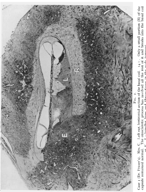
FIG . 2 . Cas e i (Dr . Eraser's) . Mrs . C . Lef t ear , horizonta l sectio n o f th e basa l coil . 1 4 x . Onl y a smal l portio n (R ) o f th e capsul e remaine d normal . Th e otoscleroti c proces s ha s involve d al l th e res t o f i t an d break s int o th e basa l coi l a t K . L = lamella r bon e an d fibrous-tissu e i n th e scal a tympani .
Bone Formation in the Scala Tympani
divided at the internal acoustic meatus, the Vllth nerve being held out of the way ; this was done on both sides.
16.5.32. Patient was very sick (noises still present). 18.5.32. Patient died, apparently of pneumonia.
Copy of the Surgeon's Notes.—•" Her operation, though not a particularly easy one, had no serious complications of any sort, in fact blood pressure at the outset was 114 and its termination the same figure. Each acoustic nerve was severed and each facial nerve preserved.
" The immediate post-operative recovery was quite satisfactory though persistent vomiting was a troublesome factor. The circula-tion after operacircula-tion showed progressive failure. I think both the persisting vomiting and poor myocardium were probably dependent on her unsatisfactory general condition, due to the prolonged use of morphia and other powerful sedatives. She recovered so well in a general way as to be able to write quite freely to tell us that the noises had gone and we were able to verify the fact that there was no trace of facial weakness. Death was due to progressive myocardial failure, aggravated I think by vomiting."
Histological Examination.—On both sides (Figs. 1 and 2) the greater part of the labyrinthine capsule is altered. Instead of three normal layers we find a very extended otosclerotic disease of the bone. This includes both windows which are completely closed. The foot-plate of the stapes is very much thickened and deformed and so is the labyrinthine wall which shows exostotic protuberances under the mucous membrane of the middle ear. The changes of the otic capsule lead even to a certain alteration of the labyrinthine hollow spaces. The superior coils of the cochlea and the vestibulum are flattened. By large otosclerotic areas in the wall of the internal meatus its lumen is narrowed. The diseased bone is mostly sclerotic, some areas are fibrous with numerous blood vessels. The bone process is therefore, in the greater part of the labyrinthine capsule, not very active. In some few regions the cellular and fibrous bone marrow with numerous osteoclasts and blood vessels prove a greater activity of the disease. The most striking alteration, however, is in the basal coil of the cochlea on both sides. The scala tympani is filled up with bone and fibrous tissue from the closed window to the end of the turn. The bone consists of lamellar as well as otosclerotic bone. It is easy to observe that the lamellar bone masses are higher up in the coil and cover in a certain way the otosclerotic tissue which enters from a big area of the round window-niche into the scala tympani. The endosteal layer which usually forms a certain limit, is destroyed, so that the otosclerotic bone tissue penetrates the cochlea unrestrainedly. The fibrous tissue fills up the remainder of the scala tympani. The border line between the otosclerotic areas and the endosteal layer of the
F. R. Nager and the late J . S. Fraser
labyrinthine capsule were examined with special care. With the exception of the basal coil, where the bone formation has been described, only the connective tissue of the spiral ligament showed a thickening of its fibres, even with some bony tissue on the marginal portions, whereas the central parts were atrophic. The cochlear duct is well preserved on both sides, the vestibular membrane is not dislocated. The whole endolymphatic and perilymphatic spaces contain a granular exudation, probably due to the section of the nerve and the arteria auditiva. The details of the nervous elements and the cells of Corti's organ are therefore not very clear. But on the left side there is undoubtedly a distinct atrophy of Corti's organ and the ganglion cells in the modiolus, whereas on the right side the shape of Corti's organ and the external sulcus seem still well preserved. The stria vascularis is swollen and hypertrophic, as is seen in acute inflammations of the labyrinth. Also the macula sacculi shows very queer alterations, a sort of swelling of the epithelial layer mixed with a granular exudate. The ganglion cells in the modiolus are more atrophic on the left side. The auditory nerve is not well stained and is permeated with blood which also fills the internal meatus. The section at the operation passes through the glial part of the nerve, the nerve fibres being destroyed for a long distance.
CASE II. Miss N., aged 43, was very deaf and feeble-minded since childhood. Moreover, she had many symptoms of endemic cretinism. Her deafness had to be considered as an endemic -cretinic one. She was totally deaf on the left side. On the right side there was a remnant of hearing from e2 to a4. No vestibular troubles. The histological changes are given in detail in
Nager-Meyer, Die Erkrankungen des Knochensystems, Berlin, 1927, p. 182.
The typical signs of endemic deafness are present: hyperostosis of the promontory, narrowness of both window-niches, deformity of the stapes which is adherent to the facial canal. Moreover, the patient suffered from bilateral otosclerosis, more extended on the left side. Several areas are on the promontory, on both windows and in the internal meatus. On the left (deaf) side the scala tympani contains newly-formed bone, starting from the endosteal layer of the labyrin-thine capsule. It consists of lamellar bone into which the oto-sclerotic process penetrates (Fig. 3). The lower part of the scala tympani is almost filled with bone and some connective tissue, but without any signs of inflammation. The region of the cochlear aqueduct and cochlear veins is occupied by otosclerotic process but even at close examination no signs of old inflammatory process can be found in the right labyrinth.
CASE III. Miss M., aged 69, was very deaf, had only sound-perception and a remnant of hearing on the right side from e2 to f5,
O N BON K I'OKMATIO N I N TH K SCAL A TVMI'AN I O F OTOSCI.KKOTICS-- OTOSCI.KKOTICS--V. K . NAOF U AN D TII K I.AT K J . S . I'KASK K FIG . 3 . Cas e II . Mrs . N . Inferio r (tympanic ) en d o f th e basa l coil . 2 3 X . Otoscleroti c area s (O ) i n th e capsule , o n th e roun d window . B = new-forme d bon e i n th e tympani c scala . MR=membran e o f th e roun d window . N R = nich e o f th e roun d window . [face p. 17 6
FIG . 5 . Cas e IV . Mrs . N . Wit h th e exceptio n o f a smal l are a (A ) th e whol e capsul e ha s undergon e th e otoscleroti c proces s whic h a t B break s i n th e tympani c scala . Th e res t o f th e scal a tympan i i s filled wit h lamella r bon e = L . 2 3 x .
fc.
FIG. 6.
Case V. Mr. \V. Vertical section of the tympanic wall of the labyrinth including both windows and the stapes St. 23 X . Diffuse otosclerotic changes of the whole region. The niche of the round window is filled with otosclerotic bone, which enters
Bone Formation in the Scala Tympani
the vestibular reaction being normal.* The otosclerotic areas are very extensive, involving the promontory, both windows, and the stapes. The scala tympani of the left side contains newly formed bone and connective tissue (Fig. 4) (not in the lowest part of the basal coil, but on some distance of the round window, the membrane of which is normal). In this case the newly-formed bone is also attached to a large otosclerotic focus of the labyrinthine capsule between cochlea and vestibule. On both sides the inner ear is free from inflammatory marks.
CASE IV. Mrs. N., aged 644 Large extension of the oto-sclerotic changes in the capsule and the stapes, both windows being totally closed. The scala tympani at the basal turn is filled up with lamellar and otosclerotic bone tissue which extends even to the spiral ligament. The region of the aqueduct and of the cochlear vein is occupied by otosclerotic bone tissue (Fig. 5). Moreover, the ganglion cells and the nerve fibres are atrophic. With the exception of a darker staining of the peri- and endolymph, signs of inflammation are missing in both labyrinths.
CASE V.J Mr. W., aged 25. (A coloured plate of this case has been appended to the paper of Cleminson (Proceedings of the Royal
Society of Medicine, 1927, xx, Sect, of Otology, 13-16).) The patient
was completely deaf on the left side and had a very small remainder of hearing from d1 to a2 on the right side. The vestibular reactions were normal. He suffered from blue sclerotics, but there was no distinct fragilitas ossium. The otosclerotic changes are very extended, including the region of the cochlear aqueduct. Different areas are located round the semicircular canals. Apart from the occlusion of both windows on each side there is a rather extended bone-formation in the basal coil of the left, totally deaf, cochlea (Fig. 6). It consists mostly of lamellar bone which is attached to otosclerotic areas of the labyrinthine capsule. The scala vestibuli is normal. The endosteal layer of the semicircular canal is thickened in certain places but only (Fig. 7) when otosclerotic areas are in the immediate neighbourhood. No signs of inflammation in the labyrinth, only deeper staining of the labyrinthine fluids.
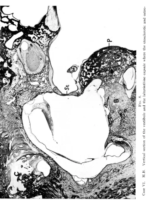
CASE VI.§ Mr. B., aged 77, was deaf and semi-cretinic. On first examination of his petrous bones the alterations seemed to belong only to otosclerosis, but there is a combination with a diffuse osteodystrophia fibrosa of Recklinghausen, and senile osteomalacia, the changes being manifold. In the right scala tympani there are
* cf. Case No. VI in the above-quoted work Nager-Meyer.
f Case No. I, Microscopic description in detail in Nager-Meyer, pp. 167 s. J Case No. II, loc. cit.
§ Details are given in Nager-Meyer, p. 133.
F. R. Nager and the late J. S. Fraser
very marked endosteotic formations along the diseased capsule, whereas the basal coil of the left side is completely filled up with lamellar and otosclerotic tissue.
Discussion
The bone formation in the scala tympani which has been found in our observations of otosclerosis seems to be very rare because, apart from the few sporadic cases mentioned, we saw it only in six petrous bones out of sixty-two cases of oto-sclerosis. The analysis of these cases indicates that the extent of the bone disease in the labyrinthine capsule of all these patients was extremely large, not in the form of circumscribed foci but more like a very diffuse affection including both windows. Moreover, the bony process itself was rather active, as the bone marrow contained many dilated blood vessels, newly-formed connective tissue, numerous osteoclasts and unripe bone tissue. It may be added that from the clinical point of view all these patients were almost totally deaf, which is easily explained by the fact of the obliteration of both windows.
As to the changes in the inner ear, we were mostly interested in the bone formation in the scala tympani. The other changes hitherto known and described by Otto Mayer and Albert
Gray7 viz. the atrophic conditions of the cells and nerve-structure of the cochlea, of the stria vascularis and the osteal tissue, etc., were of course also present. The scala tympani was almost filled up in the more advanced cases, Nos. I, III and V I ; whereas in the earlier stages there were more trabecular formations of web-like bone between which connec-tive tissue was spread out. One fact was generally observed in all these cases : the connection of this enostosis with areas of otosclerotic bone. This bone-formation was not present unless the labyrinthine wall was affected to a very large extent. With the exception of Case No. II, the lowest end of the scala tympani was filled up, including, of course, the cochlear aqueduct. In No. II, however, the round window itself was free, whereas the bone-formation started from a higher point of the diseased labyrinthine wall. It was evident that the bone tissue was mostly of the lamellar type—the lamellae being not arranged round blood vessels but more round the canals of Volkmann—and that from underneath the otosclerotic process seemed to penetrate into this enostotic tissue.
O N BON E FORMATIO N I N TH E SCAI. A TYMPAN I O F OTOSCLEROTICS—F . K . NACIK K ANI > TH K LAT E J . S . FRASK R FIG . 7 . Cas e V . Mr . W . Th e semicircula r canal= C surrounde d b y otoscleroti c bone-tissu e whic h lead s t o alteration s o f th e endostea l laye r an d ne w formatio n o f bone - an d fibrous-tissu e o f th e perilymphati c spac e V. 3 7 X . \face p. 17 8
FIG . 8 . Cas e VI . M.B . Vertica l sectio n o f th e vestibul e an d th e labyrinthin e capsul e wher e th e otoscleroti c an d osteo -dystrophi c proces s ar e t o b e seen . 1 4 x . P=Promontory , S t = Stapes , B = Ne w forme d bone-tissu e i n th e basa l coil , C = bon e cyst s i n th e capsul e (du e t o osteodystrophy) .
Bone Formation in the Scala Tympani
An early bone-formation could be detected in the semi-circular canal of Case No. II where the otosclerotic area surrounded the whole canal. The endosteal layer was thick-ened and presented a startling ossification so that these changes might be considered the same as in the scala tympani. Here, of course, the bone tissue was more web-like, whereas in the other more advanced cases in the scala tympani it was distinctly lamellar but without Haversian canals. It is exactly the same bone tissue found in obliterating labyrinthitis after purulent inflammation. It is worth mentioning that in general the inner ear of these petrous bones did not show distinct signs of old inflammation, with the exception of a darker staining of endo-labyrinthine fluids. The same observation seems to have been made by other authors, with the exception perhaps of
Lange's observation, according to which the cochlear duct
seems to be dilated.
As to the aetiology of this bone-formation, Siebenmann pointed out that it might be due to an irritation from the otosclerotic capsule. Otto Mayer,however, supposed the venous congestion produced by the bony process and the occlu-sion of the cochlear aqueduct to be the cause of the bone-formation. According to our findings the congestion alone cannot be made responsible as in both Lange's and our own case (No. II) the bone process was at a certain distance from the aqueduct. Bone-formation in the labyrinth is generally considered as a remnant of former serous or purulent labyrinthitis. In the inner ear of our case, however, other signs like ectasia of the cochlear duct, etc., were missing. We can, therefore, not accept the idea of a former labyrinth infection as a cause of these alterations. We are more inclined to believe that the otosclerotic process in the labyrinthine wall produces a certain alteration or irritation of the endosteal layer and perilymphatic space which leads to circumscribed fibrous-and bone-production in the scala tympani.
As the otosclerotic process is very active it will penetrate even this newly-formed bone. We cannot explain why this bone-formation has hitherto been found only in the inferior part of the scala tympani and in some cases in the semicircular
canals.
It is noteworthy that such bone-formation in the inner ear has been found only in otosclerotic diseases of the labyrinthine capsule, whereas all other bone affections, however extended
F. R. Nager and the late J. S. Fraser
they may be, do not lead to these alterations. It seems as if it were a peculiarity of otosclerosis which hitherto had neither an analogy nor an explanation.Summary
Six petrous bones of otosclerotic patients, with more or less extensive bone-formation in the scala tympani, are described. The otosclerotic process in all these observations was greatly extended and penetrated even into the newly-formed lamellar bone. This change in the scala tympani is considered a consequence of the irritation by the otosclerotic process of the labyrinthine wall and has not been found in any other bone disease.
REFERENCES
1
POLITZER, Zsch. f. Ohrenheilk., 1894, xxv.
2
HABERMANN, Arch. f. Ohrenheilkde., 1904, lx. 3 SIEBENMANN, Verhandlg. d. D.O.G., 1911. • MAYER, OTTO, Otosklerose, Wien, 1917.
5
LANGE, Passows Beitr., 1921, xvi. 6
KOSOKABE, Ueber die Knorpelfugen, etc. A. Kernen, Stuttgart, 1922. 7 GRAY, A., Otosclerosis, London, 1917, and / . Laryng. and Otol., 1934,
xlix.
Es werden die seltenen Befunde von Knochenneubildung in der Scala tympani von 6 Otosklerose-Felsenbeinen beschrieben. Dabei handelt es sich um sehr ausgedehnte Erkrankungen der Kapsel, wobei der otosklerotische Prozess selbst in die Scala tympani eingedrungen war. Die Knochenneubildung hier wird als Reaktion von der erkrankten Kapsel im Sinne einer umschriebenen Laby-rinthreizung aufgefasst und hat sich bisher bei keiner anderen Knochenerkrankung feststellen lassen.
L'auteur ddcrit six observations de n^oformation osseuse dans la rampe tympanique chez des otospongieux. II s'agissait dans ces cas d'une otosclerose tres etendue qui elle-meme avait penetre dans l'oreille interne. Cette neoformation osseuse est d'apres l'auteur la suite d'une irritation provoqu^e par la maladie de la capsule m&ne. Elle n'a ete observed dans aucune autre maladie osseuse, si Etendue qu'elle soit.