Extragonadal retroperitoneal germ cell tumor: evidence of origin in the testis
4
0
0
Texte intégral
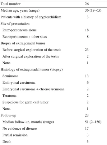
(2) 122 All patients were diagnosed as having primary extragonadal retroperitoneal germ cell tumors because testicular palpation by several physicians (e.g. medical practitioner, general surgeon or medical oncologist) before treatment was considered not to be suspicious for testicular cancer. In 25 of the 26 patients (96%) the extragonadal mass was biopsied. In one patient, surgical exploration of the extragonadal mass was waived because germ cell tumor was considered to be unequivocally proven by the elevated tumor markers at another hospital. After initial diagnosis, patients were examined by a urologist during the treatment period. Ultrasound scans were performed and evaluated by a radiologist in 20 of the 26 patients (77%) prior to surgical exploration. Hypo- and hyperechogenic lesions or microcalcifications were defined as being suspicious for active or burned-out testicular cancer. Histology of the testis was obtained by orchiectomy in 23 patients and by open biopsy of the suspicious testis in two. Twenty patients (77%) were treated with chemotherapy alone, three (11.5%) underwent radiotherapy and three (11.5%) received combined radio- and chemotherapy. Eighteen of the 23 platinum-based chemotherapy regimens (78%) were with and five without bleomycin. Four of the 23 patients received more than one regimen. In half of the patients, chemotherapy was administered before surgical exploration of the testis. Excision of a residual retroperitoneal mass was conducted in 10 patients during follow-up. Follow-up was available in 23 of the 26 patients (88%). The median follow-up was 51 months (range 2–150). Seventeen of 23 patients (74%) have no evidence of disease. Three patients (13%) are being followed regularly by computed tomography (CT) for residual tumor with a volume <2 cm in diameter. Three patients (13%) have died of disease (Table 1). Follow-up consisted of clinical evaluation, CT, chest X-ray and tumor marker determinations, which were evaluated by a radiologist, medical oncologist and urologist.. Results In the 26 patients, 13 seminomas, 10 non-seminomatous germ cell tumors and two tumors suspicious for a germ cell tumor were found at histological examination of the extragonadal mass (Table 1).The primary tumor was on the left side in 10 of 26 patients (38%), on the right side in seven (27%) and bilaterally in nine (35%). None of these patients was suspected of having a testicular tumor by the physicians initially treating them. At urological workup, palpation of the testis was unsuspicious in 11 of 26 patients (42%), atrophic in 11 (42%), atrophic and indurated in one (4%), indurated in two (8%) and enlarged in one (4%). Ultrasonographical examination was suspicious for tumor in all (20 of 20) patients examined by sonography. A suspicious finding at ultrasonographical examination was the only indication for surgical exploration in 10 of 11 patients with normal testes on palpation and supportive in 10 of 11 patients with suspicious findings on palpation. At surgical exploration of the testis, pathological findings were located ipsilaterally in 14 of 25 patients (56%) and contralaterally in three (12%) to the site of the primary tumor.. Table 1. Patient characteristics Total number. 26. Median age, years (range). 36 (19–65). Patients with a history of cryptorchidism. 3. Site of presentation Retroperitoneum alone Retroperitoneum + other sites. 18 8. Biopsy of extragonadal tumor Before surgical exploration of the testis. 23. After surgical exploration of the testis. 2. None. 1. Histology of extragonadal tumor (biopsy) Seminoma. 13. Embryonal carcinoma. 6. Embryonal carcinoma + choriocarcinoma. 2. Teratoma. 2. Suspicious for germ cell tumor. 2. None. 1. Follow-up. 23. Median follow-up, months (range). 51 (2–150). No evidence of disease. 17. Partial remission. 3. Death. 3. Of those nine patients with a bilateral primary tumor, five had pathological findings in the right testis, three in the left testis and one histology of the testis was not obtained. The histological examination of the explored 25 testes revealed scar tissue in 12 patients (48%), sclerosis in three (12%), sclerosis and fibrosis in one (4%), fibrosis in two (8%), intratubular neoplasia in four (16%) and viable tumor in three (12%) (Figure 1). Of the 11 patients with an initially non-suspicious testis on palpation, five showed scar tissue, one sclerosis, three intratubular neoplasia and two viable tumor histologically. Of the 15 patients with a suspicious finding on palpation, seven revealed atrophy in the histology, two sclerosis, two fibrosis, one sclerosis and fibrosis, one intratubular neoplasia and one viable tumor. In the group of patients receiving chemotherapy alone, testicular histology was obtained prior to and after chemotherapy in 10 patients each. After chemotherapy, two of the 10 patients still had viable tumor or intratubular neoplasia (Figure 1). One received vinblastine, bleomycin, cisplatin, cyclophosphamide, etoposide and dactinomycin. We could not retrospectively determine the chemotherapy regimen in detail for the other patient..
(3) 123. Figure 1. Histology of 25 testes explored in patients with so-called extragonadal retroperitoneal germ cell tumors and divided into those with surgical exploration of the testis before (n = 10) or after (n = 10) chemotherapy.. Discussion The most widely accepted theory of the development of extragonadal germ cell tumors suggests that these tumors originate from displaced primordial (embryonal) germ cells situated along the midline of the body [2, 3, 5, 7–12]. Whether these tumors are truly extragonadal or synchronous germ cell tumors in the testis and retroperitoneum or metastatic lesions from undetected or regressed (burned-out) testicular carcinoma remains ultimately an open question. In our series all patients with extragonadal retroperitoneal germ cell tumors had a pathological testis showing either viable tumor or lesions compatible with a burned-out testicular tumor. As early as 1927, Prym [13] had reported testicular scarring in a patient with an extragonadal tumor at autopsy. In the following years further case reports also described such lesions [14, 15]. Azzopardi et al. [16] showed that some palpably normal testes have either scar tissue or small foci of tumor on histological examination. This raised the question of whether a portion of primary extragonadal germ cell tumors may not be metastatic. In 1951, Friedmann [12] found either regressive changes or an overt tumor of the testis in 23 of 29 patients with primary retroperitoneal germ cell tumors. This was confirmed by our earlier report on 14 patients with primary retroperitoneal germ cell tumors [3]. Daugaard et al. [9], in their series, found malignant germ cells in the gonads of 42% of patients with so-called clinically primary tumors in the retroperitoneum. All of our patients had a pathological finding histologically defined as either viable tumor tissue or regressive changes. Seven patients (28%) showed some form of testicular. neoplasia: seminoma (two), teratoma (one) and intratubular neoplasia (four). In 12 (48%) of our patients the histological evaluation revealed testicular scarring. Since trauma could be excluded as a cause in all patients, the suggestion is that such scars should be considered to be residuals of burnedout primary tumors [13–15, 17, 18]. Six patients showed sclerosis, fibrosis or both. This pathological process in the testis may be interpreted in the same way as scar tissue. The differentiation of a so-called primary extragonadal retroperitoneal germ cell tumor from a primary testicular neoplasm with retroperitoneal metastases strongly depends on the aggressiveness of testicular examination. Indeed, in our series all patients were clinically evaluated with palpation of the testes by several physicians and palpation was considered not to be suspicious for testicular cancer. Urological workup, however, demonstrated a suspicious finding on palpation in 58% of the patients. Of those patients with unsuspicious testes on palpation, 10 of 11 (91%) became suspicious on ultrasonography. Ultrasound examination of the testes showed hypo- or hyperechogenic lesions and/or microcalcification in 20 of 20 patients. This is in agreement with Comiter et al. [17] who found intratesticular lesions by ultrasonographical examination in five of six patients with primary retroperitoneal extragonadal germ cell tumors. Fuchs et al. [19] and Medini et al. [20] suggested that normal testes would not require surgical exploration; however, both are small series, with five and eight patients, respectively. Furthermore, in the series of Medini et al., three patients had retroperitoneal masses only. For the other patients it is well known that mediastinal germ cell tumors are another entity and in general do not have concomitant testicular neoplasia [3, 21]. Identification of a primary testicular tumor in patients with a presumed extragonadal germ cell tumor is important because it carries the danger of persistent testicular malignancy in up to 50% of such patients despite systemic chemotherapy [3, 4, 17]. In our series, two patients still had viable tumor or intratubular neoplasia after chemotherapy. Based on our results the following conclusions may be drawn. (i) So-called primary extragonadal germ cell tumors of the retroperitoneum are probably a rare or non-existing entity. They should be considered to be metastases of a viable or burned-out testicular cancer until proven otherwise. This was found in 76% of our patients. (ii) Intensive urological workup including ultrasonographical examination of the testis, especially of those with unsuspicious findings on palpation, is mandatory. (iii) The presence of viable tumor tissue in the testis after chemotherapy (sanctuary) stresses the need for surgical exploration of all patients with extragonadal retroperitoneal germ cell tumors and a testicular abnormality. (iv) Adequate treatment of the primary testicular tumor is essential for achieving a cure..
(4) 124. References 1. Goss PE, Schwertfeger L, Blackstein ME et al. Extragonadal germ cell tumors—a 14-year Toronto experience. Cancer 1994; 73: 1971– 1979. 2. Burt ME, Javadpour N. Germ cell tumors in patients with apparently normal testis. Cancer 1981; 47: 1911–1915. 3. Böhle A, Studer UE, Sonntag RW et al. Primary or secondary extragonadal germ cell tumor. J Urol 1986; 135: 939–943. 4. Culine S, Theodore C, Terrier-Lacombe MJ et al. Primary chemotherapy in patients with nonseminomatous germ cell tumors of the testis and biological disease only after orchiectomy. J Urol 1996; 155: 1296–1298. 5. McLeod DG, Taylor HG, Skoog SJ et al. Extragonadal germ cell tumors—clinicopathologic findings and treatment experience in 12 patients. Cancer 1988; 61: 1187–1191. 6. Setchell BP. The functional significance of the blood–testis barrier. J Androl 1980; 1: 3–11. 7. Gerl A, Clemm C, Kohl P et al. Primary extragonadal germ cell tumors. Clinical manifestions, differential diagnosis and therapy. Med Klin 1994; 89: 240–244. 8. Bassetto MA, Pasini F, Franceschi T et al. Extragonadal germ cell tumor: a clinical study. Anticancer Res 1995; 15: 2751–2754. 9. Daugaard G, Rorth M, von der Maase H et al. Management of extragonadal germ cell tumors and the significance of bilateral testicular biopsies. Ann Oncol 1992; 3: 283–289. 10. Carroll PR, Whitmore WF, Richardson M et al. Testicular failure in patients with extragonadal germ cell tumors. Cancer 1987; 60: 108– 113. 11. Asif S, Uehling DT. Microscopic tumor foci in testes. J Urol 1968; 99: 776–779.. 12. Friedmann NB. The comparative morphogenesis of extragenital and gonadal germ cell tumors. Cancer 1973; 4: 265–269. 13. Prym P. Spontanheilung eines bösartigen, wahrscheinlich chorionephiteliomatösen Gewächses im Hoden. Virchows Arch Pathol Anat 1927; 265: 239–258. 14. Hailemariam S, Engeler DS, Bannwart F et al. Primary mediastinal germ cell tumor with intratubular germ cell neoplasia of the testis— further support of germ cell origin of these tumors: a case report. Cancer 1997; 79: 1031–1036. 15. Lopez JI, Angulo JC. Burned out tumor of the testis presenting as retroperitoneal choriocarcinoma. Int Urol Nephrol 1994; 26: 549– 553. 16. Azzopardi JG, Mostofi FK, Theiss EA. Lesions of testis observed in certain patients with widespread choriocarcinoma and related tumors. Am J Pathol 1961; 38: 207–225. 17. Comiter CV, Renshaw AA, Benson CB. Burned out primary testicular cancer: sonographic and pathological characteristics. J Urol 1996; 156: 85–88. 18. Heidenreich A, Neubauer S, Mostofi FK et al. Clinical stage I mature teratoma of the testis—retroperitoneal lymphadenectomy or surveillance? Urologe A 1997; 36: 440–444. 19. Fuchs E, Hatch T, Seifert A. Extragonadal germ cell tumor: the preoperative urological evaluation. J Urol 1987; 137: 993–995. 20. Medini E, Levitt SH, Jones TK, Rao Y. The management of extratesticular seminoma without gonadal involvement. Cancer 1979; 44: 2032–2038. 21. Luna MA, Valenzuela-Tamariz J. Germ-cell tumors of the mediastinum, postmortem findings. Am J Clin Pathol 1976; 65: 450–454..
(5)
Figure

Documents relatifs
Dans ma cachette, bien à l’abri, j’ai somnolé toute la journée, Dans mon antre, je suis le roi, personne ne vient me déranger.. Comme chaque jour, depuis toujours, je sors à
Bien que des contemporains et intéressés par la culture du pays voisin res- pectif, les deux poètes n’ont pas fait connaissance l’un avec l’autre ; tout au plus se sont-ils
Isolated human testicular germ cells express several alternative HIV receptors and support the attachment of HIV-1 R5 and X4 strains.. Heparan sulfate proteoglycans and mannose
Potential for virus endogenization in humans through testicular germ cell infection: the case of HIV
Altogether, these results indicate that isolated early germ cells can support low level of HIV-1 221.. DNA integration into
The germ cell (or Sertoli cell) secretome (Fig. 2, panel E) — the genes (mouse Entrez gene IDs) expressed in germ cells (or Sertoli cells) and encoding proteins actively secreted
Treves et al., “ Clinical responses in a phase II study using adoptive transfer of short- term cultured tumor in fi ltration lymphocytes in metastatic melanoma patients, ”
Il serait également pertinent, vu que les deux groupes ayant le plus haut niveau d'études sont également ceux où les personnes ont un handicap plus visible, de mener une
The purposes of this study were to: (1) determine if apoptosis of germ cells occurred in adult stallions with normal testes, and (2) describe apoptotic rates by stage of
