Exp Brain Res (2003) 153: 356–365 DOI 10.1007/s00221-003-1620-4
R E S E A R C H A RT I C L E
Michael Török . Jörg Huwyler . Heike Gutmann .
Gert Fricker . Jürgen Drewe
Modulation of transendothelial permeability and expression
of ATP-binding cassette transporters in cultured brain capillary
endothelial cells by astrocytic factors and cell-culture conditions
Received: 27 January 2003 / Accepted: 2 July 2003 / Published online: 12 September 2003 # Springer-Verlag 2003
Abstract Confluent cell monolayers of brain capillary
endothelial cells (BCEC) are used widely as an in vitro
cell culture model of the blood
–brain barrier. The present
study describes the influence of cell-culture conditions on
tight junctions, filamentous-actin cytoskeleton, and
ex-pression of ATP-binding cassette (ABC) transporters in
primary cell cultures of porcine BCEC. Astrocyte as well
as C6 glioma-conditioned cell culture medium was used in
combination with retinoic acid, dexamethasone, cyclic
adenosine monophosphate (cAMP) analogs, or
1,25-dihydroxyvitamin D
3. It was shown that C6-conditioned
medium led to a reorganization of filamentous actin and to
an improved staining of zonula occludens-associated
protein-1 (ZO-1). Further optimization of these culture
conditions was achieved with cAMP analogs and
dexa-methasone. Retinoic acid, as well as
1,25-dihydroxyvit-amin D
3, did not improve cellular tight junctions as judged
by filamentous actin, ZO-1 rearrangement, and
transcel-lular electrical resistance (TER) measurements. However,
these morphological changes did not influence the
paracellular permeability of the extracellular marker
sucrose. Expression
of
ABC
transporters
such
as
P-glycoprotein, multidrug resistance-associated protein-1
(MRP1), and MRP2 were compared by measuring
mes-senger RNA (mRNA) levels in whole-brain tissue, isolated
brain capillaries, and cultured cells. In freshly isolated
BCEC, mRNA levels of MRP2 and P-glycoprotein
dropped by two- to sevenfold, respectively, whereas
MRP1 mRNA levels were slightly increased. During cell
culture, mRNA levels of MRP1 and MRP2 decreased by
up to fivefold, while P-glycoprotein levels remained
constant. These results were unaltered by different
cell-culture conditions. In conclusion, the present study
suggests that paracellular permeability, as well as mRNA
expression of the studied ABC transporters in primary
cultures, of porcine BCEC are insensitive toward changes
in cell-culture conditions.
Keywords Blood
–brain barrier . P-glycoprotein . MRP .
Brain capillary endothelial cells
Abbreviations ACEM: Astrocyte-conditioned
endothelial medium . BCEC: Brain capillary endothelial
cells . C6CEM: C6-conditioned endothelial medium .
TER: Transcellular electrical resistance
Introduction
A continuous layer of brain microvessel endothelial cells
is central to the integrity of the blood
–brain barrier, which
controls the passage of substances from the blood to the
extracellular fluid environment of the brain. A key
component of the majority of in vitro cell culture models
of the blood
–brain barrier developed during the last years
is therefore a confluent cell monolayer of brain endothelial
cells. Primary or passaged cultures of these cells can be
obtained from mouse, rat, bovine, human, canine, or
porcine brains (for a review, see Takakura et al. 1991). In
some instances immortalized cell lines of brain endothelial
cells have been established such as rat RBE4 cells
(Durieu-Trautmann et al. 1993). A common problem
associated with cell cultures of brain endothelial cells is
the rapid dedifferentiation of these cells as soon as they are
removed from their natural environment and placed in cell
culture. A rationale for this is provided by observations
that the microenvironment of the vasculature determines
the endothelial phenotype (Stewart and Wiley 1981). As a
M. Török . H. Gutmann . J. Drewe (*)
Division of Clinical Pharmacology, University Hospital, Petersgraben 4, 4031 Basel, Switzerland e-mail: Juergen.Drewe@unibas.ch Tel.: +41-61-2653848 Fax: +41-61-2658581 J. Huwyler
Pharmaceuticals Division, F. Hoffmann-La Roche Ltd, Basel, Switzerland
G. Fricker
Institute for Pharmacy and Molecular Biotechnology, University of Heidelberg,
consequence, monolayers of brain endothelial cells have a
tendency to be of low electrical resistance and are
relatively leaky, which may lead to a poor correlation
between permeability of brain endothelial cells to various
compounds in cell culture and in vivo (Pardridge et al.
1990). During the last few years, however, much progress
has been made with respect to the development of in vitro
models of the blood
–brain barrier, with an improved
predictive value by the systematic modification of
cell-culture conditions. Since astrocyte
–endothelial cell
inter-actions are sufficient to induce the essential properties of
the blood
–brain barrier (for a review, see Abbott et al.
1992), cocultures between brain endothelial cells and
astrocytes have been established (Dehouck et al. 1995;
Raub 1996) or media conditioned by astrocytes (primary
cultures, Rubin et al. 1991; or astroglioma cell lines, Raub
et al. 1992) have been used to induce blood
–brain barrier
properties in primary cultures (Meyer et al. 1991) or
immortalized brain endothelial cells (Rist et al. 1997).
Alternative methods comprise treatment of cell cultures
with agents that elevate intracellular cyclic adenosine
monophosphate (cAMP; Rubin et al. 1991), retinoic acid
(El Hafny et al. 1997), or hydrocortisone (Franke et al.
1999). While the impact of such modifications of
cell-culture conditions on electrical resistance and permeability
of cell monolayers has been well documented,
compara-tively little is known about its consequences for
ATP-binding cassette (ABC) proteins (Klein et al. 1999), such
as the drug-efflux pumps P-glycoprotein and multidrug
resistance-associated proteins MRP1 and MRP2.
Therefore, the aim of the present study was to
systematically modify the cell-culture conditions used for
primary cultures of porcine brain capillary endothelial
cells (BCEC). Modifications included the use of two types
of astrocyte-conditioned cell culture media as well as cell
culture supplements, such as retinoic acid, dexamethasone,
cAMP analogs and 1,25-dihydroxyvitamin D
3. The impact
of these environment modifications upon cell morphology
[i.e., organization of filamentous-actin (F-actin)
cytoskel-eton and tight junctions], permeability of the extracellular
marker sucrose and mRNA expression of P-glycoprotein,
MRP1, and MRP2 were determined and directly compared
with freshly isolated brain microvessels.
Materials and methods
Isolation of porcine brain capillaries
Porcine brain capillaries were prepared as described previously for bovine brain capillaries (Pardridge et al. 1985) but with modifica-tions. In brief, cortical gray matter of one porcine brain was minced and subsequently homogenized with five up-and-down strokes using a glass homogenizer with a clearance of 0.095–0.115 mm. The homogenate was brought to 15% (w/v) dextran and centrifuged for 10 min at 5,750g at 4°C. The pellet obtained was then passed through a 150-µm mesh to remove small arterioles and tissue fragments. Brain capillaries in the effluent were further purified by passing the filtrate over a 25-ml column containing 20-ml glass beads of 0.45–0.50 mm diameter. Trapped brain capillaries were dislodged from the glass beads by stirring, washed and collected by
centrifugation (200g for 5 min at 4°C). Isolated capillaries were cryopreserved at –196°C in Dulbecco,s modified Eagle medium, supplemented with 10% (v/v) dimethylsulfoxide (DMSO) and 100 mM sucrose.
Isolation and cell culture of porcine brain capillary endothelial cells
Primary cultures of porcine BCEC were prepared as described (Huwyler et al. 1996). Cortical gray matter of six porcine brains were minced and digested enzymatically for 2 h using 0.5% (w/v) dispase. Cerebral microvessels were obtained after centrifugation in 13% (w/v) dextran and were subsequently incubated for 4.25 h in a buffer containing 0.1% (w/v) collagenase/dispase. The resulting cell suspension was supplemented with 10% (v/v) horse serum and filtered through a 150-µm nylon mesh. BCEC were isolated on a continuous 50% (v/v) Percoll gradient (Amersham Pharmacia Biotech, Uppsala, Sweden). Endothelial cells were filtered through a 35-µm nylon mesh and cryopreserved at –196°C in endothelial cell culture medium (45% v/v MEM, 45% v/v Ham,s F-12 nutrient mixture, 10% (v/v) heat-inactivated horse serum, 100 µg/ml streptomycin, 100 µg/ml penicillin G, 100 µg/ml heparin, 13 mM NaHCO3 and 10 mM HEPES) supplemented with 10% (v/v)
DMSO. Isolated endothelial cells were seeded at a density of 150,000 cells/cm2onto collagen/fibronectin-coated surfaces. Cells were grown either in endothelial cell culture medium or in medium consisting of 50% (v/v)-conditioned endothelial cell culture medium and 50% (v/v) endothelial cell culture medium. After cells had reached confluence (day 7), supplemented or unsupplemented medium, with or without serum, was added for 24 h. Supplements used were: 250 µm 8-(4-chlorophenylthio) cAMP (Sigma, Buchs, Switzerland) plus 35 µm phosphodiesterase inhibitor RO-20-1724 (Calbiochem, San Diego, Calif., USA), 10 µm dexamethasone (Sigma), 5 µm all-trans retinoic acid (Sigma), or 500 nM 1,25-dihydroxyvitamin D3(Calbiochem).
Conditioned medium
Endothelial cell culture medium was conditioned by rat astrocytes or by the rat glioma cell line C6 (American Type Culture Collection, Rockville, Md., USA). Medium (10 ml per T-75 culture flask) was added for 2 days to the confluent cells and the removed medium was sterile filtered and stored at−20°C.
Primary cultures of rat astrocytes were prepared from 1-day-old rat cortex essentially as described by Lillien et al. (1988). Small pieces of gray cortex were rubbed across a 63-µm mesh. The homogenate was then cultured for about 10 days in astrocyte cell culture medium (90% (v/v) MEM, total 33 mM D-glucose, 13 mM NaHCO3, 20 mM HEPES, 100 µg/ml streptomycin sulfate, 100 µg/
ml penicillin G, 100 µg/ml heparin, 10% (v/v) heat-inactivated fetal calf serum). Culture flasks were then shaken overnight at 37°C and the remaining cells (enriched in type-1 astrocytes) were passaged on poly-D-lysine-coated flasks. Confluent cells were treated with
astrocyte cell culture medium, supplemented with 10 µM cytosine arabinofuranoside, for 2 days to limit the growth of rapidly dividing contaminating cells. After renewed confluence, cells were treated again with cytosine arabinofuranoside. Confluent cells were subsequently used to condition endothelial cell culture medium. By immunocytochemical criteria, the cultures were approximately 95% type-1 astrocytes (Lillien et al. 1988).
C6 cells (passage 40 to 43) were plated at a concentration of 100,000 cells/cm2 in C6 cell culture medium (41.25% v/v MEM, 41.25% v/v Ham,s F-12 nutrient mixture, 13 mM NaHCO3, 20 mM
HEPES, 100 µg/ml streptomycin sulfate, 100 µg/ml penicillin G, 100 µg/ml heparin, 15% v/v heat-inactivated horse serum, 2.5% v/v heat-inactivated fetal calf serum). Confluent cells were used for up to 1 week to condition the medium.
Transcellular electrical resistance measurements
Transcellular electrical resistance (TER) across endothelial cell monolayers was measured in the growth medium using the Millicell-ERS system with chopstick electrodes (Millipore, Bedford, Mass., USA). Values were corrected for resistances of collagen/fibronectin-coated, blank filters. TER was expressed as ohm·centimeters squared.
Flux measurements
Transport experiments were performed using cell monolayers cultured on filter inserts (0.4 µM pore size, 24-well Transwell plates; Costar, Cambridge, Mass., USA) at 37°C on a rotary platform (120 rpm). Experiments were initiated by adding 0.5 ml transport buffer (142 mM NaCl, 3 mM KCl, 1.4 mM CaCl2, 1.2 mM
MgCl2, 1.5 mM K2HPO4, 4 mMD-glucose, 10 mM HEPES, pH 7.4)
containing 0.6 µCi/ml14C-sucrose (Amersham Pharmacia Biotech) to the apical chamber. The emergence of the radioactive compound in the basolateral chamber was measured after 10 min by taking a 200-µl sample from the basolateral chamber, which contained 1 ml transport buffer. Radioactivity was determined by scintillation counting. Transport was calculated from the initial concentration of tracer in the apical chamber and the final concentration of tracer in the basolateral chamber according to: Papp=dQ/dt·1/A/C0, where
dQ/dt is the rate of translocation, A is the surface of the membrane (here 1 cm2) and C0is the initial concentration of the labeled drug.
Immunocytochemistry
Confluent cultured BCEC were fixed and stained directly on the culture inserts after flux experiments. Brain capillaries were adhered to a glass slide by using a Cytospin 2 centrifuge (Shandon, Pittsburgh, Pa., USA) at 200 g for 5 min at room temperature. Tissues were fixed for 20 min in 4% (w/v) paraformaldehyde and then permeabilized in 0.5% (v/v) Triton X-100 for 5 min. Tissues were incubated for 1 h at 37°C in primary antiserum diluted in 3% (w/v) bovine serum albumin in phosphate-buffered saline (PBS), followed by incubation for 1 h at room temperature in fluorochrome-conjugated secondary antibody diluted in 3% (w/v) bovine serum albumin in PBS. Filamentous-actin was stained by addition of 3 U/ ml phalloidin-rhodamine (Molecular Probes, Eugene, Ore., USA), nuclei were stained using 100 µg/ml 4',6-diamidino-2-phenylindole or 60 µg/ml propidium iodide. Microscopy specimens were mounted in FluoroSave (Calbiochem, San Diego, Calif., USA).
Primary antibodies used for these experiments included rabbit polyclonal anti-von Willebrand factor (6 µg/ml; Sigma), rabbit polyclonal anti-ZO-1 (10 µg/ml; Zymed, San Francisco, Calif., USA), murine monoclonal anti-glial fibrillary acidic protein (1 µg/ ml; Boehringer Mannheim, Mannheim, Germany), and murine monoclonal antibody C219 directed against P-glycoprotein (5 µg/ ml; Signet, Dedham, Mass., USA). Secondary antibodies used included conjugated rabbit anti-mouse (15 µg/ml) and Cy2-conjugated goat anti-rabbit (15 µg/ml; both obtained from Jackson Immuno Research, West Grove, Pa., USA).
To determine the staining morphology of F-actin and ZO-1 in brain capillary endothelial cells under different culture conditions, a scoring system was used. For F-actin, scores from 1 to 5 were assigned and, for ZO-1, scores from 1 to 3 (Figs. 2, 3). Scores were assigned independently by two scientists, in a blind manner and specimens were presented in a random order.
Fluorescence microscopy
Confocal microscopy was performed with a Zeiss LSM 510 inverted laser scanning microscope, equipped with He/Ne 543 nm, He/Ne 633 nm, and Ar 488 nm lasers. Objectives used were a Zeiss
Plan-Neofluar ×40 oil-immersion objective with a numerical aperture of 1.3 or a Zeiss Plan-Neofluar ×63 oil-immersion objective with a numerical aperture of 1.25. Optical sections 0.35 µM thick were scanned through the z-plane of the sample. Data analysis, three-dimensional reconstruction from series of consecutive optical sections, and shadow projections were calculated on an O2 Workstation (Silicon Graphics, Mountain View, Calif., USA) with Imaris software (version 2.7; Bitplane, Zürich, Switzerland). Classical fluorescence microscopy was performed with a Zeiss Axiophot fluorescence microscope.
Real-time quantitative PCR
Real-time quantitative PCR analysis was performed with the TaqMan assay using an ABI PRISM 7700 Sequence Detector (Applied Biosystems, Rotkreuz, Switzerland), a combined thermo-cycler and fluorescence detector. A dual-labeled fluorogenic probe complementary to a sequence within each PCR product was added to the PCR reaction. The fluorescent dye at the 5' end of the probe (6-carboxyfluorescein) served as reporter, and its emission was quenched by the second fluorescent dye at the 3' end of the probe (6-carboxytetramethylrhodamine). During elongation, the 5' to 3' exonuclease activity of the Taq DNA polymerase cleaved the probe, thus releasing the reporter from the quencher. Primers and probe were designed according to the guidelines of Applied Biosystems, and with the help of Primer Express 1.0 software (Applied Biosystems). Primers were custom synthesized by Life Technologies (Paisley, Scotland), probes by Eurogentec (Seraing, Belgium). The primers and probes used were:
A. GAPDH: GGTGAAGGTCGGAGTGAACG and
CGA-CAATGTCCACTTTGCCA with the probe CGCCTGGTCAC-CAGGGCTGC
B. pgp1A: GTGGAATGATCTTCAATGGTAGCA and
AAGCGCTCATCAACTGTGACC with the probe TTGAT-TAAATGCCCAGATTTTAACACAAATATTAGACAAC C. ZO-1: AAGGCGGATGGCGCTACAAG and
TTCAC-TACCTGGGGCTGACAGG with the probe ACCTTGATTTG-CATGATGATCGTCTGTCCT
D. MRP1: GACCCTTGATTGCCACGTG and
TGGGCTGTGGGAAGTCGT with the probe
CCTCCACTTTGTCCATCTCAGCCAAGAG
E. MRP2: TGTGGGCTTTGTTCTGTCCA and CAGCCA-CAATGTTGGTCTCG with the probe CTCAATATCACA-CAAACCCTGAACTGGCTG
Quantitative data was normalized to GAPDH. Results were expressed relative to the control mean value, which was set as 1.
Total RNA was extracted using the RNeasy Mini Kit (Qiagen, Hilden, Germany) according to the manufacturer,s protocol. After DNase I digestion, RNA was quantified using a GeneQuant photometer (Pharmacia, Uppsala, Sweden). Its integrity was checked by ethidium bromide agarose gel electrophoresis. The purity of the RNA preparations was high, as demonstrated by the 260 nm to 280 nm ratio (range 1.8–2.0). One microgram of total RNA was reverse transcribed by Superscript II (Gibco, Basel, Switzerland) according to the manufacturer,s protocol using random hexamers as primers. Complimentary DNA (cDNA; 25 ng total RNA) was amplified in a 25-µl volume containing 12.5 µl of the ×2 TaqMan universal PCR master mix (Applied Biosystems), 225-nM probe, and 900 nM of each primer. Cycling conditions were 10 min, 95°C initial denaturation and activation of AmpliTaq Gold DNA polymerase, followed by 40 cycles of 15 s 95°C denaturation, 1 min 60°C combined annealing and primer extension. Mathematical analysis was performed as recommended by Applied Biosystems (see user bulletin 2, ABI PRISM 7700 Sequence Detection System, downloadable at http://www.appliedbiosystems.com/support).
Statistical analysis
For statistical comparison, data groups were compared by analysis of variance (ANOVA). If this analysis revealed significance, pair-wise comparisons between groups were performed by a two-tailed Student,s t-test with Bonferroni,s correction. Significance level was set to P≤0.05.
Results
Cell cultures and cytochemistry
Primary cultures of porcine brain microvessel endothelial
cells were cultured on Transwell filter inserts under
different cell-culture conditions (Table 1). Cell culture
media were either used without pretreatment or
condi-tioned using primary cultures of type-1 astrocytes or C6
astroglioma cells. Where indicated, supplements were
added to the cell cultures 24 h prior to the experiment.
Brain capillary endothelial cells in situ are known to
form high-resistance tight junctions. Visualization of
F-actin and the ZO-1, which are both associated with the
junctional complex in freshly isolated brain microsvessels,
reveals a characteristic belt-like distribution of these
proteins (Fig. 1). This pattern of staining for F-actin
(Fig. 2A) and ZO-1 (Fig. 3A) was only weakly retained in
brain capillary endothelial cells under standard cell-culture
conditions. However, modification of cell-culture
condi-tions such as astrocyte-conditioned endothelial medium
(ACEM)
and
C6-conditioned
endothelial
medium
(C6CEM; Table 1) led to a substantial reorganization of
F-actin and a reinforced staining of ZO-1. The degree of
reorganization (reinforcement) was described by a score
that ranged, for F-actin, between 1 (poor) and 5 (very
high) and for ZO-1 between 1 (poor) and 3 (very high).
Examples for the different scores and the associated
cell-culture conditions are given for F-actin in Fig. 2 and for
ZO-1 in Fig. 3. Figures 2 and 3 were obtained by
conventional fluorescence microscopy and represent the
situation encountered by the evaluating scientists. The
scores that were obtained using different cell-culture
conditions are summarized in Table 2. The higher the
score, the higher the degree of differentiation was
considered, since a situation closer to intact brain
capil-laries was reached (Fig. 1). F-actin scores showed that
growing in C6CEM resulted in highest scores, whereby
cells grown in ACEM still needed the addition of
chlorophenylthio-cAMP/RO-20-1724 or dexamethasone
to enhance the score further. All supplements in
combina-tion with endothelial medium (EM) did not lead to an
improvement of the staining pattern compared with EM
alone. Comparable results were also shown with ZO-1
scores. There was a statistically significant correlation
between F-actin scores and ZO-1 scores, as demonstrated
by least-square regression analysis (Pearson coefficient
R=0.833). In summary, only
chlorophenylthio-cAMP/RO-20-1724 and dexamethasone were capable of inducing
changes in F-actin and ZO-1 morphology, but only in
combination with ACEM (for F-actin) or alone (for ZO-1).
Highest scores were observed in the group of cells grown
in C6CEM.
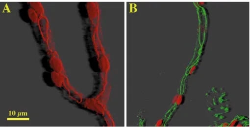
Fig 1A, B Distribution of tight junction-associated proteins in freshly isolated brain capillaries. Staining of A filamentous actin with phalloidin-rhodamine (red) and B zonula occludens-asso-ciated protein-1 (ZO-1; green). To visualize the location of individual cells in capillaries, nuclei were costained with pro-pidium iodide (red) Filamentous actin and ZO-1 staining is pre-dominantly organized at the cell borders. Images were rendered using three-dimensional image processing and shadow projec-tion
Table 1 Cell culture conditions Condition Abbreviation Exposure of cells
Standard endothelial cell culture medium EM 7 days
Astrocyte-conditioned medium ACEM 7 days
C6-conditioned medium C6CEM 7 days
250 µM chlorophenylthio-cAMP + 35 µM ro-20-1724 AMP 24 h
10 µM dexamethasone DEX 24 h
5 µM all-trans retinoic acid RET 24 h
Fig 2A-E F-actin staining with phalloidin-rhodamine in conflu-ent brain capillary endothelial cells. A Score 1 (cells grown in EM); B score 2 (cells grown in EM); C score 3 (cells grown in ACEM and 10 µM dexametha-sone for 24 h); D score 4 (cells grown in C6CEM and 10 µM dexamethasone for 24 h); E score 5 (cells grown in C6CEM). Size bar is the same for all images
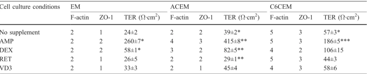
Table 2 Filamentous actin (F-actin) and zonula occludens-associated protein-1 (ZO-1) morphology scores and transcellular electrical resistance (TER) measurements 24 h after confluence. TER values are expressed as mean ± SEM of n≥4 determinations
Cell culture conditions EM ACEM C6CEM
F-actin ZO-1 TER (Ω·cm2) F-actin ZO-1 TER (Ω·cm2) F-actin ZO-1 TER (Ω·cm2)
No supplement 2 1 24±2 2 2 39±2* 5 3 57±3*
AMP 2 2 260±7* 4 3 415±8** 5 3 186±5***
DEX 2 2 58±1* 3 2 82±5** 4 2 106±15
RET 2 1 26±5 2 2 29±1** 5 3 44±3
VD3 2 1 33±3 2 1 45±4 4 3 58±6
Transcellular electrical resistance
Transcellular electrical resistance measurements 24 h after
confluence (Table 2) showed that cells grown in ACEM or
C6CEM, without supplement, showed approximately a
doubling in their TER values compared with monolayers
grown in EM. These alterations were statistically
signif-icant. Addition of chlorophenylthio-cAMP/RO-20-1724
significantly raised the TER value by a factor of ~10 for
EM and ACEM and a factor of ~3 for C6CEM. To a lesser
extent, dexamethasone led to a further increase of TER
(factor ~2) compared with their respective media in
absence of supplement. No alteration of the TER value
was observed in the presence of all-trans retinoic acid or
1,25-dihydroxyvitamin D
3.
Fig 3A-D Immunocytochem-ical staining of the tight junc-tions-associated protein ZO-1 in confluent brain capillary endo-thelial cells. A Score 1 (cells grown in EM); B score 2 (cells grown in ACEM); C score 3 (cells grown in C6CEM); D control using only secondary antibody. Size bar is the same for all images
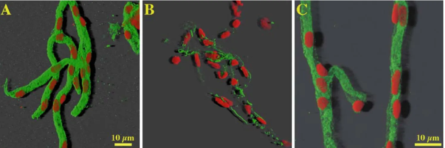
Fig 4A-C Colocalization of P-glycoprotein with brain capillary endothelial cells in freshly isolated porcine brain microvessels. Staining of A endothelial-specific von Willebrand factor (green); B astrocyte-specific glial fibrillary acidic protein (GFAP; green); and C P-glycoprotein antibody C219 (green). To visualize the location
of individual cells in capillaries, nuclei were costained with propidium iodide (red). Size bar is the same for A and B. Images were rendered using three-dimensional image processing and shadow projection
Transendothelial permeability of sucrose
Subsequent to TER measurements, permeability of the
extracellular marker [
14C]sucrose across filters with
con-fluent cells was determined. Interestingly, the apparent
permeability (P
app) of sucrose did not improve using
different cell-culture conditions (e.g., EM: P
app=7.1±0.6;
ACEM: P
app=9.0±0.6; C6CEM: P
app=9.6±0.6; units are
10
−4cm/min; n=5, mean ± SE). Therefore all permeability
coefficients were in the range of the standard cell-culture
condition (EM).
Expression of ABC transporters
Additional studies were initiated to determine the impact
of different cell-culture conditions on the expression of the
ABC transporters P-glycoprotein and the multidrug
resis-tance-associated proteins MRP1 and MRP2.
Immunohis-tochemical analysis of freshly isolated brain capillaries
using a monoclonal antibody (C219) specific for
P-glycoprotein revealed colocalization of the protein with
brain microvessel endothelial cells (Fig. 4).
To evaluate the level of expression of the ABC
transporters, real-time quantitative reverse
transcriptase-polymerase chain reaction (RT-PCR) was performed. Total
RNA was extracted from brain tissue, isolated brain
capillaries, freshly isolated BCEC and BCEC grown for
different time periods in standard EM. Expression levels of
pgp1A, MRP1, and MRP2 were normalized to the internal
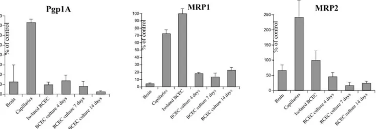
standard GAPDH (Fig. 5). The mRNA levels of all three
ABC transporters were markedly enriched in freshly
isolated brain microvessels when compared with
whole-brain tissue. There was a reduction in mRNA levels in
isolated endothelial cells when compared with intact brain
capillaries for P-glycoprotein and MRP2. There was a
further decrease in mRNA levels of MRP2 only when
freshly isolated endothelial cells were kept in culture,
whereas the mRNA levels of P-glycoprotein and MRP1
did not change during cell culture. To assess whether the
expression pattern of these transporters is altered under
different cell-culture conditions, cDNA of cells cultured
using different cell-culture conditions was assayed by
quantitative real-time PCR. Surprisingly, endothelial cells
expressed under all specified culture conditions
demon-strated the same expression pattern as cells grown in
standard EM.
Discussion
Primary cultures of BCEC form confluent monolayers that
retain many morphological and biochemical properties and
are polarized like their in vivo counterparts as previously
described (Huwyler et al. 1996, 1997; Török et al. 1998;
Gutmann et al. 1999). However, the limitation of cultured
primary brain capillary endothelia is higher than desirable
paracellular permeability, attributed to alterations in the
development of tight intracellular junctions. It has been
shown that the microenvironment can influence the
permeability properties of endothelial cells in situ (Stewart
and Wiley 1981; Janzer and Raff 1987) There are at least
four different cell types that constitute and/or are in close
contact with the brain capillaries, i.e., endothelial cells,
astrocytes, neurons, and pericytes (Pardridge 1993). The
interactions between these cells are multifaceted and
complex. Recent reports are indicative of a dynamic
control of brain microcirculation as a consequence of
neuron to astrocyte signaling (Zonta et al. 2003). Pericytes
are present from the onset of blood
–brain barrier formation
(Korn et al. 2002) and can modulate the endothelial cell
phenotype (Allt and Lawrenson 2001). Recent reports
indicate that endothelial cells and pericytes reorganize in
capillary-like structures when cocultured with astrocytes
(Ramsauer et al. 2002), indicative of intimate interactions
between these cell types. With respect to the aspect of
cellular permeability of cultured endothelial cells,
astro-cyte-derived factors seem to play a central role (Janzer and
Fig 5 Relative quantification of pgp1A, MRP1 and MRP2 mRNA by quantitative RT-PCR. GAPDH was used as an internal standard to correct for equal loading. Values were normalized to isolated
brain capillary endothelial cells (BCEC), which was set at 100%. Values represent mean ± SD of four determinations
Raff 1987). We therefore decided to focus this report on
astrocyte
–endothelial cell interactions. ACEM (Rubin et
al. 1991) and medium that was conditioned by C6 cells, a
glioma cell line, were used (Raub et al. 1992). In addition,
supplements were investigated, which have been shown to
improve cultured BCEC monolayers. We used the cAMP
analog chlorophenylthio-cAMP in combination with the
cAMP-specific phosphodiesterase inhibitor RO-20-1724
(Rubin et al. 1991), dexamethasone (Grabb and Gilbert
1995), or all-trans retinoic acid (Lechardeur et al. 1995).
1,25-Dihydroxyvitamin D
3was shown to regulate
differ-entiation in many cell types, including normal,
immorta-lized, and tumor cells (Reese et al. 1981; Tsao and Batist
1988; Bouillon et al. 1995). Supplement concentrations
used corresponded to effective concentrations published in
the previously cited publications.
Tight junctions probably represent the major functional
component of the blood
–brain barrier and depend very
much on adherens junctions. For example, it has been
shown that antibodies against E-cadherin, the Ca
2+-dependent epithelial cell adhesion molecule, block
tight-junction formation (Gumbiner and Simons 1986). The
presumption is that the normal course of events after cell
contact is cadherin-dependent cell adhesion followed by
tight-junction assembly. To assess the degree of
differen-tiation of cultured BCEC, the degree of tight- and
adherens-junction formation was determined. To judge
tight junctions, the ZO-1 pattern was assessed. Adherens
junctions were rated by the distribution of F-actin, which
closely interacts with the adherens junctions
,
catenin
proteins (Staddon and Rubin 1996; Rubin and Staddon
1999). A scoring system was introduced in order to
generate an ordinal mean to judge the staining patterns. A
high score was assigned to a high degree of differentiation.
Highest scores represented patterns such as the ones
observed in intact brain capillaries. In these capillaries,
distribution of ZO-1 showed an intense staining localized
at the cell-to-cell contact area, whereas F-actin showed a
weak intracellular distribution. This contrasts to cells
grown in EM which showed a disordered F-actin staining
that is associated with weak ZO-1 staining. Culturing in
ACEM alone did not change the F-actin and ZO-1
patterns, whereas C6CEM resulted in a rearrangement of
F-actin to the cell borders and enhanced ZO-1 signals.
BCEC cultures could be further improved by increasing
intracellular cAMP levels and by addition of
dexameth-asone. These supplements were able to raise the TER
value in EM- and ACEM-cultured BCEC and also to
enhance the F-actin and ZO-1 score in cells grown in
ACEM. This suggests that astrocytic factors and cAMP or
dexamethasone use complementary pathways to tighten
the intracellular junctions. The junction modulation by
cAMP implies the existence of one or more proteins
capable of being phosphorylated by protein kinase A,
whose state of phosphorylation controls tight and adherens
junctions (Rubin and Staddon 1999). It should be noted
that F-actin scores were highly correlated with ZO-1
scores. No further improvement of tight junctions in
BCEC could be observed when supplemented with
all-trans retinoic acid or 1,25-dihydroxyvitamin D
3.
Surprisingly, when monolayer permeability was
as-sessed, using the extracellular marker sucrose, cells grown
under different culture conditions showed no statistically
significant differences. This could be due to the fact that
TER measures the flux of ions, whereas sucrose represents
a larger molecule.
It has been shown that ABC transporters play a
functional part in establishing a barrier between the
blood and the brain (Schinkel et al. 1996; Regina et al.
1998; Miller et al. 2000; Fricker et al. 2002; Löscher and
Potschka 2002). Our studies demonstrate colocalization of
P-glycoprotein with the endothelial marker von
Wille-brand factor and not with fragments of astrocyte foot
processes (Fig. 4). Recent publications, however, suggest
some expression of P-glycoprotein and MRP1 by
astro-cytes (Declèves et al. 2000; Golden and Pardridge 2000).
To determine the impact of different culture conditions on
the expression of ABC transporters such as
P-glycopro-tein, MRP1, and MRP2, mRNA levels were assessed by
quantitative RT-PCR. Compared with humans, pigs have
four class I P-glycoprotein genes (pgp1A to pgp1D), but
with only pgp1A being expressed (Childs and Ling 1996).
pgp1A mRNA was highly enriched in cerebral capillaries
compared with whole-brain tissue. However, in isolated
BCEC, pgp1A mRNA was decreased. pgp1A expression
did not substantially change during culture in EM, nor did
it change in different cell-culture conditions. Therefore, an
enhanced tight-junction morphology does not necessarily
correlate with the preservation of P-glycoprotein. Our
findings correspond with those obtained by a recent
publication, where levels of immunoreactive
P-glycopro-tein were markedly decreased in cultures of BCEC. This
effect was less pronounced in cocultures of BCEC with
astrocytes (Gaillard et al. 2000). However, since
P-glycoprotein is lost during the preparation of capillaries,
in vitro experiments are likely to underestimate the
influence of this carrier. The same holds true for MRP1
and MRP2, since mRNA is reduced during cell culture in
the present study. It is interesting to note that other
endothelial cell types, such as immortalized rat cell lines
(El Hafny et al. 1997), or human umbilical vein
endothe-lial cells (Hayashi et al. 1997) are more sensitive to
cell-culture conditions, which results in upregulation of
P-glycoprotein expression.
Expression of MRPs in BCEC has only been reported
recently (Huai-Yun et al. 1998; Miller et al. 2000; Zhang et
al. 2000; Fricker et al. 2002). Using primary cultured
bovine brain capillary endothelial cells, RT-PCR shows
the presence of MRP1, whereas MRP2 is absent (Zhang et
al. 2000). However, using immunostaining of
P-glycopro-tein and MRP2 in isolated capillaries from rat, pig (Miller
et al. 2000), and fish (Miller et al. 2002) brains, both
multidrug transporters were localized to the luminal
surface of the capillary endothelium. It is interesting to
note that MRP2 mRNA levels are about 100 times lower
than MRP1 mRNA levels (estimated by threshold-cycle
differences on real-time quantitative RT-PCR).
The present study shows that the morphology of tight
junctions and intracellular actin of cultured BCEC can be
modulated by astrocyte-conditioned medium in
combina-tion with elevated intracellular cAMP levels or
dexameth-asone. These changes have an impact on the TER, but not
on the paracellular flux of molecules, at least with the size
of sucrose. ABC transporters such as P-glycoprotein,
MRP1, and MRP2 are expressed in cultured BCEC and in
intact brain capillaries of porcine origin. However, mRNA
levels of these transporters are lower. This could lead to an
underestimation of these carriers when assessing uptake or
drug-interactions of different P-glycoprotein substrates.
Monolayers of primary porcine BCEC can be used
nevertheless as a robust cell culture model of the blood
–
brain barrier, since it is insensitive toward changes in
cell-culture conditions.
Acknowledgements This work was supported by the Swiss National Science Foundation (grant 32-052918.97). We thank U. Behrens for excellent technical assistance. We would also like to thank S. Brill and Dr. P. Roberts for careful revision of this manuscript
References
Abbott NJ, Revest PA, Romero IA (1992) Astrocyte-endothelial interaction: physiology and pathology. Neuropathol Appl Neurobiol 18:424–433
Allt G, Lawrenson JG (2001) Pericytes: cell biology and pathology. Cells Tissues Organs 169:1–11
Bouillon R, Okamura WH, Norman AW (1995) Structure–function relationships in the vitamin D endocrine system. Endocr Rev 16:200–257
Childs S, Ling V (1996) Duplication and evolution of the P-glycoprotein genes in pig. Biochim Biophys Acta 1307:205– 212
Declèves X, Regina A, Laplanche JL et al. (2000) Functional expression of P-glycoprotein and multidrug resistance-asso-ciated protein (Mrp1) in primary cultures of rat astrocytes. J Neurosci Res 60:594–601
Dehouck MP, Dehouck B, Schluep C, Lemaire M, Cecchelli R (1995) Drug transport to the brain: comparison between in vitro and in vivo models of the blood–brain barrier. Eur J Pharm Sci 3:357–365
Durieu-Trautmann O, Federici C, Creminon C et al. (1993) Nitric oxide and endothelin secretion by brain microvessel endothelial cells: regulation by cyclic nucleotides. J Cell Physiol 155:104– 111
El Hafny B, Chappey O, Piciotti M, Debray M, Boval B, Roux F (1997) Modulation of P-glycoprotein activity by glial factors and retinoic acid in an immortalized rat brain microvessel endothelial cell line. Neurosci Lett 236:107–111
Franke H, Galla HJ, Beuckmann CT (1999) An improved low-permeability in vitro-model of the blood–brain barrier: transport studies on retinoids, sucrose, haloperidol, caffeine and manni-tol. Brain Res 818:65–71
Fricker G, Nobmann S, Miller DS (2002) Permeability of porcine blood brain barrier to somatostatin analogues. Br J Pharmacol 135:1308–1314
Gaillard PJ, Sandt IC van der, Voorwinden LH et al. (2000) Astrocytes increase the functional expression of P-glycoprotein in an in vitro model of the blood–brain barrier Pharm Res 17:1198–1205
Golden PL, Pardridge WM (2000) Brain microvascular P-glyco-protein and a revised model of multidrug resistance in brain. Cell Mol Neurobiol 20:165–181
Grabb PA, Gilbert MR (1995) Neoplastic and pharmacological influence on the permeability of an in vitro blood–brain barrier. J Neurosurg 82:1053–1058
Gumbiner B, Simons K (1986) A functional assay for proteins involved in establishing an epithelial occluding barrier: iden-tification of a uvomorulin-like polypeptide. J Cell Biol 102:457–468
Gutmann H, Török M, Fricker G et al. (1999) Modulation of multidrug resistance protein expression in porcine brain capillary endothelial cells in vitro. Drug Metab Dispos 27:937–941
Hayashi Y, Nomura M, Yamagishi S et al. (1997) Induction of various blood–brain barrier properties in non-neural endothelial cells by close apposition to cocultured astrocytes. Glia 19:13– 26
Huai-Yun H, Secrest DT, Mark KS et al. (1998) Expression of multidrug resistance-associated protein (MRP) in brain micro-vessel endothelial cells. Biochem Biophys Res Commun 243:816–820
Huwyler J, Drewe J, Klusemann C, Fricker G (1996) Evidence for P-glycoprotein-modulated penetration of morphine-6-glucuro-nide into brain capillary endothelium. Br J Pharmacol 118:1879–1885
Huwyler J, Fricker G, Török M, Schneider M, Drewe J (1997) Transport of clonidine across cultured brain microvessel endothelial cells. J Pharmacol Exp Ther 282:81–85
Janzer RC, Raff MC (1987) Astrocytes induce blood–brain barrier properties in endothelial cells. Nature 325:253–257
Klein I, Sarkadi B, Varadi A (1999) An inventory of the human ABC proteins. Biochim Biophys Acta 1461:237–262
Korn J, Christ B, Kurz H (2002) Neuroectodermal origin of brain pericytes and vascular smooth muscle cells. J Comp Neurol 442:78–88
Lechardeur D, Schwartz B, Paulin D, Scherman D (1995) Induction of blood–brain barrier differentiation in a rat brain-derived endothelial cell line. Exp Cell Res 220:161–170
Lillien LE, Sendtner M, Rohrer H, Hughes SM, Raff MC (1988) Type-2 astrocyte development in rat brain cultures is initiated by a CNTF-like protein produced by type-1 astrocytes. Neuron 1:485–494
Löscher W, Potschka H (2002) Role of multidrug transporters in pharmacoresistance to antiepileptic drugs. J Pharmacol Exp Ther 301:7–14
Meyer J, Rauh J, Galla HJ (1991) The susceptibility of cerebral endothelial cells to astroglial induction of blood–brain barrier enzymes depends on their proliferative state. J Neurochem 57:1971–1977
Miller DS, Nobmann SN, Gutmann H et al. (2000) Xenobiotic transport across isolated brain microvessels studied by confocal microscopy. Mol Pharmacol 58:1357–1367
Miller DS, Graeff C, Droulle L, Fricker S, Fricker G (2002) Xenobiotic efflux pumps in isolated fish brain capillaries. Am J Physiol Regul Integr Comp Physiol 282:191–198
Pardridge WM (1993) Brain drug delivery and blood–brain barrier transport. Drug Delivery 1:83–101
Pardridge WM, Eisenberg J, Yamada T (1985) Rapid sequestration and degradation of somatostatin analogues by isolated brain microvessels. J Neurochem 44:1178–1184
Pardridge WM, Triguero D, Yang J, Cancilla PA (1990) Comparison of in vitro and in vivo models of drug transcytosis through the blood–brain barrier. J Pharmacol Exp Ther 253:884–891 Ramsauer M, Krause D, Dermietzel R (2002) Angiogenesis of the
blood–brain barrier in vitro and the function of cerebral pericytes. FASEB J 16:1274–1276
Raub TJ (1996) Signal transduction and glial cell modulation of cultured brain microvessel endothelial cell tight junctions. Am J Physiol Cell Physiol 271:495–503
Raub TJ, Kuentzel SL, Sawada GA (1992) Permeability of bovine brain microvessel endothelial cells in vitro: barrier tightening by a factor released from astroglioma cells. Exp Cell Res 199:330–340
Reese DH, Fiorentino GJ, Claflin AJ, Malinin TI, Politano VA (1981) Rapid induction of alkaline phosphatase activity by retinoic acid. Biochem Biophys Res Commun 102:315–321 Regina A, Koman A, Piciotti M et al. (1998) Mrp1 multidrug
resistance-associated protein and P-glycoprotein expression in rat brain microvessel endothelial cells. J Neurochem 71:705– 715
Rist RJ, Romero IA, Chan MW et al. (1997) F-actin cytoskeleton and sucrose permeability of immortalised rat brain microvas-cular endothelial cell monolayers: effects of cyclic AMP and astrocytic factors. Brain Res 768:10–18
Rubin LL, Staddon JM (1999) The cell biology of the blood–brain barrier. Annu Rev Neurosci 22:11–28
Rubin LL, Hall DE, Porter S et al. (1991) A cell culture model of the blood–brain barrier. J Cell Biol 115:1725–1735
Schinkel AH, Wagenaar E, Mol CA, Deemter L van (1996) P-glycoprotein in the blood–brain barrier of mice influences the brain penetration and pharmacological activity of many drugs. J Clin Invest 97:2517–2524
Staddon JM, Rubin LL (1996) Cell adhesion, cell junctions and the blood–brain barrier. Curr Opin Neurobiol 6:622–627
Stewart PA, Wiley MJ (1981) Developing nervous tissue induces formation of blood–brain barrier characteristics in invading endothelial cells: a study using quail-chick transplantation chimeras. Dev Biol 84:183–192
Takakura Y, Audus KL, Borchardt RT (1991) Blood–brain barrier: transport studies in isolated brain capillaries and in cultured brain endothelial cells. Adv Pharmacol 22:137–165
Török M, Huwyler J, Drewe J, Gutmann H, Fricker G (1998) Transport of the β-lactam antibiotic benzylpenicillin and the dipeptide glycylsarcosine by brain capillary endothelial cells in vitro. Drug Metab Dispos 26:1144–1148
Tsao MS, Batist G (1988) Induction ofγ-glutamyl transpeptidase activity by all-trans retinoic acid in cultured rat liver epithelial cells. Biochem Biophys Res Commun 157:1039–1045 Zhang Y, Han H, Elmquist WF, Miller DW (2000) Expression of
various multidrug resistance-associated protein (MRP) homo-logues in brain microvessel endothelial cells. Brain Res 876:148–153
Zonta M, Angulo MC, Gobbo S. (2003) Neuron-to-astrocyte signaling is central to the dynamic control of brain microcir-culation. Nat Neurosci 6:43–50