Lars Husmann Bernhard A. Herzog Nina Burkhard Fuminari Tatsugami Ines Valenta Oliver Gaemperli Christophe A. Wyss Ulf Landmesser Philipp A. Kaufmann Received: 16 October 2008 Revised: 12 December 2008 Accepted: 1 January 2009
Published online: 24 February 2009
# European Society of Radiology 2009
Body physique and heart rate variability
determine the occurrence of stair-step
artefacts in 64-slice CT coronary angiography
with prospective ECG-triggering
Abstract The purpose of this study was to describe and characterize the frequency and extent of stair-step artefacts in computed tomography coronary angiography (CTCA) with prospective electrocardiogram (ECG)-triggering and to identify their deter-minants. One hundred and forty three consecutive patients (55 women, mean age 57±13 years) underwent 64-slice CTCA using prospective ECG-triggering. Occurrence of stair-step artefacts in CTCA of the thoracic wall and the coronary arteries was deter-mined and maximum offset was mea-sured. If stair-step artefacts occurred in both cases, a difference between thoracic wall and coronary artery offset of 0.6 mm or greater was attributed to additional motion of the heart. Mean effective radiation dose was 2.1±0.7 mSv (range 1.0– 3.5 mSv). Eighty-nine patients (62%) had stair-step artefacts in CTCA of the
coronary arteries (mean offset of 1.7± 1.1 mm), while only 77 patients had thoracic wall stair-step artefacts (mean offset of 1.0±0.3 mm; significantly different, P<0.001). Stair-step arte-facts in CTCA of the thoracic wall were determined by BMI and weight (P<0.01), while artefacts in CTCA of the coronary arteries were associated with heart rate variability (P<0.05). Stair-step artefacts in CTCA with prospective ECG-triggering are deter-mined by (a) motion of the entire patient during table travel, particularly in large patients and (b) by motion of the heart, particularly when heart rates are variable.
Keywords Stair-step artefacts . Low dose . Computed tomography coronary angiography . Prospective ECG-gating . Prospective
ECG-triggering
Introduction
New computed tomography coronary angiography (CTCA) acquisition protocols with prospective electrocardiogram (ECG)-triggering have recently been introduced [1] and shown to offer a tremendous reduction of radiation dose [2– 14], which appears to be a major breakthrough for non-invasive imaging of the coronary arteries. When using this technique with a 64-slice CT system, the z-coverage during image acquisition is only 40 mm, thus three to five data sets need to be acquired subsequently to image the entire heart. Between two adjacent data sets, the CT table needs to travel rapidly and the heart will usually beat twice [2]. If the patient
(and his heart) position relative to the table remains unchanged during table travel and throughout consecutive heart beats then perfect alignment of two data sets is feasible. However, stair-step artefacts due to misalignment of two adjacent data sets may occur when the position of the patient on the CT table changes during acceleration and deceleration during table travel, when the patient breathes, or when the heart does not return to the exact same position within the thorax as during the previous data acquisition.
In order to optimize future CT parameters, the purpose of this study was to describe and characterize the frequency and extent of stair-step artefacts in CTCA with prospective ECG-triggering and to identify their determinants. L. Husmann . B. A. Herzog .
N. Burkhard . F. Tatsugami . I. Valenta . O. Gaemperli . C. A. Wyss .
U. Landmesser . P. A. Kaufmann (*) Nuclear Cardiology,
University Hospital Zurich, Ramistrasse 100, 8091 Zurich, Switzerland e-mail: [email protected] Tel.: +41-44-2554196 Fax: +41-44-2554414 P. A. Kaufmann
Zurich Center for Integrative Human Physiology, University of Zurich, Zurich, Switzerland
Materials and methods Patients
One hundred and forty three consecutive patients with suspected (n=122) or known (n=21) CAD referred to CTCA were prospectively enrolled in the present study if none of the following exclusion criteria were present: hypersensitivity to iodinated contrast agent, renal insuffi-ciency (creatinine levels >150 µmol/L, or >1.7 mg/dL), non-sinus rhythm, or heart rates above 65 bpm when beta-blocker medication was not feasible; in fact, 11 additional patients did not meet the inclusion criteria because CTCA was not feasible due to non-sinus rhythm (n=6), or because heart rate could not be sufficiently reduced with beta-blocker medication (n=5). In addition, 13 patients were not included because they did not follow the breathing commands properly.
The study protocol was approved by the institutional review board and written informed consent was obtained.
CT data acquisition and postprocessing
All patients were thoroughly instructed about the course of events during the examination. A single dose of 2.5 mg isosorbide dinitrate sublingual (Isoket, Schwarz Pharma, Monheim, Germany) was administered to all patients 2 min before the scan. In addition, metoprolol (5 to 20 mg) (Beloc, AstraZeneca, London, UK) was administered intravenously before the CTCA examination if necessary to achieve a target heart rate below 65 bpm. Furthermore, the inspiration level (shallow, normal or deep inspiration) with the lowest and most stable heart rate was individually determined, and breathing commands were practised repetitively before obtaining CT data. For CTCA, 80 mL iodixanol (Visipaque 320, 320 mg/mL, GE Heathcare, Buckinghamshire, UK) was injected at a flow rate of 5 mL/s into an antecubital vein via an 18-gauge catheter followed by 50 mL saline solution. Bolus tracking was performed with a region of interest placed into the ascending aorta, and image acquisition was started 4 s after the signal density reached a predefined threshold of 120 Hounsfield units (HU).
All CTCA examinations were performed with a Light-Speed VCT XT scanner (GE Healthcare) and prospective triggering using a commercially available protocol (Snap-Shot Pulse, GE Healthcare) and the following CT parameters: slice acquisition 64×0.625 mm, smallest x-ray window (only 75% of the RR cycle), z-coverage 40 mm with an increment of 35 mm, gantry rotation time 350 ms, body mass index (BMI) adapted tube voltage (100 kV, BMI<25 kg/m2; 120 kV, BMI≥25 kg/m2) and effective tube current (450 mA, BMI<22.5 kg/m2; 500 mA, BMI 22.5–25 kg/m2; 550 mA, BMI 25–27.5 kg/m2, 600 mA, BMI 27.5–30 kg/m2; 650 mA, BMI 30–40 kg/m2; 700 mA,
BMI>40 kg/m2). The anatomical range for CTCA extended from just below the tracheal bifurcation to the diaphragm, choosing three to five blocks (field of view 11–18 cm). By choosing the smallest possible window at only one distinct end-diastolic phase of the RR cycle (i.e. 75%) we ascertained the lowest achievable effective dose delivery; the effective dose of CTCA was calculated as the product of the dose–length product (DLP) times a conver-sion coefficient for the chest (k=0.017 mSv mGy−1cm−1) as previously suggested [15, 16]. Heart rate variability was assessed as the standard deviation of the heart rate throughout the scan as previously reported [17]. All patients were carefully monitored during the examination in an effort to assure that breathing commands were adequately followed. All images were transferred to an external workstation (AW 4.4, GE Healthcare).
CT image analysis
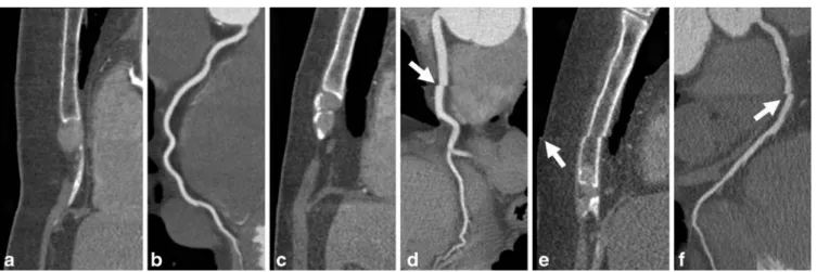
First, two readers in consensus determined if stair-step artefacts occurred on sagittal reconstructed images of the thoracic wall or if all data sets (three to five sequential image stacks) were perfectly aligned (Fig. 1a, b and c). Second, both readers determined whether stair-step artefacts occurred on axial, coronal, sagittal and multi-planar reconstructed images of any coronary artery segment [18] (Fig. 1d, e and f). Furthermore, one reader used an electronic caliper tool to measure the maximum offset between two data sets (i.e. the maximum offset of an anatomic structure in a stair-step artefact) for the thoracic wall and coronary segment with the most severe stair-step artefact. Finally, the differences between the offset in the thoracic wall and the offset in the coronary arteries (in millimetres) were calculated. A difference larger than 0.6 mm (i.e. larger than the isotropic spatial resolution) was considered to be relevant (Fig.1c and d).
Statistical analysis
Quantitative variables were expressed as mean ± standard deviation, and categorical variables as frequencies or percentages. Differences in the occurrence of stair-step artefacts in CTCA of the thoracic wall and coronary arteries were tested for significance by usingχ2tests for compar-ison of cross tables. Measurement differences compared between the maximum offset in a stair-step artefact in CTCA of the thoracic wall and in the coronary arteries in a patient were determined using the Wilcoxon signed rank test. Mann–Whitney U tests were used to determine differences in heart rate, heart rate variability, BMI and weight in groups of patients with or without stair-step artefacts in CTCA of the thoracic wall and in patient groups without or with (or with pronounced) stair-step artefacts in the coronary arteries. A P value of less than 0.05 was
considered to indicate statistical significance. SPSS soft-ware (SPSS 15.0, Chicago, ILL, USA) was used for statistical testing.
Results
CTCA was successfully performed in 143 patients; demographics are given in Table1.
The mean heart rate of the study population was 57.6± 6.1 bpm (range 44–75 bpm), the heart rate variability 1.5± 1.0 bpm (range 0.2–5.3 bpm), the mean BMI was 25.6± 3.7 kg/m2 (range 18.2–38.8 kg/m2) and the mean weight was 74.9±14.2 kg (range 46–115 kg). Thirty-six of 143 patients (25%) were on beta-blocker medication as part of their baseline medication; additional beta-blockers were administered intravenously to 96 patients (67%) for heart rate control before CTCA. The mean DLP from CTCA was 125.8±40.7 mGy cm (range 58.3–207.9 mGy cm) result-ing in an estimated mean applied radiation dose of 2.1± 0.7 mSv (range 1.0–3.5 mSv).
Eighty-nine of 143 patients (62%) had stair-step artefacts in CTCA of the coronary arteries with a mean offset of 1.7±1.1 mm, while only 77 of them (54%) had thoracic wall stair-step artefacts (mean offset of 1.0±0.3 mm). Thus, stair-step artefacts were significantly more frequent in the coronary arteries than in the thoracic wall (P<0.001), with a significant offset difference of 0.5±1.1 mm (P<0.001). In 41 patients the offset difference was 0.6 mm or greater suggesting that the stair-step artifact was predominately caused by motion of the heart, rather than by motion of the entire thorax.
Determinants of stair-step artefacts
The occurrence of stair-step artefacts in CTCA of the thoracic wall was associated with higher BMI and weight (P<0.01, Figs.1e, f and2), but not with heart rate or heart rate variability (P=0.64 and P=0.06, respectively).
On the other hand, the occurrence of stair-step artefacts in CTCA of the coronary arteries (either more pronounced Fig. 1 Sagittal reformations of the thoracic wall (a, c and e) and
curved multiplanar reformations of the right coronary artery (b, d and f) in three different patients. a and b demonstrate perfect alignment of the thoracic wall and of the coronary arteries in a patient with a mean heart rate of 59 bpm, a heart rate variability of 1.0 bpm, a BMI of 23.4 kg/m2 and a weight of 62 kg. c and d demonstrate perfect alignment of the thoracic wall, but a stair-step artefact (maximum offset 2.6 mm, arrow) in the CTCA of coronary
arteries in a patient with a mean heart rate of 54 bpm, a heart rate variability of 3.0 bpm, a BMI of 25.8 kg/m2and a weight of 79 kg. e and f demonstrate stair-step artefacts in the CTCA of the thoracic wall (maximum offset 1.5 mm, arrow) and in the right coronary artery (maximum offset 2.0 mm, arrow) in an obese patient with a mean heart rate of 50 bpm, a heart rate variability of 1.6 bpm, a BMI of 33.6 kg/m2and a weight of 85 kg
Table 1 Patient demographics
Number of patients 143
Age in years (mean±SD) 57±13
Female 55
Male 88
Body mass index in kg/m2(mean±SD) 26±4
Weight in kg 75±14
Coronary risk factors
Smokers 49
Hypertension 76
Diabetes 11
Positive family history 46
Dyslipidemia 65
Clinical symptoms
None 36
Typical angina 22
Atypical chest pain 68
than in the thoracic wall or exclusively occurring in the heart) were associated with high heart rate variability (P< 0.05, Figs. 1c, d and3), but not with heart rate (P=0.62, Fig.3), BMI or weight (P=0.12 and P=0.09, respectively).
Discussion
The description and characterization of stair-step artefacts in CTCA with prospective ECG-triggering and the validation of their determinants are essential for identifying the patient population that will benefit most from this new scanning technique. Our study adds the following results to the previous knowledge [2–12] on CTCA with prospective ECG-triggering: (i) Stair-step artefacts are more frequent and more severe in the coronary arteries than in the thoracic wall. (ii) Thoracic wall stair-step artefacts are always accompanied by artefacts in the coronary arteries and are determined by motion of the entire patient during table travel, which occurs particularly in large patients. (iii)
Stair-step artefacts which occur predominantly in the coronary arteries are determined by heart rate variability.
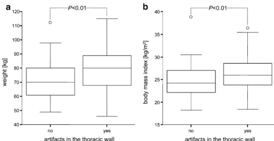
When the heart rate varies during CTCA, dispropor-tional shortening and prolongation of the cardiac phases occurs [19–21]. In CTCA with prospective ECG-triggering data acquisition is performed at a fixed percentage interval of the cardiac cycle, and with varying heart rate imaging will be performed in slightly different phases of the cardiac cycle. If this happens, perfect alignment of consecutive data sets is not feasible, and coronary artery stair-step artefacts will occur [17]. This is line with results from Shapiro et al. reporting a significant though weak relation between heart rate variability and the occurrence of stairs-step artefacts in a 64-slice CTCA, using a helical CT protocol [22]. The latter may account for the fact that stair-step artefacts were considerably less frequent in those patients (18 of 150 patients) compared with our study population (41 of 143 patients). In helical CTCA table travel is continuous during image acquisition, and therefore an impact of body physique on the occurrence of stair-step Fig. 2 Box plots demonstrate
mean weight (a) and mean BMI (b) in patients with and without stair-step artefacts in CTCA of the thoracic wall. Mean weight and mean BMI significantly differed between both groups (P <0.01). Box = 1st to 3rd quar-tiles, midline = median, whis-kers = minimum and maximum values, circle = mild outlier, asterisk = extreme outlier
Fig. 3 Box plots demonstrate mean heart rate variability (a) and mean heart rate (b) in patients with and without stair-step artefacts in CTCA of the coronary arteries. Mean heart rate variability significantly dif-fered between both groups (P< 0.05), while the mean heart rate did not (P = not significant, n. s.). Box = 1st to 3rd quartiles, midline = median, whiskers = minimum and maximum values, circle = mild outlier,
artefacts appears to be less likely, further explaining the higher prevalence of stair-step artefacts in the present study compared with the previously mentioned study [22]. However, the higher prevalence of stair-step artefacts caused by cardiac motion in our study may also have been favoured by our low-dose scanning protocol with a narrow acquisition window (no “padding”), precluding image reconstruction in another phase [9,23,24].
Stair-step artefacts in CTCA can also be secondary to respiratory motion during data acquisition [25, 26] and then they affect both the thoracic wall and the coronary arteries. In the present study respiratory motion was carefully excluded as a cause for stair-step artefacts, and we found that stair-step artefacts in CTCA with prospec-tive ECG-triggering that occur simultaneously in the thoracic wall and in the coronary arteries are more frequent in heavy and obese patients. This is most likely caused by relative motion of the patient versus the CT table during rapid acceleration and deceleration of the table, required to move the patient between blocks of acquisition in the prospective triggering mode. This effect is more pronounced in heavy and particularly obese patients with unfavourable body fat distribution, aggravating the potential for stair-step artefacts.
Notably, the occurrence of stair-step artefacts may vary with different CT systems. Current dual-source scanners [8, 11] have a smaller detector width, which may increase the likelihood of stair-step artefacts, as more scanning steps are need to cover the entire heart. On the other hand, the latest generations of CT systems with 256 and more slices have a large detector width allowing them to cover the entire heart
within one heart beat. Thus, avoiding the need for table movement, prospective triggering can be successfully used in such devices [27] to reduce radiation dose without stair-step artefacts caused by heart rate variability and table movement.
We acknowledge the following limitations to our study. Quantification of stair-step artefacts is only possible in the x- and y-plane, while no measurements are feasible in the z-plane. However, as a result of the curved anatomy of the thoracic wall and the coronary arteries, one can reasonably assume that the measured offset in the x- and y-plane provides an adequate estimate for the extent of stair-step artefacts in all three dimensions.
Furthermore, we did not assess the impact of stair-step artefacts on the diagnostic accuracy of CTCA by compar-ing our findcompar-ings with the reference standard invasive coronary angiography. However, first reports on diagnostic accuracy of CTCA with prospective ECG-triggering [7–9] have demonstrated that the new technique is equally as accurate as CTCA with retrospective ECG-gating. There-fore, the impact of stair-step artefacts on diagnostic accuracy appears to be of limited relevance. Nonetheless, further studies are required to confirm this hypothesis.
In conclusion, stair-step artefacts in CTCA with prospective ECG-triggering are determined (a) by motion of the entire patient during table travel, particularly in large patients and (b) by motion of the heart, particularly when heart rates are variable. Our findings suggest that even a perfectly regular heart rate may not entirely eliminate stair-step artefacts, as these can only be fully avoided by eliminating the need for table travel.
References
1. Hsieh J, Londt J, Vass M et al (2006) Step-and-shoot data acquisition and reconstruction for cardiac x-ray com-puted tomography. Med Phys 33:4236– 4248
2. Husmann L, Valenta I, Gaemperli O et al (2008) Feasibility of low-dose coro-nary CT angiography: first experience with prospective ECG-gating. Eur Heart J 29:191–197
3. Husmann L, Valenta I, Kaufmann PA (2008) Coronary angiography with low-dose computed tomography at 1.4 mSv. Herz 33:75
4. Earls JP, Berman EL, Urban BA et al (2008) Prospectively gated transverse coronary CT angiography versus retro-spectively gated helical technique: im-proved image quality and reduced radiation dose. Radiology 246:742–753
5. Hirai N, Horiguchi J, Fujioka C et al (2008) Prospective versus retrospective ECG-gated 64-detector coronary CT angiography: assessment of image quality, stenosis, and radiation dose. Radiology 248:424–430
6. Shuman WP, Branch KR, May JM et al (2008) Prospective versus retrospective ECG gating for 64-detector CT of the coronary arteries: comparison of image quality and patient radiation dose. Ra-diology 248:431–437
7. Herzog BA, Husmann L, Burkhard N et al (2008) Accuracy of low-dose com-puted tomography coronary
angiography using prospective electro-cardiogram-triggering: first clinical experience. Eur Heart J 29(24):3037– 3042
8. Scheffel H, Alkadhi H, Leschka S et al (2008) Low-dose CT coronary angiog-raphy in the step-and-shoot mode: diagnostic performance. Heart 94:1132–1137
9. Maruyama T, Takada M, Hasuike T et al (2008) Radiation dose reduction and coronary assessability of prospective electrocardiogram-gated computed tomography coronary angiography. JACC 52:1450–1455
10. Kaufmann PA (2008) Low-dose com-puted tomography coronary angiogra-phy with prospective triggering: a promise for the future. J Am Coll Cardiol 52:1456–1457
11. Stolzmann P, Leschka S, Scheffel H et al (2008) Dual-source CT in step-and-shoot mode: noninvasive coronary an-giography with low radiation dose. Radiology 249:71–80
12. Klass O, Jeltsch M, Feuerlein S et al (2008) Prospectively gated axial CT coronary angiography: preliminary ex-periences with a novel low-dose tech-nique. Eur Radiol. doi:10.1007/ s00330-008-1222-4
13. Stolzmann P, Scheffel H, Schertler T et al (2008) Radiation dose estimates in dual-source computed tomography coronary angiography. Eur Radiol 18:592–599
14. Achenbach S, Anders K, Kalender WA (2008) Dual-source cardiac computed tomography: image quality and dose considerations. Eur Radiol 18:1188– 1198
15. Einstein AJ, Moser KW, Thompson RC, Cerqueira MD, Henzlova MJ (2007) Radiation dose to patients from cardiac diagnostic imaging. Circulation 116:1290–1305
16. Hurwitz LM, Reiman RE, Yoshizumi TT et al (2007) Radiation dose from contemporary cardiothoracic multide-tector CT protocols with an anthropo-morphic female phantom: implications for cancer induction. Radiology 245:742–750
17. Leschka S, Wildermuth S, Boehm T et al (2006) Noninvasive coronary angi-ography with 64-section CT: effect of average heart rate and heart rate vari-ability on image quality. Radiology 241:378–385
18. Austen WG, Edwards JE, Frye RL et al (1975) A reporting system on patients evaluated for coronary artery disease. Report of the Ad Hoc Committee for Grading of Coronary Artery Disease, Council on Cardiovascular Surgery, American Heart Association. Circula-tion 51:5–40
19. Kovacs SJ Jr (1985) The duration of the QT interval as a function of heart rate: a derivation based on physical principles and a comparison to measured values. Am Heart J 110:872–878
20. Husmann L, Leschka S, Desbiolles L et al (2007) Coronary artery motion and cardiac phases: dependency on heart rate implications for CT image recon-struction. Radiology 245:567–576 21. Herzog C, Abolmaali N, Balzer JO et al
(2002) Heart-rate-adapted image re-construction in multidetector-row car-diac CT: influence of physiological and technical prerequisite on image quality. Eur Radiol 12:2670–2678
22. Shapiro MD, Pena AJ, Nichols JH et al (2008) Efficacy of pre-scan beta-blockade and impact of heart rate on image quality in patients undergoing coronary multidetector computed tomography angiography. Eur J Radiol 66:37–41
23. Schoenhagen P (2008) Back to the future: coronary CT angiography using prospective ECG triggering. Eur Heart J 29:153–154
24. Brodoefel H, Burgstahler C, Tsiflikas I et al (2008) Dual-source CT: effect of heart rate, heart rate variability, and calcification on image quality and diagnostic accuracy. Radiology 247:346–355
25. Nakanishi T, Kayashima Y, Inoue R, Sumii K, Gomyo Y (2005) Pitfalls in 16-detector row CT of the coronary arteries. Radiographics 25:425–438 discussion 438–440
26. Choi HS, Choi BW, Choe KO et al (2004) Pitfalls, artifacts, and remedies in multi- detector row CT coronary angiography. Radiographics 24:787– 800
27. Rybicki FJ, Otero HJ, Steigner ML et al (2008) Initial evaluation of coronary images from 320-detector row com-puted tomography. Int J Cardiovasc Imaging 24:535–546