Distribution and density of a- and /?-adrenergic receptor binding
sites in the bovine mammary gland
B Y HARALD M. HAMMON, RUPERT M. BRUCKMAIER,
ULRICH E. HONEGGER* AND JURG W. BLUMf
Institut fiir Tierzucht der Universitdt Bern, CH-3012 Bern, Schweiz * Pharmakologisches Institut der Universitat Bern, G'H-3010 Bern, Schweiz
(Received 7 January 1993 and accepted for publication 7 May 1993)
SUMMARY. Radioreceptor binding studies were designed to localize and determine
the number of a- and /S-adrenergic receptors in the mammary gland of lactating cows. 3H-prazosin, 3H-rauwolscine and 3H-dihydroalprenolol were used for the regional characterization of ar, a2- and /?-adrenergic receptors by competitive
inhibition of binding of 3H-ligands with unlabelled adrenergic agonists and
antagonists. The a.x-, cc2- and /?2-adrenergic receptor subtypes could thus be demonstrated in the regions of the teats, large mammary ducts and parenchyma. Tissues of the teat wall, of the large mammary ducts above the gland cistern and of the mammary parenchyma were prepared to determine the density of a1; a2- and /?-receptors by saturation binding assays using 3H-prazosin, 3H-rauwolscine and 3 H-dihydroalprenolol respectively. Binding to high affinity sites was reversible within minutes and saturable. Equilibrium was reached within minutes. The number of ar and a2-adrenergic receptors decreased from the teat to the mammary ducts to the parenchyma. Most of the a.x- and a2-adrenergic receptors were found in the teat wall, whereas in the parenchyma a-adrenergic receptors were absent or barely detectable. The density of /?-adrenergic receptors was similar in the teat wall and the large mammary ducts, but much lower in the parenchyma. Thus, a.x-, a2- and /?-adrenergic receptors were found mainly in the milk purging system and hardly at all in mammary parenchyma. Inhibition of milk removal by a-adrenergic stimulation is possibly due to constriction of teat wall and to constriction of the mammary ducts, whereas enhanced milk flow after /?-adrenergic stimulation is possibly due to relaxation not only of the teat sphincter and teat wall, but probably also of the large mammary ducts.
The sympathetic nervous system is involved in peripheral modulation of milk removal. Milk yield was decreased during adrenalin, noradrenaline or a-specific adrenergic agonist (a-Ag) administration. The inhibition of milk removal by the a-Ag phenylephrine was shown to be dose-dependent (H. M. Hammon, unpublished observations). The effects were not mediated by inhibition of endogenous oxytocin release during milking (Lefcourt & Akers, 1984; Blum et al. 1989). Furthermore, the inhibition of milk let-down by adrenergic drugs could be abolished by a-, but not by /?-adrenergic blockade (Blum et al. 1989). Contraction of both teat and gland cistern was observed using ultrasonography after a-Ag administration (Bruckmaier & Blum, 1992). Inhibition of milk removal by premilking electroshocks was abolished by
48 H . M. H A M M O N AND OTHERS
a-adrenergic blockade (Lefcourt & Akers, 1982; Blum et al. 1989). These studies support the notion that catecholamines mediate their inhibitory effects on milk removal through stimulation of a-adrenergic receptors located in the bovine mammary gland (Blum et al. 1989; Bruckmaier et al. 1991), most likely on smooth muscle cells not only in teats, but also in mammary ducts (Ziegler & Mosimann, 1960). On the other hand, enhanced milk flow after administration of /?-adrenergic agonists (/?-Ag) was ascribed to relaxation of the teat muscles and muscles around milk ducts leading from the alveolar tissue to the gland cistern (Hamann, 1981; Bruckmaier et al. 1991). Binding studies using radiolabelled adrenergic compounds as ligands indicated the existence of both a2- and a2-adrenergic receptor subtypes and of the /?2-adrenergic receptor subtype, most likely on smooth muscles in the wall of the bovine teat, thus confirming radioligand binding studies on smooth muscles of the teat (Roets et al. 1984; Roets & Peeters, 1985, 1986). Adrenergic receptor density in the teat muscles was related to milkability of lactating cows (Roets et al. 1989).
Because previous in vivo investigations suggested a- and /?-adrenergic effects not only in the teat, but also in other regions of the udder (Bruckmaier et al. 1991; Bruckmaier & Blum, 1992), the goal of the present investigations was the determination of a- and /?-adrenergic receptors not only in the teat, but also in the region of the large mammary ducts and in the mammary parenchyma. The possible importance of the different regions of the mammary gland for inhibition of milk removal mediated by a-adrenergic receptors and enhancement of milk flow mediated by /?-adrenergic receptor interaction could thus be evaluated.
MATERIALS AND METHODS
Preparation of membranes
Tissues from three regions of lactating udders were used: the muscular layer of the teat (teat region), tissue around the gland cistern including the large mammary ducts (duct region), and mammary parenchyma from the dorsal udder in which mammary ducts were not visible macroscopically (parenchyma region). The material was removed immediately after slaughter and transferred on ice. The tissues of duct and parenchyma regions were minced into small pieces with scissors and put into ice cold 50 mM-Tris-HCl buffer, pH 7-4. The Tris-HCl buffer used in our assays contained 6 mM-MgCl2, essential for binding of the radiolabelled ligand (Moor et al. 1988), and 1 mM-EGTA to chelate metals without affecting ligand binding (Parini et al. 1987). Teats were prepared as described by Roets et al. (1984). Internal mucosa and epidermis were removed, and the sphincter area of the teat was cut off and discarded. The tissue was suspended in ~ 10-15 volumes of buffer. All subsequent manipula-tions were carried out at 4 °C. The material was homogenized four times for 20 s at low speed (8000 rev./min) with an Ultra-Turrax homogenizer (T 25, Janke & Kunkel, Staufen, Germany). The homogenate was centrifuged at 500 g for 10 min; the supernatant was passed through two layers of cheesecloth and then centrifuged at 49000 £ and 4 CC for 30 min (Moor et al. 1988). The resulting supernatant was decanted completely and the remaining pellet was suspended in ice cold buffer (as above) by a motor-driven Glass-Teflon homogenizer.
The protein concentration was determined using a kit (BCA Protein Assay Reagent; Pierce, Rockford, IL, USA) and the membrane suspension was then adjusted to 200-300 /tg protein/100 fi\. Specific 3H-ligand binding was plotted against protein concentration and was shown to increase with increasing membrane protein concentration, as expected (results not shown). Protein concentration was
kept constant (200 or 300 /tg/100 /A) throughout. While membrane suspensions used for competitive binding assays were frozen in liquid nitrogen and stored at —80 °C, saturation binding assays were carried out immediately after preparation of the membrane suspension. Freezing of membranes did not affect binding as such, but preliminary studies demonstrated some reduction in the number of binding sites (Shi
et al. 1989). Binding studies
aj-Adrenergic receptor binding was tested by use of 3H-prazosin, a highly <xr selective antagonist (Shi et al. 1989), a2-adrenergic receptor binding with 3 H-rauwolscine, an a2-selective antagonist (Shi et al. 1989), and /?-adrenergic binding with 3H-dihydroalprenolol (3H-DHA), a non-selective /?-adrenergic antagonist (Hancock et al. 1979). Saturation binding assays were performed to determine binding capacity. Competitive binding assays with different specific unlabelled ligands were performed to characterize the receptor subtype.
Saturation binding assays. These assays were carried out to investigate the
distribution of 3H-prazosin (n = 7), 3H-rauwolscine (n = 4) and 3H-DHA (n = 7) binding sites in teat, duct and parenchyma regions, where n is the number of udders. Membrane suspensions (100 /tl) were incubated with increasing concentrations of the 3H-labelled ligand with or without unlabelled ligand (50 fi\) at 37 °C for 15 min with constant shaking to determine total and non-specific binding. All drugs were diluted in the Tris-HCl buffer described above. The binding reaction was terminated by adding 1 ml of ice cold buffer and the material was immediately passed through glass fibre filters (diam. 20 mm; Whatman GF/C, Whatman International, Maidstone ME14 2LE, UK or MN GF-3; Macherey-Nagel, Diiren, Germany), using a vacuum filtration manifold (Holzel, Dorfen, Germany). Filters were rapidly washed three times with 5 ml saline (9 g/1), dried and placed into plastic vials. Scintillation cocktail (3 ml; OptiPhase 'HiSafe' I I ; FSA Laboratory, Loughborough LEU, UK) was added and the bound 3H-activity was measured in a liquid scintillation counter (BETAmatic, Kontron, Switzerland).
Specific binding of the 3H-ligand was calculated. For testing a:- and a2-adrenergic receptor binding, 10 /*M phentolamine (final concentration) was used as the unlabelled ligand (or competitor), whereas for evaluation of the /?-adrenergic receptor binding 1 /tM-( + )-propranolol (final concentration) was used as competitor. Specific binding represented 60-95 % of the total binding depending on mammary tissue site and on the type of the 3H-labelled ligand.
Specific binding was expressed as fmol bound 3H-ligand per mg of membrane
protein. Equilibrium binding data were plotted as a function of 3H-ligand
concentration (saturation curve). Maximal binding capacities (i?max) a nd equilibrium dissociation constants (A'D) were calculated by a curve-fitting computer program (Motulsky, 1987).
Competitive binding assays. These experiments were performed by incubating
membrane preparations (100/tl) with a fixed concentration of 3H-labelled ligand (50 /tl) and a series of competitors (non-radioactive a- or /?-Ag or blocking agents) at eight or ten concentrations. Final concentrations of 3H-prazosin, 3H-rauwolscine and 3
H-DHA were 1, 5 and 2 nivi respectively. The procedures used to determine radiolabelled binding were similar to those described for saturation binding assay but used membrane suspensions obtained from the duct region of several udders.
Binding kinetics. 3H-ligand binding kinetics were studied using a single
50 H . M. H A M M O N AND OTHERS
incubation times. Final concentrations of 3H-prazosin, 3H-rauwolscine and 3H-DHA
were 1, 5 and 2 nM respectively. Assays with 3H-prazosin and 3H-DHA were
performed at 37 °C. Assays with 3H-rauwolscine were carried out at 25 °C to
maintain the reversibility of specific binding. In assays with 3H-prazosin and 3 H-DHA the competitor was added after 10 min of binding reaction, in assays with 3 H-rauwolscine after 6 min.
Drugs
3
H-prazosin (81 Ci/mmol), 3H-rauwolscine (84 Ci/mmol) and 3H-DHA (83 Ci/
mmol) were purchased from Amersham International pic (Aylesbury HP20 2TP, UK); (— )-noradrenaline-L-hydrogen tartrate, dopamine-HCl and (+ )-isoproterenol from Pluka AG (Buchs, Switzerland); (+ )-propranolol from Imperial Chemical Industries (Macclesfield, UK); ( —adrenalin( + bitartrate, ( —propranolol, ( + )-propranolol, prazosin-HCl, ( —)-alprenolol hydrate, atenolol, ( — )-phenylephrine-HC1, fenoterol-HBr, yohimbine-HCl and clonidine-HCl from Sigma (St Louis, MO 63178, USA). Phentolamine mesylate and rauwolscine-HCl were purchased from Research Biochemicals Inc. (Natick, MA, USA). Dihydroergocryptine and dihydro-ergotamine were kindly donated by Sandoz AG (Basle) and clenbuterol by Bayer AG (Wuppertal, Germany).
Solutions were prepared freshly before each assay and substances were dissolved in Tris-HCl buffer (see above). If necessary, especially when using lipophilic agents (rauwolscine, yohimbine or prazosin), ethanol (980 ml/1) was added to promote dissolution. In this quantity (50 IHM final concentration in the incubation media), ethanol does not modify binding (Roets et al. 1984).
Mathematical and statistical evaluation
For data analysis we used the GraphPAD computer program (Motulsky, 1987). Weighted least squares curve fitting of binding values was performed to analyse saturation and competitor displacement studies. For statistical evaluation of the results, the SAS program package (SAS, 1990, release 6.04) was employed. Competitor displacement and receptor density were tested for significance of difference
(P < 0-05) by means of Wilcoxon's two-sample test. All values are presented as
means+ SEM.
RESULTS
Binding kinetics
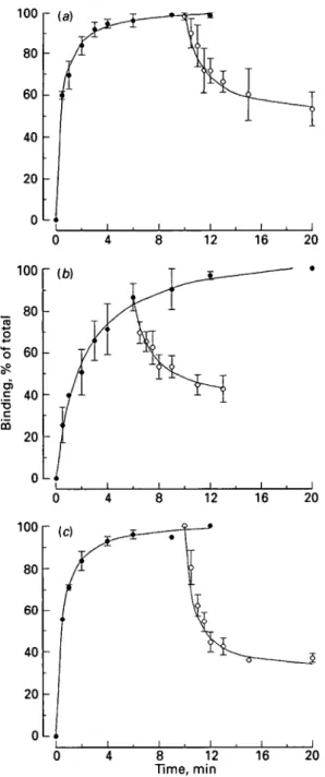
3
H-prazosin, 3H-rauwolscine and 3H-DHA binding was rapid and steady state
conditions were reached within minutes (Fig. 1). There was dissociation of binding within minutes after addition of competitors at 37 °C for 3H-prazosin and 3H-DHA, but only at 25 °C for 3H-rauwolscine.
Competitive binding assays
The i^-values and Hill coefficients (h) for inhibition of specific 3H-prazosin binding by adrenergic competitors are shown in Table 1. For 3H-prazosin binding the
Kt value of the unlabelled ax-antagonist prazosin was lowest while the a2-selective antagonist yohimbine and the a2-selective agonist clonidine had ~ 1000-fold higher
Ki values than unlabelled prazosin (P < 0'05). The non-selective a-adrenergic antagonist phentolamine and the a-adrenergic agents dihydroergotamine and dihydroergocryptine were ~ 30-50 times less potent than unlabelled prazosin for inhibiting binding (P < 0'05). TheiQ value of phenylephrine, an aragonist, was very high compared with the other a-adrenergic competitors. Isoproterenol, a
/?-51
100 80 60 40 20 0L* 12 16 20 12 16 20 0 8 12 Time, min 16 20Fig. 1. Binding of (a) 3H-prazosin, (6) 3H-rauwolseine and (c) 3H-dihydroalprenolol to membranes of mammary duct tissue from the udders of three cows: # , association; O, dissociation. The competitor was (a) 10/<M-phentolamine added at 10 min, (b) 10/tM-phentolamine added at 6 min, (c) 1/tM-propanolol added at 10 min; total binding of the radiolabelled ligand was taken as 100%.
adrenergic agonist, displaced 3H-prazosin binding only at very high concentrations. The order of potency for inhibition of 3H-prazosin binding was prazosin > phentol-amine > dihydroergotphentol-amine > dihydroergocryptine > yohimbine > clonidine > adrenalin > noradrenaline > phenylephrine > isoproterenol > dopamine. Most of the Hill coefficients were close to one, except for phentolamine, a non-selective a-antagonist.
52 H. M. HAMMON AND OTHERS
Table 1. Inhibition of specific 3H-prazosin binding by adrenergic agonists and antagonists (competitors) on membranes of the duct region of the udder
(Values for K( and h are mean + SEM for n udders) Competitor Prazosin Phentolamine Dihydroergotamine Dihydroergocryptine Yohimbino Clonidine Adrenalin Noradrenaline Phenylephrine Isoproterenol Dopamine a, b , c . d , e, f, g , h \ l «( 1. ,i. Competitor dissociation constant (K,), M 10±0-2x lO"9" 2-7 + 0-3 x lO"8" 5-0 ± 2-0 x lO"8" 6-2 + 1-7 x 10~8b 1-1+0-2 x 1O"60 l-3±01 x 10~6c 3-7 + 0-5 x lO"6" 6-7±0-7xl0"6 e 2-2±0-3xl0"5 f 8-8+l-3xlO"5 g 5-l+O-4xlO-4h I I H T V\ J~\l 1 T" / I A tVk tV^ *~\B1 O i l T"\ f» *•£"* *1 HI »"fcH Potency ratio 10000 00370 0-0200 00161 00009 00008 00003 00001 0-0000 00000 00000 Hill coefficient of plot (k) 0-90 ±010 0-64 ±003 0-83 ±007 0-87 ±006 0-80 ±0-04 0-84 ±011 0-80 ±009 0-79 ± 0 0 3 0-79 ± 0 0 4 0-91 ± 0 0 8 0-87 + 0-06 « 6 5 6 6 5 5 5 5 6 5 4
Table 2. Inhibition of specific 3H-rauivolscine binding by adrenergic agonists and antagonists (competitors) on membranes of the duct region of the udder
(Values for Kf and h are mean + SEM for n udders) Competitor Rauwolscine Clonidine Phentolamine Yohimbine Prazosin Adrenalin Noradrenaline Phenylephrine Dopamine Isoproterenol
s.b.cd. e.f j \ [e a n s without common superscript letters were significantly different (P < 0-05).
Table 3. Inhibition of specific 3H-dihydroalprenolol binding by adrenergic agonists and antagonists (competitors) on membranes of the duct region of the udder
Competitor dissociation constant (/f,), M 3-1+0-4X10⁻⁸" 5-2 + 0-9 xlO-8"" 5-4 +3-0 x 10"8ol) 8-4+l-6xlO-8 b 3-3±0-6xl0-6° 3-5 + 0-7 xlO"6 c l-0±0-2xlO"5 d 4-1 +1-6 x 10"5d 1-3 + 0-2 x 10"46 2-5 ± 0-2 x 10"4' Potency ratio 10000 0-5960 0-5740 0-3690 00090 0-0089 00030 00008 00002 00001 Hill coefficient of plot (h) 110±019 0-68 ±006 0-73 ±0-08 0-70 ±0-07 100 ±0-24 0-70 ±0-05 0-85 ± 0 0 5 0-98 + 011 0-87 ±0-03 0-99 ±0-15 n 5 5 5 5 4 5 5 5 5 5 Competitor (— )-Propranolol Alprenolol (+ )-Propranolol Clenbuterol ( + )-Propranolol Fenoterol Isoproterenol Adrenalin Noradrenaline Atenolol Dopamine a.b.c.d, e. f. g AT™.
(Values for Kt and h are Competitor dissociation constant (Kt), M 3-5 +0-5 x 10~9a 3-7 + 0-7 x lO"90 8-6±0-8xl0-9b 4-9 ± 0-7 x 10"80 3-2±O-5xlO-'d 4-8+1-1 x 10~7<1 9-l+2-6xlO"7 d 2-2 + 0-3 x 10"6e 1-7 + 0-3 x 10"5' 3-9±l-3xlO~5f 3-8+1-9X10"48
mean + SEM for n Potencj' ratio 10000 10000 0-4444 0-0816 00124 00084 00044 00018 0-0002 0-0001 00000 udders) Hill coefficient of plot (h) 0-72 ±0-08 0-91 ± 0 1 3 0-98 ±0-08 0-99 ±006 1 22±013 0-94 ±0-05 0-88 ±0-03 0-86 + 014 1-24 + 0-28 0-73 ±0-08 0-95 ±010 n 5 5 5 5 5 5 5 5 5 4 4
4 0 r (a)
53
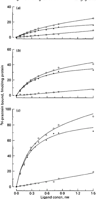
60 r (W 0 0 0-3 0-6 0-9 Ligand concn, nM 1-2 1-5Fig. 2. Representative saturation plot for binding of the a-antagonist 3H-prazosin to suspensions of membrane preparations of (a) parenchyma, (b) mammary duct and (c) teat regions of one bovine udder: O, total binding; • , non-specific binding; A, specific binding. Each point is the mean of triplicate determinations.
The a2-adrenergic competitors rauwolscine, clonidine, and yohimbine had low, and similar, Kt values for displacement of specific 3H-rauwolscine binding (Table 2). Inhibition potency of the non-selective a-adrenergic antagonist phentolamine was of
the same order of magnitude, whereas the arantagonist prazosin was about 100
times less potent than unlabelled rauwolscine for inhibition of 3H-rauwolscine binding (P < 0-05). Displacement by isoproterenol of 3H-rauwolscine binding was in agreement with displacement of 3H-prazosine from its binding sites. The order of potency for inhibition of 3H-rauwolscine binding was rauwolscine > clonidine >
54 H. M. HAMMON AND OTHERS
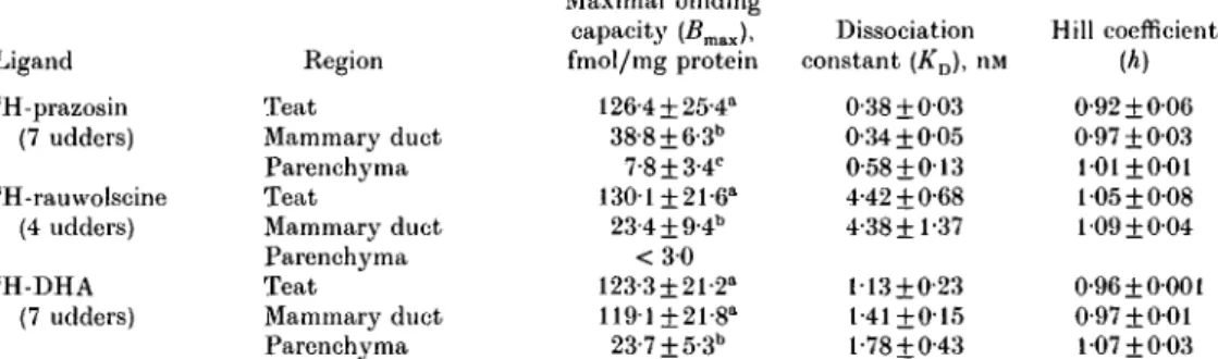
Table 4. Values from saturation assays of sH-prazosin, sH-rauwolscine and 3 H-dihydroalprenolol binding on membranes of different regions of the udder
(Values are means + SEM for no. of udders shown)
Ligand 3 H-prazosin (7 udders) 3 H-rauwolscine (4 udders) 3 H-DHA (7 udders) Region Teat Mammary duct Parenchyma Teat Mammary duct Parenchyma Teat Mammary duct Parenchyma Maximal binding capacity (Bmax), fmol/mg protein 126-4 ±25-4a 38-8+ 6-3" 7-8±3-4c 130-1 ±21-6" 23-4+ 9-4" < 3 0 123-3±21-2" 119-1 ±21-8" 23-7±5-3b Dissociation constant (KD), nM 0-38 ±003 0-34 ±005 0-58 + 013 4-42 + 0-68 4-38+1-37 113 + 0-23 1-41+015 l-78±0-43 Hill coefficient (A) 0-92 ±006 0-97 + 003 101 ±001 105 ±0-08 109 ±004 0-96 ±0001 0-97 ±001 107 ±003 a.t>.c j ie a n s without common superscript letters were significantly different between regions within ligand (P < 0-05).
phentolamine > yohimbine > prazosin > adrenalin > noradrenaline > phenyl-ephrine > dopamine > isoproterenol. Hill coefficients were in the same range as in 3H-prazosin binding studies.
The unlabelled non-selective /^-antagonists alprenolol and propranolol had the lowest Kt values for specific 3H-DHA binding sites, followed by the /?2-adrenergic agonist clenbuterol (Table 3). The /?2-agonist fenoterol and the non-selective /?-agonist isoproterenol were about 100-fold more potent than the /^-ant/?-agonist atenolol for inhibition of 3H-DHA binding (P < 0-05). The order of potency for inhibition of 3H-DHA binding was alprenolol > propranolol > clenbuterol > fenoterol > isoproterenol > adrenalin > noradrenaline > atenolol > dopamine. Inhibition of 3H-DHA binding was investigated with different isomers of propranolol. The order of potency was dependent on stereospecificity: (— propranolol > (+ )-propranolol (P < 0-05) > (+ )-)-propranolol (P < 0-05; Table 3). Most of the substances competed for 3H-DHA binding with Hill coefficients close to one, except for (— )-propranolol and atenolol.
Saturation binding assays
The specific binding of 3H-prazosin to membrane suspensions was a saturable process (Fig. 2). There was a decrease in the density of 3H-prazosin binding sites from the teat region to the duct region to the parenchyma region (P < 0-05; Table 4). KD values of the three udder regions were in the same range. Hill coefficients were all close to one.
The specific binding of 3H-rauwolscine to membrane suspensions was a saturable process. There was a decrease in the number of 3H-rauwolscine binding sites from the teat to the duct region (P < 0'05; Table 4). In the parenchyma region no specific 3 H-rauwolscine binding could be measured. KD values of the teat and duct region were in the same range. Hill coefficients were all close to one.
The specific binding of 3H-DHA was saturable for each udder region. Numbers of binding sites for 3H-DHA were similar in the teat and duct regions, but markedly lower in the parenchyma region (P < 0-05; Table 4). KD values of the three udder regions were of the same order. However, iCD values for 3H-prazosin, 3H-rauwolscine and 3H-DHA binding were significantly different (P < 0-05). All Hill coefficients were close to one.
DISCUSSION
Kinetic experiments demonstrated the reversibility of 3H-prazosin, 3
H-rauwolscine and 3H-DHA binding. Association of 3H-rauwolscine had to be studied at 25 °C and an excess of unlabelled phentolamine had to be added after only 6 min because 3H-rauwolscine binding was no longer reversible with incubation for longer times or at 37 °C. This was surprising, because competitive binding assays with simultaneous addition of rauwolscine and phentolamine at 37 °C caused inhibition of 3
H-rauwolscine binding.
Three different adrenergic receptor subtypes (tXj, a2, /?2) could be identified by inhibition of specific 3H-prazosin, 3H-rauwolscine and 3H-DHA binding by different subtype-specific drugs.
From the characteristics of Scatchard and Hill plots, 3H-prazosin, 3H-rauwolscine and 3H-DHA saturation binding indicated interaction with only a single binding site, identified as ar, a2- and /?2-adrenergic receptor subtypes respectively. K^, values calculated for 3H-prazosin, 3H-rau\volscine and 3H-DHA binding were significantly different from each other. On the other hand, KD values of the same 3H-ligand for teat, duct and parenchyma region were of the same order. Numbers of ar, a2- and /?2-adrenergic receptors of membranes obtained from the teat region agreed well with the results of Roets et al. (1984) and Roets & Peeters (1985, 1986). Our studies demonstrate that there are additional regions in the bovine mammary gland containing <Xj-, a2- and /?2-adrenergic receptors, localized in the area around the gland cistern including the large mammary ducts. However, it is important to note that the parenchyma region contained only very small numbers of adrenergic receptors. Thus, adrenergic receptors were mainly present in the milk purging system of the udder.
That a-adrenergic receptors were specifically localized on smooth muscle cells of mammary ducts cannot be stated with absolute certainty. If we assume their presence, several in vivo studies on inhibition of milk removal after a-Ag administration and in stress situations may be explained. The connection between mammary parenchyma and gland cistern may be interrupted completely or in part by a-adrenergic receptor-stimulated smooth muscle contractions of the mammary ducts. Although oxytocin is released (Blum et al. 1989) and may cause myoepithelial contraction, no milk or only small amounts are transported to the gland cistern and only milk already available in the teat and gland cistern can be removed during milking, a view supported by studies on effects of the a-Ag phenylephrine on milk removal and intramammary pressure before and after teat stimulation (Bruckmaier
et al. 1991). Because the number of a-adrenergic receptors in particular was very low
in mammary parenchyma, the influence of the adrenergic system on this region of the mammary gland is probably relatively small.
It has been proposed that under stress conditions or after a-Ag administration oxytocin may not reach the receptors of myoepithelial cells because of decreasing blood flow to the udder due to vascular contraction after catecholamine administration through interaction with a-adrenergic receptors in the vascular bed (Dhondt et al. 1976; Lefcourt & Akers, 1984; Gorewit & Aromando, 1985). However, only a very small increase of oxytocin concentration in blood is necessary to elicit milk ejection (Schams et al. 1984). Furthermore, vasoconstriction under stress conditions is hardly the main reason for peripheral inhibition of milk removal, because our study demonstrated only small numbers of a- and /?-adrenergic receptors in the mammary parenchyma. From in vivo studies (Bruckmaier et al. 1991), it also
56 H . M. H A M M O N AND OTHERS
appears unlikely that a-Ag have an effect on the responsiveness of myoepithelial cells to oxytocin. Because of the significantly higher density of a-adrenergic receptors in teat tissue and the region of the large mammary ducts, peripheral disturbed milk removal by a-Ag is primarily caused by constriction of the milk purging system of the udder.
It has been shown that administration of /?-Ag has a favourable effect on milk flow as a consequence of teat muscle relaxation (Bernabe & Peeters, 1980; Hamann, 1981; Vandeputte-Van Messom et al. 1986). Through interaction with the numerous /?-adrenergic receptors in the large mammary ducts, probably localized on smooth muscles, relaxation of the large mammarj' ducts is likely. Thus, after /?-Ag administration, transport of milk from parenchyma tissue to the cistern is facilitated by enlarged mammary ducts and milk withdrawal accelerated after milk ejection (Blum et al. 1989; Bruckmaier et al. 1991). Mielke et al. (1991) have suggested the possibility of milk ejection after administration of /?-Ag. However, lack of increased intramammary pressure by the administration of a /?-Ag contradicts these findings (Bruckmaier et al. 1991).
We conclude that a- and /?-adrenergic receptors are present not only in teat walls, but also around the gland cistern and large milk ducts. Thus, the modulation of milk ejection and milk removal by the adrenergic system occurs not only during the milk's passage through the teat cavity, but also very probably during its passage from gland parenchyma to the gland cistern.
These studies have been supported by the Swiss National Science Foundation (Grant no. 32-28781.90).
REFERENCES
BERNABE, J. & PEETERS, G. 1980 Studies on the motility of smooth muscles of the teats in laetating cows. Journal of Dairy Research 47 259-275
BLUM, J. W., SOHAMS, D. & BRUCKMAIER, R. 1989 Catecholamines, oxytocin and milk removal in dairy cows. Journal of Dairy Research 56 107-177
BRUCKMAIER, R. M. & BLUM, J. W. 1992 B-mode ultrasonography of mammary glands of cows, goats and sheep during a- and /?-adrenergie agonist and oxytocin administration. Journal of Dairy Research 59 151-159 BRUCKMAIER, R., MAYER, H. & SCHAMS, D. 1991 Effects of a- and /J-adrenergic agonists on intramammary
pressure and milk flow in dairy cows. Journal of Dairy Research 58 411—419
DHONDT, G., HOUVENAGHEL, A., FEYS-VAN D E BROECK, L. & PEETERS, G. 1976 [Adrenergic receptors in
blood vessels in the udder of laetating cows.] Zentralblatt fur Veterinarmedizin A 23 331-337
GOREWIT, R. C. & AROMANDO, M. C. 1985 Mechanisms involved in the adrenalin-induced blockade of milk ejection in dairy cattle. Proceedings of the Society for Experimental Biology and Medicine 180 340-347 HAMANN, J. 1981 [The influence of a /?2-mimetic substance (Planipart) on the milking behaviour of cows.]
Tierarztlkhe Umschau 36 287-290
HANCOCK, A. A., DELEAN, A. L. & LEFKOWITZ, R. J. 1979 Quantitative resolution of beta-adrenergic receptor subtypes by selective ligand binding: application of a computerized model fitting technique. Molecular Pharmacology 16 1-9
LETOOURT, A. M. & AKERS, R. M. 1982 Endocrine responses of cows subjected to controlled voltages during milking. Journal of Dairy Science 65 2125-2130
LEFCOURT, A. M. & AKERS, R. M. 1984 Small increases in peripheral noradrenaline inhibit the milk-ejection response by means of a peripheral mechanism. Journal of Endocrinology 100 337-344
MIELKE, H., MARKOV, A., FURLL, B., KASKOUS, S. & PONCE, P. 1991 [Studies on the action of the /9-receptor
agonist isoprcnaline on mammary gland alveoli in laetating cows with particular reference to its effect on the secretory function of epithelial cells.] Monatshefte fur Veterinarmedizin 46 427—430
MOOR, M., HONEOGER, U. E. & VVIESMANN, U. N. 1988 Organospeeific, qualitative changes in the phospholipid composition of rats after chronic administration of the antidepressant drug desipramine. Biochemical Pharmacology 37 2035-2039
MOTULSKY, H. J. 1987 GraphPAD (version 2.0). Philadelphia. PA: Institute of Scientific Information PARIS i, A., HOMCY, C. J. & GRAHAM, R. M. 1987 Structural properties of the a,-adrenergic receptor: studies
with membrane and purified receptor preparations. Circulation Research 61 (Suppl.) 1-100-1-104. ROETS, E. & PEETERS, G. 1985 Identification and characterization of 3H-Prazosin binding to a,-adrenoceptors
ROETS, E. & PEETERS, G. 1986 A comparison of the binding characteristics of the a2-adrenoceptor antagonists 3
H-yohimbine and 3H-rauwolscine in bovine teat muscles. Archives Internationales de Pharmacodynamie et de
Therapie 279 212-222
ROETS, E., PEETERS, G. & LEYSEN, J. E. 1984 Identification of/?-adrenoceptors in bovine teat muscles by 3 H-dihydroalprenolol binding. Archives Internationales de Pharmacodynamie et de Therapie 270 203-214
ROETS, E., VANDEPUTTE-VAN MESSOM, G., BURVENICH, C. & PEETERS, G. 1989 Relationship between numbers
of <x2- and /S2-adrenoeeptors in teat tissue and blood cells and milkability of primiparous cows. Journal of
Dairy Science 72 3304-3313
SAS 1990 SAS User's Guide: Statistics. Cary, NO: SAS Institute Inc.
SCIIAMS, D., MAYER, H., PROKOPP, A. & WORSTORFF, H. 1984 Oxytocin secretion during milking in dairy cows with regard to the variation and importance of a threshold level for milk removal. Journal of Endocrinology
102 337-343
Sin, A. G., AHMAD, S., KWAN, C. Y. & DANIEL, E. E. 1989 Characterization of a-adrenoceptor subtypes by
[3H |prazosin and [3H]rauwolscine binding to canine venous smooth muscle membranes. Canadian Journal of
Physiology and Pharmacology 67 1067-1073
VANDEPUTTE-VAN MESSOM, G., BURVENICH, C. & PEETERS, G. 1986 Involvement of /?2-adrenoceptors in teat
sphincter function in the lactating cow. Archives Internationales de Physiologie et de Biochimie 94 p. 34 ZIEOLER, H. & MOSIMANN, W. 1960 Anatomie und Physiologie der Einderniilchdruse. Berlin: Paul Parey