CONTROVERSIES IN NUCLEAR CARDIOLOGY: ANTAGONIST
Hybrid cardiac imaging: More than the sum of its
parts?
Oliver Gaemperli, MD,aand Philipp A. Kaufmann, MDa,b
The constant technological developments in noninva-sive cardiac imaging over the past few decades have contributed toward our pathophysiologic understanding of many conditions. Particularly in coronary artery disease (CAD), management is based on the assessment of both the presence of coronary stenoses and their hemodynamic consequences.1,2Hence, noninvasive imaging helps guide therapeutic decisions by providing complementary infor-mation on coronary morphology and on myocardial perfu-sion and metabolism, using several imaging tools.3,4This imaging includes nuclear techniques such as single-photon emission computed tomography (SPECT) or positron emis-sion tomography (PET), computed tomography (CT) tech-niques such as electron-beam CT (EBCT) or multislice CT (MSCT), and cardiac magnetic resonance (CMR).
Advances in image-processing software and the advent of hybrid scanners have paved the way for the fusion of image datasets from different modalities, giving rise to multimodality or hybrid imaging. This technology avoids the mental integration of functional and morphologic im-ages, and facilitates a comprehensive interpretation of combined datasets. The interest in hybrid imaging has rapidly spread to cardiac applications, and has changed the landscape of noninvasive cardiac imaging by bringing different clinical specialties (eg, cardiology, radiology, and nuclear medicine) closer together.5In addition, this interest has driven the development and production of dedicated hybrid scanners in an effort to simplify image coregistration and improve patient throughput for specialized cardiac imaging centers (ie, hardware-based image coregistration). However, given the high costs associated with such devices, an attractive alternative for hybrid imaging consists of the “offline,” software-based fusion of images obtained from nondedicated standalone scanners (software-based image coregistration). Here, we focus on comparing hardware-based versus software-hardware-based image coregistration for
car-diac hybrid imaging and their respective advantages and drawbacks.
WHAT IS CARDIAC HYBRID IMAGING? The hallmark of hybrid imaging is the combined or fused imaging of two datasets, where both modalities contribute equally to image information. However, the term “hybrid imaging” has been used in other contexts, raising confusion about its exact meaning.
Some authors have referred to the X-ray-based atten-uation correction of myocardial perfusion imaging (MPI) as hybrid imaging.6However, in such a setting, CT images do
not provide added anatomical or functional information, but are used merely to improve the image quality of the other modality (ie, PET or SPECT). In fact, whereas attenuation correction by68Germanium sources, as used in the previous generation of PET scanners, provided the same information, such imaging was not perceived as hybrid, probably be-cause attenuation correction does not contribute to topo-graphic image information. Similarly, the parametric maps obtained from low-dose CT do not provide image informa-tion beyond that needed for attenuainforma-tion correcinforma-tion.7,8Others used the term “hybrid imaging” for the mere side-by-side analysis of MPI and CT images.9To avoid confusion, we suggest the use of “hybrid imaging” to describe any combination of structural and functional information beyond that offered by attenuation correction or side-by-side analysis, by fusion of the separate data sets into one image (Figure 1). Thus, this definition would not include attenuation-corrected images without integrating ana-tomical information. Similarly, the separate acquisition of structural information as well as functional data (eg, perfusion) on two separate scanners or on one hybrid device would allow a mental integration of side-by-side evaluation, but only a fusion of both pieces of informa-tion would result in a hybrid image.
INTEGRATED SCANNERS VERSUS SOFTWARE FUSION
The added value of hybrid imaging involves the spatial correlation of structural and functional information on the fused images, which facilitates a comprehen-sive interpretation of coronary lesions and their patho-physiologic relevance. For cardiac applications, a
three-From the Cardiovascular Center,aUniversity Hospital Zurich, Zurich,
Switzerland, and Zurich Center for Integrative Human Physiology,b
University of Zurich, Zurich, Switzerland.
Reprint requests: Philipp A. Kaufmann, MD, Cardiovascular Center, University Hospital Zurich, NUK C 32, Raemistrasse 100, CH-8091 Zurich, Switzerland;[email protected].
J Nucl Cardiol 2008;15:123-6. 1071-3581/$34.00
Copyright © 2008 by the American Society of Nuclear Cardiology. doi:10.1016/j.nuclcard.2007.12.002
dimensional display of fused images (generated by a volume-rendering technique10) is of greater value
com-pared with oncologic or neurologic applications, because it allows the best evaluation of myocardial territories and their respective serving coronary-artery branches. Thus, an important prerequisite of hybrid imaging comprises accurate image coregistration, because misalignment may result in erroneous allocations of perfusion defects and coronary-artery territories.
From a computational perspective, image coregis-tration can be achieved by a software-based or hardware-based approach.11Hardware-based image coregistration
permits the acquisition of coregistered anatomical and functional images using hybrid scanners (such as PET/CT or SPECT/CT devices), with the capability to perform nuclear and CT image acquisition almost simul-taneously with the patient’s position fixed. Inherently, image fusion is performed fully or semi-automatically by superposition of image datasets. With software-based coregistration, image datasets can be obtained on stan-dalone scanners, and fused manually through the use of
landmark-based coregistration techniques. Intuitively, the hardware-based approach appears preferable, be-cause manual coregistration may be hampered by issues of accuracy and user interaction. Thus, to date, hybrid PET/CT devices have been widely used for whole-body PET/CT imaging, predominantly in oncology.
However, in contrast to whole-body PET/CT, the routine use of fully automated, hardware-based image coregistration for cardiac hybrid applications is limited by organ-specific characteristics. Despite fixation of the patient’s position and orientation, minor beat-to-beat variations in the heart’s position may interfere with accurate image coregistation. Furthermore, CT image acquisition and analysis require electrocardiographic gat-ing, and images are generally reconstructed in mid-diastolic phases to obtain optimal image quality.12 By
contrast, to ensure sufficient quality of SPECT images, a nongated dataset is used, resulting in a slight mismatch of ventricular size between CT and SPECT images. Finally, the position of the heart is highly susceptible to respiratory motion. Whereas a CT scan is performed
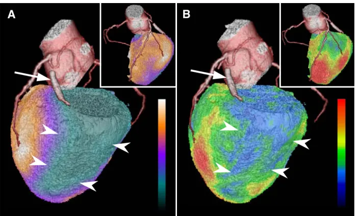
A
B
Figure 1. A, Three-dimensional (3D) volume-rendered (VR) fusion image generated from computed tomography (CT) angiography and myocardial perfusion SPECT with99mTechnetium-tetrofosmin
at rest. Arrow denotes an occluded stent in the proximal left circumflex artery (LCX), and
arrowheads demarcate the associated myocardial scar. Inset: Anterior view shows normal perfusion
of the anterior wall. B, A 3D VR fusion image of the same patient as in A, generated from CT angiography and PET with18Fluoro-deoxyglucose. Arrowheads indicate a lack of viability in the
posterolateral scar, associated with occlusion of the LCX-stent (arrow). Inset: Anterior view shows normal metabolism of the anterior wall.
124 Gaemperli and Kaufmann Journal of Nuclear Cardiology
during a single inspiratory breath hold, SPECT images are acquired during normal breathing, without account-ing for respiratory motion unless respiratory gataccount-ing is conducted. In fact, whole-body PET/CT studies showed significant misalignments of the heart between superim-posed PET and CT images acquired during inspira-tion.13,14
These factors contribute to the notion that, despite the integration of high-end CT devices (with the capability to perform state-of-the-art coronary CT angiography) with nuclear scanners to form dedicated cardiac hybrid scanners, manual-image coregistration may remain indispensable. Published reports on X-ray-based attenuation correction indicate that automated coregistration of CT and SPECT images is often unreliable, and manual correction for misalignment is needed in the vast majority of cases.7,15
Dedicated cardiac fusion software packages are now com-mercially available, allowing software-based hybrid imag-ing with excellent interobserver reproducibility and short processing durations.16The full integration of these fusion
software packages into regular post-processing applications for CT angiography will allow users to further minimize time expenditure and improve workflow for hybrid imaging by avoiding repeated actions, such as coronary-artery track-ing from CT angiography images.16
DO WE NEED HYBRID SCANNERS FOR HYBRID IMAGING?
Despite the widespread use of coronary CT angio-graphy and MPI with SPECT or PET, both techniques vary considerably in their image-acquisition times. Whereas CT coronary angiography with the newest generation 64-slice or dual-source CT devices is per-formed in⬍12 seconds,12emission scans for
stress-and-rest gated SPECT with 99mTechnetium-based radiotrac-ers at standard doses take at least 45 minutes.17 This
discrepancy between emission and transmission scan times determines that high-end CT facilities constituting the CT component of hybrid cardiac scanners will be blocked by long emission scan times, and therefore will operate at low capacity. Many advances in nuclear medicine, such as newly developed dedicated cardiac detectors systems18 and novel image reconstruction
al-gorithms,19 may contribute to reduce emission scan
times considerably. However, to date, in hybrid scanners with high-end CT facilities, the rather long emission scan times preclude operating the high-end CT device at full capacity. In addition, despite the promise of hybrid cardiac imaging, the first clinical experiences with hybrid SPECT/CT imaging showed that in a typical population referred for a noninvasive workup of CAD, only a minority benefited from hybrid imaging, compared with side-by-side interpretation of MPI and CT.3 Thus, at
present, from the standpoint of patient throughput, a dedicated cardiac hybrid scanner is less profitable than two standalone devices for normal-volume nuclear diag-nostic centers. Nonetheless, it will depend on the indi-vidual setting of each institution to determine the type of approach (ie, software-based fusion or hybrid scanner) that is best tailored for its particular purpose, and highly specialized cardiac centers may prefer hybrid scanners for integrative cardiac imaging.
FUTURE PERSPECTIVES
Atherosclerotic disease accounts for the majority of fatalities reported in industrialized countries. Despite major advances in the treatment of CAD patients, a large number of victims of the disease who are apparently healthy die suddenly without prior symptoms. The rec-ognition of the role of vulnerable plaque has opened new avenues of opportunity in the field of cardiovascular medicine.20Hybrid technology has the unique potential
to enable the detection and quantification of the burden of calcified and noncalcified plaques, the quantification of vascular reactivity and endothelial health, the identi-fication of flow-limiting coronary stenoses, and poten-tially, the identification of high-risk plaques by using a fusion of morphology and biology with molecularly targeted PET imaging.21 By such means, in the future,
hybrid imaging may allow for the easy and comprehen-sive noninvacomprehen-sive assessment of coronary plaque burden, its pathophysiologic relevance, and biological plaque activity, thus providing accurate individual risk esti-mates, on which further management decisions can be based.
At present, however, accurate hybrid imaging to integrate and correlate functional and anatomical data is most efficiently achieved by the software fusion of data acquired on two separate scanners. Thus, integrated scanners are nice to have, but are not a must for hybrid imaging.
References
1. Topol EJ, Nissen SE. Our preoccupation with coronary luminol-ogy. The dissociation between clinical and angiographic findings in ischemic heart disease. Circulation 1995;92:2333-42. 2. Silber S, Albertsson P, Aviles FF, Carnici PG, Colombo A, Hamm
C, et al. Guidelines for percutaneous coronary interventions. The Task Force for Percutaneous Coronary Interventions of the Euro-pean Society of Cardiology. Eur Heart J 2005;26:804-47. 3. Gaemperli O, Schepis T, Valenta I, Hushmann L, Scheffel H,
Duerst V, et al. Cardiac image fusion from stand-alone SPECT and CT: clinical experience. J Nucl Med 2007;48:696-703.
4. Namdar M, Hany TF, Koepfli P, Siegrist PT, Burger C, Wyss CA, et al. Integrated PET/CT for the assessment of coronary artery disease: a feasibility study. J Nucl Med 2005;46:930-5.
Journal of Nuclear Cardiology Gaemperli and Kaufmann 125
5. Gourtsoyiannis N, McCall I, Reiser M, Silberman B, Bischof Delaloye A, Carrio I, et al. White paper of the European Society of Radiology (ESR) and the European Association of Nuclear Med-icine (EANM) on multimodality imaging. Eur Radiol 2007;17: 1926-30.
6. Sampson UK, Dorbala S, Limaye A, Kwong R, Di Carli MF. Diagnostic accuracy of rubidium-82 myocardial perfusion imaging with hybrid positron emission tomography/computed tomography in the detection of coronary artery disease. J Am Coll Cardiol 2007;49:1052-8.
7. Schepis T, Gaemperli O, Koepfli P, Ruegg C, Burger C, Leschka S, et al. Use of coronary calcium score scans from stand-alone multislice computed tomography for attenuation correction of myocardial per-fusion SPECT. Eur J Nucl Med Mol Imag 2007;34:11-9.
8. Koepfli P, Hany TF, Wyss CA, Namdar M, Burger C, Konstan-tinidis AV, et al. CT attenuation correction for myocardial perfu-sion quantification using a PET/CT hybrid scanner. J Nucl Med 2004;45:537-42.
9. Hong EC, Kimura-Hayama ET, Di Carli MF. Hybrid cardiac imaging: complementary roles of CT angiography and PET in a patient with a history of radiation therapy. J Nucl Cardiol 2007;14:617-20. 10. Fishman EK, Ney DR, Heath DG, Corl FM, Horton KM, Johnson
PT. Volume rendering versus maximum intensity projection in CT angiography: what works best, when, and why. Radiographics 2006;26:905-22.
11. Bax JJ, Beanlands RS, Klocke FJ, Knuuti J, Lammertsma AA, Schaefers MA, et al. Diagnostic and clinical perspectives of fusion imaging in cardiology: is the total greater than the sum of its parts? Heart 2007;93:16-22.
12. Leschka S, Scheffel H, Desbiolles L, Plass A, Gaemperli O, Valenta I, et al. Image quality and reconstruction intervals of dual-source CT coronary angiography: recommendations for ECG-pulsing windowing. Invest Radiol 2007;42:543-9.
13. Gilman MD, Fischman AJ, Krishnasetty V, Halpern EF, Aquino SL. Hybrid PET/CT of the thorax: when is computer registration necessary? J Comput Assist Tomogr 2007;31:395-401.
14. Gould KL, Pan T, Loghin C, Johnson NP, Guha A, Sdringola S. Frequent diagnostic errors in cardiac PET/CT due to misregistra-tion of CT attenuamisregistra-tion and emission PET images: a definitive analysis of causes, consequences, and corrections. J Nucl Med 2007;48:1112-21.
15. Goetze S, Wahl RL. Prevalence of misregistration between SPECT and CT for attenuation-corrected myocardial perfusion SPECT. J Nucl Cardiol 2007;14:200-6.
16. Gaemperli O, Schepis T, Kalff V, Namdar M, Valenta I, Stefani L, et al. Validation of a new cardiac image fusion software for three-dimensional integration of myocardial perfusion SPECT and stand-alone 64-slice CT angiography. Eur J Nucl Med Mol Imag 2007;34:1097-106.
17. Hansen CL, Goldstein RA, Berman DS, Churchwell KB, Cooke CD, Corbett JR, et al. Myocardial perfusion and function single photon emission computed tomography. J Nucl Cardiol 2006;13: e97-120.
18. Patton JA, Slomka PJ, Germano G, Berman DS. Recent technologic advances in nuclear cardiology. J Nucl Cardiol 2007;14:501-13. 19. Borges-Neto S, Pagnanelli RA, Shaw LK, Honeycutt E, Shwartz
SC, Adams GL, et al. Clinical results of a novel wide beam reconstruction method for shortening scan time of Tc-99m cardiac SPECT perfusion studies. J Nucl Cardiol 2007;14:555-65. 20. Naghavi M, Libby P, Falk E, Casscells SW, Litovsky S,
Rum-berger J, et al. From vulnerable plaque to vulnerable patient: a call for new definitions and risk assessment strategies: Part I. Circula-tion 2003;108:1664-72.
21. Di Carli MF, Hachamovitch R. New technology for noninvasive evaluation of coronary artery disease. Circulation 2007;115: 1464-80.
126 Gaemperli and Kaufmann Journal of Nuclear Cardiology