Localised in vivo proton spectroscopy
G.G. McKinnon, R Boesiger
Institut of Biomedical Engineering and Medical Informatlcs,
University and Zürich
.'
i
In this paper we discuss a method for performing localised in vivo spectroscopy. The method is known äs "volume selective refocusing" [3]; and is particularly suited to proton spectroscopy of the human brain. In vivo proton brain spectroscopy is still very much in the development phase. However it is potentially a valuable tool for monitoring the metabolic levels; particularly that of lactate.
The main technical problems, relating to in vivo proton spectroscopy, arise from the fact that the interesting compounds have very small signal levels. To obtain a reasonable signal to noise level, in a time of say 15 mins, at 1.5 tesla magnetic field strength, the measured volume has to be at least about 8 cc. Further, the signal from water is about 1000 to 10000 times stronger in amplitude than that from the more interesting signals such äs lactate. To suppress the water signal, however, one can make use of the 200 Hz (at 1.5 tesla) difference in resonance frequencies between water and lactate.
The laboratory spectroscopist typically works with homogeneous samples. The whole body spectroscopist, on the other hand, deals with patients, which are anything but homogeneous. The whole body spectroscopist, in contrast to the laboratory spectroscopist, needs to be able to make separate spectroscopic measurements from different parts of the body. For instance, he or she may want to compare the spectrum from a volume of tissue within a tumor, with that from healthy tissue nearby. To do this some method for performing localised spectroscopy is needed.
There are two main methods of localised spectroscopy. The simplest makes use of small coils for the reception of the rf signals. The sensitivity profile of the coil ensures that the measured signal is largely from that tissue in the immediate neighbourhood. By repositioning the coil, other volumes of interest can be measured and compared. A more flexible method, however, makes use ofrf pulses and magnetic field gradients; and functions much in the way
that slice selection is achieved'in magnetic resonance-imaging. This technique is much more flexible because the size and position of the selected volume can be chosen at will - simply by varying the centre . frequency and bandwidth of the rf pulses, and the strength of the field gradients. These later procedures are generally referred to by the term "Volume localised (br selective) spectroscopyV
90°
echo
| [ (3*180»)
n
Gv~>
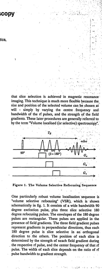
Figure 1. The Volume Selective Refocusing Sequence
One particularly robust volume localisation sequence is "volume selective refocusing" (VSR), which is shown schematically in fig. 1. It consists of a wide bandwidth 90 degree excitation pulse, plus three slice selective 180 degree refocusing pulses. The envelopes of the 180 degree pulses are rectangular. These pulses are applied in the presence of field gradients. The three field gradient pulses represent gradients in perpendicular directions, thus each 180 degree pulse is slice selective in an orthogonal direction to. the others. The position of each slice is determined by the strength of neach field gradient during . the respective rf pulse, and the center frequency of that rf pulse. The width of each slice depends on the ratio of rf pulse bandwidth to gradient strength.
Not every sequence consisting of three slice selective pulses can be used for obtaining localised spectra. VSR can however. The reason is that every sequence consisting of an excitation pulse followed by an odd number of 180 degrec pulses results in the creation of a so called "echo Signal", irrespective of the time intervals between the pulses. What happens, then, is that immediately after the excitation pulse all the spin vectors are rotating in phase. However äs time progresses, the microscopic and macroscopic magnetic field inhomogeneities cause the individual spins to rotate at slightly different rates. Eventually they become so dephased that the NMR signal disappears. The effect of an odd number of 180 degree pulses is to negate the phase of the individual spin vectors. Thus at a later predeterminable point in time, due to the continued influence of the field inhomogeneities, all the spins again achieve a period of coherence. This rephasing leads to increased signal level, and is denoted by the term "echo".
If the field gradients are placed symmetrically with respect to their corresponding rf pulses, the resulting echo is localised to the "intersection volume" of the three perpendicular slices. In this manner localised spectra can be obtained. "Dephasing" and "rephasing" are relative terms, so there is always a small amount of signal present from outside of the selected volume. This unwanted signal can be suppressed further by increasing the length, or strength, (or both) of the field gradient pulses - provided that the symmetry of these pulses is preserved.
We have tested the spatial localisation of VSR using a phantom consisting of a 27 cc cube filled with cyclohexane placed in 10 litres of water. As cyclohexane and water have different resonance frequencies, the individual contributions can be separately identified. Choosing a "selection volume" of 8 cc centred on the cyclohexane cube, the resulting spectrum contained 97% cyclohexane and only 3% water. This represents a suppression factor, for the total signal outside the selection volume, of 40000. VSR, in addition to giving good spatial localisation, is also a very robust procedure. By this it is meant that the adjustment of the rf pulse amplitudes is not very critical. These two points are very important considerations when one is performing volume localised spectroscopy in patients. To obtain good results; one has to firstly optimize the rf pulse amplitudes; and secondly shim the volume of interest so that the magnetic field within this volume is äs homogeneous äs possible.
As VSR spectroscopy is generally preceded by an imaging sequence, we simply use those rf optimisation settings. Shimming is performed interactively by observing the volume localised echo signal, and adjusting the currents to the shim coils such that this echo signal is äs long äs possible. Because each single measured echo contains very little signal from outside the volume of interest, the shimming can also be performed relatively quickly.
A further attractive feature of VSR is that the 90 degree excitation pulse can be replaced by any other type of "excitation sequence". Practically this means that water suppression, using the (again very robust) binomial pulse method [1], is easy to incorporate. Also multiple quantum excitation sequences can be used to allow one to separate overlapping peaks in the spectrum [3,4]. We won't deal with the multiple quantum excitation sequences here, however the binomial excitation is very important. The binomial pulse we use consists of four impulses separated in time by a period T. The amplitude ratio of these impulses is (l -3: 3: -1). Now, to a good approximation, the excitation response in the frequency domain is simply the Fourier transform of the time domain pulse form. In this case the excitation response is given by 3sin ( ) - 5 (3 ). This has a null at f=0, and a maximum at f=l/2T. Thus by adjusting the centre frequency, and interpulse spacing, the binomial pulse can be made to maximally excite the, say, lactate resonance without exciting the water resonance. Using a binomial excitation, water suppression factors of over 1000 can be easily achieved.
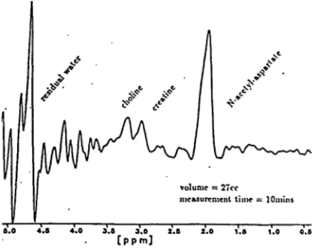
volume =: 27cc
mcftsurement lim* = lOmins 3.8 3.0 2.8
[ p p m ]
Figure 2. Localised Proton Spectrum from a Normal Brain An example of an in vivo localised proton spectrum is shown in fig. 2. This is from a normal volunteen Here there is no lactate to be seen - it would appear at about 1.33 ppm. However measurements on excised brain tissue [4] show lactate levels almost equal in amplitude with the N-acetyl-aspartate peak. Thus these type of measurements could become very sensitive diagnostic tools.
References:
[1] Höre, P.J., J.Magn.Reson.,55, 283,
[2] Luyten, P.R., den Holländer, J.A. Kadiology, IM, 795, (1986).
[3] McKinnon, G.C., Boesiger Pn Magn.Res, in Med., 6,
343, (1988).
[4] McKinnon, G.C, Boesiger PM Magn.Res. in Mai.,
submitted,(198S).