INTERNATIONAL JOURNAL OF SYSTEMATIC BACTERIOLOGY, Jan. 1995, p. 9-16 Copyright
0
1995, International Union of Microbiological Societies 0020-7713/95/$04,00+0Vol.
45,
No. 1Bacillus themoamylovorans sp. nov., a Moderately Thermophilic
and Amylolytic Bacterium
Y.
COMBET-BLANC,l* B. OLLIVIER,l C. STREICHER,l B. K. C. PATEL,' P. P. DWIVEDI,'
B. POT,3 G. PRENSIER,4
ANDJ.-L. GARCIA'
Laboratoire de Microbiologie ORSTOM, Universitd de Provence, 13331 Marseille Cedex 3, and Laboratoire de Microbiologie,
Universiti Blaise Pascal, 631 77 Aubi2re Cedex, France; Faculty of Science and Technology, Grifith University, Brisbane,
Queensland 41 11, Australia2; and La boratorium voor Microbiologie, Universiteit Gent, B-9000 Ghent, Belgium3
A moderately thermophilic, facultatively anaerobic, amylolytic bacterium was isolated from palm wine, a
tropical alcoholic beverage that was sampled in Senegal. The cells were gram positive, catalase positive,
non-spore forming, rod shaped, and slightly motile with peritrichous flagella. The strain which we examined
did not possess cytochrome and produced
L-(+)-lactate, acetate, ethanol, and formate but not hydrogen during
carbohydrate fermentation. Growth occurred at pH values ranging from 5.4 to 8.5, and optimum growth
occurred at around pH 7.0. The optimum temperature for growth was around
5O"C,
and the upper temperature
limit for growth was 58°C. The guanine-plus-cytosine content of the DNA was 38.8
k0.2 mol%. A sequence
analysis of the 16s rRNA gene revealed that the new organism is closely related phylogenetically to members
of genus Bacillus. Despite the lack of spores, we propose that on the basis of phylogenetic characteristics, the
new isolate should be classified as a new Bacillus species, Bacillus thermoamylovorans. The type strain is strain
DKP
(=Collection of Institut Pasteur CNCM 1-1378).
Palm wine is the collective name of alcoholic beverages that
result from spontaneous fermentation of the sap of any species
of tree belonging to the large family Palmae. This traditional
beverage is prepared in the tropical areas of Africa, Asia, and
South America where palm trees are common (27). The sap is
collected from a newly made slit at the base of an immature
male inflorescence on a tree. Leaking sap is collected in a
calabash, through a channel made from a plaited palm leaf
(48). This method allows daily collection of about 10 liters of
sap per tree (16). The sap contains about 10 to 15% (wt/vol)
sucrose (12, 45), amino acids (51 , and vitamins (2, 6, 38, 49).
High densities of bacteria
(10 to 4
Xlo8 cells ml-l) and
yeasts (2
x
lo5 to
lo8 cells m1-l) (45) give palm wine a milky
flocculent appearance (29). The bacteria and yeasts isolated
from palm wine belong almost exclusively to the genera
Lactobacillus and Saccharomyces, respectively (30, 41, 45, 46,
51), which are both adapted to the high osmotic pressures and
acidity that occur in fermented beverages (beer, grape wine,
and cider) (21). Microbiological studies of palm wine have
shown that the stage of fermentation markedly affects the
diversity of the microflora.
During the early stages of production, when the sap has a
neutral pH (pH 7.0 to 7.2) and contains a high concentration of
sucrose, numerous species belonging mostly to the genera Leu-
conostoc, Lactobacillus, Streptococcus, Saccharomyces, Schizosac-
charomyces, Candida, and Rchia have been isolated from palm
wine
(30, 41, 51); a few Clostndium or Bacillus spp. and gram-
negative Sewatia and Klebsiella spp. have also been isolated
(30, 45). This diverse microbiota ferments the sucrose within
about 30 h (12,38), and this fermentation leads to acidification
of the medium through the production of organic acids, mostly
lactic acid. The decrease in pH results in (i) the selection of
acidophilic species and (ii) the disappearance of gram-negative
2
*
Corresponding author. Mailing address: Laboratoire de Microbi- ologie ORSTOM, UniversitC de Provence, Case 87, 3 Place Victor- Hugo, 13331 Marseille Cedex 03, France. Phone: 33-91106478. Fax: 33-91106481.bacteria, as well as pathogenic bacteria, that are not adapted to
low pH values (45).
After about 4 days of fermentation, Lactobacillus species
tend to disappear, while members of the genera Streptococcus
and Micrococcus become dominant along with yeasts
(30, 41,
45). Later, the accumulation of ethanol and the low pH of the
medium favor the growth of acetic acid-producing bacteria,
which oxidize ethanol to acetic acid. Despite the selectivity of
the ecosystem, some Clostridium and Bacillus spp. survive in
this environment, probably because of their ability to sporu-
late. As the mesophilic bacteria involved in palm wine produc-
tion have already been studied (6, 30, 41, 45, 46, 51) and as
thermophilic lactobacilli belonging to the subgenus Ther-
mobacterium have been isolated previously from grape wine by
Barre
(5),
we focused on the thermophilic anaerobic flora in
palm wine. During this study we isolated two thermophilic
species, Clostridium thermopalmarium (23) and a thermophilic
amylolytic bacterium whose characteristics are described and
discussed in this paper.
MATERIALS AND METHODS
Origin of the isolate. Strain DKPT (T = type strain) was isolated from a sample of palm wine collected in Rufisque (20 km east of Dakar), Senegal.
Culture methods and media. Hungate's anaerobic techniques (17, 25) were
used in the experiments described below. Strain DKPT was cultured on a basal medium containing (per liter) 5.0 g of yeast extract (Difco Laboratories, Detroit, Mich.), 5.0 g of Biotrypcase (bioMCrieux, Craponne, France), 1.0 g of KH2P04,
0.4 g of MgCl, * 6H,O, 1.0 g of NH,Cl, 5.0 mg of FeS04
-
7H,O, 25 ml of amineral solution (4), 1 ml of a trace element solution (4), and 1 ml of Tween 80. The p H of this medium was adjusted to 7.5 with 10 M KOH, and the medium was boiled and cooled under a stream of 0,-free N, at room temperature. It was then distributed into 60-ml serum bottles (20 ml of medium per bottle) and Hungate tubes (4.5 ml of medium per tube) (28). After the medium was autoclaved at 110°C for 45 min, energy sources were injected into the serum bottles and Hungate tubes from separately sterilized stock solutions to give a final concen- tration of 0.3% (wt/vol).
A vitamin-free, chemically defined medium was used to determine vitamin
requirements. This medium contained (per liter) 1.0 g of KH,P04, 0.4 g of
MgCl, * 6 H,O, 0.2 g of NH4C1, 5.0 mg of FeS04 * 7H20, 25 ml of a mineral
solution (4), 1 ml of a trace element solution (4), 1 ml of Tween 80, 0.26 mmol of DL-cysteine, 0.26 mmol of L-lysine, 0.26 mmol of L-tryptophan, 0.26 mmol of L-( +)-asparaghe, 0.26 mmol of L-glutamine, 0.26 mmol of DL-tyrosine, 0.26 mmol of DL-phenylalanine, 0.26 mmol of DL-threonine, 0.26 mmol of DL-aspartic
acid, 0.26 mmol of L-isoleucine, 0.26 mmol of DL-serine, 0.26 mmol of DL-valine, 0.26 mmol of L-histidine-HCL, 0.26 mmol of DL-leucine, 0.26 mmol of glycine, 0.26 mmol of m-alanine, 0.26 mmol of DL-proline, 0.26 mmol of L-arginine, 0.26 mmol of DL-methionhe, and 0.26 mmol of L-glutamic acid (all amino acids from Sigma Chemical Co., St. Louis, Mo.). The p H of the medium was adjusted to 7.5 with 10 M KOH. The medium was then distributed under a stream of 0,-free N, into Hungate tubes (4.5 ml of medium per tube) and autoclaved at 110°C for 40 min. Glucose, vitamins, and purine and pyrimidine bases were injected into the Hungate tubes from filter-sterilized solutions.
Unless indicated otherwise, all experiments were conducted at the optimum temperature for growth (50°C) and pH 7.5.
Enrichment and isolation procedure. Enrichment cultures were obtained by
adding 10 ml of palm wine, 1 ml of 10% NaHCO,, and 0.2 ml of 2% Na,S 9H,O to a serum bottle containing 20 ml of basal medium supplemented with 0.3% glucose. After 24 h at 50"C, a I-ml sample was injected into another serum bottle containing basal medium. This process was repeated twice. Pure cultures were obtained by repeated application of the roll tube method of Hungate (17). Single colonies were removed, preparations were diluted serially in Hungate tubes containing 4.5 ml of basal medium supplemented with 0.3% glucose, and the tubes were incubated for 24 h at 50°C. Strain DKPT was stored in liquid growth medium at room temperature and was used for further characterization.
Sporulation test. Two agar-based media were used to test for sporulation.
Medium A contained (per liter) 5.0 g of Bacto Peptone (Difco), 3.0 g of meat extract (Difco), 5 mg of MnSO,, and 16 g of agar (Difco).
Medium B, which was used for plates, was the same as the basal medium except that 1% glucose and 1.6% agar were added. The p H of medium A and the pH of medium B were adjusted to pH 7.2 with 10 M KOH. After autoclaving at 110°C €or 45 min, the media were distributed into plates. The plates were inoculated with 0.3-ml portions of an overnight liquid culture and then incubated at 50°C for up to 5 days to determine the presence of spores.
The heat resistance of cells was determined in basal medium supplemented with 0.3% glucose. After 1, 2, and 20 days of incubation at 50"C, duplicate cultures were heated at 80°C for 5 and 10 min and subcultured into fresh medium (inoculum, 20% [vol/vol]), and the resulting preparations were incubated 48 h at 50°C. In addition, the viability of cells after 1, 2, and 20 days of incubation was checked by subculturing before heating.
Analytical techniques. Bacterial growth was monitored by measuring the
increase in turbidity at 600 nm in anaerobic Hungate tubes inserted directly into a Shimadzu model UV 160A spectrophotometer. The effect of oxygen on the growth and metabolism of strain DKPT was determined in flasks which contained basal medium supplemented with 1% glucose and were agitated. Hungate tubes containing basal medium supplemented with 0.3% glucose with air or 0,-free N, in the headspace were also used. Flasks and Hungate tubes were inoculated (inoculum, lo%, [vol/vol]) with overnight cultures and incubated for 48 h at 50°C. The fermentation pattern was determined in duplicate by using basal medium containing the carbohydrate being tested at a final concentration of 0.3% (wt/vol) and 0.017% bromothymol blue. Stock carbohydrates were sterilized separately by filtration.
The vitamin requirements were determined by using the vitamin-free chemi- cally defined medium supplemented with glucose at a final concentration of 0.3% (wt/vol). A vitamin solution, an adenine-guanine-uracil solution, and a xanthine solution were prepared as described by Kogosa et al. (37) and were sterilized separately by filtration. The final concentrations of the vitamins and purine and pyrimidine bases tested were as follows: nicotinic acid, thiamine-HCL, calcium D-pantothenate, and riboflavin, 992 pg liter-'; p-aminobenzoic acid, 551 pg liter-'; pyridoxine, 1,984 pg liter-'; vitamin B,,, 1 pg liter-'; m-biotin, 10 pg liter-'; folic acid, 1 wg liter-'; and adenine, guanine, uracil, and xanthine, 5.157 mg liter-'. Actively growing cultures obtained after three successive transfers in basal medium were used for inoculation. Inocula were obtained from suspen- sions of organisms that had been washed twice in saline. For each growth factor, the test was performed in triplicate.
The temperature range for growth was determined by using thermostatically controlled water baths. Catalase activity was tested with a 3% (vol/vol) hydrogen peroxide solution by using a pellet resulting from centrifugation of 1.5 ml of an overnight culture. Aerobiosis was achieved by shaking cultures growing in basal medium containing 1% glucose. Indole production and ammonium production were assayed with Kovks and Nessler's reagents (Sigma), respectively. Nitrate reduction was determined with Griess's reagent (Sigma). H,S was measured photometrically as colloidal CuS after reaction with a mixture containing 50 mM HCI and 5 mM CuSO, (9).
Glucose and fermentation product contents were determined by high-perfor- mance liquid chromatography (HPLC), using a model Analprep 93 pump (Touzart et Matignon, Vitry sur Seine, France) and a type O R H 801 column (Interaction Chemicals, Inc., Mountain View, Calif.) equipped with a differential refractometer detector (Knauer, Berlin, Germany). A 2 0 - 4 portion of a cell-free supernatant was injected jnto the column, which was maintained at 35°C. A 25 mM H,S04 solution was used as the solvent at a flow rate of 0.7 ml
-
min-'. Hydrogen was quantified by using a Girdel model Serie 30 gas chromatograph equipped with a thermal conductivity detector and a stainless steel column (1 m by 3.2 mm) packed with Carbosphere SS (60/80 mesh); the column temperature was 150"C, the carrier gas was He (lo5 Pa), the injector and detector temperature was 210"C, and the power of the filament was 90 mA. CO, and 0, contents weredetermined with a Chrompack model CP 9000 gas chromatograph equipped with a thermal conductivity detector and two stainless steel columns (1.5 m by 2.2 mm). One column was packed with molecular sieve 5A (60180 mesh), and the other column was packed with silica gel (GC grade); the column temperature was 60°C the carrier gas was He (lo5 Pa), the injector temperature was 70"C, the detector temperature was 150"C, and the power of the filament was 90 mA. L-( +)-Lactic dehydrogenase and D-( -)-lactic dehydrogenase (Boeringer Mann- heim, Mannheim, Germany) were used to assess the stereoisomerism of the
lactic acid produced by the fermentation of glucose.
For cytochrome analysis, 3 g of wet cells suspended in 10 ml of 20 mM Tris hydrochloride buffer (pH 7.6) was sonicated eight times at 0.5 cycle * s-' for 2
min. The suspension was centrifuged at 30,000 X g for 20 min at 5°C to remove the cell debris. The resulting cell extract was separated into a supernatant fraction and a particulate fraction by centrifugation at 140,000 X g for 2 h. The resulting dark gelatinous pellet was resuspended in the same buffer; this represented the particulate fraction. Both the soluble fraction and the particulate fraction were examined for cytochromes by determining their air-oxidized and dithionite-reduced spectra (300 to 600 nm), as well as their redox difference spectra (dithionite-reduced spectrum minus air-oxidized spectrum), with a Shimadzu model UV 300 spectrophotometer.
Morphological characteristics. Morphological properties were determined by
phase-contrast microscopy, using slides coated with a thin layer (0.5 mm) of purified agar (Difco). For electron microscopy preparations were negatively stained with 4% (wt/vol) uranyl acetate in distilled water. For transmission electron microscopy cells were fixed for 1 h in 0.07 M sodium cacodylate buffer (pH 7.3) containing 1.2% (wt/vol) glutaraldehyde and 0.05% ruthenium red. After the samples were washed in cacodylate buffer containing ruthenium red, they were postfixed with 1% OsO, in cacodylate buffer. The samples were embedded in Epon, and ultrathin sections were stained with 2% uranyl acetate in 50% ethanol and then with lead citrate. Micrographs were taken with a JEOL model 1200 CX electron microscope.
DNA base composition. The guanine-plus-cytosine (G+C) content of the
DNA was determined by workers at the Deutsche Sammlung von Mikroorgan- ismen und Zellkulturen, Braunschweig, Germany. After disruption with a French pressure cell, the DNA was isolated and purified by chromatography on hydroxyapatite. The G + C content was determined by HPLC as described by Meshbah et al. (26); nonmethylated lambda DNA (Sigma) was used as the internal standard.
SDS-PAGE. Approximately 90 mg of cells was harvested from three bottles
that contained basal medium supplemented with 0.3% of glucose and had been incubated at 50°C for 48 h. Whole-cell protein extracts were prepared and sodium dodecyl sulfate (SDS)-polyacrylamide gel electrophoresis (PAGE) was performed as described by Pot et al. (33). Registration of the protein electro- phoretic patterns, normalization of the densitometric traces, grouping of strains by using the Pearson product moment correlation coefficient, and an unweighted pair group using mathematical average cluster analysis were performed by the techniques described by Pot et al. (33), using the GELCOMPAR software package (version 2.0) (52). The protein profile of strain DKPT was compared with a database consisting of normalized protein fingerprints derived from reference strains belonging to almost all previously described species of lactic acid bacteria (32).
16s rRNA sequence studies. Purification of genomic DNA, amplification, and
purification of the 16s rKNA gene from isolate DKP?' were performed by using a technique described previously (24, 35). The purified PCR product was sequenced directly. Sequencing was performed with an ABI automated DNA sequencer by using a Prism dideoxy terminator cycle sequencing kit as recom- mended by the manufacturer (Applied Biosystems, Ltd.). The primers used for sequencing have been described previously (35). The 16s ribosomal DNA sequence obtained from the sequencing data was aligned, by using sequence editor ae2, with the sequences of various members of the bacterial phylum whose 16s rRNA sequences were obtained from the Ribosomal RNA Database Project and from GenBank (22). Positions of sequence and alignment uncertainty were omitted from the analysis, and the pairwise evolutionary distances for 1,128 nucleotides were computed from levels of similarity by using the Olsen correc- tion parameter (31) of Jukes and Cantor (18). Dendrograms were constructed from evolutionary distances by using the program of De Soete (10). A transver- sion analysis was performed by using the program DNAPARS implemented in the PHYLIP package (13). Tree topology was reexamined by using 100 boot- strapped data sets; the SEEQBOOT, DNADIST, FITCH, and CONSENSE programs available on TKEECON (50) were used for this purpose.
Nucleotide sequence accession numbers. The strain DKPT 16s rRNA se-
quence which we determined has been deposited in the GenBank database under accession number L27478. The EMBL accession numbers of the 16s rRNA sequences of Sporosarcina urea and Saccharococcus themophilus are X62173 and X70430, respectively.
RESULTS
Colony morphology.
After 48 h of growth on basal medium,
strain
DKPT colonies were small, white, lens shaped with
smooth edges, and
2 to
3 mm in diameter.
VOL. 45, 1995 BACILLUS THERMOAMYLOVORANS SP. NOV. 11
FIG. 1. (A) Phase-contrast photomicrograph of strain DKPT. Bar = 10 pm. (B) Ultrathin transverse section of isolate DKPT, showing the cell wall structure. Bar = 0.5 pm. (C) Ultrathin longitudinal section of isolate DIPT, showing the septum. Bar = 0.5 pm.
Cell morphology.
Strain DKPT cells were straight rods that
occurred either singly or in short chains of two to four cells
(Fig.
1A). Although the cells possessed peritrichous flagella,
they exhibited only very slight motility as determined by
phase-contrast microscopy. The cells were 0.45 to 0.5 pm wide
and 3.0 to 4.0 pm long. The Gram stain reaction was positive,
but cells in older cultures lost the ability to retain the Gram
stain. Thin sections revealed a typical gram-positive cell enve-
lope profile (Fig. 1B and C). Strain DKPT did not grow on solid
medium
A, which was used to induce sporulation of bacilli.
Reference strain for normalisation
La cto ba cillus pa raca s e i
Strain DKP
Lac to b
a
cillus a cidop h ilu s
L a
c
to
ba cillus bre vis
Reference strain for normalisation
FIG. 2. Profiles obtained by electrophoretic analysis of whole-cell proteins of the following organisms: strain DKPT, Lactobacillus paracasei, Lactobacillus
acidophilus, and Lactobacillus brevis.
Strain DKPT and
Bacillus coagulans, which was used as a
standard, exhibited good growth on solid medium B. After 48
h,
B. coagulans sporulated, but strain DKPT did not. In
addition 24- and 48-h and 20-day-old cultures that had been
incubated at 50°C exhibited no heat resistance when they were
heated at 80°C for
5
or 10 min, indicating that no spores were
present.
DNA base composition.
The average G + C content of strain
DKPT DNA, based on three determinations, was 38.8
20.2
mol%.
Growth conditions and metabolic properties.
In medium
containing glucose as the energy source, strain DKPT required
yeast extract or Biotrypcase for growth. No growth was ob-
served when gelatin or casein replaced yeast extract. However,
a small amount of growth was observed in the vitamin-free
chemically defined medium when glucose was used as an
e n e r q source. Colonies grew on plates incubated in air. Strain
DKP also grew in liquid medium that had been agitated and
in anaerobic medium with
0, in the headspace; in the latter
medium the 0, was completely consumed and CO, was
produced. Strain DKPT also grew under strict anaerobic
conditions in a prereduced medium. This indicated that strain
DKPT is a facultative anaerobe. During anaerobic growth on
glucose, strain DKPT produced lactate, formate, acetate, and
ethanol as the only end products. No H, was produced by
strain DKPT even when it was cocultured with an hydrog-
enotrophic methanogen. At the end of the time course the
fermentation balance measured was: 1.0 glucose
+0.89 lactate
+
1.0 formate
+
0.56 acetate
+
0.57 ethanol.
Our data accounted for a level of carbon recovery of 99%.
The molar ratio of acetate, ethanol, and formate was 1:1:2. The
lactate produced was 96% L-( +)-lactate. Under aerobic con-
ditions, strain DKPT produced lactate, acetate, and CO, but
not formate during glucose metabolism, and acetate produc-
tion increased compared with acetate production under anaer-
obic conditions. Under aerobic conditions, the end products of
glucose metabolism were:
1.0 glucose
+=0.6 lactate
+
1.21
acetate
+
0.06 ethanol
+
1.21 CO,.
However, strain DKPT lacked cytochromes but was positive
for catalase activity. Nitrate reduction and sulfate reduction
were negative, and indole and H,S were not produced. In
medium containing arginine as an energy source, NH, was
produced but no growth occurred.
Under anaerobic conditions, growth was optimal at approx-
imately 50°C; 58°C was the highest temperature at which
growth occurred. The organism grew at pH values ranging
from 5.4 to 8.5; the optimum pH was 7.0. In basal medium
containing glucose as the energy source at pH 7.0 and 50"C, the
maximum doubling time was 40 min.
Fermentation of sugars.
The following sugars (final concen-
tration, 0.3% [wt/vol]) were fermented within 24 h: L-arabi-
nose, D-ribose, D-glucose, D-fructose, D-mannose, L-rhamnose,
amygdalin, arbutin, esculin, salicin, cellobiose, maltose, treha-
lose, starch, glycogen, gentiobiose, and gluconate. D-Xylose,
D-galactose, N-acetyl-D-glucosamine, lactose, sucrose, melezi-
tose, and D-turanose were fermented slowly (within 48 to 96 h).
Glycerol, erythritol, D-arabinose, L-xylose, ribitol, L-sorbose,
galactitol, inositol, D-mannitol, D-glucitol, a-methyl-D-manno-
side, a-methyl-D-glucoside, melibiose, inulin, raffinose, xylitol,
D-lyxose, D-tagatose, D- and L-fucose, D-arabitol, and L-arabitol
were not fermented.
Vitamin requirements.
Thiamine, DL-biotin, and purine and
pyrimidine bases (adenine, guanine, uracil, xanthine) stimu-
lated the growth of strain DKPT but were not essential.
Vitamin BIZ, pyridoxine, nicotinic acid, p-aminobenzoic acid,
calcium D-pantothenate, folic acid, and riboflavin had no
significant effect
on
growth.
SDS-PAGE.
The electrophoretic patterns of the soluble
cellular proteins, as determined by the PAGE method (Fig. 2),
showed that strain DKPT is not similar to any previously
described
Lactobacillus or Camobacten'um species and that it
does not belong to the genus
Enterococcus, Lactococcus, or
Vagococcus. Strain DKPT was also compared with the most
relevant species of the genera
Leuconostoc, Pediococcus, and
Tetragenococcus and with a number of representative strains
belonging to the genera
Streptococcus and Bacillus, including
Bacillus stearothermophilus, Bacillus thermoglucosida, and Ba-
cillus thennoleovora strains and two Bacillus kaustophilus
strains. Low correlation values were obtained, indicating that
strain DKPT does not belong to any of the species mentioned
above (data not shown).
16s rRNA sequence.
Using 10 primers, we determined an
almost complete sequence consisting of 1,542 bases for the 16s
ribosomal DNA gene of isolate DKPT. Sequence positions 8 to
1542
(Eschen'chia coli numbering as described by Winker and
Woese [53]) had 70 of the
70 signature nucleotides and/or
nucleotide pairs which indicate that an organism is a member
of the domain
Bacteria (53). The results of a sequence align-
ment, followed by a phylogenetic analysis of the rRNA gene
sequence in which representatives of the families of the
domain
Bacteria were used, indicated that strain DKPT belongs
to the subphylum containing gram-positive bacteria with DNA
G + C contents of less than 55 mol%, which includes members
of the genera
Bacillus (groups 1 to
5 as defined by Ash et al.
[3]),
Sporolactobacillus, Sporosarcina, and Saccharococcus.
Further analysis indicated that the position of isolate DKPT is
equidistant from members of
Bacillus groups 1, 2, and 5, with
levels of similarity ranging from 93.9% for members of group
1
(Bacillus cereus, Bacillus subtilis, Bacillus circulans, Bacillus
pantothenticus) and group
5 (B. kaustophilus, B. stearother-
mophilus, Bacillus thermoglucosidasius, and Saccharococcus
thennophilus) to 93.4% for members of group 2 (Bacillus
smithii, Bacillus globisporus, and Sporosarcina urea). Members
of
group 3
(Bacillus polymyxa) and Bacillus amylolyticus) and
group
4
(Bacillus aneurinolyticus) were more distantly related
(level of similarity, 89.1%). Figure 3 is a dendrogram gener-
VOL.
45, 1995 BACILLUSTHERMOAMYLOVORANS SP.
NOV. 13Isolate
DKP
B.
B . k a u s t o p h il u s st e a r o t h e r m o p h i l u s B . t h e r m o g l u c o s i d a s i u s S a c . t h e r r n o p h i l u s-
B . p a n t 0 t h e n tic us ~ B.
a n e u r in o ly t i c ii s B . p o l y m y x a B . amylolyticus B . laterosporus b A . c y c l o h e p t a n i c u s 1 0 %FIG. 3. Dendrogram showing the position of isolate DKPT among represen- tatives of the genus Bacillus and related bacteria. The dendrogram was derived
from the similarity matrix shown in Table 1. Abbreviations: B., Bacillus; Sac., Saccharococcus; Spl., Sporolactobacillus; A ., Alicyclobacillus; S., Sporosarcina.
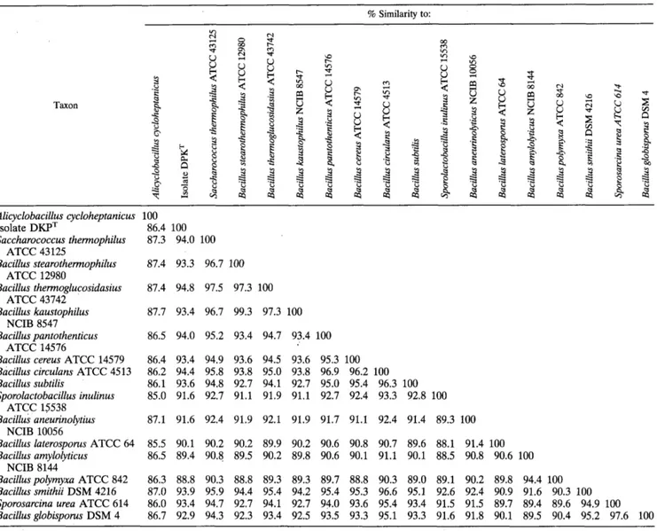
ated by the method of De Soete (10) from the evolutionary
distance matrix (Table 1) and shows these relationships. The
G + C content of the
16s rRNA of isolate DKPT was
55.5
mol%; this value is similar to the values determined for
members of groups 1 to 5 but slightly lower than the values
determined for members of group 5 (average G + C content, 58
mol%). A transversion analysis, implemented in the PHYLIP
package, yielded results similar to those shown in Fig. 3,
indicating that the results were not biased by the G + C content
of the 16s ribosomal DNA gene used in the analysis.
The following evidence supports the hypothesis that strain
DKPT occupies an intermediate position in the cluster con-
taining members of
Bacillus groups 1, 2, and 5. (i) Only two of
the nine signature nucleotides that are found in members of
the thermophilic bacilli belonging to grou 5 as defined by
Rainey et al. (34) were found in strain DKP’,
indicating a lack
of close affiliation. And (ii) certain nucleotides, nucleotide
pairs, and nucleotide stretches were found in strain DKPT
exclusively or occurred rarely in the 62 members of the genus
Bacillus and related genera (e.g., the genus Sporosarcina)
whose 16s rRNA sequences are available from the Ribosomal
Database Project; these nucleotides, nucleotide pairs, and
nucleotide stretches include C at position 189, GAUAA at
positions 193.3 to 193.7, GAUGGC at positions 198 to 203,
GCCAUCACUU at positions 214 to 224, U at position 264, U
and G at positions 458 and 474, UCUUGCGGCUCAACCG
CAA at positions 610 to 628), G and C at positions 835 and
851), U and A at positions 999 and 1041), G and C at positions
118 and 1155, and UCUAG at positions 1120 to 1124
(E. coli
numbering as described by Winker and Woese [53]). A boot-
strap analysis of the data performed by using programs in the
TREECON package and the PHYLIP package revealed low
levels of relatedness with members of all three groups; the
levels of relatedness with group 1,2, and
5
strains were 54, 22,
and 36%, respectively.
DISCUSSION
Strain DKPT, which was isolated from a palm wine sample
harvested near Dakar, Senegal, is a moderately thermophilic
facultative anaerobe. Its type of metabolism, its low G + C
content (38.8 mol%), and its typical gram-positive cell wall
(Fig. 1B) suggest that strain DKPT could belong to the genus
Bacillus or the genus Lactobacillus (15, 44).
The morphological and physiological characteristics and
nutritional requirements of strain DKPT were consistent with
the description of the genus
Lactobacillus (19, 42). Like
lactobacilli, strain DKPT is non-spore forming, saccharolytic,
and chemoorganotrophic. It requires Biotrypcase or yeast
extract for good growth. It produces lactate, acetate, ethanol,
and formate but not hydrogen during sugar fermentation.
Furthermore, NH, is produced during arginine fermentation,
as described previously for heterofermentative lactobacilli (1,
19). The ratio of the molar yields of acetate, ethanol, and
formate is 1:1:2; this fermentation ratio is typical of the
phosphoroclastic split of pyruvate into formate and acetyl
phosphate, where acetyl phosphate produces acetate and eth-
anol. Unpublished data have shown that the lactate concen-
tration varies considerably according to the culture conditions.
In an acidic medium, the yield of lactate increases concomi-
tantly with decreases in the yields of acetate, ethanol, and
formate. Under glucose-limiting conditions in a continuous
culture, a decrease in lactate production and an increase in the
production of acetate, ethanol, and formate occur. These
results are consistent with those reported previously for lactic
bacteria (7, 11, 36, 47). Furthermore, like members of the
genus
Lactobacillus, strain DKPT lacks cytochrome. Neverthe-
less, it has been demonstrated that under aerobic conditions,
lactobacilli reoxidize NADH,, with oxygen serving as the final
electron acceptor, since they possess flavin-containing oxidases
and peroxidases (19). This process probably results in an
increase in acetate production by strain DKPT in aerobiosis, as
described previously for
Lactobacillus plantarum (40).
Despite the phenotypic similarities between strain DKPT
and the genus
Lactobacillus, there are important differences
between these taxa. (i) Strain DKPT possesses peritrichous
flagella, which is unusual in lactobacilli (19, 42). (ii) Strain
DKPT is catalase positive. (iii) Strain DKPT exhibits optimal
growth in neutrophilic media, whereas lactobacilli grow best in
slightly acidic media (pH
5 or less), with optimal growth
occurring at pH 5.5 to 6.2 and growth often reduced at neutral
or slightly alkaline pH values (19). (iv) Strain DKPT exhibits an
optimum temperature for growth of 50°C and the upper
temperature limit for growth is 58°C; most lactobacilli grow
best at mesophilic temperatures and have an upper tempera-
ture limit for growth of around 40°C (the so-called “thermo-
philic” lactobacilli may have an upper temperature limit of
55”C, but no strain capable of growth at temperatures above
55°C is known [19]). In addition, a comparison of the electro-
phoretic pattern of the soluble cellular proteins of strain DKPT
with representative patterns of lactic acid bacteria (Fig. 2; data
not shown) also indicated that strain DKPT does not belong to
any previously described species of lactic bacteria.
The genus
Sporolactobacillus, which contains only one spe-
cies,
Sporolactobacillus inulinus (20), is phylogenetically closely
related to the genera
Bacillus and Lactobacillus. However,
Sporolactobacillus inulinus differs from strain DKPT by its
mesophilic and homolactic characteristics. Moreover, this spe-
cies produces
D-(-)-lactate and does not ferment entoses.
The genus
Sporolactobacillus differs from strain DKP and the
genus
Lactobacillus by spore production and from the genus
Bacillus by its lack of catalase and cytochrome. The taxonomic
position of
Sporolactobacillus inulinus has been discussed on
the basis of the results of a 16s rRNA sequence analysis (14,
43,44), and despite the lack of heme proteins such as catalase
and cytochrome, this organism is considered a member of the
family
Bacillaceae.
TABLE 1. 16s rRNA similarity matrbf % Similarity to: Taxon
B
Y
L d-
m Y Alicyclobacillus cycloheptanicus 100 Isolate DKPT 86.4 100 Saccharococcus thermophilus 87.3 94.0 100 Bacillus stearothermophilus 87.4 93.3 96.7 100 Bacillus thermoglucosidasius 87.4 94.8 97.5 97.3 100 Bacillus kaustophilus 87.7 93.4 96.7 99.3 97.3 100 Bacillus pantothenticus 86.5 94.0 95.2 93.4 94.7 93.4 100Bacillus cereus ATCC 14579 86.4 93.4 94.9 93.6 94.5 93.6 95.3 100
Bacillus circulans ATCC 4513 86.2 94.4 95.8 93.8 95.0 93.8 96.9 96.2 100
Bacillus subtilis 86.1 93.6 94.8 92.7 94.1 92.7 95.0 95.4 96.3 100
Sporolactobacillus inulinus 85.0 91.6 92.7 91.1 91.9 91.1 92.7 92.4 93.3 92.8 100
Bacillus anetlrinolytius 87.1 91.6 92.4 91.9 92.1 91.9 91.7 91.1 92.4 91.4 89.3 100
Bacillus laterosporus ATCC 64 85.5 90.1 90.2 90.2 89.9 90.2 90.6 90.8 90.7 89.6 88.1 91.4 100
Bacillus amylolyticus 86.5 89.4 90.8 89.5 90.2 89.8 90.6 90.1 91.1 90.1 88.5 90.8 90.6 100
Bacilluspolyrnyxa ATCC 842 86.3 88.8 90.3 88.8 89.3 89.3 89.7 88.8 90.3 89.0 89.1 90.2 89.8 94.4 100
Bacillus srnithii DSM 4216 87.0 93.9 95.9 94.4 95.4 94.2 95.4 95.3 96.6 95.1 92.6 92.4 90.9 91.6 90.3 100
Sporosarcina urea ATCC 614 86.0 93.4 94.7 92.7 94.1 92.7 94.0 93.6 95.4 93.4 91.5 91.5 89.7 89.4 89.6 94.9 100
Bacillusglobisporus DSM 4 86.7 92.9 94.3 92.3 93.4 92.5 93.5 93.3 95.1 93.3 91.6 91.8 90.1 89.5 90.4 95.2 97.6 100 ATCC 43125 ATCC 12980 ATCC 43742 NCIB 8547 ATCC 14576 ATCC 15538 NCIB 10056 NCIB 8144
a Values were determined by using Olsen's modification of the method of Jukes and Cantor (see Materials and Methods). Most of the sequences used in this analysis were obtained from the Ribosomal Data Project, version 3.0 (22). The Sporosarcina urea and Saccharococcus thermophilus sequences were obtained from the EMBL. A total of 1,128 unambiguous nucleotides were used in the analysis.
An analysis of the
16s rRNA gene of strain DKPT revealed
that this organism is closely related phylogenetically to mem-
bers of the genus
Bacillus
(Fig.
3) as defined by Ash et al. (3);
the position of this isolate is equidistantly from the position of
members of groups 1,2, and 5. However, a detailed analysis of
the signature nucleotides found in members of these three
groups did nQt reveal the precise position of isolate DKPT in
the cluster containing members of groups 1,2, and 5, indicating
that strain
DKPT is not closely affiliated with any of these three
groups. Phenotypically, members of the genus
Bacillus
and
strain DKPT are similar since
Bacillus
species may be faculta-
tive anaerobes and usually produce catalase.
Bacillus
species
use oxygen as the final electron acceptor; oxygen can be
replaced by alternative electron acceptors in some species
(8).
However, strain
DKPT differs from the previously described
Bacillus
species since it is non-spore forming and does not
contain heme proteins such as cytochromes. The possibility
that strain DKPT produces specific spores which do not exhibit
all of the characteristics of
Clostridium
spores, as described in
Thennoanaerobacter Jinnii
(39), was ruled out since strain
DKPT did not exhibit any heat resistance at 80°C.
According to Stackebrandt and Woese (44), the genera
Bacillus, Lactobacillus,
and
Streptococcus
and a few other
genera seem to
form
a unit, microaerophilic to aerobic in
phenotype, which is relatively coherent phylogenetically and is
a subbranch
of
the clostridia. Since the primitive atmosphere
of the earth was anaerobic, it has been suggested that among
the genera
Clostridium, Bacillus,
and
Lactobacillus,
the genus
Clostridium
is the most ancient. This hypothesis is consistent
with the fact that almost all species belonging to this genus are
strictly anaerobic and is supported by the results of
16s rRNA
studies (44). Accordingly, it has been hypothesized that the
genus
Bacillus
is the most recent genus, because almost all
Bacillus
species are facultatively anaerobic.
The genus
Lacto-
bacillus
probably appeared between the genera
Clostridium
and
Bacillus.
Lactobacilli and, more generally, lactic bacteria
possess metabolism which is intermediate between anaerobio-
sis and aerobiosis. We postulate that strain DKPT, because of
its phylogenetic and phenotypic characteristics, could be a
primitive member of the genus
Bacillus.
We propose that strain DKPT should be placed in the genus
VOL. 45, 1995
BACILLUS
THERMOAMYLOVORANSSP.
NOV. 15Description of Bacillus thermoamylovorans sp. nov.
Bacillus
thermoamylovorans (ther.mo.a.my.1o.vo’rans. Gr. adj. thermos,
hot; Gr. n.
amylum, starch; L. v. vorare, to devour; M. L. adj.
thermoamylovorans, utilizing starch at high temperatures).
Gram-positive, straight, rod-shaped eubacteria; cells are 0.45
to
0.5
pm wide and 3 to 4 pm long, and occur singly or in short
chains. Slightly motile by means of peritrichous flagella.
Spores are not detected under a range of conditions; cells
are killed by heating at 80°C for 5 min.
Facultative anaerobe. Catalase positive. No cytochrome is
produced. NO,- and
SO,,- are not reduced. Indole and H,S
are not produced.
Heterolactic fermentation of hexoses occurs; the end prod-
ucts are lactate, acetate, ethanol, and formate but not H,.
Chemoorganotrophic. Biotrypcase or yeast extract is re-
quired for luxuriant growth. Vitamins and nucleic acid deriv-
atives stimulate growth but are not essential.
Moderately thermophilic. The optimum temperature for
growth is about 50°C; the upper temperature limit for growth
is 58°C.
Neutrophilic. The optimal pH for growth ranges from 6.5 to
7.5; the pH range for growth is 5.4 to 8.5.
The G + C content of the DNA is 38.8
?0.2 mol%.
The results of a 16s rRNA sequence analysis indicate the
position of this organism is equidistant from the positions of
members of groups 1, 2, and
5 of the genus Bacillus.
Isolated from palm wine, an African alcoholic beverage, in
Rufisque (near Dakar), Senegal.
The type strain is DKP (= Collection of Institut Pasteur
CNCM 1-1378).
ACKNOWLEDGMENTS
We are indebted to
K.
D. Jahnke, Deutsche Sammlung von Mikro- organismen und Zellkulturen, for the G+
C content determination. We thankP.
Roger, A. Haemmerle, J.-L. Cayol, and M. Ramakrishna for revising the manuscript.B.
P.
is indebted to the Biotechnology (BRIDGE) T-Project on Lactic Acid Bacteria of the Commission of European Communities (contract BIOT-CT91-0263). We thank the Australian Research Council for financial assistance (to B.K.C.P.).1. 2. 3. 4. 5. 6. 7. 8. 9. 10. 11. 12. REFERENCES
Abdelal, A. G. 1979. Arginine catabolism by microorganisms. Annu. Rev. Microbiol. 6967-70.
qjayi, 0. A, E. 0. Fakiya, and G. 0. Oladopo. 1989. Industrial processing of palm juice and riboflavin loss. Food Chem. 3489-94.
Ash, C., J. A. E. Farrow, S. Walksbanks, and M. D. Collins. 1991. Phylogenetic heterogeneity of the genus Bacillus revealed by comparative
analysis of small-subunit-ribosomal RNA sequences. Lett. Appl. Microbiol.
Balch, W. E., G. E. Fox, L. J. Magrum, and R. S. Wolfe. 1979. Methanogens: reevaluation of a unique biological group. Microbiol. Rev. 43:260-296. Barre, P. 1978. Identification of thermobacteria and homofermentative, thermophilic, pentose-utilizing lactobacilli from high temperature ferment- ing grape musts. J. Appl. Bacteriol. M125-129.
Bassir, 0.1968. Some Nigerian wines. West Afr. J. Biol. Appl. Chem. 104245. Carlson, J., and C. J. Griffith. 1974. Fermentation products and bacterial yields in glucose-limited and nitrogen-limited cultures of streptococci. Arch. Oral Biol. 191105-1109.
Claus, D., and R. C. W. Berkeley. 1986. The genus Bacillus, p. 1105-1127. In P. H. A. Sneath, N. S. Mair, M. E. Sharpe, and J. G. Holt (ed.), Bergey’s manual of systematic bacteriology, vol. 2. The Williams & Wilkins Co., Baltimore.
Cord-Ruwisch, R. 1985. A quick method for the determination of dissolved
and precipitated sulfides in cultures of sulfate-reducing bacteria. J. Micro-
biol. Methods 433-36.
De Soete, G. 1983. A least square algorithm for fitting additive trees to proximity data. Psychometrika 48621-626.
De Vries, W., W. M. C. Kapteijn, E. G. van der Beek, and A. H. Stoutham- mer. 1970. Molar growth yields and fermentation balances of Lactobacillus casei 13 in batch cultures and in continuous cultures. J. Gen. Microbiol.
Eze, M. O., and A. U. Ogan. 1988. Sugars of the unfermented sap and the 13:202-206.
63: 333-345.
wine from the oil palm, Elaeis guinensis, tree. Plant Foods Hum. Nutr.
13. Felsentein, J. 1993. PHILIP (phylogenetic inference package), version 3.51~. Departement of Genetics, University of Washington, Seattle.
14. Fox, G. E., K. J. Pechmann, and C. R. Woese. 1977. Comparative cataloging of the 16s ribosomal ribonucleic acid: molecular approach to procaryotic systematics. Int. J. Syst. Bacteriol. 2244-57.
15. Fox, G. E., E. Stackebrandt, R. B. Hespell, J. Gibson, J. Malinoff, T. A. Dyer, R. S. Wolfe, W. E. Balch, R. S. Tanner, L. J. Magrum, L. B. Zablen, R. Blakemore, R. Gupta, L. Bonen, B. J. Lewis, D. A. Stahl, K. R. Luehrsen, K. N. Chen, and C. R. Woese. 1980. The phylogeny of procaryotes. Science 209457463.
16. Heller, R. 1985. Stwes, p. 764-767. In J. Bersani, J. Gall, H. Schweizer, and
M. Lardy (ed.), Encyclopaedia universalis, vol. 16. Encyclopzdia universalis publisher, Paris.
17. Hungate, R. E. 1969. A roll-tube method for the cultivation of strict anaerobes. Methods Microbiol. 3B:117-132.
18. Jukes, T. H., and C. R. Cantor. 1969. Evolution of protein molecules, p. 21-132. In H. N. Munro (ed.), Mammalian protein metabolism. Academic
Press, New York.
19. Kandler, O., and N. Weiss. 1986. The genus Sporolactobacillus, p. 113S1141. In
P. H. A. Sneath, N. S. Mair, M. E. Sharpe, and J. G. Holt (ed.), Bergey’s manual
of systematic bacteriology, vol. 2. The Williams & W i h s Co., Baltimore. 20. Kandler, O., and N. Weiss. 1986. The genus Lactobacillus, p. 1209-1219. In
P. H. A. Sneath, N. S. Mair, M. E. Sharpe, and J. G. Holt (ed.), Bergey’s
manual of systematic bacteriology, vol. 2. The Williams & Wilkins Co., Baltimore.
21. Kunkee, R. E., and M. A. Amerine. 1970. Yeast in wine-making, p. 10. In A. H. Rose (ed.), The yeast, vol. 3. Academic Press, London.
22. Larsen, N. A., G. J. Olsen, B. L. Maidak, M. J. McCaughey, R. Overbeek, T. J. Marsh, and C. R. Woese. 1993. The Ribosomal Database Project. Nucleic Acids Res. 21:3021-3023.
23. Lawson Anani Soh, A., H. Ralambotiana, B. Ollivier, G. Premier, E. Tine, and J. L. Garcia. 1991. Clostridium thermopalmarium sp. nov., a moderately
thermophilic butyrate-producing bacterium isolated from palm wine in Senegal. Syst. Appl. Microbiol. 14:135-139.
24. Love, C. A, B. K. J. Patel, P. D. Nichols, and E. Stackebrandt. 1993.
Desulfotomaculum australicum sp. nov., a thermophilic sulfate-reducing
bacterium isolated from the Great Artesian Basin of Australia. Syst. Appl. Microbiol. 16244-250.
25. Macy, J. M., J. E. Snellen, and R. E. Hungate. 1972. Use of syringe methods for anaerobiosis. Am. J. Clin. Nutr. 251318-1323.
26. Meshbah, M., U. Premachandran, and W. Whitman. 1989. Precise measure- ment of the G + C content of deoxyribonucleic acid by high-performance liquid chromatography. Int. J. Syst. Bacteriol. 39159-167.
27. Misge, J. 1985. Palmales, p. 454-457. In J. Bersani, J. Gall, H. Schweizer, and
M. Lardy (ed.), Encyclopaedia universalis, vol. 13. Encyclopzdia universalis publisher, Paris.
28. Miller, T. L., and M. J. Wolin. 1974. A serum bottle modification of the Hungate technique for cultivating obligate anaerobes. Appl. Microbiol.
29. Okafor, N. 1975. Preliminary microbiological studies on the preservation of palm wine. J. Appl. Bacteriol. 381-7.
30. Okafor, N. 1975. Microbiology of Nigerian palm wine with particular reference to bacteria. J. Appl. Bacteriol. 3881-88.
31. Olsen, G. J. 1988. Phylogenetic analysis using ribosomal RNA. Methods Enzymol. 164:793-812.
32. Pot, B., W. Ludwig, K. Kersters, and K. H. Schleifer. 1993. The taxonomy of lactic acid bacteria, p. 13. In L. De Vuys and E. J. Vandamme (ed.),
Bacteriocins of lactic acid bacteria: microbiology, genetics and applications. Blackie Academic & Professional, London.
33. Pot, B., P. Vandamme, and K. Kersters. 1993. Analysis of electrophoretic whole organism protein fingerprints, p. 493. In M. Goodfellow and A. G.
O’Donnell (ed.), Chemical methods in prokaryotic systematics. J. Wiley & Sons Ltd., Chichester, United Kingdom.
34. Rainey, F. A., N. L. Ward, H. W. Morgan, R. Toalster, and E. Stackebrandt. 1993. Phylogenetic analysis of anaerobic thermophilic bacteria: aid for their reclassification. J. Bacteriol. 17547724779.
35. Redburn, A. C., and B. K. C. Patel. 1993. Phylogenetic analysis of Desulfo- tomaculum thermobenzoicum using polymerase chain reaction-amplified 16s rRNA-specific DNA. FEMS Microbiol. Lett. 113531-86.
36. Rhee, S. K., and M. Y. Pack 1980. Effect of environmental pH on fermentation balance of Lactobacillus bulgaricus. J. Bacteriol. 144217-221. 37. Rogosa, M., J. G. Franklin, and K. D. Perry. 1961. Correlation of the vitamin
requirements with cultural and biochemical characters of Lactobacillus spp. J. Gen. Microbiol. 29473-482.
38. Rokosu, A. A., and J. J. Nwisienyi. 1980. Variation in the components of palm wine during fermentation. Enzyme Microb. Technol. 12:63-65. 39. Schmid, U., H. Giesel, S. M. Schoberth, and H. Sahm. 1986. Thermoanaer-
obacter finnii spec. nov., a new ethanologenic sporogenous bacterium. Syst.
Appl. Microbiol. 8:80-85.
40. Sedewitz, B., K. H. Schleifer, and F. Gotz. 1984. Physiological role of 38121-126.
pyruvate oxidase in the aerobic metabolism of Lactobacillus plantarum. J. Bacteriol. 160:462-465.
41. Shamala, T. R., and K. R. Sreekantiah. 1988. Microbiological and biochem- ical studies on traditional Indian palm wine fermentation. Food Microbiol.
42. Sharpe, M. E. 1992. The genus Lactobacillus, p. 1653-1679. In M. P. Starr, H. Stolp, H. G. Truper, A. Balows, and H. G. Schlegel (ed.), The pro- caryotes, vol. 2. Springer Verlag, Berlin.
43. Stackebrandt, E., V. J. Fowler, and C. R. Woese. 1983. A phylogenetic
analysis of lactobacilli, Pediococcus pentosaceus and Leuconostoc mesen- teroides. Syst. Appl. Microbiol. 4326-337.
44. Stackebrandt, E., and C. R. Woese. 1981. The evolution of procaryotes.
Symp. SOC. Gen. Microbiol. 32:l-31.
45. Tchiendji, C. 1985. Ph.D. thesis. Ecole Nationale SupCrieure d’Agronomie et des Industries Alimentaires, Universitt de Nancy, France.
46. Theivendirarajah, K., and R. K. Chrystopher. 1987. Microflora and micro- bial activity in palmyrah (Borassus flabellqer) palm wine in Sri Lanka. 5~157-162.
MIRCEN J. 323-31.
47. Thomas, T. D., D. C. Ellwood, and V. M. C. Longyear. 1979. Change from
homo- to heterolactic fermentation by Streptococcus lactis resulting from glucose limitation in anaerobic chemostat cultures. J. Bacteriol. 138109-117. 48. Tulley, P. 1965. How to tap an oil palm. Nigerian Field 3028-37. 49. Ukhun, M. E., and E. N. Dibie. 1991. The ascorbic acid contents of selected
marketed foods and influence of water activity (a,,,) during storage. Food Chem. 41:277-283.
50. Van de Peer, Y., and R De Wachter. 1992. TREECON: a software package
for the construction and drawing of evolutionary trees. Comput. Appl. Biosci. 9177-182.
51. Van Pee, W., and J. G. Swings. 1971. Chemical and microbiological studies on Congolese palm wines. East A f r . Agric. For. J. 36311-314.
52. Vauterin, L., and P. Vauterin. 1992. Computer-aided objective comparison
of electrophoresis patterns for grouping and identification of microorgan-
isms. Eur. J. Microbiol. 1:37-41.
53. Winker, S., and C. R. Woese. 1991. A definition of the domains Archaea, Bacteria, and Eucalya in terms of small subunit ribosomal RNA character- istics. Syst. Appl. Microbiol. 14305-310.