Publisher’s version / Version de l'éditeur:
The Molecular Immunology of Complex Carbohydrates-3, 2011
READ THESE TERMS AND CONDITIONS CAREFULLY BEFORE USING THIS WEBSITE.
https://nrc-publications.canada.ca/eng/copyright
Vous avez des questions? Nous pouvons vous aider. Pour communiquer directement avec un auteur, consultez la
première page de la revue dans laquelle son article a été publié afin de trouver ses coordonnées. Si vous n’arrivez pas à les repérer, communiquez avec nous à PublicationsArchive-ArchivesPublications@nrc-cnrc.gc.ca.
Questions? Contact the NRC Publications Archive team at
PublicationsArchive-ArchivesPublications@nrc-cnrc.gc.ca. If you wish to email the authors directly, please see the first page of the publication for their contact information.
Archives des publications du CNRC
This publication could be one of several versions: author’s original, accepted manuscript or the publisher’s version. / La version de cette publication peut être l’une des suivantes : la version prépublication de l’auteur, la version acceptée du manuscrit ou la version de l’éditeur.
For the publisher’s version, please access the DOI link below./ Pour consulter la version de l’éditeur, utilisez le lien DOI ci-dessous.
https://doi.org/10.1007/978-1-4419-7877-6_36
Access and use of this website and the material on it are subject to the Terms and Conditions set forth at
Polysialic acid bioengineering of cancer and neuronal cells by N-acyl
sialic acid precursor treatment
Jennings, H. J.; Zou, W.; Pon, R. A.
https://publications-cnrc.canada.ca/fra/droits
L’accès à ce site Web et l’utilisation de son contenu sont assujettis aux conditions présentées dans le site
LISEZ CES CONDITIONS ATTENTIVEMENT AVANT D’UTILISER CE SITE WEB.
NRC Publications Record / Notice d'Archives des publications de CNRC:
https://nrc-publications.canada.ca/eng/view/object/?id=9dd4d43f-e2eb-4172-80bb-5cdd6c9c4905 https://publications-cnrc.canada.ca/fra/voir/objet/?id=9dd4d43f-e2eb-4172-80bb-5cdd6c9c4905
bioengineering of cancer and neuronal cells by N-acyl sialic
acid precursor treatment. In “The Molecular Immunology of
Complex Carbohydrates”, ed., A. Wu, p757-768 (2010).
POLYSIALIC ACID BIOENGINEERING OF CANCER AND
NEURONAL CELLS BY N-ACYL SIALIC ACID PRECURSOR
TREATMENT
1Harold J. Jennings
2, Wei Zou and Robert A. Pon
Institute for Biological Sciences, National Research Council of Canada,
Ottawa, Ontario, Canada K1A 0R6
1
The abbreviations used in this manuscript are the following: PSA, polysialic acid; NPrPSA, N-propionyl polysialic acid; NBuPSA, N-butanoyl polysialic acid;, ManPr, N-propionyl mannosamine; ManBu, N-butanoyl mannosamine; mAb, monoclonal antibody; KLH, keyhole limpet hemocyanin; HSA, human serum albumin; CE-MS, capillary electrophoresis mass spectrometry; NCAM, neural cell adhesion molecule; NT2, NTera 2/ cl.D1 teratocarcinoma cell line; NeuAc, N-acetyl neuraminic acid; NeuPr, N-propionyl neuraminic acid; NeuBu, N-butanoyl neuraminic acid; s.c., subcutaneous; i.p., intraperitoneal.
N-acetyl neuraminic acid, the most prominent member of the acidic sugar family known as sialic acids, is ubiquitous on the surface of eukaryotic cells, where as a glycoconjugate substituent it is involved in numerous important biological processes.
Polysialic acid (PSA), an α-(2-8) linked homopolymer of N-acetyl neuraminic acid, is
equally as important biologically however its expression is more restricted to embryonic tissues (1), regions of brain plasticity (2), and as an oncofetal antigen in several cancers of neuroectodermal origin (3;4). PSA, by virtue of its large polyanionic charge, functions as an anti-adhesive thereby negatively regulating cell-cell interactions (5), and is implicated in processes such as neurogenesis (6), synaptogenesis (7), axonal pathfinding (8), and cell migration (6). The limited expression of PSA in normal adult tissue increases its potential as a cancer determinant where it has been found in abundance in such malignancies as Wilm’s tumor (9), small cell and non-small cell lung cancer (10;11), neuroblastoma (12), and rhabdomyosarcoma (13). Several lines of evidence further implicate the anti-adhesive nature of PSA with primary tumor shedding and metastasis (14-16), where its expression is negatively associated with favourable prognoses (10).
The permissiveness of the enzymes involved in sialic acid biosynthesis and sialoside and polysialic acid formation have been exploited for the bioengineering of cell surface molecules (17), and this procedure has been used successfully in the study of biological processes (17), the chemotargeting of mammalian cells (18) and the immunotargeting of tumor cells (19). This latter strategy tackles the often observed and inherent problem of poor immunogenicity of the natural saccharide markers of cancer cells, including PSA (20), by virtue of the formation of de novo non-natural, non-self sialylglycoconjugates. When an exogenous source of the sialic acid precursor N-propionyl mannosamine (ManPr) was provided in vitro, both rat and mouse PSA positive leukemic cell lines expressed N-propionyl polysialic acid (NPrPSA) on their surfaces (Figure 1) and its expression was both time and dose dependent (20). The expression of
de novo NPrPSA, with the concomitant reduction of surface PSA on the bioengineered cells, could be followed by flow cytometric analysis using the respective serologically distinct mAbs 13D9 and 735 (21). Furthermore, specific cancer cell cytotoxicity was achieved by treating the bioengineered cells with the NPrPSA-specific mAb 13D9 in the presence of complement, and cytotoxicity was shown to be dependent on the amount of
NPrPSA expressed on the cells. The bioengineering strategy using sialic acid precursor treatment to target experimental tumors has also been validated in vivo using a solid tumor mouse model. Administration of ManPr in conjunction with mAb 13D9 to mice with pre-existing solid tumors resulted in delayed tumor growth and overall reductions in tumor size, but not tumor eradication.
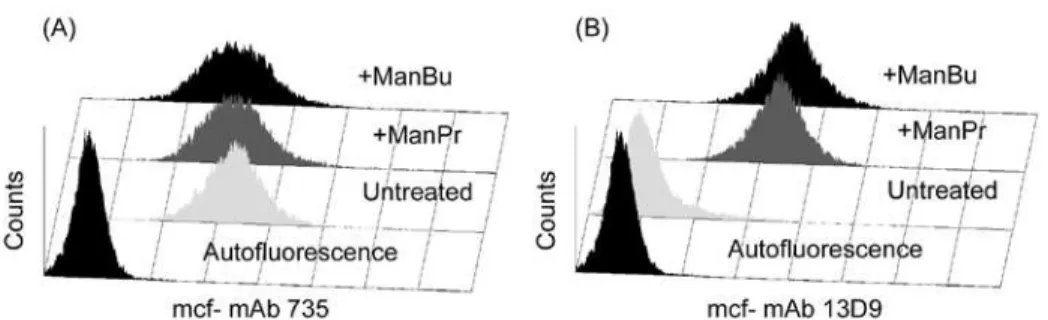
Figure 1. Effects of ManPr and ManBu on PSA expression in RMA-s tumor cells. RMA-s cells in log phase growth were treated with 10 mM ManPr (+ManPr), ManBu (+ManBu), or medium alone (untreated) for 3 days followed by surface staining with mAbs 735 (A) and 13D9 (B) and analysis by flow cytometry. Gates were set on viable cells only, as determined through propidium iodide exclusion, and 10,000 events were acquired. RMA-s autofluorescence appears within the first decade in each histogram.
There was evidence however, that sialic acid precursor therapy, along with specific targeting by mAb, may be able to control tumor shedding and micrometastases since in our model, there was no evidence of tumor burden within the spleens of treated animals; unlike untreated controls or those treated with mAb 13D9 alone (Table 1).
Table 1. Antibodies to NPrPSA control tumor migration in ManPr treated mice. RMA-s cells (1.0 x10⁶) were transplanted s.c. into syngeneic C57BL/6 mice and established for 5d prior to daily i.p. injections (8d) with ManPr (5mg) and mAb 13D9 (200 ug); 13D9 alone; or with PBS buffer. Spleens were removed (d25), and tumor presence detected via limiting dilution analysis of splenic cells.
Group # Mice with Splenic tumors % Metastasis
mAb 13D9 + ManPr 0/5 0
PBS control 4/5 80
Extrapolating from these results, it is probable that a cancer control strategy using similar approaches may be most effective, not for tissue based tumor eradication, but as an adjuvant therapy for control of micrometastases; the importance of which cannot be underestimated.
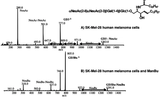
The range of modified N-acyl mannosamine precursors that have been used in sialo-bioengineering studies is fairly extensive (17), and we were able to use this to our advantage to target human GD3- expressing melanoma cells (SK-MEL-28) with either polyclonal antisera or the derived monoclonal antibody 2A; both generated from a synthetic therapeutic cancer conjugate vaccine construct (NBuGD3-KLH) (22) (for GD3 structure- see Figure 2).
αNeuAc(2-8)αNeuAc(2-3)βGal(1-4)βGlc(1-O C13H27 HN OH C23H47 O 161.0 318.0 503.0 637.0 744.0 805.0 972.0 1291.0 200 300 400 500 600 700 800 900 1000 1100 1200 1300 1400 m/z 179.0 290.0 493.0 581.0 647.0 777.0 1263.0 200 300 400 500 600 700 800 900 1000 1100 1200 1300 1400 m/z 971.0 NeuAc NeuBu NeuBu-NeuBu NeuAc-NeuAc GD3 2-GD3Bu 2-GD3 - NeuAc GD3Bu-NeuBu NeuBu NeuAc 809.0 Gal Glc
A) SK-Mel-28 human melanoma cells
B) SK-Mel-28 human melanoma cells and ManBu
αNeuAc(2-8)αNeuAc(2-3)βGal(1-4)βGlc(1-O C13H27 HN OH C23H47 O 161.0 318.0 503.0 637.0 744.0 805.0 972.0 1291.0 200 300 400 500 600 700 800 900 1000 1100 1200 1300 1400 m/z 179.0 290.0 493.0 581.0 647.0 777.0 1263.0 200 300 400 500 600 700 800 900 1000 1100 1200 1300 1400 m/z 971.0 NeuAc NeuBu NeuBu-NeuBu NeuAc-NeuAc GD3 2-GD3Bu 2-GD3 - NeuAc GD3Bu-NeuBu NeuBu NeuAc 809.0 Gal Glc 161.0 318.0 503.0 637.0 744.0 805.0 972.0 1291.0 200 300 400 500 600 700 800 900 1000 1100 1200 1300 1400 m/z 179.0 290.0 493.0 581.0 647.0 777.0 1263.0 200 300 400 500 600 700 800 900 1000 1100 1200 1300 1400 m/z 971.0 NeuAc NeuBu NeuBu-NeuBu NeuAc-NeuAc GD3 2-GD3Bu 2-GD3 - NeuAc GD3Bu-NeuBu NeuBu NeuAc 809.0 Gal Glc
A) SK-Mel-28 human melanoma cells
B) SK-Mel-28 human melanoma cells and ManBu
Figure 2. CE-MS analysis of native and modified GD3 from ManBu treated SK-MEL-28 melanoma cells. GD3 was extracted from untreated and ManBu treated (1mg/ml) SK-MEL-28 cells and was analyzed by capillary electrophoresis- mass spectrometry using selective ion scanning techniques (m/z 290 native NeuAc and m/z 318 for NeuBu). MS-MS fragmentation patterns confirmed GD3 on untreated cells (A) and NBuGD3 on ManBu treated cells (B).
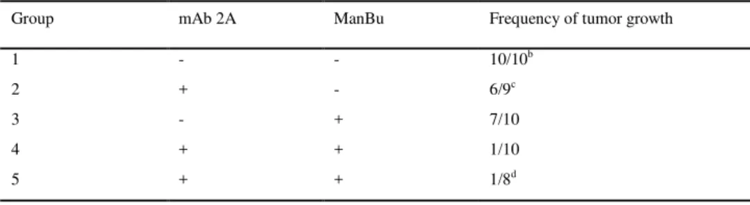
In this case N-butanoyl mannosamine (ManBu) was selected over ManPr as the sialo-modifying precursor of choice in order to optimize specificity and minimize cross reactions with native GD3 sugar residues. When treated with exogenous ManBu, the SK-MEL-28 melanoma cells efficiently metabolized this sialic acid precursor resulting in non-natural NBuGD3 being incorporated into their surface membranes as determined both by flow cytometric analysis with mAb 2A (22) and by direct physical determination using CE-MS analysis (Figure2). Similar to what was observed with bioengineered PSA expressing tumors, NBuGD3– specific antibodies became potent cytotoxic reagents in the presence of complement against SK-MEL-28 cells engineered to express NBuGD3 molecules. Moreover, studies in vivo also indicated that treatment with a combination of precursor (ManBu) and mAb 2A could prevent nude mice from being grafted with SK-MEL-28 melanoma cells (Table 2), but was not as effective on previously established tumors. Again this result corroborates our and other’s (23) view that this approach may be more relevant for the control of tumor shedding and metastasis; a phenomenon that we are currently investigating.
Table 2. Effect of mAb 2A and/ or ManBu on SK-MEL-28 tumor graftinga. SK-MEL-28 cells (1.0 x 10⁷) were transplanted subcutaneously into Balb/c nu/nu mice and subsequently treated 3d later with daily injections (2 weeks) of mAb 2A (200 ug/ mouse) + ManBu (5 mg/ mouse); mAb 2A alone; ManBu alone; or medium control.
Group mAb 2A ManBu Frequency of tumor growth
1 - - 10/10b
2 + - 6/9c
3 - + 7/10
4 + + 1/10
5 + + 1/8d
a 1.0 x 10E7 SK-MEL-28 cells were transplanted s.c. into BALB/c nu/nu mice. Administration of mAb 2A (200 ug/ mouse) and/ or ManBu (5 mg/mouse) was started 3d after tumor grafting and continued 5d. week for 2 weeks
b
tumor growth was recorded at d30 and d45. The size of tumors at d45= 4-7 mm c one mouse died
d administered intraperitoneally
Several groups have reported success in using ManPr as a precursor to incorporate NPrPSA into cell surfaces (19;24), but unlike our work with GD3, there is considerable debate as to the effects of using ManBu as a sialic acid precursor. It was originally reported that ManBu precursor treatment effectively halted PSA surface expression on both cancer cells and human neurons (25), thus suggesting that ManBu could not only be an important reagent in studying the role of PSA-NCAM, but more relevant to our studies, that it could function as a potential cancer therapeutic independent of the use of vaccines. More recently (26), it was reported that ManBu treatment of PSA-expressing cells abrogated PSA expression in human NT2 neurons but not tumor cells. Because of these mixed results and the potential impact they could have had on the further development of our PSA immunotargeting strategy, we decided to re-examine this issue.
Flow cytometric analysis was used to detect NBuPSA on the surface of cancer and neuronal cells using mAb13D9 as the antibody probe (27). In earlier studies (28), it was shown that mAb 13D9 specifically recognizes extended helical NPrPSA epitopes, requiring at least nine NeuNPr residues for binding to occur (29), but whether it bound to NBuPSA had not been established. However more recently (27), in vitro screening experiments using PSA, NPrPSA and NBuPSA as binding antigens confirmed that mAb13D9 does not recognize PSA, and additionally, demonstrated that recognition of NBuPSA was virtually indistinguishable from that of NPrPSA, thus indicating that mAb 13D9 could be used as a probe to detect NBuPSA in the absence of NPrPSA (Figure3).
0.001 0.01 0.1 1 0.0 0.5 1.0 1.5 2.0 2.5 NPrPSA-HSA NBuPSA-HSA NAcPSA-HSA mAb 735 (ug/ml) OD 405 0.001 0.01 0.1 1 0.0 0.5 1.0 1.5 2.0 2.5 NPrPSA-HSA NBuPSA-HSA NAcPSA-HSA mAb 13D9 (ug/ml) OD 405 A) B)
Figure 3. Binding characteristics of mAbs 735 and 13D9. The specificities of mAbs 735 (A) and 13D9 (B) were determined by indirect ELISA against HSA conjugates of PSA (■), NPrPSA (▲), and NBuPSA (▼).
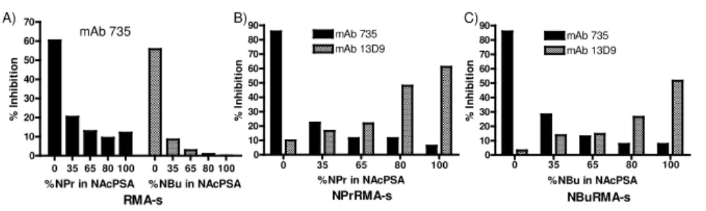
When the mouse leukemic cell line RMA-s was subjected to ManBu precursor treatment, flow cytometric analysis in the presence of mAb 13D9 revealed the expression of newly formed NBuPSA on its surface, consistent with the expression of NPrPSA generated in response to ManPr treatment as we had observed previously (Figure 1). Regardless of whether the tumor cells were treated with ManPr or ManBu, there was a continued presence of native PSA detected with mAb 735 (21), indicating that precursor treatment did not abrogate the expression of PSA on cancer cells as observed by others (25) (Figure 1). The nature of the neo-surface epitopes formed upon either ManPr or ManBu precursor treatment was probed since these polymers arise from in vivo metabolic and biosynthetic processes that can result in the formation of unknown membrane bound mixed polysaccharides. Furthermore, the binding specificities of either mAb 735 or 13D9 towards such mixed polymer substrates are unknown. Using competitive inhibition flow cytometry experiments with a series of small synthetic PSA inhibitors that contain mixed ratios of N-acetyl to N-butanoyl neuraminic acid (NeuAc:NeuBu), only those inhibitors with > 80% NeuBu residues were effective inhibitors of 13D9 binding to the ManBu treated tumor cells (Figure 4).
Figure 4. Surface epitope specificity on bioengineered RMA-s tumor cells. Mixed polysaccharide inhibitors of defined length and composition (NPr:NAcPSA; NBu:NAcPSA) were prepared (27) and used in competitive inhibition studies. RMA-s tumor cells were cultured for 3d with media alone (A), 10 mM ManPr (B), or ManBu (C), stained with either mAbs 735 (A-C) or 13D9 (B, C) that were pretreated with 1 ug of the indicated mixed inhibitor, and evaluated by flow cytometry. The % inhibition was calculated relative to the mean channel fluorescence values obtained from each respective mAb without polysaccharide inhibitors.
0 35 65 80 100 0 35 65 80 100 0 10 20 30 40 50 60 70 %NPr in NAcPSA mAb 735 RMA-s %NBu in NAcPSA % I nhi bi ti on 0 35 65 80 100 0 10 20 30 40 50 60 70 80 90 mAb 735 mAb 13D9 NPrRMA-s %NPr in NAcPSA % I n h ib it io n 0 35 65 80 100 0 10 20 30 40 50 60 70 80 90 mAb 735 mAb 13D9 NBuRMA-s %NBu in NAcPSA % I n h ib it io n A) B) C)
Conversely, only the homogeneous NeuAc containing inhibitor was able to interfere with mAb 735 binding to the same treated tumor cells. This result indicated that native PSA and de novo NBuPSA exist as discrete polysaccharides on the surface of ManBu treated cells and not as a single polysaccharide composed of varying levels of mixed NeuAc to NeuBu residues. An identical inhibition profile was obtained using mixed inhibitors composed of NeuPr: NeuAc residues on ManPr treated tumor cells supporting this conclusion (Figure 4). Although flow cytometric analysis indicated a relatively high surface density of de novo NBuPSA and residual PSA, strong cytotoxicity responses of ManBu treated tumor cells in the presence of either mAbs 13D9 or 735 and complement illustrated that the abundances of these two surface polysaccharides were sufficient to behave as functional determinants (Figure 5).
mAb 735 mAb 13D9 0 10 20 30 40 50 60 4 2 1 0.5 0.25 0.125 0.063 0.031 0.004 0.016 mAb (ug) 0.008 mAb treatment % S p eci fi c cyt o to xi ci ty mAb 735 mAb 13D9 0 10 20 30 40 50 60 4 2 1 0.5 0.25 0.125 0.063 0.031 0.004 0.016 mAb (ug) 0.008 mAb treatment % S p eci fi c cyt o to xi ci ty
(A) RMA-s cells- non-treated (B) RMA-s cells- ManBu treated
Figure 5. Complement dependent cytotoxicity of native and ManBu precursor treated RMA-s tumor cells with mAbs 735 and 13D9. RMA-s cells labeled for 1h with Na2[51Cr]O4 were combined with graded amounts of mAbs 735 or 13D9 in the presence of 5% baby rabbit complement for 4h at 37°C. Released Cr-51 was measured by scintillation counting and the % specific cytotoxicity calculated. Maximum release was determined in the presence of cetrimide and spontaneous release in the presence of medium alone and was consistently < 20%.
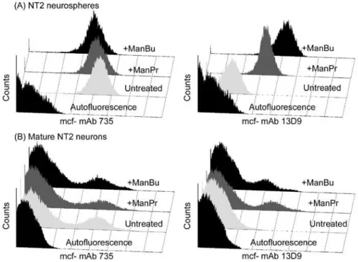
Although our studies indicated that an array of tumor cells were amenable to bioengineering with various N-acyl mannosamine sialic acid precursors, other groups (25;26) reported that ManBu precursor treatment had the effect of abrogating PSA expression on human NTera 2 (NT2) derived neurons. This observation has significant
implications on neural regeneration and function studies as PSA is intimately involved in these processes (30). However in our hands, we detected de novo NBuPSA on ManBu treated neuronal cells using our mAb 13D9 as an antibody probe (27). Neuronal cells were generated from the parent NTera 2 progenitor cell line and were isolated at different stages of maturation. Following incubation with both ManPr and ManBu, immature neurospheres were shown to homogeneously express NPrPSA and NBuPSA respectively using mAb13D9 as the antibody probe (Figure 6).
Figure 6. The effects of ManPr and ManBu sialic acid precursors on PSA expression on NT2 neurons at different stages of maturation. NT2 neurons (neurospheres (A) and matrigel matured neurons (B) (27) were treated with 10 mM ManPr (+ManPr), ManBu (+ManBu), and medium alone (Untreated) for 3d followed by surface staining with mAbs 735 and 13D9 and evaluation by flow cytometry. Histogram profiles are based on viable cells and autofluorescence was adjusted to appear within the first logarithmic decade of each histogram. Note the homogeneity of either mAb 735 or 13D9 surface staining within the neurosphere fraction (A) as opposed to matrigel matured neurons (B).
Also as shown in Figure6, in similar experiments using mAb735 as the antibody probe, the presence of PSA on the surface of the bioengineered neurospheres was still detected, thus demonstrating that PSA expression was not abrogated by treatment with ManBu as had been observed by others (25;26). Flow cytometric analyses of maturing neurons demonstrated that PSA expression, and thus the ability to bioengineer it with sialic acid precursors, rapidly decreased with maturation and resided only in the immature neurosphere phenotype. This observation, along with changes in the relative levels of the different polysialyltransferases at play in this cell type (26), might help to explain the mixed results obtained previously.
SUMMARY
It is clear from our studies and others that the use of non-natural sialic acid precursor molecules is an effective manner to manipulate surface sialoglycoconjugates, and that this strategy can further take advantage of polysialyltransferase promiscuity to incorporate non-natural sialic acids into surface PSAs. The ability to manipulate cellular PSA, whether in its natural state as in neuronal expression or as an abnormal cancer determinant, remains a key goal both in the fields of neurobiology and oncology. As we were not able to substantiate the PSA abrogating effects of ManBu, which potentially could have been used as a stand alone therapeutic, the strategy of converting sialic and polysialic acids into their non-natural derivatives through bioengineering approaches reinforces this method as a manner to effectively disturb the natural setting of PSA and in particular, to specifically target this relevant cancer determinant. There are questions that remain to be addressed in this approach to cancer therapy such as the relative turnover of sialylated/ polysialylated glycoconjugates on tumor versus normal cells; the effect(s) of modifying all cellular sialic acids when targeting the oncofetal PSA antigen; the role that PSA plays in metastasis; and finally the pharmacokinetics and toxicity of therapeutic sialic acid precursor molecules. Yet in spite of these lingering questions, the use of small molecules to bioengineer cellular sialic and polysialic acid glycoconjugates and an ability to specifically target them, remains an exciting therapeutic avenue that holds a great deal of promise.
REFERENCES
1. Szele, F. G., Dowling, J. J., Gonzales, C., Theveniau, M., Rougon, G., Chesselet, M. F. (1994) Pattern of expression of highly polysialylated neural cell adhesion molecule in the developing and adult rat striatum. Neuroscience 60, 133-144.
2. Theodosis, D. T., Rougon, G., Poulain, D. A. (1991) Retention of embryonic features by an adult neuronal system capable of plasticity: polysialylated neural cell adhesion molecule in the hypothalamo-neurohypophysial system. Proc.Natl.Acad.Sci.U.S.A 88, 5494-5498.
3. Troy, F. A. (1992) Polysialylation: from bacteria to brains. Glycobiology 2, 5-23.
4. Lantuejoul, S., Moro, D., Michalides, R. J., Brambilla, C., Brambilla, E. (1998) Neural cell adhesion molecules (NCAM) and NCAM-PSA expression in neuroendocrine lung tumors. Am.J.Surg.Pathol. 22, 1267-1276.
5. Rutishauser, U. (1996) Polysialic acid and the regulation of cell interactions. Curr.Opin.Cell Biol. 8, 679-684.
6. Petridis, A. K., El Maarouf, A., Rutishauser, U. (2004) Polysialic acid regulates cell contact-dependent neuronal differentiation of progenitor cells from the subventricular zone. Dev.Dyn. 230, 675-684. 7. El Maarouf, A., Rutishauser, U. (2003) Removal of polysialic acid induces aberrant pathways, synaptic
vesicle distribution, and terminal arborization of retinotectal axons. J.Comp Neurol. 460, 203-211. 8. Rutishauser, U., Landmesser, L. (1996) Polysialic acid in the vertebrate nervous system: a promoter of
plasticity in cell-cell interactions. Trends Neurosci. 19, 422-427.
9. Roth, J., Zuber, C., Wagner, P., Taatjes, D. J., Weisgerber, C., Heitz, P. U., Goridis, C., Bitter-Suermann, D. (1988) Reexpression of poly(sialic acid) units of the neural cell adhesion molecule in Wilms tumor. Proc.Natl.Acad.Sci.U.S.A 85, 2999-3003.
10. Tanaka, F., Otake, Y., Nakagawa, T., Kawano, Y., Miyahara, R., Li, M., Yanagihara, K., Inui, K., Oyanagi, H., Yamada, T., Nakayama, J., Fujimoto, I., Ikenaka, K., Wada, H. (2001) Prognostic significance of polysialic acid expression in resected non-small cell lung cancer. Cancer Res. 61, 1666-1670.
11. Komminoth, P., Roth, J., Lackie, P. M., Bitter-Suermann, D., Heitz, P. U. (1991) Polysialic acid of the neural cell adhesion molecule distinguishes small cell lung carcinoma from carcinoids. Am.J.Pathol. 139, 297-304.
12. Livingston, B. D., Jacobs, J. L., Glick, M. C., Troy, F. A. (1988) Extended polysialic acid chains (n greater than 55) in glycoproteins from human neuroblastoma cells. J.Biol.Chem. 263, 9443-9448. 13. Soler, A. P., Johnson, K. R., Wheelock, M. J., Knudsen, K. A. (1993) Rhabdomyosarcoma-derived cell
lines exhibit aberrant expression of the cell-cell adhesion molecules N-CAM, N-cadherin, and cadherin-associated proteins. Exp.Cell Res. 208, 84-93.
14. Scheidegger, E. P., Lackie, P. M., Papay, J., Roth, J. (1994) In vitro and in vivo growth of clonal sublines of human small cell lung carcinoma is modulated by polysialic acid of the neural cell adhesion molecule. Lab Invest 70, 95-106.
15. Daniel, L., Durbec, P., Gautherot, E., Rouvier, E., Rougon, G., Figarella-Branger, D. (2001) A nude mice model of human rhabdomyosarcoma lung metastases for evaluating the role of polysialic acids in the metastatic process. Oncogene 20, 997-1004.
16. Cheung, I. Y., Vickers, A., Cheung, N. K. (2006) Sialyltransferase STX (ST8SiaII): a novel molecular marker of metastatic neuroblastoma. Int.J.Cancer 119, 152-156.
17. Keppler, O. T., Horstkorte, R., Pawlita, M., Schmidt, C., Reutter, W. (2001) Biochemical engineering of the N-acyl side chain of sialic acid: biological implications. Glycobiology 11, 11R-18R.
18. Mahal, L. K., Yarema, K. J., Bertozzi, C. R. (1997) Engineering chemical reactivity on cell surfaces through oligosaccharide biosynthesis. Science 276, 1125-1128.
19. Liu, T., Guo, Z., Yang, Q., Sad, S., Jennings, H. J. (2000) Biochemical engineering of surface alpha 2-8 polysialic acid for immunotargeting tumor cells. J.Biol.Chem. 275, 32832-32836.
20. Krug, L. M., Ragupathi, G., Ng, K. K., Hood, C., Jennings, H. J., Guo, Z., Kris, M. G., Miller, V., Pizzo, B., Tyson, L., Baez, V., Livingston, P. O. (2004) Vaccination of small cell lung cancer patients with polysialic acid or N-propionylated polysialic acid conjugated to keyhole limpet hemocyanin. Clin.Cancer Res. 10, 916-923.
21. Frosch, M., Gorgen, I., Boulnois, G. J., Timmis, K. N., Bitter-Suermann, D. (1985) NZB mouse system for production of monoclonal antibodies to weak bacterial antigens: isolation of an IgG antibody to the polysaccharide capsules of Escherichia coli K1 and group B meningococci. Proc.Natl.Acad.Sci.U.S.A 82, 1194-1198.
22. Zou, W., Borrelli, S., Gilbert, M., Liu, T., Pon, R. A., Jennings, H. J. (2004) Bioengineering of surface GD3 ganglioside for immunotargeting human melanoma cells. J.Biol.Chem. 279, 25390-25399. 23. Livingston, P. O., Hood, C., Krug, L. M., Warren, N., Kris, M. G., Brezicka, T., Ragupathi, G. (2005)
Selection of GM2, fucosyl GM1, globo H and polysialic acid as targets on small cell lung cancers for antibody mediated immunotherapy. Cancer Immunol.Immunother. 54, 1018-1025.
24. Charter, N. W., Mahal, L. K., Koshland, D. E., Bertozzi, C. R. (2000) Biosynthetic incorporation of unnatural sialic acids into polysialic acid on neural cells. Glycobiology 10, 1049-1056.
25. Mahal, L. K., Charter, N. W., Angata, K., Fukuda, M., Koshland, D.-E. J., Bertozzi, C. R. (2001) A small-molecule modulator of poly-alpha 2,8-sialic acid expression on cultured neurons and tumor cells. Science 294, 380-381.
26. Horstkorte, R., Muhlenhoff, M., Reutter, W., Nohring, S., Zimmermann-Kordmann, M., Gerardy-Schahn, R. (2004) Selective inhibition of polysialyltransferase ST8SiaII by unnatural sialic acids. Exp.Cell Res. 298, 268-274.
27. Pon, R. A., Biggs, N. J., Jennings, H. J. (2007) Polysialic acid bioengineering of neuronal cells by N-acyl sialic acid precursor treatment. Glycobiology 17, 249-260.
28. Jennings, H. J. (2003) Polysialic acid vaccines. In Carbohydrate-based Drug Discovery (Wong, C. H., ed) pp. 357-380, Wiley-VCH, Weinham, Germany.
29. MacKenzie, C. R., Jennings, H. J. (2003) Characterization of polysaccharide conformational epitopes by surface plasmon resonance. Methods Enzymol. 363, 340-354.
30. Bruses, J. L., Rutishauser, U. (2001) Roles, regulation, and mechanism of polysialic acid function during neural development. Biochimie 83, 635-643.