Supporting Information
Excitonic Channels from Bio-Inspired Templated Supramolecular
Assembly of J-aggregate Nanowires
Surendra B. Anantharaman,1,2 Daniel Messmer,3 Amin Sadeghpour,4, 5, 6 Stefan Salentinig,5 Frank Nüesch,1,2 Jakob Heier1,*
1 Laboratory for Functional Polymers, Swiss Federal Laboratories for Materials Science and Technology (Empa), Überlandstrasse 129, CH-8600 Dübendorf, Switzerland.
2 Institut des matériaux, École Polytechnique Fédérale de Lausanne (EPFL), CH-1015 Lausanne, Switzerland.
3 Laboratory of Polymer Chemistry, Department of Materials, ETH Zurich, Vladimir-Prelog-Weg 5, CH-8093 Zürich, Switzerland.
4 Center for X-ray Analytics, Swiss Federal Laboratories for Materials Science and Technology (Empa), Überlandstrasse 129, CH-8600 Dübendorf, Switzerland.
5 Laboratory for Biointerfaces and 6 Laboratory for Biomimetic Membranes and Textiles , Swiss Federal Laboratories for Materials Science and Technology (Empa), Lerchenfeldstrasse 5, CH-9014 St. Gallen, Switzerland.
Corresponding Author
S1. Details concerning dendronized polymer synthesis and characterization
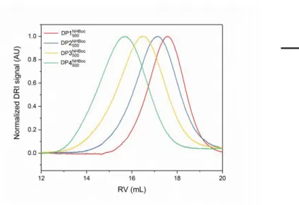
The dendronized polymers (DPs) employed in this work were prepared in a bottom-up approach, building the side chains by adding exactly one layer of dendritic monomers, i.e. DP1 served as the precursor to DP2, DP2 as the precursor for DP3, etc.1,2 This mode of synthesis guarantees 1) that main-chain length within such a homologous series of DPs is essentially conserved, facilitating comparisons between DPs of differing g, and 2) that the dendritic side-chains are virtually defect-free, as quantified using a colorimetric defect labeling approach.3,4 The N-protected precursor polymers of samples used in this publication have been characterized elsewhere; DP1 has a molar mass of 259 kDa (Pn ≈ 500) as determined by GPC (Figure. S1), and chain length is conserved throughout the homologous DP series as evidenced by AFM imaging.5 GPC data does not provide a reliable measure of chain lengths particularly for high g DPs, as they feature very large molar masses per repeat unit (several kDa) and appropriate molar mass standards are not available; the results in Fig. S1 rely on universal calibration using PMMA standards.
Figure S1. Normalized GPC curves (DMF with 0.1 % LiBr, 45 °C, DRI detector) of the
N-protected precursors DP1-DP4 to the polyelectrolytes DP1-DP4 used in this publication, constituting a homologous series of DPs and corresponding numerical data (based on universal calibration using PMMA standards).
DP Mn [kDa] Đ
DP1 259 1.72 DP2 665 1.57 DP3 1686 1.51 DP4 3805 1.26
S2. Optimization conditions for obtaining monolayer DP on solid substrates
In preliminary explorations, the adsorption of DP4 was investigated in order to arrive at suitable conditions for the coating of other silicate surfaces. Mica was chosen as a substrate for these studies as it is atomically flat, permitting facile observation of adsorbed features by AFM, and as qualitatively it behaves quite similar to e.g. glass in that the surfaces exposes net negative charges, providing good adsorption of the polycationic dendronized polymers. The g = 4 representative of the series was selected as due to its large diameter (ca. 5 nm) it can without any difficulty be observed in AFM. A number of parameters pertaining to the adsorption process was investigated, with the aim of developing a procedure which reliably provides densely coated substrates, as the goal was initially to prepare substrates suitable for the deposition and controlled growth of J-aggregates rather than to prepare self-assembled structures in solution.
Generally, AFM samples described below were prepared by submerging a freshly cleaved platelet of mica (ca. 5 mm x 5 mm, Plano GmbH) into the solution in question, then gently agitating the vessel for the indicated duration. Thereafter, the platelet was removed from solution, rinsed with copious amounts of pure solvent or buffer, affixed to a magnetic specimen disk (12 mm, Ted Pella Inc.) by means of a small piece of double-sided tape, and dried in a stream of dry N2 for at least 1 h. AFM was performed on a NanoScope Multimode IIIa (Digital Instruments) using a 10 μm x 10 μm “E” scanner (Digital Instruments) using silicon cantilevers with a typical resonance frequency of 300 MHz and a typical spring constant of 26 N m-1 (OMCL-160TS-R3, Olympus). Images were processed using NanoScope Analysis (Bruker) and Fiji.6
1. Solvent
The polyelectrolytic DPs are soluble in MeOH and water, as well as in a number of polar aprotic solvents such as DMSO or DMF, which due to their very high boiling points are however difficult to remove, particularly from strongly interacting surfaces such as mica or glass. A suitable solvent
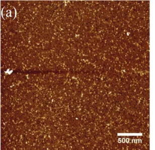
was evaluated first by immersing mica specimen into fairly concentrated solutions of DP4 (ca. 100 mg L-1 in MeOH or deionized water) and then rinsing with the respective solvent. As is evident from Fig. S1, this resulted in a fairly well-behaved monolayer of the dendronized polymer in the case of the aqueous solution, however for the methanolic solution no dense coverage was observed, and only individual polymers or small aggregates remained behind after washing.
(a)
(b)
Figure S2. AFM height images of DP4 deposited from 100 mg L-1 solutions in a) deionized water
and b) methanol. The samples were removed from the solution and rinsed after 100 min. This observation agrees with the generally better solubility of DP4 and its lower g analogues in methanol than in water: Likely, a significant portion of initially deposited DP molecules is washed off by rinsing with methanol, whereas the dense layer deposited from aqueous solution remains in place.
2. pH
For the purpose of synthesizing covalent DP-enzyme conjugates, as well as for the immobilization of theses conjugates, slightly basic aqueous buffers are usually employed as solvents.13 In order to provide quantitatively improved solubility of the DPs discussed here and in order to fully protonate the peripheral amines, more acidic solutions in 100 mM acetate buffers of pH = 4 and pH = 5 were prepared. Using solutions containing 100 mg L-1 of DP4, both buffers were found to result in the
deposition of dense layers of DPs. However, the layers in the case of pH = 4 were much more regular, appearing as essentially densely packed monolayers of DP chains with the occasional protruding coil or chain-crossover (Fig. S2).
(b)
(a)
Figure S3. AFM height images of DP4 deposited from 100 mg L-1 solutions in 100 mM acetate
buffer at pH = 4 during 1 h. a) overview image, b) zoom revealing structural details.
3. Immersion time
Initially, fairly long immersion of the mica platelets in DP4 solutions was used. It was found that exposure times as short as 5 min (Fig. S3a) are entirely sufficient in order to produce complete adlayers of DP at 100 mg L-1, and that prolonged exposure is not necessary.
(c)
(b)
(a)
Figure S4. AFM height images of DP4 deposited from 100 mg L-1 solutions in 100 mM acetate
buffer at pH = 4 with a) 5 min, b) 10 min and c) 20 min immersion time
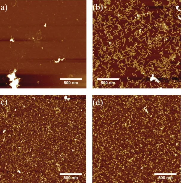
4. Concentration
At 100 mg L-1, a fair amount of polymer is employed; considering the quite strong adhesion in the case of aqueous solutions and the fact that in drop-casting of solutions fairly dense surface coverage may be achieved already at concentrations of around 10 mg L-1 for DPs comparable to DP4, the polymer concentration was varied. While very low concentrations employed for drop-casting were insufficient (1 mg L-1 – 20 mg L-1 Fig. S4a&b) – likely in part due to loss of material by adsorption to the walls of the vessel in which the solution was prepared – concentrations starting at 50 mg L-1 (Fig. S4c&d) produced complete adlayers.
(a)
(b)
(c)
(d)
Figure S5. AFM height images of mica substrates immersed for 15 min into solutions (100 mM
acetate buffer, pH = 4) containing a) 10 mg L-1, b) 20 mg L-1, c) 50 mg L-1 and d) 100 mg L-1 DP4.
5. Ionic strength
The buffer solutions employed up to this point were not adjusted for ionic strength; Fig. S5 displays the result of such adjustments to ionic strengths of 20mM, 50 mM, 100 mM, and 200 mM by addition of NaCl to the acetate buffer. Rather than leading to a further improvement of the homogeneity of the adlayers, increasing ionic strength appears to be counterproductive, likely due to shielding of the peripheral ammonium cations of the DPs which reduce adhesion and DP interactions: instead of homogenous adlayers, large aggregates appear and coverage becomes much
looser than in the standard buffer. Additionally, large aggregate structures – possibly salt deposits – can be found in all samples where NaCl was added. No adjustment of ionic strength therefore appears necessary or helpful.
(a)
(b)
(c)
(d)
Figure S6. AFM height images of mica substrates immersed into solutions of DP4 (50 mg L-1 in
acetate buffer, pH = 4), adjusted with NaCl to ionic strengths of a) 20 mM, b) 50 mM, c) 100 mM, d) 200 mM.
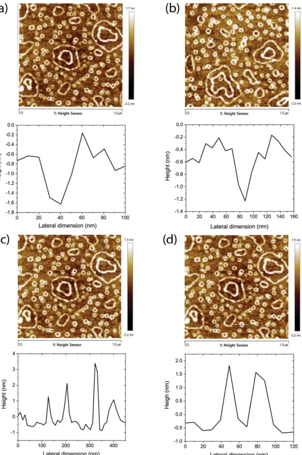
Figure S7. AFM images and profiles of different sections of DP1 deposited on glass substrates. (a)
and (b): Scans through defects in the DP1 monolayer show that the DP1 monolayer is about 1 nm thick. (c) Profile through the larger ring features. The height of these ring features is up to 4 nm on top of the base monolayer. (d) Profile through a small toroidal ring. The ring features are about 2 nm high.
Figure S8. Attenuance of monomers and J-aggregates deposited on glass substrates
pre-functionalized with (a) DP1, (b) DP2, (c) DP3 and (d) DP4.
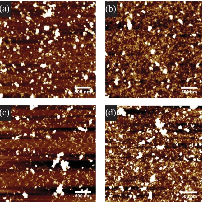
Figure S9. AFM height images of DP2 (a) and DP4 (b) functionalized glass with J-aggregates
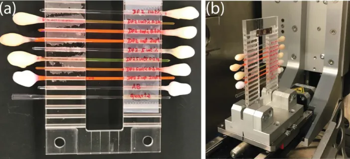
Figure S10. SAXS measurement set-up for quartz capillaries filled with DP2 (1 wt% and 5 wt%)
with 0.2 mM, 0.6 mM and 2 mM dye solution (a) loaded in the measurement set-up (b).
References
1. B. Zhang, R. Wepf, K. Fischer, M. Schmidt, S. Besse, P. Lindner, B. T. King, R. Sigel, P. Schurtenberger, Y. Talmon, Y. Ding, M. Kroeger, A. Halperin and A. D. Schlueter,
Angewandte Chemie-International Edition, 2011, 50, 737-740.
2. A. Kuchler, D. Messmer, A. D. Schluter and P. Walde, in Nanoarmoring of Enzymes:
Rational Design of Polymer-Wrapped Enzymes, ed. C. V. Kumar, 2017, vol. 590, pp.
445-474.
3. L. J. Shu, I. Gossl, J. P. Rabe and A. D. Schluter, Macromolecular Chemistry and Physics, 2002, 203, 2540-2550.
4. B. Zhang, H. Yu, A. D. Schlueter, A. Halperin and M. Kroeger, Nature Communications, 2013, 4.
5. D. Messmer, M. Kroger and A. D. Schluter, Macromolecules, 2018, 51, 5420-5429. 6. J. Schindelin, I. Arganda-Carreras, E. Frise, V. Kaynig, M. Longair, T. Pietzsch, S.
Preibisch, C. Rueden, S. Saalfeld, B. Schmid, J.-Y. Tinevez, D. J. White, V. Hartenstein, K. Eliceiri, P. Tomancak and A. Cardona, Nature Methods, 2012, 9, 676.