INVITED REVIEW
Biomarkers in renal cancer
Holger Moch&John Srigley&Brett Delahunt&Rodolfo Montironi&Lars Egevad&Puay Hoon Tan
Received: 11 November 2013 / Revised: 6 January 2014 / Accepted: 14 January 2014 / Published online: 1 February 2014 # Springer-Verlag Berlin Heidelberg 2014
Abstract Treatment options for primary and metastatic renal cancer are increasing. Accurate data from the pathological examination of renal cancer specimens aid clinicians in stratifying patients for surveillance and adjuvant therapies. This review focuses on biomarkers in diagnosis, prognosis and prediction of the biologic behavior of renal tumors which should be recorded in pathology reports and which are under investigation. Special emphasis is given to the use of immunohisto-chemical markers in differential diagnosis of various renal tumor subtypes. The relevance of cytogenetic and molecular findings is also discussed. The review in-cludes the 2012 International Society for Urological Pathology Consensus conference recommendations.
Keywords RCC . Staging . Grading .
Immunohistochemistry . Biomarker . Molecular pathology
Introduction
Biomarkers in renal cell carcinoma (RCC) are of help in pathological diagnosis, classifying new entities, and include predictive and prognostic markers. Immunohistochemistry allows the characterization of renal tumor subtypes, whereas novel molecular analyses are mainly used to characterize specific molecular pathways involved in different tumor sub-types. They are also useful to identify potential therapeutic targets. This article will review the key prognostic factors with an emphasis on the use of immunohistochemical markers in routine practice. The review is intended to complement the International Society of Urological Pathology (ISUP) confer-ence on renal cancer held in Vancouver in 2012 [1,2].
Old and new subtypes of renal cell neoplasms
In adults, the main morphotype of RCC, when correctly classified, is the strongest prognostic biomarker in addition to tumor stage and grade [3,4]. The current World Health Organization Classification of Renal Tumors was established 2004 [5]. In adults, the main subtypes are clear cell, papillary, chromophobe, collecting duct, and unclassified RCC, but various other subtypes have been recognized in the 2004 WHO classification, including mixed epithelial and stromal tumors (MEST), mucinous tubular and spindle-cell carcinoma ( MTS C ), a nd tr an s l oc a t ion ca nc e r. Onc ocytom a , angiomyolipoma, and metanephric adenoma are considered benign renal neoplasms. At the ISUP conference, a consensus was reached to recognize five entities as new distinct epithelial tumors: tubulocystic RCC, acquired cystic disease-associated
H. Moch (*)
Institute of Surgical Pathology, University Hospital Zurich, Schmelzbergstrasse 12, CH-8091 Zürich, Switzerland e-mail: holger.moch@usz.ch
J. Srigley
Department of Pathology and Molecular Medicine, McMaster University, Hamilton, ON, Canada
B. Delahunt
Department of Pathology and Molecular Medicine, University of Otago, Wellington, New Zealand
R. Montironi
Section of Pathological Anatomy, Polytechnic University of the Marche Region, School of Medicine, United Hospitals, Ancona, Italy L. Egevad
Department of Oncology-Pathology, Karolinska Institutet, Stockholm, Sweden
P. H. Tan
Department of Pathology, Singapore General Hospital, Singapore, Singapore
RCC, clear cell (tubulo) papillary RCC, the MiT family trans-location RCCs (including t(6;11) RCC), and hereditary leiomyomatosis RCC syndrome-associated RCC. Thyroid-like follicular RCC, succinate dehydrogenase B deficiency-associated RCC and ALK translocation RCC were regarded as emerging new entities (Figs.1,2and3) [1]. Subtyping of papillary RCC (types 1 and 2) provides additional prognostic information. For the time being, the oncocytic variant of papillary RCC is not considered as a distinct entity. Clear cell (tubulo)papillary RCC is associated with a more favorable outcome. Multicystic clear cell RCC is considered as a neo-plasm of low malignant potential within the group of clear cell RCC. The hybrid oncocytic chromophobe tumor occurs in Birt-Hogg-Dubé Syndrome, in renal oncocytosis and also as a sporadic neoplasm. Hybrid oncocytic chromophobe tumors are indolent tumors grouped, at least for the time being, in the chromophobe RCC category. Epitheloid angiomyolipoma is nowadays considered a potentially malignant variant of angiomyolipoma, because metastatic disease has been reported in some patients with this specific subtype of angiomyolipoma. Cystic nephroma and mixed epithelial and stromal tumor are seen as a morphological spectrum within a single tumor type [1].
Perinephric fat invasion as a staging parameter
The 2009 TNM system has minor changes compared to the 2002 TNM system, notably in clarifying that infiltration of renal sinus (peripelvic) fat is part of perinephric tissue involve-ment in the pT3 tumor category [6,7]. Perinephric fat invasion is prognostically relevant, as it is associated with a significant decrease in 5-year overall survival [8]. Perinephric fat inva-sion was defined at the 2012 ISUP conference as either the tumor touching the fat or extending with irregular tongues into the perinephric tissue, with or without desmoplasia [9]. Im-portantly, recent studies have shown the relevance of renal
sinus invasion in renal cancer [10, 11]. The frequent renal sinus invasion in early-stage tumors has been overlooked for many years, but has significant prognostic implications. Renal sinus invasion is present when the tumor is in direct contact with the fatty tissue or the loose connective tissue of the sinus. Involvement of any endothelial lined spaces within the renal sinus is considered as renal sinus invasion, regardless of the size of the vascular space [9].
Tumor grade, sarcomatoid differentiation, and necrosis as prognostic biomarkers of renal cancer
The Fuhrman grading system has been in use for over 30 years and is still the most widely utilized grading system for renal cancer [12]. Nonetheless, it has not evolved over the years to accommodate our increasing understanding of the nature and biological spectrum of renal carcinoma. Furthermore, its use has become controversial, as it is increasingly recognized that its application is not without problems [13,14]. In addition, the grading system has not been validated for many of the morphotypes of renal cell carcinoma. In view of the problems associated with the application of Fuhrman grading, a new grading system was endorsed at the recent ISUP consensus conference [3]. Nucleolar prominence defines grades 1 to 3 of clear cell and papillary RCCs, whereas extreme nuclear pleo-morphism or sarcomatoid and/or rhabdoid differentiation de-fines grade 4 tumors. The ISUP grading system was endorsed as a prognostic parameter for clear cell renal cell carcinoma. In addition, a consensus was reached that chromophobe RCC should not be graded [15–17], as irregular nuclei, prominent nucleoli, and nuclear pleomorphism are inherently present in chromophobe RCC, even in cases with good prognosis [3].
Different grading systems for chromophobe RCC have been proposed [15], but these were not endorsed at the 2012 ISUP consensus conference. Sarcomatoid and/or rhabdoid differentiation and tumor necrosis were accepted as useful
Fig. 1 Mucinous tubular and spindle-cell carcinoma showing classical pattern with elongated tubules, spindle-cell areas, and stromal mucin
additional histopathologic prognostic parameters [4]. Sarcomatoid or rhabdoid differentiation should be mentioned in pathology reports of all RCC subtypes. For a correct tumor classification, the underlying carcinoma subtype should be reported, because this can be recognized for all main subtypes. If the underlying carcinoma subtype cannot be recognized in sarcomatoid RCC, the tumor should be classified as a grade 4 unclassified carcinoma with a sarcomatoid component. Tumor necrosis is considered of prognostic significance and the amount of necrosis should be quantified in clear cell RCC. In contrast, intratumoral microvascular invasion should not be included as a staging criterion for RCC [4,9].
RCC is considered to be susceptible to the innate and adaptive immune responses of the host because a small subset of patients with advanced disease achieves spontaneous or immunotherapy-induced complete remission. The cellular and molecular mechanisms underlying these complete remis-sions are not completely understood, although a variety of inflammatory cells including T and Natural killer (NK) cells
have been identified in RCC lesions. Local immunoregulatory processes may have an impact on disease progression and therefore on survival of patients with primary clear cell renal cell carcinoma. Earlier data have shown that a high number of tumor-infiltrating lymphocytes and/or macrophages are asso-ciated with poor prognosis [18–20].
Differential diagnosis of renal cell neoplasms using immunohistochemical biomarkers
Most renal cancer subtypes have a characteristic immunohis-tochemical staining profile that helps in the correct tumor classification. It has been shown that upregulation of CD10 and Pax 2 expression is due to VHL inactivation in clear cell RCC [21,22], while CAIX is also consistently expressed due to its regulation by the VHL protein [23,24]. Papillary RCC type 1 stains for vimentin, broad spectrum keratins, CK7, AMACR and RCC marker, but not for CD117, kidney-specific cadherin, and parvalbumin. Papillary RCC type 2 has variable staining patterns. Whereas papillary type 1 RCC frequently show diffuse CK7 immunoreactivity, papillary type 2 RCC are less often positive for CK7, in a staining pattern comparable to that of clear cell RCC with only focal CK7 expression. Chromophobe RCC are negative for vimentin (Table1), but show diffuse reactivity for E-cadherin, kidney-specific cadherin, parvalbumin, CD117, EMA, broad spec-trum keratins, and CK7 [1,4]. To differentiate between benign oncocytoma and chromophobe RCC, specifically its eosino-philic variant, is difficult as these tumors have overlapping histological and immunohistochemical characteristics. CK7 is the best marker to differentiate between these two tumor entities. Most chromophobe RCCs show membranous CK7 expression in tumor cell clusters or in the majority of tumor cells while oncocytoma is typically negative or at most focally positive in scattered cells [25,26]. Other biomarkers useful for this differential diagnosis are summarized in Table2.
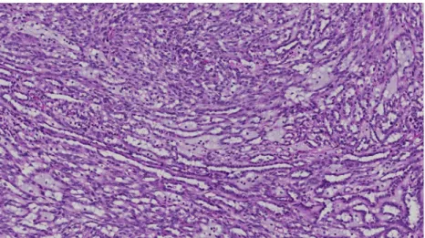
Fig. 2 New tumor entity: leiomyomatous renal cell carcinoma. Note nests and tubules lined by clear cells embedded in smooth muscle
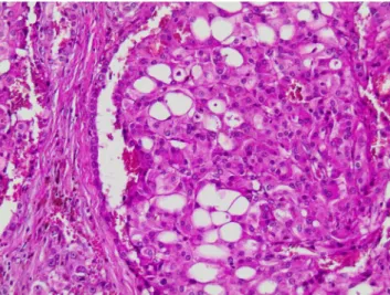
Fig. 3 New tumor entity: Renal cell carcinoma in end-stage renal dis-ease. Note cytoplasmic vacuolation
Collecting duct carcinomas often show immunoreactivity for EMA, CK7, high molecular weight keratin, Pax 2 and/or Pax 8 [27] (Table 3). Pax 2 and/or Pax 8 are frequently expressed in different renal carcinoma subtypes and represent useful markers in the differential diagnosis of a primary renal carcinoma, although expression patterns need to be interpreted in conjunction with other markers. For instance, expression of Pax 8 has been reported in primary pancreatic neuroendocrine tumors, which can mimic metastatic RCC [28]. Distinction from RCC will therefore require additional support, including the lack of expression of neuroendocrine markers for a diag-nosis of RCC. There is consensus that TFE3 and TFEB immonostains should be requested in order to diagnose RCC in a young patient or when histological appearances are sug-gestive of the translocation subtype of RCC [4].
Epitheloid angiomyolipoma closely resembles RCC [3, 29]. Positive immunoreactivity for HMB45, melan-A and SMA, and absence of expression of keratins, helps in the diagnosis of angiomyolipoma [30]. Metanephric adenoma can be mistaken for type 1 papillary RCC. The former shows positive immunostaining for WT1, CD57, and absence of
reactivity for AMACR [31]. AMACR, CK7, WT1, and CD57 are therefore useful to distinguish metanephric adeno-ma from papillary RCC (Table4).
Molecular prognostic biomarkers
Biomarkers for potential prognostication of RCC include molecules in intracellular pathways and/or specific DNA al-terations. Most of them have not entered clinical practice. Clear cell RCC is characterized by loss of chromosome 3p and mutations of the von Hippel Lindau (VHL) gene [32].VHL mutations have been reported in the majority of sporadic clear cell renal carcinomas [33]. These mutations can impact on the hypoxia inducible factor (HIF) pathway and their existence provides the theoretical molecular explanation for the success of HIF targeted treatment strategies in some patients with clear cell RCC (see below) [34,35]. Dysregulation of HIF leads to upregulation of the expression of downstream molecules, including Carbonic anhydrase 9 (CAIX). One study found an association between CAIX expression and grade of clear cell RCC [36]. Diminished CAIX expression was indepen-dently correlated with poor survival in advanced renal cell cancer patients [37]. Some other chromosomal abnormalities may have prognostic value, such as loss of chromosome 9p in clear cell RCC, which is associated with a significantly poorer cancer specific survival [38,39]
A relatively new finding is the frequent polybromo-1 (PBRM1) and BAP 1 mutation on chromosome 3p in a sur-prisingly high percentage of clear cell RCC [40]. Loss of PBRM1 protein expression product BAF180 was shown to be associated with advanced tumor stage and worse patient outcome [41]. All of these newly identified mutations target
Table 2 Biomarker in chromophobe RCC and oncocytoma
Biomarker Chromophobe RCC Oncocytoma
CK7 + −/focal + MOC31 + − EpCam + − Caveolin-1 + − EABA − + CD82 + − S100A1 − + Parvalbumin + + Ksp cadherin + + CD117 + +
Table 3 Biomarker for poorly differentiated carcinomas
Antibody RCC, unclassified Collecting duct carcinoma Urothelial carcinoma CK7 −/+ + + CK20 − − (rarely focal+) +/− P63 − − (rarely+) + RCC +/− − − Vimentin +/− + −/+ CD10 +/− − −/+ CK5/6 − − +/− INI1 + + + Ulex-1 − + −/+ PAX8 +/− + −/rarely + PAX2 +/− +/− −
GATA3 −/(rarely focal +) − +
Table 1 Biomarkers in clear cell and chromophobe RCC
Biomarker Clear cell RCC Chromophobe RCC
CK7 − (focal) + RCC marker + − CD10 + − Vimentin + − CD117 − + Parvalbumin − + E-cadherin − + EMA + + MUC1 + + CK20 − − AMACR − −
genes which are responsible for chromatin remodeling. Future studies will help to understand the tumor suppressor gene network betweenVHL and the other genes on chromosome 3p. Compared to other solid tumors, intratumoral heterogene-ity of driver gene mutations is very pronounced within prima-ry RCC as well as between primaprima-ry tumors and their metas-tases [42,43]. The process of clonal evolution of renal cancer cells is very complex, and this has potential consequences for tumor progression, development of resistance against targeted therapies, with clinical impact.
Papillary RCC is characterized by numerical abnormalities, often trisomies of chromosomes 7 and 17 [44]. Chromophobe RCCs harbor multiple numerical losses of chromosomes 1, 2, 6, 10, and 17 [45]. Translocation RCCs are defined by trans-locations involving chromosome Xp11.2, resulting in TFE3 gene fusions [46]. Another variant of translocation associated RCC is characterized by fusion of theTFEB gene on chromo-some 6p with the alpha gene on 11q12, which leads to expression of the TFEB protein [47,48].
Cytogenetics and novel molecular technologies are rarely used in renal carcinoma diagnosis, but have contributed to tumor classification, understanding the histogenesis and the genotype/ phenotype correlation in RCC. Some groups recommend a panel of fluorescence in situ hybridization (FISH) probes for the differential diagnosis of RCC. For instance, numerical abnor-malities of chromosomes 7 and 17 on FISH favor a diagnosis of papillary RCCs over clear cell RCC. The value of FISH in diagnosing a translocation RCC with equivocal immunohisto-chemical results has recently been emphasized [49].
Predictive markers in renal cancer
Predictive markers provide information on whether a particu-lar cancer will respond to or be resistant to a specific type of therapy. In contrast to other solid tumors, e.g., lung and colorectal cancer or melanoma, use of predictive biomarkers
for clinical stratification and management planning has not yet entered routine practice in metastatic renal cancer [4,50,51]. In 2007, six new agents, which target complex molecular pathways regulating tumor angiogenesis and cell proliferation and survival, have been approved. These treatments have significantly improved survival times in metastatic renal cell carcinoma [52]. Patients with advanced clear cell RCC receive VEGF pathway antagonists, e.g., sunitinib or pazopanib. Analysis of VHL mutation status, plasma CAIX, VEGF, sVEGFR2, tissue inhibitor of metalloproteinase 1 (TIMP-1), and Rasp21 has been performed in the TARGET trial com-paring sorafenib with placebo in advanced RCC [53]. No predictive markers were identified [54, 55]. Choueiri and colleagues evaluated tumor CAIX expression using immuno-histochemistry in patients treated with antiangiogenic thera-pies [56]. While CAIX expression was neither prognostic nor predictive of response to sunitinib, for sorafenib-treated pa-tients, high CAIX expression (>85 %) was associated with decreased tumor size in response to treatment.
MicroRNAs (miRNAs, miRs) are small (19–25 nucleo-tides), non-coding RNAs that play an important role in apopto-sis, survival, proliferation, and differentiation processes, by the post-transcriptional regulation of gene-expression. An increas-ing body of evidence suggests that micro RNA’s play a crucial role, not only as oncogenic or tumor-suppressive molecules in cancer initiation, progression and metastasis, but also in resis-tance to chemotherapy or other systemic therapies. Therefore, miRNA’s might constitute potential prognostic and/or predic-tive biomarkers. Prognostic miRNA expression patterns have recently been identified in RCC tissues [57,58]. High levels of miR-210 were noticed in chemotherapy resistant ccRCC pa-tients [59]. Downregulation of miR-141 was correlated with lack of response to sunitinib treatment in ccRCC patients, when compared to the expression profile in responding patients [60]. Gamez-Pazo and other groups provided evidence that the re-sponse of metastatic ccRCC patients to TKI inhibitors is corre-lated with a miRNA profile in patient serum [61–63].
Table 4 Biomarker for renal tu-mors with papillary or tubulopapillary architecture Antibody Papillary RCC Collecting duct carcinoma Metanephric adenoma
Mucinous tubular and spindle-cell carcinoma Clear cell papillary RCC CK7 + + − (only focal) + + (diffuse) CD10 + − − −/+ − RCC + − − variable −/+ AMACR + − −/+ + − EMA/MUC1 + + − (may be focal) + + WT1 − − + − − CD57 − No data + − No data Ulex-1 − + − − No data
Conclusion
Correct tumor classification, histologic grading and systemat-ic examination of radsystemat-ical nephrectomy or tumorectomy spec-imen are essential for patient management. Tumor stage, tumor type, and tumor grade are robust and independent histopathological prognostic factors, which should be routinely included in pathology reports. Evidence for other prognostic biomarker is lacking. In contrast to other cancers, e.g., lung and colorectal carcinomas or melanoma there are at present no predictive molecular biomarker, suitable for routine use. Future investigation of recently identified novel molecular alterations and tumor suppressor networks in clear cell RCC will help to clarify the biological relevance of different molecular signaling pathways in predicting therapy response.
References
1. Srigley JR, Delahunt B, Eble JN et al (2013) The International Society of Urological Pathology (ISUP) Vancouver classification of
renal neoplasia. Am J Surg Pathol 37:1469–1489
2. Delahunt B, Egevad L, Montironi R et al (2013) International Society of Urological Pathology (ISUP) consensus conference on renal neoplasia: rationale and organization. Am J Surg
Pathol 37:1463–1468
3. Delahunt B, Cheville JC, Martignoni G et al (2013) The International Society of Urological Pathology (ISUP) grading system for renal cell carcinoma and other prognostic parameters. Am J Surg Pathol 37:
1490–1504
4. Tan PH, Cheng L, Rioux-Leclercq N et al (2013) Renal tumors: diagnostic and prognostic biomarkers. Am J Surg
Pathol 37:1518–1531
5. Eble J, Sauter G, Epstein J et al (2004) Tumours of the kidney, tumours of the urinary system and male genital organs. WHO clas-sification of tumours. IARC, Lyon
6. UICC: TNM Classification of Malignant Tumours (ed 7th), Wiley-Blackwell, 2009
7. UICC (2002) TNM classification of malignant tumours, 6th edn. Wiley, New York
8. Moch H, Artibani W, Delahunt B et al (2009) Reassessing the current UICC/AJCC TNM staging for renal cell carcinoma. Eur Urol 56:
636–643
9. Trpkov K, Grignon DJ, Bonsib SM et al (2013) Handling and staging of renal cell carcinoma: the International Society of Urological Pathology consensus (ISUP) conference recommendations. Am J
Surg Pathol 37:1505–1517
10. Bonsib SM (2005) T2 clear cell renal cell carcinoma is a rare entity: a
study of 120 clear cell renal cell carcinomas. J Urol 174:1199–1202,
discussion 1202
11. Bonsib SM (2006) Renal lymphatics, and lymphatic involvement in sinus vein invasive (pT3b) clear cell renal cell carcinoma: a study of
40 cases. Mod Pathol 19:746–753
12. Fuhrman S, Lasky L, Limas C (1982) Prognostic significance of morphologic parameters in renal cell carcinoma. Am J Surg Pathol
6:655–663
13. Delahunt B (2009) Advances and controversies in grading and stag-ing of renal cell carcinoma. Mod Pathol: Off J U S Can Acad Pathol,
Inc 22(2):S24–S36
14. Delahunt B, Sika-Paotonu D, Bethwaite PB et al (2011) Grading of clear cell renal cell carcinoma should be based on nucleolar
promi-nence. Am J Surg Pathol 35:1134–1139
15. Paner GP, Amin MB, Alvarado-Cabrero I et al (2010) A novel tumor grading scheme for chromophobe renal cell carcinoma: prognostic utility and comparison with Fuhrman nuclear grade. Am J Surg
Pathol 34:1233–1240
16. Cheville JC, Lohse CM, Sukov WR et al (2012) Chromophobe renal cell carcinoma: the impact of tumor grade on outcome. Am J Surg
Pathol 36:851–856
17. Amin MB, Paner GP, Alvarado-Cabrero I et al (2008) Chromophobe renal cell carcinoma: histomorphologic characteristics and evaluation of conventional pathologic prognostic parameters in 145 cases. Am J
Surg Pathol 32:1822–1834
18. Dannenmann SR, Thielicke J, Stockli M et al (2013) Tumor-associated macrophages subvert T-cell function and correlate with reduced survival in clear cell renal cell carcinoma. Oncoimmunology 2:e23562
19. Morra L, Rechsteiner M, Casagrande S et al (2011) Relevance of periostin splice variants in renal cell carcinoma. Am J Pathol 179: 1513–1521
20. Sconocchia G, Spagnoli GC, Del Principe D et al (2009) Defective infiltration of natural killer cells in MICA/B-positive renal cell carci-noma involves beta(2)-integrin-mediated interaction. Neoplasia 11: 662–671
21. Boysen G, Bausch-Fluck D, Thoma CR et al (2012) Identification and functional characterization of pVHL-dependent cell surface pro-teins in renal cell carcinoma. Neoplasia 14:535–546
22. Luu VD, Boysen G, Struckmann K et al (2009) Loss of VHL and hypoxia provokes PAX2 up-regulation in clear cell renal cell carci-noma. Clin Cancer Res 15:3297–3304
23. Dahinden C, Ingold B, Wild P et al (2010) Mining tissue microarray data to uncover combinations of biomarker expression patterns that improve intermediate staging and grading of clear cell renal cell cancer. Clin Cancer Res: Off J Am Assoc Cancer Res 16:88–98 24. Patard JJ, Pouessel D, Bensalah K et al (2008) Targeted therapy in
renal cell carcinoma. World J Urol 26:135–140
25. Skinnider BF, Folpe AL, Hennigar RA et al (2005) Distribution of cytokeratins and vimentin in adult renal neoplasms and normal renal tissue: potential utility of a cytokeratin antibody panel in the
differ-ential diagnosis of renal tumors. Am J Surg Pathol 29:747–754
26. Skinnider BF, Amin MB (2005) An immunohistochemical approach to the differential diagnosis of renal tumors. Semin Diagn
Pathol 22:51–68
27. Zhou M, Yang XJ, Lopez JI et al (2009) Renal tubulocystic carcino-ma is closely related to papillary renal cell carcinocarcino-ma: implications
for pathologic classification. Am J Surg Pathol 33:1840–1849
28. Sangoi AR, Ohgami RS, Pai RK et al (2011) PAX8 expression reliably distinguishes pancreatic well-differentiated neuroendocrine tumors from ileal and pulmonary well-differentiated neuroendocrine tumors and pancreatic acinar cell carcinoma. Mod Pathol: Off J U S
Can Acad Pathol, Inc 24:412–424
29. Mete O, van der Kwast TH (2011) Epithelioid angiomyolipoma: a morphologically distinct variant that mimics a variety of
intra-abdominal neoplasms. Arch Pathol Lab Med 135:665–670
30. Eble JN (1998) Angiomyolipoma of kidney. Semin Diagn
Pathol 15:21–40
31. Olgac S, Hutchinson B, Tickoo SK et al (2006) Alpha-methylacyl-CoA racemase as a marker in the differential diagnosis of metanephric
adenoma. Mod Pathol: Off J U S Can Acad Pathol, Inc 19:218–224
32. The Cancer Genome Atlas Network (2013) Comprehensive molecular
33. Moch H (2013) An overview of renal cell cancer: pathology and
genetics. Semin Cancer Biol 23:3–9
34. Shen C, Kaelin WG Jr (2013) The VHL/HIF axis in clear cell renal
carcinoma. Semin Cancer Biol 23:18–25
35. Rechsteiner MP, von Teichman A, Nowicka A et al (2011) VHL gene mutations and their effects on hypoxia inducible factor HIFalpha: Identification of potential driver and passenger mutations. Cancer
Res 71:5500–5511
36. Genega EM, Ghebremichael M, Najarian R et al (2010) Carbonic anhydrase IX expression in renal neoplasms: correlation with tumor
type and grade. Am J Clin Pathol 134:873–879
37. Bui MH, Seligson D, Han KR et al (2003) Carbonic anhydrase IX is an independent predictor of survival in advanced renal clear cell carcinoma: Implications for prognosis and therapy. Clin Cancer
Res: Off J Am Assoc Cancer Res 9:802–811
38. Moch H, Presti JC Jr, Sauter G et al (1996) Genetic aberrations detected by comparative genomic hybridization are associated with
clinical outcome in renal cell carcinoma. Cancer Res 56:27–30
39. Schraml P, Struckmann K, Bednar R et al (2001) CDKNA2A Mutation analysis, protein expression, and deletion mapping of chro-mosome 9p in conventional clear-cell renal carcinomas: evidence for a second tumor suppressor gene proximal to CDKN2A. Am J Pathol 158:593–601
40. Varela I, Tarpey P, Raine K et al (2011) Exome sequencing identifies frequent mutation of the SWI/SNF complex gene PBRM1 in renal carcinoma. Nature 469:539–542
41. Pawlowski R, Muhl SM, Sulser T et al (2013) Loss of PBRM1 expression is associated with renal cell carcinoma progression. Int J Cancer J Int Cancer 132:E11–E17
42. Xu X, Hou Y, Yin X et al (2012) Single-cell exome sequencing reveals single-nucleotide mutation characteristics of a kidney tumor. Cell 148:886–895
43. Gerlinger M, Rowan AJ, Horswell S et al (2012) Intratumor hetero-geneity and branched evolution revealed by multiregion sequencing. N Engl J Med 366:883–892
44. Kovacs G, Fuzesi L, Emanual A et al (1991) Cytogenetics of papil-lary renal cell tumors. Genes Chromosom Cancer 3:249–255 45. Speicher M, Schoell B, Du Manoir S et al (1994) Specific loss of
chromosomes 1, 2, 6, 10, 13, 17, and 21 in chromophobe renal cell carcinomas revealed by comparative genomic hybridization. Am J
Pathol 145:356–364
46. Argani P, Antonescu CR, Illei PB et al (2001) Primary renal neo-plasms with the ASPL-TFE3 gene fusion of alveolar soft part sarco-ma: a distinctive tumor entity previously included among renal cell
carcinomas of children and adolescents. Am J Pathol 159:179–192
47. Argani P, Hawkins A, Griffin CA et al (2001) A distinctive pediatric renal neoplasm characterized by epithelioid morphology, basement membrane production, focal HMB45 immunoreactivity, and t(6;11)(p21.1;q12) chromosome translocation. Am J Pathol 158:
2089–2096
48. Argani P, Lae M, Hutchinson B et al (2005) Renal carcinomas with the t(6;11)(p21;q12): clinicopathologic features and demonstration of
the specific alpha-TFEB gene fusion by immunohistochemistry,
RT-PCR, and DNA PCR. Am J Surg Pathol 29:230–240
49. Green WM, Yonescu R, Morsberger L et al (2013) Utilization of a TFE3 break-apart FISH assay in a renal tumor consultation service.
Am J Surg Pathol 37:1150–1163
50. Eichelberg C, Junker K, Ljungberg B et al (2009) Diagnostic and prognostic molecular markers for renal cell carcinoma: a critical appraisal of the current state of research and clinical applicability.
Eur Urol 55:851–863
51. Algaba F, Akaza H, Lopez-Beltran A et al (2011) Current pathology
keys of renal cell carcinoma. Eur Urol 60:634–643
52. Fisher R, Gore M, Larkin J (2013) Current and future systemic
treatments for renal cell carcinoma. Semin Cancer Biol 23:38–45
53. Pena C, Lathia C, Shan M et al (2010) Biomarkers predicting out-come in patients with advanced renal cell carcinoma: results from Sorafenib phase III treatment approaches in renal cancer global evaluation trial. Clin Cancer Res: Off J Am Assoc Cancer Res 16:
4853–4863
54. Tang PA, Vickers MM, Heng DY (2011) Clinical and molecular prognostic factors in renal cell carcinoma: what we know so far. Hematol/Oncol Clin N Am 25:871–891
55. Minner S, Rump D, Tennstedt P et al (2012) Epidermal growth factor receptor protein expression and genomic alterations in renal cell carcinoma. Cancer 118:1268–1275
56. Choueiri TK, Regan MM, Rosenberg JE et al (2010) Carbonic anhydrase IX and pathological features as predictors of outcome in patients with metastatic clear-cell renal cell carcinoma receiving vascular endothelial growth factor-targeted therapy. BJU Int 106: 772–778
57. Heinzelmann J, Henning B, Sanjmyatav J et al (2011) Specific miRNA signatures are associated with metastasis and poor prognosis in clear cell renal cell carcinoma. World J Urol 29:367–373 58. Slaby O, Redova M, Poprach A et al (2012) Identification of
MicroRNAs associated with early relapse after nephrectomy in renal cell carcinoma patients. Genes Chromosom Cancer 51:707–716 59. Nakada C, Tsukamoto Y, Matsuura K et al (2011) Overexpression of
miR-210, a downstream target of HIF1alpha, causes centrosome amplification in renal carcinoma cells. J Pathol 224:280–288 60. Berkers JHMG, Wolter O, Beuselinck P, Schoffski B, Roskams P,
Joniau TAD, Van Poppel S, Lerut , E.S.M.: MicroRNA-141 expres-sion in clear cell renal cell carcinoma is linked with Sunitinib response. European Urology, Supplements
61. Redova M, Poprach A, Nekvindova J et al (2012) Circulating miR-378 and miR-451 in serum are potential biomarkers for renal cell carcinoma. J Transl Med 10:55
62. Gamez-Pozo A, Anton-Aparicio LM, Bayona C et al (2012) MicroRNA expression profiling of peripheral blood samples predicts resistance to first-line sunitinib in advanced renal cell carcinoma
patients. Neoplasia 14:1144–1152
63. Wu X, Weng L, Li X et al (2012) Identification of a 4-microRNA signature for clear cell renal cell carcinoma metastasis and prognosis. PloS One 7:e35661